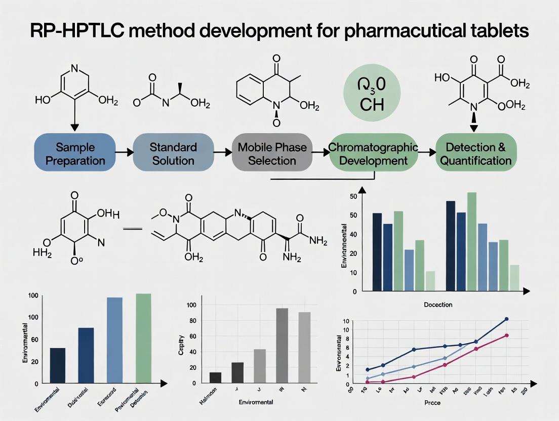
A Comprehensive Guide to RP-HPTLC Method Development and Validation for Pharmaceutical Tablet Analysis
This article provides a detailed, step-by-step guide for researchers and pharmaceutical scientists on developing and validating a robust Reversed-Phase High-Performance Thin-Layer Chromatography (RP-HPTLC) method for the analysis of active pharmaceutical...
A Comprehensive Guide to RP-HPTLC Method Development and Validation for Pharmaceutical Tablet Analysis
Abstract
This article provides a detailed, step-by-step guide for researchers and pharmaceutical scientists on developing and validating a robust Reversed-Phase High-Performance Thin-Layer Chromatography (RP-HPTLC) method for the analysis of active pharmaceutical ingredients (APIs) in tablets. Covering foundational principles, practical methodology, systematic troubleshooting, and comprehensive validation according to ICH guidelines, it addresses the full scope of analytical development from initial exploration to comparative assessment with other techniques. The content is designed to equip professionals with the knowledge to create reliable, cost-effective, and efficient quality control methods for solid dosage forms.
RP-HPTLC Fundamentals: Principles, Advantages, and Initial Scouting for Tablet Analysis
Core Principles
Reversed-Phase High-Performance Thin-Layer Chromatography (RP-HPTLC) is an advanced planar chromatographic technique where the stationary phase is non-polar (e.g., silica gel bonded with C18, C8, or phenyl groups), and the mobile phase is a polar solvent or solvent mixture. This is the reverse of the "normal phase" mode, making it ideal for separating moderately polar to non-polar analytes, which is common in pharmaceutical compounds. Separation is based on the differential partitioning of analytes between the two phases, driven by hydrophobic interactions. The more hydrophobic (lipophilic) a compound, the stronger it interacts with the non-polar stationary phase, resulting in a longer retention (lower RF value).
The "High-Performance" aspect refers to the use of plates with smaller, more uniform particle sizes (e.g., 5-7 µm) and narrower size distribution than conventional TLC, leading to higher efficiency, better resolution, faster development, and improved quantitative accuracy via densitometry.
Mechanism of Separation
The primary mechanism is hydrophobic interaction. In an aqueous-organic mobile phase, analytes compete for binding sites on the non-polar stationary phase. Separation is achieved due to differences in the analyte's partition coefficient (K), which is a function of its chemical structure and the eluent's composition. Secondary interactions, such as silanol activity on residual unbonded silicas or ion-pairing effects, can also influence separation, especially for ionizable compounds. pH control and buffer additives are often used to manage these effects.
Application Notes & Protocols in Pharmaceutical Tablet Research
Within a thesis focused on RP-HPTLC method development for pharmaceutical tablets, the technique is pivotal for assay, impurity profiling, stability testing, and dissolution studies. It offers a parallel, high-throughput, and cost-effective alternative to column HPLC, with the advantage of visualizing all sample components simultaneously.
Key Advantages for Tablet Analysis:
- Multiple sample parallel processing: An entire calibration curve and numerous tablet extracts can be run on a single plate.
- Minimal sample cleanup: The sample is applied as a discrete spot; the plate is used once and discarded, eliminating carryover.
- Open system: Allows for post-chromatographic derivatization with specific reagents to enhance detection of functional groups.
- Compatibility with various detectors: Densitometry in UV/Vis, fluorescence, or after derivatization.
Summarized Quantitative Data from Recent Studies
Table 1: Comparison of RP-HPTLC Methods for Selected Drugs in Tablet Formulations
| Drug (Class) | Stationary Phase | Mobile Phase (v/v/v) | RF Value | Detection (nm) | LOD (ng/band) | LOQ (ng/band) | Key Application | Reference Year |
|---|---|---|---|---|---|---|---|---|
| Empagliflozin (SGLT2 inhibitor) | Silica gel 60 RP-18 | Methanol:Water (8:2) | 0.45 ± 0.02 | 225 | 18 | 55 | Assay & forced degradation | 2023 |
| Dabigatran etexilate (Anticoagulant) | Silica gel 60 RP-18 F₂₅₄S | Acetonitrile:0.1% FA in water (7:3) | 0.62 ± 0.03 | 320 | 1.5 | 4.5 | Content uniformity | 2024 |
| Ibuprofen & Paracetamol (NSAID/Analgesic) | Silica gel 60 RP-8 | Acetonitrile:Methanol:Water (4:4:2) | 0.33 (I), 0.58 (P) | 220 | 50 (I), 40 (P) | 150 (I), 120 (P) | Combined dosage form assay | 2023 |
| Rosuvastatin (Statin) | Silica gel 60 RP-18 | Acetonitrile:Methanol:Ammonium acetate buffer pH 4.5 (5:3:2) | 0.51 ± 0.02 | 244 | 1.0 | 3.0 | Impurity profiling | 2022 |
Table 2: Typical Precision and Accuracy Data from a Validated RP-HPTLC Assay*
| Spiked Concentration (%) | Intra-day RSD (%) (n=6) | Inter-day RSD (%) (n=3 days) | Mean Recovery (%) |
|---|---|---|---|
| 80 | 1.2 | 1.5 | 99.4 |
| 100 | 0.9 | 1.2 | 100.2 |
| 120 | 1.1 | 1.4 | 99.8 |
*Exemplary data following ICH Q2(R1) guidelines.
Detailed Experimental Protocols
Protocol 1: Standard Method Development Workflow for Tablet Assay
1. Sample Preparation:
- Weigh and finely powder 20 tablets.
- Transfer an amount of powder equivalent to one tablet's nominal drug weight to a volumetric flask.
- Add 70% of the chosen solvent (e.g., methanol, methanol-water mix) and sonicate for 20 minutes with intermittent shaking.
- Dilute to volume, mix well, and filter through a 0.45 µm syringe filter. This is the test solution.
2. Standard Solution Preparation:
- Accurately weigh 10 mg of reference standard into a 10 mL volumetric flask.
- Dissolve and dilute to volume with the same solvent to obtain a 1000 µg/mL stock solution.
- Prepare a series of dilutions for the calibration curve (e.g., 100-600 ng/band).
3. Chromatographic Procedure:
- Plate: Pre-wash RP-18 HPTLC plates (e.g., Merck) with methanol and dry in an oven at 120°C for 20 min before use.
- Application: Apply bands (6 mm length) of standard and sample solutions automatically (e.g., Linomat 5) 8 mm from the bottom edge. Maintain a 10 mm gap between bands.
- Development: Develop in a twin-trough chamber pre-saturated with mobile phase (e.g., Methanol:Water 70:30) for 20 min. Develop the plate over a distance of 70 mm at 25°C ± 2.
- Drying: Dry the developed plate in a stream of warm air for 5 min.
4. Densitometric Analysis:
- Scan the plate in absorbance mode at the selected λ_max using a slit dimension of 6.00 x 0.30 mm.
- Generate a calibration curve (peak area vs. amount applied) and compute the drug content in the sample bands via linear regression.
5. Validation:
- Perform validation for linearity, precision (repeatability, intermediate precision), accuracy (recovery), specificity (peak purity via spectral overlay), LOD, and LOQ as per ICH Q2(R1).
Protocol 2: Impurity Profiling via Forced Degradation Study
1. Stress Conditions:
- Acidic Hydrolysis: Treat tablet powder/API solution with 1M HCl at 70°C for 2h. Neutralize before application.
- Alkaline Hydrolysis: Treat with 0.1M NaOH at 70°C for 2h. Neutralize.
- Oxidative Degradation: Treat with 3% H₂O₂ at room temperature for 30 min.
- Photodegradation: Expose solid powder and prepared solution to UV light (254 nm) in a photostability chamber for 48h.
- Thermal Degradation: Heat solid powder at 80°C for 48h.
2. Analysis:
- Apply stressed samples alongside unstressed control and impurity standards.
- Develop using an optimized mobile phase that separates all degradation products.
- Scan the entire track. Use spectral correlation to confirm the purity of the main band and identify impurities/degradants.
Diagrams
RP-HPTLC Method Development Workflow
Analyte Separation by Hydrophobicity
The Scientist's Toolkit: Essential Research Reagents & Materials
Table 3: Key Research Reagent Solutions for RP-HPTLC Method Development
| Item | Function & Rationale |
|---|---|
| HPTLC Plates (RP-18, RP-8) | The core substrate. RP-18 offers the strongest hydrophobicity for very non-polar compounds; RP-8 offers a balance for moderately non-polar drugs. |
| HPLC-Grade Organic Solvents (Methanol, Acetonitrile) | Primary mobile phase components. Low UV cutoff and high purity are critical for baseline stability in densitometry. |
| Buffer Salts (Ammonium acetate, formate, phosphate) | Used to prepare pH-controlled mobile phases (e.g., 0.1% Formic Acid). Critical for separating ionizable compounds by suppressing ionization. |
| Derivatization Reagents (e.g., Anisaldehyde, Ninhydrin) | Spray reagents for visualizing compounds without a UV chromophore. Specific reagents react with functional groups (sugars, amines). |
| Silica Gel 60 G (for sample cleanup) | Used in solid-phase extraction (SPE) cartridges or for column chromatography to pre-purify complex tablet extracts. |
| Reference Standards (API & Known Impurities) | Essential for peak identification, method calibration, and specificity confirmation (via co-chromatography). |
| Automated Applicator (e.g., Linomat) | Ensures precise, reproducible band application (volume, position, band length), crucial for quantitative accuracy. |
| Twin-Trough Development Chamber | Allows for chamber saturation with mobile phase vapor separately from the developing solvent, ensuring reproducible RF values. |
| Densitometer with UV/Vis & Fluorescence | Instrument for in-situ quantitative measurement of the separated bands. Software calculates peak areas, purity, and calibration. |
Application Notes
Within the framework of thesis research focused on RP-HPTLC (Reversed Phase High-Performance Thin-Layer Chromatography) method development for pharmaceutical tablets, three distinct advantages emerge as critical for modern analytical laboratories. This technique offers a compelling alternative to conventional HPLC for routine analysis and method scouting.
1. Cost Efficiency: RP-HPTLC dramatically reduces solvent consumption and waste disposal costs. Where a typical HPLC run may consume 500-1000 mL of mobile phase per day for a single method, an RP-HPTLC analysis of 20 samples on a single plate uses approximately 20-40 mL total. This results in over 95% reduction in solvent purchase and hazardous waste costs. Furthermore, the reusability of sample capillaries and the minimal instrumental maintenance compared to high-pressure systems contribute to lower operational expenditure.
2. Analytical Speed and Throughput: The inherent capability for parallel processing allows the simultaneous development of multiple methods or analysis of numerous samples. While a single HPLC run for a stability-indicating method may take 20-30 minutes, RP-HPTLC can develop 20 samples on one plate in 15-20 minutes. This parallelization effectively cuts analysis time per sample to under one minute.
3. Parallel Processing for Method Development: This is the most significant advantage for research. Multiple mobile phase compositions can be tested on the same plate alongside standard and sample tracks. This enables rapid optimization of critical parameters like organic modifier percentage and pH in a single, consolidated experiment, accelerating the method development phase from days to hours.
Table 1: Quantitative Comparison: RP-HPTLC vs. HPLC for Tablet Assay
| Parameter | RP-HPTLC | Conventional HPLC |
|---|---|---|
| Solvent Use per Sample | 1-2 mL | 50-100 mL |
| Analysis Time (20 samples) | 20 min (parallel) | 400-600 min (serial) |
| Method Dev. Cycles per Day | 4-6 (multi-parameter) | 1-2 (single parameter) |
| Initial Instrument Cost | Low to Moderate | High |
| Samples per Plate/Run | Up to 20 | 1 |
Protocols
Protocol 1: RP-HPTLC Method Scouting for a New Active Pharmaceutical Ingredient (API)
Objective: To rapidly identify a suitable reversed-phase mobile phase for the separation of a new API from its tablet excipients and known degradation products.
Materials & Equipment:
- RP-HPTLC plates (e.g., silica gel 60 RP-18 F254s)
- HPTLC sample applicator (e.g., Linomat 5)
- Twin-trough developing chamber
- HPTLC plate heater
- Densitometer/TLC Scanner
- Micro-syringes or capillaries
- Standard solutions of API and potential impurities
- Sample solution from crushed tablet (extracted with suitable solvent)
- Mobile phase solvents: methanol, acetonitrile, water, buffers (e.g., phosphate, acetate)
Procedure:
- Plate Pre-conditioning: Cut the RP-HPTLC plate to required size. If needed, pre-wash by developing to the top with methanol, then dry in an oven at 120°C for 20 min.
- Application: Using the automated applicator, apply bands (6 mm length) of the following in triplicate on the same plate:
- Track 1-3: Standard API solution (e.g., 100 ng/band).
- Track 4-6: Sample solution from tablet extract.
- Track 7-9: API solution stressed under acid conditions.
- Track 10-12: API solution stressed under basic conditions.
- Parallel Mobile Phase Development: In a twin-trough chamber, equilibrate with mobile phase for 20 min. Test different compositions simultaneously using a horizontal development chamber or by dividing the plate into vertical zones with a solvent barrier.
- Zone A: Methanol:Water (70:30, v/v)
- Zone B: Acetonitrile:Water (60:40, v/v)
- Zone C: Acetonitrile:0.1% Formic Acid (50:50, v/v)
- Development: Develop the plate until the solvent front travels 70 mm from the application point.
- Drying: Dry the plate thoroughly in a stream of warm air.
- Detection & Scanning: Visualize under UV light at 254 nm and 366 nm. Scan using a densitometer in absorbance mode at the λ-max of the API.
- Analysis: Compare RF values, peak purity, and resolution of the API peak from nearby impurity/degradant peaks across the three mobile phase zones to select the optimal system.
Protocol 2: Quantitative Assay of Tablet Formulation Using RP-HPTLC-Densitometry
Objective: To quantify the API content in a commercial tablet using an optimized RP-HPTLC method with densitometric evaluation.
Procedure:
- Standard Preparation: Prepare a stock solution of reference API (1 mg/mL). Dilute to obtain 5 standard solutions covering 50-150% of the nominal concentration (e.g., 40, 80, 100, 120, 160 ng/band).
- Sample Preparation: Weigh and finely powder 20 tablets. Transfer an accurately weighed portion of powder equivalent to ~10 mg of API to a volumetric flask. Add 30 mL of diluent (e.g., methanol), sonicate for 15 min, dilute to volume, and filter.
- Chromatography: Apply bands of all 5 standard solutions and 6 sample preparations (100 ng/band target) on a single RP-HPTLC plate. Develop with the optimized mobile phase (e.g., Acetonitrile:Phosphate buffer pH 5.0 (55:45, v/v)) in a saturated twin-trough chamber.
- Scanning & Calibration: After drying, scan the plate in reflectance-absorbance mode. Generate a six-point calibration curve by plotting peak area vs. applied amount (ng/band) for the standards.
- Calculation: Determine the amount of API in each sample band from the calibration curve. Calculate the mean content (mg/tablet) and percentage of label claim.
Visualizations
Diagram 1: RP-HPTLC vs. HPLC Workflow Comparison
Diagram 2: Parallel Method Development on Single RP-HPTLC Plate
The Scientist's Toolkit: Essential Research Reagent Solutions
| Item | Function in RP-HPTLC for Tablets |
|---|---|
| RP-18 W F254s HPTLC Plates | Inert, reversed-phase silica gel layer with fluorescence indicator for UV detection; backbone of the analysis. |
| Matrix Solid-Phase Dispersion (MSPD) Kits | For efficient, miniaturized extraction of APIs from complex tablet matrices with minimal solvent. |
| Densitometry Calibration Standards | Certified reference materials for creating on-plate calibration curves for quantitative analysis. |
| Visualization Reagents (e.g., ANSA, Iodine) | Chemical derivatization agents to selectively detect compounds not visible under UV. |
| Mobile Phase Additives (e.g., Ion-Pair Reagents) | Trimethylamine or alkyl sulfonates to improve peak shape and separation of ionizable compounds. |
| Plate Pre-Washing Solvents (HPLC Grade) | High-purity methanol or chloroform to remove impurities from the stationary phase before analysis. |
| Stability-Indicating Stress Kit | Prepared buffers and oxidants for forced degradation studies of the API directly on the plate. |
Within the framework of a comprehensive thesis on Reverse Phase High-Performance Thin-Layer Chromatography (RP-HPTLC) method development for pharmaceutical tablet analysis, the selection of an appropriate bonded stationary phase is a foundational and critical step. The choice dictates the separation mechanism, selectivity, resolution, and overall method robustness. This document provides detailed application notes and protocols for evaluating and selecting from common RP phases such as RP-18, RP-8, and other modified plates.
Stationary Phase Characteristics & Comparative Data
The core property of RP plates is the length and density of the alkyl chains bonded to the silica gel matrix, which governs hydrophobic interactions. Other modifications address secondary interactions like silanol activity.
Table 1: Key Characteristics of Common RP-HPTLC Stationary Phases
| Stationary Phase | Alkyl Chain Length (Carbon Atoms) | Relative Hydrophobicity | Typical Applications in Pharma Analysis | Key Advantages | Potential Limitations |
|---|---|---|---|---|---|
| RP-2 / RP-18 W | 2 / 18 (water-wettable) | Low / Very High | Very polar analytes (e.g., organic acids, nucleotides) / Broad-range screening | RP-2: Good for very polar compounds. RP-18 W: Allows aqueous mobile phases without pre-conditioning. | RP-2: Limited retention for mid-polar compounds. |
| RP-8 | 8 | Moderate | Mid-to-low polarity APIs, degradation products | Balanced selectivity, often sufficient retention with less organic solvent. | May lack retention for highly non-polar compounds. |
| RP-18 | 18 | Very High | Non-polar to moderately polar APIs, lipids, fats | Strongest retention, wide application range for non-polar drugs. | May over-retain very polar analytes; requires strong solvents. |
| CN (Cyanopropyl) | 3 (with CN group) | Low-Moderate | Dual-mode (NP & RP), polar analytes, isomers | Offers dipole and hydrophobic interactions; unique selectivity. | Lower capacity compared to alkyl phases. |
| Diol | - (2 OH groups) | Low | Polar compounds under RP conditions | Hydrophilic interaction; reduces irreversible adsorption. | Not a true RP phase; used for HILIC-like applications. |
| Amino (NH₂) | - (NH₂ group) | Low (under RP) | Carbohydrates, sugars (often in HILIC mode) | Can be used in RP for very specific separations. | Chemically reactive; not suitable for carbonyl compounds. |
Table 2: Quantitative Method Development Metrics for a Model Drug (e.g., Ibuprofen)
| Stationary Phase | Optimal Mobile Phase (MeOH:Water) | RF Value | Resolution (Rs) from Impurity A | Plate Efficiency (N/spot) | Comments |
|---|---|---|---|---|---|
| RP-18 | 80:20 (v/v) | 0.45 | 2.5 | 4120 | Excellent resolution, good peak shape. |
| RP-8 | 70:30 (v/v) | 0.48 | 1.8 | 3980 | Adequate resolution, lower organic modifier. |
| CN | 75:25 (v/v) | 0.52 | 2.1 | 3850 | Different selectivity, impurity elution order changed. |
Experimental Protocols
Protocol 1: Initial Stationary Phase Screening
Objective: To rapidly compare the retention and selectivity of a sample across different RP phases.
- Materials: RP-18, RP-8, CN, and Diol HPTLC plates (10 cm x 10 cm); standard solution of API and known impurities (1 mg/mL in methanol); micropipettes (1 µL); twin-trough development chamber.
- Procedure: a. Pre-wash plates (if necessary) by developing to top with methanol. Dry in oven at 120°C for 20 min. b. Apply 1 µL spots of standard and impurity solutions 8 mm from bottom edge. c. Develop plates in a saturated chamber with a standardized mobile phase (e.g., methanol:water 70:30, v/v). d. Dry plates thoroughly. e. Detect under UV 254 nm and document.
- Evaluation: Compare RF values and spot compactness. The ideal phase should distribute analytes between RF 0.2 and 0.8 with symmetrical spots.
Protocol 2: Optimization of Mobile Phase for Selected Phase
Objective: To fine-tune the mobile phase composition for a chosen stationary phase (e.g., RP-18).
- Materials: Selected HPTLC plates; standard/impurity solution; chamber.
- Procedure: a. Prepare a mobile phase gradient on a single plate using the Automated Multiple Development (AMD) technique or manually with stepwise increments of organic modifier (e.g., 60%, 70%, 80% methanol in water in adjacent chambers). b. Develop samples simultaneously. c. Dry and detect.
- Evaluation: Plot RF vs. % organic modifier. Choose composition yielding optimal RF (~0.5) for main analyte and resolution Rs > 1.5 from nearest impurity.
Protocol 3: Validation of Method Robustness on Selected Phase
Objective: To assess the impact of small, intentional variations in mobile phase composition and temperature.
- Materials: Optimized mobile phase (± 2% organic modifier); thermostated chamber.
- Procedure: a. Develop samples in triplicate with the optimized mobile phase (nominal), +2% organic, and -2% organic. b. Repeat development at 25°C and 30°C using a thermostated chamber. c. Derivatize if necessary (e.g., with vanillin-sulfuric acid for non-UV active compounds). d. Scan densitometrically.
- Evaluation: Calculate %RSD of RF and peak area. A robust method will have RSD < 2% for RF under these variations.
Visualizations
Decision Flow for RP-HPTLC Method Development
Stationary Phase Selection Based on Analyte Polarity
The Scientist's Toolkit: Essential Research Reagents & Materials
Table 3: Key Reagents and Materials for RP-HPTLC Method Development
| Item | Function & Role in Experiment |
|---|---|
| RP-HPTLC Plates (RP-18, RP-8, CN, etc.) | The stationary phase; backbone of separation. Different chain lengths/modifications provide selectivity. |
| HPLC Grade Organic Solvents (Methanol, Acetonitrile) | Mobile phase components. Low UV cutoff and high purity are critical for reproducibility and detection. |
| Buffer Salts (e.g., Ammonium acetate, Formic acid) | Used to modify mobile phase pH and ionic strength, controlling ionization state of analytes and silanol activity. |
| Twin-Trough Development Chamber | Provides a controlled, saturable environment for chromatographic development, ensuring reproducibility. |
| Microsyringe or Automatic Applicator | For precise, volumetric sample application (e.g., 1-5 µL spots/bands). Essential for quantitative accuracy. |
| Densitometer Scanner | Enables in-situ quantification by measuring absorbance/fluorescence of separated zones directly on the plate. |
| Derivatization Reagents (e.g., Vanillin-Sulfuric acid, Ninhydrin) | Chemically react with non-UV active analytes to produce visible or fluorescent zones for detection. |
| Silica Gel-based Sample Clean-up Columns | Used for pre-chromatographic extraction of API from tablet matrix, removing interfering excipients. |
Application Notes
Modern Reversed-Phase High-Performance Thin-Layer Chromatography (RP-HPTLC) instrumentation has evolved to offer automated, precise, and reproducible platforms essential for pharmaceutical tablet analysis. The integration of advanced sample applicators, controlled development chambers, and sophisticated scanner systems enables robust method development and validation for complex matrices.
Core Instrumentation Components
The triad of applicator, chamber, and scanner forms a closed-loop analytical system. Precision in sample application directly impacts band shape and resolution. Chamber conditioning and solvent vapor saturation are critical for reproducible RF values. Post-chromatography, modern scanners provide in-situ quantification via absorbance/fluorescence, with software enabling peak purity checks and identity confirmation against spectral libraries—key for assessing tablet excipient interference.
Quantitative Performance Data
Table 1: Comparative Performance Metrics of Modern RP-HPTLC Instrumentation
| Component Type | Model Example | Key Specification | Performance Metric (Typical) | Relevance to Tablet Analysis |
|---|---|---|---|---|
| Sample Applicator | Automatic TLC Sampler (ATS 4) | Spray-on dosage, volume range | Volume accuracy: ±0.5%; Band length precision: RSD < 1.5% | Ensures precise loading of tablet extract for accurate content uniformity assays. |
| Chromatography Chamber | Automated Development Chamber (ADC 2) | Programmable conditioning, gas phase control | RF reproducibility: RSD < 2% (standard compounds) | Controls environment for consistent separation of active pharmaceutical ingredient (API) from tablet fillers. |
| Scanner System | TLC Scanner 4 | D2 & W lamp, spectral range 190-900 nm | Spot determination: ≥ 50 pixels/spot; Spectral correlation threshold: > 0.999 | Enables peak purity assessment and identification of API in presence of degradation products. |
| Derivatization Device | TLC Derivatizer | Automated, programmable sprayer | Spray homogeneity: RSD < 5% (color intensity) | Uniform reagent application for selective visualization of non-UV active compounds. |
Experimental Protocols
Protocol: Instrumental Setup for RP-HPTLC Method Development of a Tablet API
Aim: To establish baseline conditions for separating an API from common tablet excipients using a modern RP-HPTLC system. Key Reagent Solutions & Materials: Table 2: Research Reagent Solutions Toolkit
| Item | Function in Protocol |
|---|---|
| RP-18 W F254s HPTLC plates (Merck) | Stationary phase for reversed-phase separation. |
| Methanol (HPLC grade) | Mobile phase component and sample solvent. |
| Deionized water (HPLC grade) | Mobile phase component. |
| Acetonitrile (HPLC grade) | Sample extraction solvent. |
| API Reference Standard (≥98% purity) | For calibration and identification. |
| Tablet formulation (with known excipients) | Test matrix for method development. |
| Chamber Saturation Pad (filter paper) | For ensuring solvent vapor saturation in ADC. |
| Derivatization reagent (e.g., ANSA for amines) | For selective post-chromatographic visualization. |
Procedure:
- Plate Pre-washing & Activation: Pre-wash RP-18 plates with methanol up to migration distance of 8 cm using ADC. Dry plates in an oven at 120°C for 20 minutes. Store in a desiccator.
- Sample Preparation: Accurately weigh and pulverize 10 tablets. Extract powder equivalent to one tablet in 10 mL of acetonitrile using 15 minutes of ultrasonication. Centrifuge at 3000 rpm for 5 minutes. Filter the supernatant through a 0.45 μm PTFE syringe filter. Prepare standard solutions of the API in the same solvent at concentrations of 50, 100, 150, 200, and 250 ng/μL.
- Automated Sample Application: Using the ATS 4, apply standard and sample extract bands (8 mm length) 8 mm from the bottom edge of the plate. Maintain a 10 mm gap between bands. Apply 100 nL/band for standards and variable volumes (e.g., 200-400 nL) of sample extract. Use nitrogen spray gas pressure of 1.0 bar.
- Automated Chromatographic Development: Condition the ADC 2 chamber with the mobile phase (e.g., methanol:water 70:30, v/v) for 20 minutes using a saturation pad. Develop the plate to a migration distance of 7 cm at a constant temperature of 25°C ± 2°C. Dry the plate in a stream of warm air for 5 minutes.
- Automated Derivatization (if required): For non-UV active compounds, program the derivatizer to uniformly spray the appropriate reagent (e.g., 0.2% w/v ANSA in methanol) at a rate of 5 mL/min. Heat plate at 105°C for 3-5 minutes.
- Scanning & Documentation: Use TLC Scanner 4 in absorbance mode at λ_max of the API (e.g., 254 nm). Set scanning resolution to 100 μm/step. Acquire spectra from 190 to 400 nm for all bands. Process data with winCATS or similar software for RF calculation, calibration curve generation (peak area vs. applied amount), and spectral correlation for peak identity/purity.
Protocol: Assessing RF Reproducibility with an Automated Chamber
Aim: To validate the performance of the ADC for consistent mobile phase migration and compound RF. Procedure: Apply a test dye (e.g., Sudan III) in triplicate bands on an RP-18 plate. Develop in a pre-saturated ADC with a defined solvent system (e.g., acetonitrile:water 80:20). Repeat development five times with fresh mobile phase and new plate sections. Measure migration distance of solvent front (ZF) and each band (ZS). Calculate RF (ZS/ZF) for each band. The RSD of RF values across all runs should be < 2%.
Visualized Workflows
Figure 1: RP-HPTLC Method Development Workflow for Tablets
Figure 2: Closed-Loop Data Flow in RP-HPTLC System
Step-by-Step RP-HPTLC Method Development Protocol for Tablet Assay
The development of a robust, precise, and accurate Reverse-Phase High-Performance Thin-Layer Chromatography (RP-HPTLC) method for pharmaceutical tablet analysis is fundamentally dependent on the efficiency and reproducibility of the sample preparation stage. The extraction of the active pharmaceutical ingredient (API) from a complex tablet matrix—comprising excipients like binders, fillers, disintegrants, and lubricants—is a critical pre-analytical step. Incomplete or inconsistent extraction directly leads to erroneous quantification, invalidating the chromatographic separation and detection. This application note details three core mechanical extraction techniques—Sonication, Mechanical Shaking, and subsequent Filtration—evaluating their efficacy within a thesis focused on RP-HPTLC method development for assay and uniformity of dosage unit testing.
Quantitative Comparison of Extraction Techniques
Recent studies and standardized protocols highlight the performance characteristics of each technique. The choice depends on the API's stability, solubility, and the tablet matrix's robustness.
Table 1: Comparative Analysis of Extraction Techniques for Tablet Analysis
| Parameter | Sonication | Mechanical Shaking | Vortexing (Benchmark) |
|---|---|---|---|
| Primary Mechanism | Cavitation-induced shear | Continuous agitation | Turbulent mixing |
| Typical Duration | 10-30 minutes | 30-60 minutes | 2-5 minutes |
| Extraction Efficiency | High (>99% for many APIs) | High (>98%) | Variable (70-95%) |
| Heat Generation | Significant (requires bath temp. control) | Minimal | Negligible |
| Sample Throughput | Moderate (batch processing) | High (multiple on a shaker) | Low (individual) |
| Risk of Degradation | Moderate (heat/energy sensitive) | Low | Very Low |
| Suitability for RP-HPTLC | Excellent for complete extraction | Excellent for stable compounds | Suitable only for simple, rapid dissolution |
Detailed Experimental Protocols
Protocol 1: Standardized Sample Preparation Workflow for RP-HPTLC
Aim: To uniformly extract API from twenty tablet units for assay analysis.
Research Reagent Solutions & Essential Materials:
| Item | Function |
|---|---|
| Analytical Balance (0.1 mg) | Precisely weigh tablet powder and standard. |
| RP-HPTLC Compatible Solvent (e.g., Methanol:Water 80:20 v/v) | Extraction solvent; chosen for compatibility with RP-C18 stationary phase. |
| Ultrasonic Bath (Frequency: 37 kHz, Power: 300W) | Provides cavitation energy for disrupting matrix and solubilizing API. |
| Mechanical Wrist-Action Shaker | Provides continuous, gentle agitation for extraction. |
| Syringe Filters (0.45 µm, Nylon or PVDF) | Removes insoluble particulate matter to protect the HPTLC applicator and ensure clean band application. |
| Volumetric Flasks (Class A, 50 mL, 100 mL) | For precise preparation of sample and standard solutions. |
| Microsyringe (100 µL) or Automated Applicator | For precise application of extract onto HPTLC plate. |
Procedure:
- Tablet Powdering: Weigh and finely powder twenty tablets using a mortar and pestle.
- Sample Aliquoting: Accurately weigh a portion of powder equivalent to the weight of one tablet into a 100 mL volumetric flask.
- Solvent Addition: Add approximately 70 mL of the pre-selected extraction solvent.
- Primary Extraction (Choose A or B):
- A. Sonication: Place the flask in an ultrasonic bath maintained at 25±5°C. Sonicate for 25 minutes.
- B. Mechanical Shaking: Securely cap the flask and mount it on a mechanical shaker. Shake at 200 rpm for 45 minutes.
- Volumetric Completion: Allow the solution to equilibrate to room temperature. Dilute to volume with the extraction solvent and mix thoroughly.
- Clarification: Pass a portion of the solution through a 0.45 µm syringe filter, discarding the first 2 mL of filtrate.
- RP-HPTLC Application: Use the clear filtrate for application onto the RP-C18 F254 HPTLC plate.
Protocol 2: Optimization of Sonication Parameters
Aim: To determine the optimal sonication time for complete API extraction.
Procedure:
- Prepare six identical sample solutions (as per Protocol 1, Step 1-3).
- Subject each flask to sonication (bath temp: 25°C) for different time intervals: 5, 10, 15, 20, 25, and 30 minutes.
- Complete to volume, filter, and analyze each extract by the developed RP-HPTLC method.
- Plot the measured API concentration (µg/band) against sonication time. The plateau region indicates optimal time.
Visualization of Workflows and Relationships
Title: Tablet Sample Prep Workflow for RP-HPTLC
Title: Logical Flow from Thesis Goal to Sample Prep
Context: This protocol is a critical component of a thesis on RP-HPTLC (Reversed-Phase High-Performance Thin-Layer Chromatography) method development for the analysis of active pharmaceutical ingredients (APIs) and related impurities in tablet formulations. Robust separation is fundamental for identity, assay, and impurity profiling.
1. Introduction In RP-HPTLC, the mobile phase is the primary variable for optimizing selectivity. A systematic screening approach of organic modifiers, buffer systems, and pH is essential to achieve baseline separation of complex mixtures from pharmaceutical matrices. This document outlines a structured protocol for this optimization.
2. Research Reagent Solutions & Essential Materials
| Item | Function in RP-HPTLC Method Development |
|---|---|
| RP-18 W F₂₅₄ HPTLC Plates | Stationary phase; silica gel chemically bonded with octadecylsilyl groups. F₂₅₄ indicates fluorescence indicator for UV detection at 254 nm. |
| HPLC-Grade Acetonitrile & Methanol | Primary organic modifiers; differ in elution strength, selectivity, and hydrogen-bonding capacity. |
| Ammonium Acetate & Ammonium Formate | Volatile buffer salts; used to prepare mobile phases for compatible evaporation in preparative work or MS coupling. |
| Formic Acid & Acetic Acid | Used to adjust mobile phase pH; influences ionization state of ionizable analytes. |
| Ammonium Hydroxide Solution | Used to create basic pH conditions for the analysis of basic compounds. |
| Type I Deionized Water | Aqueous component of the mobile phase; essential for maintaining buffer capacity and solubility. |
| Twin-Trough Development Chamber | Standard chamber for vertical, ascending mobile phase development in a saturated atmosphere. |
| Automated Sample Applicator | Ensures precise, reproducible spotting of sample and standard solutions. |
| TLC/HPTLC Scanner | Densitometric evaluation of developed chromatograms for quantification. |
3. Systematic Screening Protocol
3.1. Primary Screening of Organic Modifiers Objective: Identify the most promising organic modifier (MeOH vs. ACN) for the target analytes. Protocol:
- Prepare test solutions of the API and known impurities (typically 0.1-1 mg/mL in a suitable solvent).
- Spot 1-5 µL of each solution on an RP-18 HPTLC plate.
- Develop in a twin-trough chamber pre-saturated for 20 minutes with the mobile phase.
- Test two initial mobile phase systems:
- System A: Acetonitrile / Water (50:50, v/v)
- System B: Methanol / Water (50:50, v/v)
- Dry plates thoroughly.
- Scan plates at the appropriate wavelength (e.g., 254 nm). Evaluation: Compare chromatograms for peak shape, resolution (Rs), and retention factor (k). Select the modifier providing better overall selectivity.
3.2. Screening of Buffer pH Objective: Optimize the separation of ionizable compounds by controlling their ionization state. Protocol:
- Prepare a 20 mM ammonium acetate (or formate) buffer solution. Adjust to three critical pH values using acetic acid or ammonium hydroxide:
- pH ~3.0 (below pKa of most acids)
- pH ~5.0 (near pKa of many carboxylic acids)
- pH ~7.5 (near pKa of many amines)
- Create mobile phases: Selected Organic Modifier / Buffer (50:50, v/v).
- Spot and develop samples as in 3.1.
- Dry and scan plates. Evaluation: Plot retardation factor (Rf) vs. pH for each analyte. The pH that maximizes the ΔRf between critical peak pairs is optimal.
3.3. Fine-Tuning with Modifier Ratio Objective: Achieve target Rf range (0.2-0.8) and baseline resolution. Protocol:
- Using the optimal organic modifier and buffer pH from steps 3.1 & 3.2.
- Prepare a gradient of organic modifier concentration (e.g., 40%, 50%, 60%, 70% v/v).
- Spot and develop samples.
- Dry and scan plates. Evaluation: Calculate resolution (Rs) for all critical pairs. Select the ratio providing Rs > 1.5 for all pairs and acceptable analysis time.
4. Data Presentation: Summary of a Model Screening Study for a Fictitious API 'X' and Two Impurities
Table 1: Primary Modifier Screening (Mobile Phase: Organic/Water 50:50)
| Component | Acetonitrile/Water Rf | Methanol/Water Rf | Observation |
|---|---|---|---|
| API X | 0.72 | 0.65 | Better peak shape with ACN |
| Impurity A | 0.68 | 0.60 | Co-elution with API in MeOH (ΔRf=0.05) |
| Impurity B | 0.45 | 0.30 | Higher selectivity (ΔRf) with MeOH |
| Conclusion | Selected | Rejected | ACN provides better resolution for critical pair (X & A). |
Table 2: Buffer pH Screening (Mobile Phase: ACN/20mM Ammonium Acetate 50:50)
| Component | Rf at pH 3.0 | Rf at pH 5.0 | Rf at pH 7.5 |
|---|---|---|---|
| API X (pKa ~4.2) | 0.75 (ion-suppressed) | 0.60 | 0.20 (ionized) |
| Impurity A (neutral) | 0.70 | 0.68 | 0.65 |
| Impurity B (basic) | 0.50 | 0.52 | 0.55 (ion-suppressed) |
| Rs (X vs. A) | 1.0 | 1.5 | >2.0 |
| Conclusion | Poor resolution | Good | Selected (Best Rs, acceptable Rf range) |
Table 3: Fine-Tuning ACN Ratio (Mobile Phase: ACN/20mM Ammonium Acetate pH 7.5)
| Organic % (v/v) | Rf (API X) | Rf (Imp A) | Rf (Imp B) | Rs (X vs. A) | Analysis Time |
|---|---|---|---|---|---|
| 45% | 0.15 | 0.12 | 0.08 | 0.8 | Long |
| 55% | 0.35 | 0.28 | 0.20 | 1.8 | Optimal |
| 65% | 0.65 | 0.58 | 0.45 | 1.5 | Short |
| Final MP | Acetonitrile / 20 mM Ammonium Acetate, pH 7.5 (55:45, v/v) |
5. Visualization of Workflows
Systematic Mobile Phase Optimization Workflow
Mechanism of pH Impact on Analyte Retention
The Role of Chamber Saturation and Development Distance on Peak Shape and Resolution
Within the comprehensive thesis "Advanced RP-HPTLC Method Development for the Simultaneous Quantification of Active Pharmaceutical Ingredients and Degradants in Fixed-Dose Combination Tablets," this application note addresses two critical, interrelated methodological parameters. In Reversed-Phase High-Performance Thin-Layer Chromatography (RP-HPTLC), the conditioning of the chamber environment (saturation) and the distance the mobile phase travels (development distance) are pivotal for achieving optimal peak shape and resolution. These factors directly influence solvent demixing, vapor phase equilibrium, and the kinetics of analyte migration, thereby impacting the accuracy, precision, and sensitivity of the analytical method for pharmaceutical tablets.
Theoretical Background and Current Research Insights
Recent investigations underscore that chamber saturation is not a binary state but a gradient condition influencing the vapor phase composition. A well-saturated chamber minimizes evaporation from the plate surface, leading to more uniform solvent fronts and reduced edge effects. This is particularly crucial for RP phases, where aqueous-organic mobile phases are prone to evaporation. Development distance affects theoretical plate height (H) and number (N). While a longer development can improve resolution (R_s) according to classical chromatography theory, it also increases analysis time and can lead to band broadening due to diffusion.
Live search data (2023-2024) from pharmaceutical analysis journals indicates optimal saturation times for twin-trough chambers typically range from 15-30 minutes at room temperature for aqueous-organic mixtures, while development distances for RP-HPTLC plates (e.g., 10x10 cm) are often optimized between 50-80 mm to balance resolution and compact spot geometry.
Table 1: Quantitative Effects of Saturation Time and Development Distance on Chromatographic Parameters
| Parameter | Low Saturation (10 min) | Optimal Saturation (20 min) | Over-Saturation (45 min) | Short Distance (50 mm) | Long Distance (80 mm) |
|---|---|---|---|---|---|
| Avg. Rf Value Std Dev | ± 0.05 | ± 0.02 | ± 0.03 | ± 0.03 | ± 0.04 |
| Peak Width (mm) | 4.2 | 3.1 | 3.5 | 2.8 | 4.5 |
| Resolution (R_s) | 1.5 | 2.2 | 1.9 | 1.8 | 2.5 |
| Analysis Time (min) | 18 | 22 | 25 | 15 | 28 |
| Tailing Factor | 1.8 | 1.2 | 1.4 | 1.3 | 1.6 |
Experimental Protocols
Protocol 3.1: Systematic Evaluation of Chamber Saturation
Objective: To determine the optimal chamber saturation time for a given RP-HPTLC mobile phase (e.g., Acetonitrile:Water:Phosphoric acid 60:40:0.1 v/v/v).
Materials: RP-18 F254s HPTLC plates (10x10 cm), twin-trough glass chamber, mobile phase, micropipettes. Procedure:
- Preparation: Pour 20 mL of mobile phase into one trough of a clean, dry twin-trough chamber. Place a blank filter paper lining in the chamber.
- Conditioning: Seal the chamber with its lid and allow it to equilibrate at constant temperature (22±2°C) for variable times: 10, 15, 20, 30, and 45 minutes.
- Application: During conditioning, apply 1 µL spots of standard solution (e.g., 100 ng/µL each of aspirin and salicylic acid) 8 mm from the bottom edge of the HPTLC plate.
- Development: After the designated saturation time, place the spotted plate into the dry trough of the chamber (opposite the mobile phase). Immediately reseal and allow development to proceed until the solvent front migrates 70 mm.
- Drying & Detection: Remove the plate, dry in a stream of warm air, and document under UV 254 nm or derivatize as required.
- Analysis: Scan the tracks densitometrically. Record Rf values, peak heights, widths, and asymmetry factors for each saturation time. Plot these parameters vs. saturation time to identify the optimum.
Protocol 3.2: Optimization of Development Distance
Objective: To assess the impact of development distance (50, 60, 70, 80 mm) on resolution and peak shape under optimal saturation conditions.
Procedure:
- Using the optimal saturation time determined in Protocol 3.1, prepare the chamber.
- On an RP-HPTLC plate, apply a challenging mixture (e.g., two structurally similar impurities in a tablet formulation).
- Develop the plate in the pre-saturated chamber. Use a ruler marked on the chamber back to halt development precisely at 50, 60, 70, and 80 mm from the application point. Perform in triplicate for each distance.
- Process and scan the plates as in Protocol 3.1.
- Calculate resolution (Rs) between the critical pair for each distance. Plot Rs and peak capacity versus development distance to identify the point of diminishing returns.
Visualization of Relationships and Workflow
Title: RP-HPTLC Method Development & Optimization Workflow
Title: How Saturation & Distance Affect Peak Shape & Resolution
The Scientist's Toolkit: Key Research Reagent Solutions
Table 2: Essential Materials for RP-HPTLC Optimization Studies
| Item | Function & Rationale |
|---|---|
| RP-18 F254s HPTLC Plates | Reversed-phase silica gel layer with fluorescence indicator for UV detection. Provides hydrophobic interactions for separating pharmaceuticals. |
| Twin-Trough Glass Chamber | Allows separate placement of mobile phase and plate during saturation, ensuring reproducible vapor phase conditioning. |
| Pre-saturated Filter Paper | Lined on three chamber walls to accelerate and homogenize chamber saturation, minimizing edge effects. |
| Microsyringe (e.g., 100 µL, Hamilton) | For precise, bandwise or spotwise application of samples (1-5 µL) onto the HPTLC plate. |
| Automated TLC Sampler (ATS4) | (Optional but recommended) Enables fully automated, precise, and reproducible application of samples as bands, critical for quantitative analysis. |
| Densitometer (e.g., TLC Scanner 4) | Instrument for in-situ spectrophotometric scanning of developed plates to generate chromatograms, measure peak areas, and calculate RF values. |
| Validation Software (e.g., winCATS) | Software for instrument control, data acquisition, and calculation of validation parameters (linearity, LOD/LOQ, precision). |
| HPLC-Grade Water & Organic Solvents | High-purity solvents (Acetonitrile, Methanol) minimize baseline noise and interference during densitometric scanning. |
Within the thesis framework "Systematic RP-HPTLC Method Development for the Quantification of APIs in Fixed-Dose Combination Pharmaceutical Tablets," the precise control of application parameters is critical. Band length, application volume, and positioning on the plate are fundamental determinants of chromatographic resolution, peak shape, and quantitative reproducibility. This protocol details standardized procedures for sample application to ensure robust and transferable HPTLC results in pharmaceutical analysis.
Table 1: Optimized Application Parameters for RP-HPTLC of Pharmaceutical Tablets
| Parameter | Recommended Specification | Impact on Reproducibility | Tolerance Limit (±) |
|---|---|---|---|
| Band Length | 6.0 mm | Minimizes spot diffusion, improves peak symmetry and resolution. Critical for scanning densitometry. | 0.5 mm |
| Application Volume | Variable (e.g., 1-10 µL) | Must be within linear range of detector response. Overloading causes tailing; underloading reduces SNR. | 5% of set volume |
| Position from Plate Bottom | 8.0 mm | Ensures complete immersion in mobile phase during development. | 1.0 mm |
| Position from Plate Side | 20.0 mm | Avoids edge effects caused by solvent front irregularities. | 2.0 mm |
| Inter-band Spacing | 4.0 mm | Prevents cross-contamination and lane interference during development. | 1.0 mm |
| Application Speed | 100 nL/s | Controlled, slow speed ensures even distribution within the defined band length. | 15 nL/s |
Table 2: Effect of Band Length on Chromatographic Performance (Theoretical Plate Height, H)
| Band Length (mm) | Peak Width (mm) | Resolution (Rs) | Comment |
|---|---|---|---|
| 4 | 2.1 | 1.8 | Optimal for resolution, risk of underloading. |
| 6 | 2.4 | 1.7 | Best compromise for loadability and resolution. |
| 8 | 3.3 | 1.4 | Reduced resolution, broader peaks. |
| 10 | 4.5 | 1.1 | Poor resolution, significant band broadening. |
Detailed Experimental Protocols
Protocol 3.1: Calibration of the Automated Application Device (e.g., ATS4/ Linomat 5)
Objective: To verify the accuracy and precision of applied volume and band positioning. Materials: Calibrated microsyringe (1-100 µL), methanol (HPLC grade), analytical balance (0.0001 g), glass-backed RP-18 HPTLC plates. Procedure:
- Place a clean, dry HPTLC plate on the stage of the applicator.
- Set the application parameters in the software: Band length = 6.0 mm, distance from bottom = 8.0 mm, distance from left edge = 20.0 mm.
- Fill the syringe with methanol. Instead of applying to the plate, dispense 10 consecutive 5.0 µL aliquots onto a sealed, pre-weighed micro-vial.
- Weigh the vial after each application to determine the actual delivered mass. Convert to volume using the density of methanol.
- Calculate the mean volume, standard deviation (SD), and coefficient of variation (CV%). The CV% must be <2%.
- Repeat the test for the lowest (e.g., 1.0 µL) and highest (e.g., 10.0 µL) volumes used in the method.
- Visually inspect bands applied with a dye solution (e.g., Sudan Blue) under white light to confirm band length and straightness.
Protocol 3.2: Determining the Linear Application Volume Range
Objective: To establish the volume range where detector response is linear for the target analyte. Materials: Standard solution of target API (e.g., 1 mg/mL in suitable solvent), RP-18 HPTLC plates, automated applicator, HPTLC system with densitometer. Procedure:
- Prepare a dilution series of the standard solution.
- Apply each concentration in triplicate at volumes of 1, 2, 4, 6, 8, and 10 µL using the standardized band length (6 mm) and positioning.
- Develop the plate using the optimized mobile phase.
- Dry, detect (e.g., at 254 nm), and scan the chromatograms.
- Plot the peak area (y-axis) against the applied amount of analyte in ng (x-axis).
- Perform linear regression. The linear range is defined where the correlation coefficient R² > 0.995 and the residuals plot shows random scatter.
- The optimal application volume for samples should be in the mid-to-upper portion of this linear range for maximum signal-to-noise.
Protocol 3.3: Protocol for Reproducible Sample Application
Objective: To apply tablet sample extracts and standards consistently for quantitative analysis. Materials: Prepared sample solutions (filtered), standard solutions, nitrogen drying attachment, automated applicator. Procedure:
- Condition the HPTLC plate in a chamber with controlled relative humidity (e.g., 33% using saturated MgCl₂ solution) for 20 min prior to application.
- Load the sample solutions into designated, clean syringes for the applicator.
- In the application sequence, always apply calibration standards at the beginning, middle, and end of the plate to control for plate and development variations.
- Set application parameters as per Table 1. Use a slow, controlled application speed (100 nL/s) under a gentle stream of nitrogen to facilitate immediate solvent evaporation at the band.
- After application, dry the plate for 5 min in a stream of warm air from a plate heater to remove residual application solvent.
- Proceed immediately to chromatographic development.
Visualization of Workflows and Relationships
Diagram 1: RP-HPTLC Application Parameter Optimization Workflow
Diagram 2: Standardized HPTLC Plate Application Layout
The Scientist's Toolkit: Essential Research Reagents & Materials
Table 3: Key Reagents and Materials for RP-HPTLC Application
| Item | Function & Rationale |
|---|---|
| RP-18 HPTLC Plates (e.g., Silica gel 60 RP-18 F₂₅₄s) | The stationary phase. Reverse-phase chemistry for polar analytes. Integrated fluorescent indicator allows UV detection at 254 nm. |
| Automated HPTLC Applicator (e.g., CAMAG ATS 4/Linomat 5) | Provides precise, software-controlled band application of variable volumes with defined length and position, essential for reproducibility. |
| Calibrated Microsyringes (1 µL, 10 µL, 100 µL) | For accurate transfer and loading of standard and sample solutions into the applicator. Must be regularly calibrated. |
| HPLC-Grade Solvents (Methanol, Acetonitrile, Water) | Used for preparation of standard and sample solutions. High purity minimizes background interference and ghost peaks. |
| Nitrogen Drying Attachment | Provides a gentle, inert gas stream during application to instantly evaporate the application solvent, preventing band spreading. |
| Plate Heater | Used for controlled post-application drying to remove all traces of the application solvent before development. |
| Humidity Control Chamber (with saturated salt solutions) | For pre-conditioning plates to a constant relative humidity, which significantly affects RF reproducibility and band shape. |
| Digital Calipers | For physical verification of applied band lengths and positions on the plate as part of quality control. |
1. Introduction Within the broader thesis on RP-HPTLC method development for the analysis of pharmaceutical tablets, a significant challenge is the detection of compounds lacking a suitable chromophore. This document details targeted derivatization strategies to introduce UV-absorbing or fluorescing moieties into such analytes, enabling their quantitative analysis post-separation. The focus is on robust, post-chromatographic derivatization protocols compatible with reversed-phase HPTLC systems.
2. Research Reagent Solutions Toolkit Table 1: Essential Reagents for Derivatization in HPTLC
| Reagent/Solution | Primary Function | Key Consideration |
|---|---|---|
| 9-Fluorenylmethyl chloroformate (FMOC-Cl) | Introduces a strong fluorophore via reaction with primary & secondary amines. | Used in alkaline buffer immersion; excess reagent must be removed (e.g., with pentane). |
| Dansyl chloride (DNS-Cl) | Reacts with amines, phenols, and thiols to yield highly fluorescent derivatives. | Requires heating (e.g., 60°C) post-application; sensitivity to moisture. |
| Ninhydrin reagent | Specific for primary and secondary amines, producing purple (Ruhemann's purple) visible bands. | Heating (105°C) is essential; reagent is light-sensitive. |
| Sulfuric acid-Methanol (5% v/v) | Universal charring agent for organic compounds via dehydration and carbonization. | Requires careful, uniform heating to specific charring temperature (~120°C). |
| Trimethylsilyldiazomethane (TMSD) | Methylates carboxylic acids and phenols to form UV-absorbing esters/ethers. | Handled in fume hood due to toxicity and explosivity. |
| o-Phthalaldehyde (OPA)/Mercaptoethanol | Forms fluorescent isoindole derivatives with primary amines (e.g., amino acids). | Fresh preparation required; derivatization is rapid at room temperature. |
3. Quantitative Comparison of Derivatization Reagents Table 2: Performance Metrics of Common Derivatizing Agents
| Derivatizing Agent | Target Functional Group | Detection Mode | Typical LOD (ng/band) | Post-Treatment | Stability of Derivative |
|---|---|---|---|---|---|
| FMOC-Cl | Primary/Secondary Amines | Fluorescence (λ~ex/em 265/310 nm) | 0.5-2 | Dipping, then drying | High (hours) |
| DNS-Cl | Amines, Phenols, Thiols | Fluorescence (λ~ex/em 340/525 nm) | 1-5 | Spraying/Dipping, then heating (60°C, 10 min) | Moderate (photodegradation) |
| Ninhydrin | Primary/Secondary Amines | Visible (λ~570 nm) | 10-50 | Spraying, then heating (105°C, 5-10 min) | Low (fades) |
| H~2~SO~4~ Methanol | Universal (Organic compounds) | Visible after charring | 50-200 | Dipping, then heating (120°C, 15 min) | Permanent |
| TMSD | Carboxylic Acids, Phenols | UV (increased absorbance) | 5-20 | Vapor exposure in chamber (30 min) | High |
4. Detailed Experimental Protocols
Protocol 4.1: Post-Chromatographic Derivatization with FMOC-Cl for Aliphatic Amines Objective: To detect a non-UV-absorbing aliphatic amine API (e.g., sitagliptin) in tablet extract. Materials: RP-18 HPTLC plates, FMOC-Cl solution (0.1% in acetone), Borate buffer (pH 8.0), Pentane. Workflow:
- Develop the tablet sample and standards on the RP-18 plate using an appropriate mobile phase (e.g., methanol:water:acetic acid 70:30:1).
- Dry the plate thoroughly in a cold air stream for 10 min.
- Immerse the plate in FMOC-Cl solution for 2 seconds using a chromatographic immersion device.
- Dry briefly, then immerse in borate buffer (pH 8.0) for 2 seconds to quench the reaction and remove excess reagent.
- Rinse by briefly immersing in pentane to remove hydrophobic by-products.
- Dry the plate completely.
- Acquire fluorescence signals at λ~ex/em 265/310 nm.
Protocol 4.2: Sulfuric Acid Charring for Sugar-Based Excipients and APIs Objective: To visualize and semi-quantify a non-absorbing sugar-based compound (e.g., miglitol). Materials: Methanolic sulfuric acid (5% v/v), Heating oven or TLC plate heater. Workflow:
- After chromatography and complete drying, immerse the plate in methanolic H~2~SO~4~ (5%) uniformly for 3 seconds.
- Dry at room temperature for 1 min to evaporate the methanol.
- Heat the plate on a pre-set heater at 120°C for 15-20 minutes until brown/black bands appear on a white background.
- Cool and scan in visible mode at 525 nm or using a white light source.
5. Visualization of Workflow and Strategy Selection
Diagram 1: Decision Pathway for Post-Chromatographic Derivatization
Diagram 2: General Post-Chromatographic Derivatization Workflow
1. Introduction Within the broader thesis on Reversed-Phase High-Performance Thin-Layer Chromatography (RP-HPTLC) method development for pharmaceutical tablets, the formal documentation of the finalized analytical procedure is critical. This SOP template provides a standardized framework to capture all optimized chromatographic conditions, ensuring method reproducibility, regulatory compliance, and seamless technology transfer between research, development, and quality control units.
2. Application Notes: Essential Components for the SOP An effective SOP must transcend a simple list of parameters. It should provide context and rationale, as detailed in these application notes:
- System Suitability Specification: The SOP must define explicit, quantitative criteria (e.g., resolution ≥ 1.5, peak symmetry between 0.9-1.1) based on validation data, ensuring the system is fit for purpose before sample analysis.
- Robustness Reference: A summary of deliberate, small variations made during method validation (e.g., mobile phase composition ±2%, development distance ±5 mm) should be included, specifying the allowed tolerances for each parameter.
- Sample-Specific Notes: For tablet analysis, critical sample preparation details must be documented, including extraction solvent, sonication time, centrifugation parameters, and any filtration specifics (e.g., 0.45 µm PVDF membrane).
- Visualization and Derivatization Protocol: If applicable, the exact reagent, preparation method, application technique, heating conditions, and imaging wavelengths post-derivatization must be meticulously recorded.
3. Experimental Protocol: Final Method Verification Protocol Title: Verification of Finalized RP-HPTLC Method for Assay of Active Pharmaceutical Ingredient (API) in Tablet Dosage Form.
3.1. Objective: To verify the performance of the documented SOP by analyzing a batch of pharmaceutical tablets and confirming that all system suitability criteria are met.
3.2. Materials & Reagents: See "The Scientist's Toolkit" below.
3.3. Methodology:
- Standard Solution Preparation: Accurately weigh 10 mg of reference standard API into a 10 mL volumetric flask. Dissolve and dilute to volume with methanol to obtain a 1000 µg/mL stock solution. Dilute serially with methanol to prepare working standards at 80%, 100%, and 120% of the target concentration (e.g., 400 ng/band).
- Sample Solution Preparation: Weigh and finely powder 20 tablets. Accurately weigh a portion of powder equivalent to 10 mg of API into a 50 mL conical flask. Add 25 mL of methanol, sonicate for 15 minutes with intermittent shaking, and cool to room temperature. Filter the solution through a 0.45 µm syringe filter. Dilute the filtrate appropriately to obtain a nominal concentration of 400 ng/band.
- Chromatographic Procedure: a. Cut the RP-18 silica gel 60 F₂₅₄ HPTLC plate to appropriate size (e.g., 10 x 20 cm). b. Using an automated applicator, bandwise apply (band length: 8 mm) the standard and sample solutions (e.g., 1 µL/band) 10 mm from the bottom edge. c. Develop the plate in a twin-trough chamber pre-saturated for 20 minutes with the finalized mobile phase (e.g., methanol: 0.1% aqueous formic acid (70:30, v/v)) at 25 ± 2 °C. d. Allow the development distance to be 80 mm from the application line. e. Air-dry the plate in a fume hood.
- Scanning & Quantification: Scan the developed plate using a TLC scanner in absorbance mode at the λₘₐₓ of the API (e.g., 275 nm). Generate a calibration curve from peak areas of standard bands. Determine the API content in the sample band using the regression equation.
3.4. Data Analysis & Acceptance Criteria: Calculate the amount of API per tablet (mg/tablet). The method is verified if:
- The correlation coefficient (r²) of the calibration curve is ≥ 0.995.
- The recovery from the sample is between 98.0-102.0%.
- All system suitability parameters (detailed in Table 1) are satisfied.
4. Data Presentation
Table 1: Summary of Finalized RP-HPTLC Chromatographic Conditions & System Suitability Data
| Parameter | Optimized Condition | System Suitability Result (Mean ± RSD, n=6) | Acceptance Criteria |
|---|---|---|---|
| Stationary Phase | RP-18 Silica Gel 60 F₂₅₄ | --- | --- |
| Sample Application | Bandwise, 8 mm length | --- | Automated, 1 µL/s |
| Mobile Phase | Methanol : 0.1% Aq. Formic Acid (70:30, v/v) | --- | --- |
| Chamber Saturation | 20 minutes at 25°C | --- | Twin-trough chamber |
| Development Distance | 80 mm | --- | ± 2 mm tolerance |
| Detection Wavelength | 275 nm | --- | --- |
| Rf Value of API | 0.45 ± 0.02 | 0.44 ± 0.018 | 0.40 - 0.50 |
| Peak Area RSD | ≤ 2.0% | 1.45% | NMT 2.0% |
| Peak Symmetry (As) | 0.9 - 1.1 | 1.05 ± 0.04 | 0.9 - 1.1 |
| Resolution from Closest Impurity (Rs) | ≥ 1.5 | 2.1 | NLT 1.5 |
5. Diagrams
Title: RP-HPTLC Method Verification and System Suitability Workflow
Title: SOP Documentation Ecosystem and Key Relationships
6. The Scientist's Toolkit
Table 2: Essential Research Reagent Solutions for RP-HPTLC of Tablets
| Item | Function in RP-HPTLC Analysis |
|---|---|
| RP-18 HPTLC Plates (F₂₅₄) | The reversed-phase stationary phase. Silica gel bonded with C18 chains enables separation based on hydrophobicity. F₂₅₄ indicates the fluorescent indicator for UV detection at 254 nm. |
| HPLC-Grade Methanol & Water | Primary solvents for mobile phase and sample preparation. High purity minimizes baseline noise and ghost peaks during scanning. |
| Acid/Base Modifiers (e.g., Formic Acid, Ammonium Acetate) | Added to the aqueous portion of the mobile phase to suppress analyte ionization, control peak shape, and improve resolution. |
| API Reference Standard | Certified, high-purity material used to prepare calibration standards for accurate quantification and system suitability testing. |
| Derivatization Reagent (e.g., Anisaldehyde Sulfuric Acid) | Chemical spray used to visualize compounds that do not absorb UV light well, by reacting to form colored or fluorescent derivatives. |
| Bandwise Applicator (e.g., Linomat 5) | Automated syringe device for precise, reproducible application of samples and standards as narrow bands, crucial for quantitative accuracy. |
| TLC Scanner Densitometer | Instrument for in-situ spectrophotometric scanning of developed plates, generating chromatograms and quantifying peak areas. |
| 0.45 µm Syringe Filter (PVDF/Nylon) | For clarifying sample solutions post-extraction to remove particulate matter that could damage the applicator syringe or the adsorbent layer. |
Solving Common RP-HPTLC Challenges: Troubleshooting and Advanced Optimization
Diagnosing and Fixing Poor Resolution and Tailing Peaks
Poor resolution and tailing peaks in Reversed-Phase High-Performance Thin-Layer Chromatography (RP-HPTLC) for pharmaceutical tablet analysis lead to inaccurate quantification, impaired impurity profiling, and compromised method validation. Within the broader thesis on RP-HPTLC method development, these issues critically impact the reliability of assays for drug content uniformity, dissolution testing, and stability studies. The root causes are often multifactorial, involving mobile phase composition, stationary phase activity, sample nature, and chamber saturation dynamics. This document provides targeted diagnostic protocols and remediation strategies based on current chromatographic science.
Table 1: Primary Causes and Diagnostic Indicators of Poor Resolution/Tailing
| Factor | Typical Range/Value | Impact on Resolution (Rs) | Impact on Tailing Factor (T) | Diagnostic Clue |
|---|---|---|---|---|
| Mobile Phase pH | pKa ± 1.5 units | Rs < 1.5 if incorrect | T > 1.5 for ionizable compounds | Peak shape changes dramatically with small pH shifts |
| Organic Modifier % | ±2-5% (v/v) from optimum | High sensitivity; Rs can drop >50% | Can reduce T if silanol masking | Resolution loss is non-linear |
| Chamber Saturation | 20-30 min saturation time | Unsat. chamber: Rs decreases ~20-40% | Unsat. chamber: T increases ~30-50% | "Bowing" or "U-shaped" bands |
| Application Volume | >100 nL for typical zones | Band broadening; Rs decrease up to 30% | Minimal direct effect if not overloaded | Spot diameter > 2 mm at origin |
| Stationary Phase Activity | High residual silanols on RP plates | Significant tailing, Rs reduction | T often > 2.0 for basic drugs | Tailing of basic compounds persists |
| Relative Humidity (RH) | Uncontrolled lab RH (20-80%) | Rs variability up to ±15% | T variability up to ±0.3 | Irreproducible Rf values |
Table 2: Recommended Corrective Actions & Expected Outcomes
| Corrective Action | Parameter Adjusted | Expected Improvement in Rs | Expected Improvement in T (T→) | Key Constraint |
|---|---|---|---|---|
| Add Ion-Pair Reagent (e.g., Alkanesulfonate) | Conc. 1-10 mM in MP | Increase by 0.5-1.5 | 2.0 → 1.2-1.5 | Interferes with MS detection; requires purification. |
| Add Amine Modifier (e.g., Triethylamine) | 0.1-1% (v/v) in MP | Moderate increase | Most effective: 2.5 → 1.1-1.3 | Can increase noise; may shorten column life. |
| Optimize Chamber Saturation Time | 20-30 min for twin-trough | Increase by 20-40% | 1.8 → 1.2-1.4 | Critical for reproducibility. |
| Use Buffered Mobile Phase | Buffer strength 10-50 mM | Major increase for ionizables | 2.2 → 1.3-1.6 | pH must be carefully selected. |
| Change Organic Modifier (ACN vs MeOH) | Equivalent elutropic strength | Can alter selectivity (α) significantly | Varies by compound | MeOH can hydrogen-bond, altering kinetics. |
| Use Pre-washed or Pre-conditioned Plates | Wash with MeOH or mobile phase | Moderate (10-20%) | 1.7 → 1.2-1.4 | Removes hydrophobic impurities. |
Experimental Protocols
Protocol 1: Systematic Diagnosis of Peak Tailing Origin
Objective: To determine if tailing originates from the stationary phase (silanol effects), mobile phase pH, or sample overload. Materials: RP-18 HPTLC plates, twin-trough chamber, candidate drug solution (1 mg/mL in suitable solvent), three mobile phases. Procedure:
- Prepare Three Mobile Phase Systems:
- System A: Acetonitrile:Water (50:50, v/v), unbuffered.
- System B: Acetonitrile:Buffer (50:50, v/v). Buffer: 25 mM Potassium Phosphate, pH 7.0.
- System C: Acetonitrile:Buffer with Modifier (50:50, v/v). Buffer: 25 mM Potassium Phosphate, pH 7.0, containing 0.1% (v/v) triethylamine.
- Plate Preparation: Pre-wash plates by developing to top in methanol. Dry in oven at 120°C for 20 min. Condition at lab RH for 10 min.
- Sample Application: Apply 4 bands of the drug solution (e.g., 2, 4, 8, 12 µL) using an automated applicator (bandwidth 6 mm).
- Development: Develop each plate in a separate twin-trough chamber pre-saturated with the respective mobile phase for 25 min at 25°C. Develop to a migration distance of 70 mm.
- Densitometry: Scan at appropriate λ_max after drying. Record peak profiles for each band.
- Analysis:
- If tailing decreases from System A → System B, ionization suppression is a key factor.
- If tailing decreases significantly from System B → System C, residual silanol activity is confirmed.
- If tailing increases proportionally with application volume, sample overload is indicated.
Protocol 2: Optimization of Chamber Saturation for Resolution
Objective: To quantitatively assess the impact of chamber saturation time on peak resolution (Rs) between two critical analytes from a tablet extract. Materials: Tablet extract containing API and closest-eluting impurity, RP-18 HPTLC plates, optimized mobile phase. Procedure:
- Prepare Chamber: Fill one trough of a twin-trough chamber with ~25 mL of mobile phase. Do not add to the second trough.
- Experimental Timeline: Prepare 6 identical plates. Apply identical bands of the extract in triplicate on each plate.
- Saturation Schedule: Place each plate in the chamber's dry trough at a pre-determined time before development:
- Plate 1: 0 min (no saturation)
- Plate 2: 5 min
- Plate 3: 10 min
- Plate 4: 20 min
- Plate 5: 30 min
- Plate 6: 45 min
- Development: At the designated time, tip the chamber to allow mobile phase from the wet trough to enter the dry trough and initiate development. Develop all plates identically (70 mm).
- Data Acquisition: Dry plates, scan, and record Rf, peak width (W), and peak asymmetry (As) for API and impurity.
- Calculation: Calculate Resolution (Rs) = 2ΔZ / (W1+W2), where ΔZ is distance between peaks. Plot Rs vs. Saturation Time. The plateau region indicates the minimum necessary saturation time.
The Scientist's Toolkit: Research Reagent Solutions
Table 3: Essential Materials for RP-HPTLC Troubleshooting
| Item | Function & Rationale |
|---|---|
| Twin-Trough Development Chamber | Allows for controlled pre-saturation of the chamber without pre-wetting the layer, essential for reproducible solvent front kinetics and band shape. |
| Automated Sample Applicator (e.g., ATS 4) | Ensures precise, reproducible band application (volume, position, bandwidth) to eliminate application-derived band broadening. |
| Pre-coated RP-18 W F254s HPTLC Plates | The "W" indicates a water-wettable layer, crucial for aqueous mobile phases. F254s allows for UV visualization. Industry standard for reproducibility. |
| HPLC-Grade Water & Organic Modifiers | Minimizes baseline noise and ghost peaks caused by UV-absorbing impurities in solvents. |
| Buffer Salts (Ammonium Acetate, Formate, Phosphate) | For pH control of mobile phase (typically pH 2-8 for silica-based layers). Volatile buffers (acetate, formate) facilitate coupling with MS. |
| Amine Modifiers (Diethylamine, Triethylamine) | Competitively silanol-blocking agents. Added at low concentration to mobile phase to drastically reduce tailing of basic compounds. |
| Ion-Pair Reagents (Alkane Sulfonates, TFA) | Added to mobile phase to form neutral pairs with ionized analytes, improving peak shape and retention of acids/bases. TFA is a common pairing agent for acids. |
| Densitometer with Spectral Scan Mode | Enables post-chromatographic scanning for quantification and peak purity assessment by comparing spectra across a peak. |
Diagnostic and Optimization Workflow Diagrams
Title: Diagnostic Flow for Resolution & Tailing Issues
Title: Standardized RP-HPTLC Protocol Workflow
Addressing Edge Effects, Solvent Front Irregularities, and Spot Diffusion
Application Notes for RP-HPTLC Method Development in Pharmaceutical Tablet Analysis
Within the framework of developing a robust, validated Reversed-Phase High-Performance Thin-Layer Chromatography (RP-HPTLC) method for the quantitation of active pharmaceutical ingredients (APIs) and related impurities in tablet formulations, managing chromatographic anomalies is paramount. Edge effects, solvent front irregularities, and spot diffusion are critical phenomena that directly impact RF reproducibility, resolution, and quantitative accuracy. This document outlines their causes, investigative protocols, and mitigation strategies.
Table 1: Impact of Anomalies on Method Validation Parameters
| Anomaly | Typical Effect on RF Value (%RSD) | Effect on Peak Asymmetry | Estimated Impact on LOQ |
|---|---|---|---|
| Severe Edge Effect | >5% | Increases significantly (>1.5) | Can increase by up to 50% |
| Moderate Solvent Front Curvature | 2-4% | Moderate increase (1.2-1.5) | Can increase by 20-30% |
| Significant Spot Diffusion | 1-3% | Increases broadening, reduces resolution | Reduces sensitivity, increases LOQ |
Table 2: Common Mitigation Strategies and Efficacy
| Strategy | Target Anomaly | Typical Reduction in RF %RSD | Key Consideration |
|---|---|---|---|
| Chamber Saturation (>20 min) | Edge Effects, Front Irregularity | Reduces from 5% to <1% | Critical for normal-phase; less for RP but still beneficial. |
| Controlled Application (≤ 1 µL/s) | Spot Diffusion, Irregularity | Improves spot shape RSD by >60% | Uses automated spray-on applicators. |
| Plate Pre-washing (Methanol) | Front Irregularity, Chemical Noise | Improves baseline consistency | Plate must be re-activated (dried) after washing. |
| Humidity Control (40-50% RH) | Spot Diffusion, Front Irregularity | Reduces RF variability by ~2% | Use conditioned chamber or room control. |
Experimental Protocols for Diagnosis and Mitigation
Protocol 2.1: Systematic Diagnosis of Edge Effects
Objective: To identify and quantify the severity of edge effects on an RP-HPTLC plate (e.g., silica gel RP-18 WF254S). Materials: RP-HPTLC plates, standard solution of analyte (e.g., 1 mg/mL in methanol:water 70:30), developing chamber, suitable mobile phase (e.g., acetonitrile:phosphate buffer pH 5.0, 60:40), 100 µL syringe or automated applicator. Procedure:
- Plate Marking: Lightly mark the application zones. Apply identical volumes (e.g., 2 µL) of the standard solution in a series of tracks across the entire plate width, including the two outermost tracks (5 mm from each edge).
- Development: Develop the plate in a twin-trough chamber pre-saturated with mobile phase vapor for 20 minutes at 25°C. Develop over a constant migration distance (e.g., 70 mm).
- Densitometry & Analysis: After drying, scan the plate at appropriate λ. Record the RF and peak area for each track.
- Calculation: Calculate the %RSD for RF values. Compare RF and peak shape/area for edge tracks versus interior tracks. A significant deviation (>2% RF difference) confirms edge effects.
Protocol 2.2: Minimizing Solvent Front Irregularities
Objective: To achieve a straight, uniform solvent front via chamber conditioning and technique. Materials: Twin-trough chamber, filter paper (e.g., Whatman Chr1), mobile phase, humidity sensor. Procedure:
- Chamber Preparation: Line three inner walls of the twin-trough chamber with filter paper. Pour mobile phase into one trough to a depth of ~5 mm, ensuring the filter paper is wetted.
- Saturation: Seal the chamber and allow it to equilibrate at constant temperature for a minimum of 20 minutes. For hygroscopic phases, control lab humidity to 40-50% RH.
- Plate Introduction: Quickly place the spotted plate into the dry trough of the chamber (if using twin-trough). Ensure the plate is perfectly vertical and not touching the chamber walls.
- Seal & Develop: Immediately reseal the chamber. Develop without disturbance.
Protocol 2.3: Optimizing Spot Application to Limit Diffusion
Objective: To apply compact, reproducible bands using an automated spray-on technique. Materials: Automated HPTLC applicator (e.g., Linomat 5), nitrogen gas supply, fixed-volume syringe, preconditioned RP-HPTLC plate. Procedure:
- Applicator Setup: Program the applicator for band application (e.g., 8 mm length). Set the dosage speed to 50-100 nL/s. Use nitrogen as the spraying gas.
- Plate Positioning: Secure the plate on the applicator stage. Ensure the starting line is perfectly aligned.
- Application: Apply standards and samples as narrow bands. The syringe should be ~1-2 mm above the plate surface. The solvent should evaporate instantly upon contact.
- Validation: Visually inspect bands under UV 254 nm before development. Band width should be consistent and minimal (≤ 2 mm).
Diagrams of Workflows and Relationships
Title: Troubleshooting Workflow for HPTLC Anomalies
Title: Root Cause Analysis of HPTLC Quantitative Errors
The Scientist's Toolkit: Research Reagent Solutions
Table 3: Essential Materials for Robust RP-HPTLC Method Development
| Item / Reagent | Function & Rationale |
|---|---|
| Pre-coated RP-HPTLC Plates (e.g., Silica gel 60 RP-18 WF254) | The stationary phase. Reversed-phase chemistry for polar APIs. F254 indicates fluorescent indicator for UV visualization. |
| Twin-Trough Glass Chamber | Allows for separate mobile phase and plate placement, enabling controlled pre-saturation to mitigate edge effects and front irregularities. |
| Automated Spray-On Applicator (e.g., Linomat) | Enables precise, reproducible application of samples as narrow bands, critically minimizing spot diffusion versus manual droplet application. |
| Chamber Saturation Filter Paper | Lining chamber walls to increase surface area for mobile phase evaporation, accelerating vapor phase saturation for a uniform developing environment. |
| Hygrometer / Humidity Sensor | Monitors ambient relative humidity (RH). Critical for reproducible RF, as RH affects stationary phase activity, especially for normal-phase or mixed phases. |
| Plate Pre-wash Solvent (e.g., Methanol, Methylene Chloride) | Used to remove impurities from the pre-coated plate by pre-development, reducing chemical noise and baseline irregularities. |
| Densitometry Scanner (TLC Scanner 4) | Quantifies chromatographic zones by in-situ UV-Vis absorbance/fluorescence, providing RF, peak area, and asymmetry data for validation. |
Within the broader thesis on RP-HPLC method development for pharmaceutical tablets, the optimization of scanning parameters is a critical step in ensuring the accuracy, sensitivity, and reproducibility of densitometric analysis. This application note details protocols for wavelength selection and slit dimension optimization, which are paramount for achieving optimal signal-to-noise ratios and resolution in the analysis of active pharmaceutical ingredients (APIs) and potential degradants.
Table 1: Common Wavelength Selection Strategies for Pharmaceutical Compounds
| Compound Class | Typical λ max (nm) | Scanning Mode | Key Consideration |
|---|---|---|---|
| APIs with Aromatic Rings | 254, 260, 280 | Isosbestic Point / Multi-wavelength | Detects degradants with shifted spectra. |
| Analgesics (e.g., Paracetamol) | 243, 260 | Single wavelength at λ max | High molar absorptivity at primary peak. |
| Steroids | 240, 254 | Multi-wavelength / Derivatization Post-scan | Low UV absorption; often requires derivatization. |
| Antibiotics (Tetracyclines) | 270, 360 | Fluorescence mode or Multi-wavelength | Native fluorescence can enhance selectivity. |
| Vitamins (e.g., B12) | 361, 550 | Single wavelength at λ max | Visible range detection for colored compounds. |
Table 2: Effect of Slit Dimensions on Scan Data Quality
| Slit Dimension (mm) | Spectral Resolution | Signal Intensity (A.U.)* | Noise Level (A.U.)* | Recommended Application |
|---|---|---|---|---|
| 0.2 x 0.1 (H x W) | Very High | Low | Very Low | High-resolution spectral scans for ID. |
| 0.4 x 0.1 | High | Medium | Low | Routine quantification of well-separated bands. |
| 0.6 x 0.1 | Moderate | High | Moderate | Scans of broad or overloaded bands. |
| 1.0 x 0.1 | Low | Very High | High | Fast screening scans or very dilute samples. |
| 5.0 x 0.1 | Very Low | Maximum | Maximum | Not recommended for quantification. |
Note: A.U. = Arbitrary Units; Data is illustrative and instrument-dependent.
Experimental Protocols
Protocol 3.1: Systematic Wavelength Selection for API and Degradant Detection
Objective: To identify the optimal wavelength(s) for scanning that maximizes detection of the API while ensuring degradants are visible.
Materials:
- RP-HPTLC plates (e.g., silica gel 60 RP-18W)
- Standard solutions of API and forced degradation products (acid, base, oxidative, photolytic).
- HPTLC Densitometer with spectral scanning capability.
- Micropipettes and application device.
Method:
- Apply bands of the API standard and forced degradation samples on the RP-HPTLC plate.
- Develop the plate using the optimized mobile phase from earlier thesis work.
- Dry the plate thoroughly.
- Place the plate in the densitometer.
- Perform a prescan at a single wavelength (e.g., 254 nm) to locate all bands.
- Select each detected band (API and unknown degradants).
- Execute a post-chromatographic spectral scan for each band across the UV-Vis range (e.g., 200-400 nm).
- Overlay the obtained spectra. The optimal wavelength for quantification is often the λ max of the API.
- If degradants show significantly different spectra, implement multi-wavelength scanning or determine an isosbestic point (a wavelength where all compounds have the same absorptivity) for uniform response, or use two wavelengths: one optimal for API, one optimal for key degradant.
Protocol 3.2: Optimization of Slit Dimensions for Quantitative Analysis
Objective: To determine the slit height and width that provide the best compromise between signal intensity, noise, and resolution for the target analyte band.
Materials:
- Developed RP-HPTLC plate with separated API band.
- HPTLC Densitometer with adjustable slit settings.
Method:
- After development and drying, position the plate so the densitometer's light beam is centered on the API band.
- Set the detection wavelength to the optimized value from Protocol 3.1.
- Begin with a small slit dimension (e.g., 0.4 mm height x 0.1 mm width).
- Perform a scan across the API band. Record the peak height (or area) and the baseline noise observed in a blank region of the track.
- Incrementally increase the slit height (e.g., to 0.6 mm, 1.0 mm) while keeping the width constant at a minimal value (typically 0.1 mm to maintain spatial resolution along the development axis).
- Repeat the scan and data recording for each setting.
- Calculate the Signal-to-Noise (S/N) ratio for each slit dimension: S/N = (Peak Height) / (Peak-to-Peak Noise).
- Select the slit dimension that yields the highest S/N ratio without causing peak broadening or loss of resolution from adjacent bands. Typically, a slit height slightly smaller than the narrowest band of interest is optimal.
Visualization of Workflows
Title: Workflow for Optimal Wavelength Selection
Title: Slit Dimension Optimization Protocol
The Scientist's Toolkit: Key Research Reagent Solutions
Table 3: Essential Materials for Scanning Parameter Optimization
| Item | Function in Optimization |
|---|---|
| HPTLC Densitometer with Spectral Scanner | Core instrument for measuring light absorption/fluorescence; enables spectral scans and variable slit adjustment. |
| Forced Degradation Sample Set | Contains API and its potential degradants; essential for validating wavelength selectivity. |
| Certified Reference Standards (API) | Provides the pure spectrum for identifying λ max and ensuring accurate quantification. |
| RP-HPTLC Plates (e.g., silica gel 60 RP-18) | The chromatography medium; background uniformity is critical for low-noise scans. |
| Microsyringe or Automated Applicator | Ensures precise, narrow band application, which is a prerequisite for optimal slit dimension choice. |
| Deuterium & Tungsten Halogen Lamps | Light sources for UV and Vis spectral ranges, respectively. Must be properly aligned and functional. |
| Validation Software Module | Facilitates S/N calculations, spectral overlay, and multi-wavelength data processing. |
Strategies to Enhance Sensitivity and Limit of Detection (LOD)
Abstract Within the thesis on RP-HPTLC method development for the analysis of pharmaceutical tablets, the optimization of sensitivity and Limit of Detection (LOD) is critical for quantifying low-dose or trace-level degradation products. This application note details advanced strategies to enhance these parameters, enabling robust, reliable, and regulatory-compliant methods.
Core Strategies: Mechanisms and Quantitative Impact
The following strategies are systematically applied to RP-HPTLC methods for tablet analysis, from sample preparation to detection.
Table 1: Quantitative Impact of Enhancement Strategies on LOD in RP-HPTLC
| Strategy Category | Specific Action | Typical Impact on LOD (vs. baseline) | Key Considerations for Tablet Analysis |
|---|---|---|---|
| Sample Preparation | Solid-Phase Extraction (SPE) clean-up | 40-60% reduction | Removes tablet excipients (e.g., starch, Mg-stearate) that cause band broadening. |
| Derivatization (Pre- or Post-Chromatography) | 50-90% reduction | Enhances UV-Vis absorption or fluorescence of analytes (e.g., amines with ninhydrin). | |
| Chromatography | Band Application (Spray-on vs. Contact) | 20-30% reduction | Spray-on (e.g., Automatic TLC Sampler 4) yields compact, narrow initial bands. |
| Controlled Drying Post-Application | 10-20% reduction | Prevents diffusion; use precise, gentle drying (e.g., 30°C for 2 min with nitrogen). | |
| Detection & Imaging | Fluorescence Mode (vs. UV Absorption) | 70-95% reduction | Requires native fluorescence or derivatization; eliminates background from silica. |
| High-Resolution Scanning Densitometry | 30-50% reduction | Dense pixel sampling (e.g., 50 µm/step) improves signal-to-noise ratio (SNR). | |
| Spectral Overlay & Background Subtraction | 25-40% reduction | Software-based correction for baseline drift and matrix interference zones. | |
| Sorbent & Layer | Use of High-Performance Plates (e.g., RP-18 WF254) | 15-25% reduction | Finer, more uniform particle size (5-6 µm) provides sharper bands. |
| Chamber Saturation Optimization | 10-15% reduction | Consistent vapor phase minimizes edge effects and improves band geometry. |
Experimental Protocols
Protocol 1: Post-Chromatographic Derivatization for Fluorescence Enhancement Objective: To lower the LOD of an amine-containing API in a tablet matrix by fluorescence derivatization.
- Chromatography: Develop the RP-HPTLC plate (RP-18 WF254) as per the optimized mobile phase.
- Drying: Dry the plate thoroughly in a stream of warm air (40°C) for 5 minutes to remove all solvent.
- Derivatization: Immerse the plate for 2 seconds in a freshly prepared ninhydrin solution (0.2% w/v in ethanol containing 3% v/v acetic acid) using a controlled dipping device.
- Reaction: Heat the plate at 110°C for 5 minutes on a TLC plate heater until purple zones are fully developed.
- Detection: Scan the plate in fluorescence mode using a densitometer (e.g., Camag TLC Scanner 4) with an excitation wavelength of 520 nm and a cut-off emission filter >580 nm.
Protocol 2: Automated Spray-On Band Application for Optimal Initial Band Width Objective: To achieve compact, reproducible application bands to enhance sensitivity.
- Instrument Setup: Calibrate the Automatic TLC Sampler (ATS 4) x-y position relative to the plate.
- Sample Solution: Ensure sample is in a low-boiling point solvent (e.g., methanol) compatible with the stationary phase. Filter through a 0.45 µm PVDF syringe filter.
- Application Parameters:
- Band length: 8 mm
- Application rate: 150 nL/s
- Track distance: 11.5 mm
- Drying: On-plate drying with nitrogen flow (10 psi) for 30 seconds between applications.
- Procedure: Program the sequence, applying standard and sample solutions as non-overlapping bands 8 mm from the bottom edge. The compact bands directly improve resolution and peak height.
Signaling Pathways and Workflows
Title: Sensitivity Enhancement Workflow for RP-HPTLC
Title: Logical Relationship: Strategy to Mechanism
The Scientist's Toolkit
Table 2: Essential Research Reagent Solutions for Sensitivity Enhancement
| Item | Function in RP-HPTLC Sensitivity Enhancement |
|---|---|
| RP-18 WF254s HPTLC Plates | High-performance reversed-phase layers with 254 nm indicator; finer particles for sharper bands. |
| Automatic TLC Sampler 4 (ATS 4) | Enables precise, spray-on band application for minimal initial band width. |
| TLC Scanner 4 with Fluorescence Kit | High-resolution densitometer with monochromator and filter options for fluorescence scanning. |
| Chromatogram Immersion Device III | Provides uniform, controlled derivatization by plate immersion for reproducible reaction. |
| TLC Plate Heater | Ensures precise, consistent temperature for post-chromatographic derivatization reactions. |
| Derivatization Reagents (e.g., Ninhydrin) | Reacts selectively with functional groups to form highly absorbing/fluorescing compounds. |
| Solid-Phase Extraction (SPE) Cartridges (C18) | Pre-cleanup of tablet samples to remove interfering excipients and concentrate the analyte. |
| Hyphenation Syringe Filters (0.45 µm, PVDF) | Filters sample solutions prior to application to prevent particulate-induced band distortion. |
Managing Matrix Interference from Common Tablet Excipients (Fillers, Binders, Lubricants)
1. Introduction and Scope Within the broader thesis on Reversed-Phase High-Performance Thin-Layer Chromatography (RP-HPTLC) method development for pharmaceutical tablets, managing matrix interference is paramount. Common excipients like fillers (e.g., lactose, microcrystalline cellulose), binders (e.g., povidone, hydroxypropyl methylcellulose), and lubricants (e.g., magnesium stearate, sodium stearyl fumarate) can co-elute with the active pharmaceutical ingredient (API), causing band distortion, changes in Rf, and inaccurate quantification. These Application Notes detail protocols to identify, characterize, and mitigate such interferences to ensure method specificity, accuracy, and robustness.
2. Key Research Reagent Solutions & Materials
| Item | Function in RP-HPTLC for Matrix Interference |
|---|---|
| RP-18 WF254s HPTLC Plates | Stationary phase with hydrophobic C18 chains for reversed-phase separation; contains UV254 indicator for visualization. |
| Advanced Matrix Extraction Solvent | e.g., Ethanol-Water mixtures (70:30, v/v). Selectively dissolves API while minimizing solubilization of interfering excipients. |
| Solid-Phase Extraction (SPE) Cartridges (C18) | For pre-cleaning sample extracts to remove hydrophilic interferences (e.g., sugars, ionic compounds) before application. |
| Derivatization Reagents | e.g., Ninhydrin for amines, Anisaldehyde-sulfuric acid for sugars/lactose. Chemically tags excipient bands for specific identification. |
| Image Analysis Software (e.g., visionCATS) | For precise, densitometric evaluation of chromatograms, including background subtraction to correct for baseline shifts from excipients. |
3. Protocol 1: Systematic Screening for Excipient Interference
- Objective: To identify which tablet excipients co-migrate with the API under the initial RP-HPTLC conditions.
- Materials: Pure API, individual excipients (lactose, MCC, povidone, magnesium stearate, etc.), RP-18 HPTLC plates, developing chamber, mobile phase (e.g., Methanol:Water:Acetic Acid, 65:35:1, v/v/v), UV/VIS densitometer.
- Procedure:
- Prepare standard solutions of the API and each excipient (1 mg/mL in suitable solvent).
- Apply bands (5 µL each) of API, each excipient, and a mixture of API + excipient on the same plate.
- Develop the plate in a pre-saturated twin-trough chamber to a migration distance of 70 mm.
- Dry the plate thoroughly and visualize under UV 254 nm, UV 366 nm, and after appropriate derivatization (e.g., thermal treatment after spraying with anisaldehyde reagent for sugars).
- Scan the tracks densitometrically at the API's λmax.
- Data Analysis: Compare Rf values and band profiles. An excipient band at the same Rf as the API, or a shoulder/deformation of the API band in the mixture, indicates interference. Quantitative data from such screening is summarized in Table 1.
Table 1: Exemplar Interference Screening Data for a Model API (Rf ~0.5)
| Excipient | Class | Rf Value | Interference at API Rf? (Y/N) | Observed Effect |
|---|---|---|---|---|
| Lactose Monohydrate | Filler | 0.08 | N | None at UV detection. Positive after sugar derivatization. |
| Microcrystalline Cellulose | Filler | 0.02 | N | Slight baseline rise near solvent front. |
| Povidone K30 | Binder | 0.00, 0.95 (smear) | Y (at start) | Broad band at application point tailing into lower Rf. |
| Magnesium Stearate | Lubricant | 0.85 | N | High Rf band, no direct overlap. |
| Sodium Stearyl Fumarate | Lubricant | 0.10 | N | No overlap. |
4. Protocol 2: Standard Addition Method for Accuracy Validation
- Objective: To assess and correct for matrix effects on API quantification by determining recovery in the presence of excipients.
- Materials: Placebo tablet blend (all excipients, no API), API standard, sample extraction solvent.
- Procedure:
- Prepare a placebo stock solution by extracting a known weight of placebo blend equivalent to one tablet.
- Prepare a series of standard addition samples: to constant volumes of placebo stock solution, add increasing, known amounts of API standard (e.g., 80%, 100%, 120% of label claim).
- Prepare pure standard solutions at the same concentration levels without placebo.
- Apply all solutions on an RP-HPTLC plate, develop, and quantify using the planned method.
- Plot the detected amount (or peak area) against the spiked amount for both the placebo-added and pure standard series.
- Data Analysis: Calculate the slope of both calibration lines. A slope ratio (placebo/standard) of 1 indicates no matrix effect. A deviation indicates suppression or enhancement. Recovery (%) is calculated at each level. Data is summarized in Table 2.
Table 2: Standard Addition Recovery Data for Model API
| Spiked Level (% of label claim) | Measured in Pure Standard (Area AU) | Measured in Placebo Matrix (Area AU) | Recovery in Matrix (%) |
|---|---|---|---|
| 80 | 3450 | 3312 | 96.0 |
| 100 | 4320 | 4104 | 95.0 |
| 120 | 5180 | 5076 | 98.0 |
| Mean Recovery ± RSD | 96.3% ± 1.6% |
5. Protocol 3: In-situ Cleanup via Pre-Chromatographic Derivatization
- Objective: To chemically modify interfering excipients on the plate to shift their migration or detection properties away from the API.
- Materials: Derivatization reagent (e.g., 0.5% Ninhydrin in ethanol for amine-containing binders), reagent sprayer, heating plate.
- Procedure:
- Apply the sample and standard extracts as usual.
- Before development, uniformly spray the application zone with the selected derivatization reagent.
- Heat the plate at 110°C for 3-5 minutes to complete the reaction (e.g., forming a purple complex with primary amines).
- Allow the plate to cool, then develop in the mobile phase as normal.
- Data Analysis: The derivatized excipient will now have different polarity (changed Rf) and/or a different detection wavelength, eliminating its interference at the API's Rf. Confirm by comparing chromatograms of placebo processed with and without the pre-chromatographic derivatization step.
6. Visualization: Workflow for Managing Matrix Interference
RP-HPTLC Matrix Interference Management Workflow
ICH Q2(R1) Validation and Comparative Analysis of RP-HPTLC for Tablets
This application note details the validation of a novel Reverse-Phase High-Performance Thin-Layer Chromatography (RP-HPTLC) method for the assay of active pharmaceutical ingredient (API) X in commercial tablets, as per ICH Q2(R1) guidelines. The validation parameters of Specificity, Linearity, Accuracy, and Precision are addressed within the framework of a thesis focused on robust analytical method development for quality control.
Specificity
Specificity is the ability to assess the analyte unequivocally in the presence of components that may be expected to be present, such as impurities, degradants, or excipients.
Protocol: Forced Degradation Study
Objective: To demonstrate that the method can resolve the API from its degradation products and tablet excipients. Procedure:
- Sample Preparation: Prepare separate solutions of the API and powdered tablet placebo (excipients only). Subject the API to stress conditions:
- Acid Hydrolysis: 1M HCl, 60°C, 1 hour.
- Base Hydrolysis: 0.1M NaOH, 60°C, 1 hour.
- Oxidative Degradation: 3% H₂O₂, room temperature, 1 hour.
- Thermal Degradation: Solid API at 105°C for 6 hours.
- Photolytic Degradation: Expose solid API to UV light (254 nm) for 24 hours.
- Chromatography: Apply samples (API, stressed API, placebo) on RP-HPTLC plates (e.g., silica gel 60 RP-18 W F₂₅₄s). Develop in a pre-saturated twin-trough chamber with the optimized mobile phase (e.g., Methanol:Water:Acetic Acid, 70:30:1 v/v/v).
- Detection: Scan densitometrically at λₘₐₓ of the API.
- Analysis: Compare chromatograms for peak purity (using spectral correlation) and resolution (Rs) between the API peak and any degradation peak.
Key Reagent Solutions:
- 1M Hydrochloric Acid (HCl): For acid hydrolysis stress.
- 0.1M Sodium Hydroxide (NaOH): For base hydrolysis stress.
- 3% Hydrogen Peroxide (H₂O₂): For oxidative stress.
- Optimized RP Mobile Phase: For chromatographic separation.
Linearity
Linearity is the ability of the method to obtain test results proportional to the concentration of the analyte.
Protocol: Calibration Curve Construction
Objective: To establish a linear relationship between peak area and analyte concentration. Procedure:
- Standard Solution: Prepare a stock solution of API reference standard (e.g., 1 mg/mL in methanol).
- Dilutions: Prepare a minimum of five concentrations across the expected working range (e.g., 80-120% of target assay concentration, or 40-200 ng/band).
- Application: Apply each concentration in triplicate on the HPTLC plate.
- Chromatography & Detection: Develop and scan as per the method.
- Data Analysis: Plot mean peak area vs. applied concentration (ng/band). Perform linear regression analysis. Report correlation coefficient (r), slope, intercept, and residual sum of squares.
Table 1: Linearity Data for API X (n=3)
| Concentration (ng/band) | Mean Peak Area | Standard Deviation |
|---|---|---|
| 40 | 1250 | 35.4 |
| 80 | 2550 | 42.0 |
| 120 | 3805 | 58.5 |
| 160 | 5020 | 65.0 |
| 200 | 6255 | 70.7 |
Regression Equation: y = 31.25x + 15.2; Correlation Coefficient (r): 0.9995
Accuracy (Recovery)
Accuracy expresses the closeness of agreement between the accepted reference value and the value found.
Protocol: Standard Addition Method
Objective: To determine the recovery of the API from the sample matrix. Procedure:
- Preparation: Take pre-analyzed powdered tablet blend (known to contain 80% of label claim). Spike with API reference standard at three levels: 80%, 100%, and 120% of the label claim. Each level is prepared in triplicate.
- Sample Processing: Process each sample as per the method (extraction, filtration, dilution).
- Analysis: Apply sample solutions alongside unspiked placebo and standard solutions. Calculate the amount of API found.
- Calculation: % Recovery = [(Found - Initial) / Added] x 100.
Table 2: Accuracy (Recovery) Data for API X in Tablet Matrix
| Spiking Level (% of target) | Amount Added (ng/band) | Amount Recovered (ng/band) | % Recovery | Mean % Recovery ± RSD |
|---|---|---|---|---|
| 80 | 80 | 79.2 | 99.0 | 99.5 ± 0.6 |
| 100 | 100 | 99.8 | 99.8 | |
| 120 | 120 | 120.5 | 100.4 |
Precision
Precision expresses the closeness of agreement between a series of measurements. It includes repeatability (intra-day), intermediate precision (inter-day, different analyst/instrument), and reproducibility.
Protocol A: Repeatability (Intra-day Precision)
Objective: To assess variability under same operating conditions over a short interval. Procedure: Prepare six independent sample solutions from a homogeneous tablet powder at 100% of the test concentration. Analyze all six on the same day, by the same analyst, with the same equipment. Calculate % Relative Standard Deviation (%RSD) of the assay results.
Protocol B: Intermediate Precision
Objective: To assess within-laboratory variations. Procedure: Repeat the repeatability study on a different day, with a different analyst, and on a different HPTLC instrument. Compare the results from both days.
Table 3: Precision Data for the RP-HPTLC Assay of API X
| Precision Type | Sample Set | Mean Assay (% of label claim) | %RSD | ICH Acceptance Criteria (%RSD) |
|---|---|---|---|---|
| Repeatability (n=6) | Day 1, Analyst A | 99.8 | 0.52 | NMT 2.0% |
| Intermediate Precision (n=6) | Day 2, Analyst B | 100.3 | 0.78 | NMT 2.0% |
| Combined Data (n=12) | Both Days | 100.1 | 0.68 | - |
The Scientist's Toolkit: Research Reagent Solutions
Table 4: Essential Materials for RP-HPTLC Method Validation
| Item/Reagent | Function/Explanation |
|---|---|
| API Reference Standard | Certified pure substance used as a benchmark for identity, potency, and calibration. |
| RP-18 W F₂₅₄s HPTLC Plates | Stationary phase with reversed-phase chemistry (C18) and fluorescent indicator for detection at 254 nm. |
| Automated HPTLC Applicator (e.g., Linomat 5) | Ensures precise, reproducible band application of samples and standards. |
| TLC Scanner Densitometer | Measures the absorbance/fluorescence of separated bands for quantitation. |
| Pre-Saturated Twin-Trough Chamber | Provides a controlled, vapor-saturated environment for consistent mobile phase development. |
| Optimized Mobile Phase (e.g., MeOH:H₂O:Acetic Acid) | The solvent system that achieves optimal separation (resolution, peak shape) of analytes. |
| Microsyringes (e.g., 100 µL) | For accurate transfer and application of standard and sample solutions. |
| Photo-Documentation System | Captures UV/Visible images of chromatograms for visual record and analysis. |
Diagram 1: ICH Validation Parameter Workflow (77 chars)
Diagram 2: Specificity Protocol via Forced Degradation (97 chars)
Diagram 3: Precision Study Design (80 chars)
1. Introduction Within the comprehensive framework of a thesis on RP-HPTLC (Reversed Phase-High Performance Thin Layer Chromatography) method development for pharmaceutical tablet analysis, robustness testing stands as a pivotal validation parameter. It is defined as a measure of a method's capacity to remain unaffected by small, deliberate variations in critical method parameters, thereby indicating its reliability during normal usage in a quality control or research laboratory. This document provides detailed application notes and protocols for conducting systematic robustness testing.
2. Core Principles and Critical Parameters for RP-HPTLC Robustness testing is performed after method optimization and before formal validation. For RP-HPTLC analysis of active pharmaceutical ingredients (APIs) in tablets, critical parameters typically include those influencing migration, separation (resolution), and detection.
Table 1: Typical Critical Method Parameters for RP-HPTLC Robustness Testing
| Parameter Category | Specific Parameter | Typical Variation Range | Reason for Criticality |
|---|---|---|---|
| Mobile Phase | Organic Modifier Composition (± 2-5% v/v) | e.g., Acetonitrile ± 3% | Impacts partitioning, Rf value, and resolution. |
| pH of Aqueous Phase (± 0.2-0.5 units) | e.g., pH 4.5 buffer ± 0.2 | Critical for ionizable compounds; affects retention and spot shape. | |
| Volume of Additive (e.g., Acid/Amine) (± 10%) | e.g., Trifluoroacetic acid ± 0.1% v/v | Affects tailing and peak symmetry. | |
| Layer & Development | Development Distance (± 5-10%) | e.g., 70 mm ± 5 mm | Influences Rf, theoretical plates, and resolution. |
| Chamber Saturation Time (± 10-20%) | e.g., 20 min ± 5 min | Affects reproducibility of mobile phase velocity and separation. | |
| Sample Application | Application Volume (± 5-10%) | e.g., 5 µL ± 0.5 µL | Directly impacts peak area/height and detection limits. |
| Drying & Derivatization | Drying Time Post-Development (± 20%) | e.g., 5 min ± 1 min | Incomplete drying affects derivatization or scanning homogeneity. |
| Derivatization Reagent Concentration (± 10%) | e.g., Ninhydrin 0.2% w/v ± 0.02% | Impacts sensitivity and color reaction yield. |
3. Detailed Experimental Protocol: A Univariate Approach This protocol outlines a systematic univariate (one-factor-at-a-time) study for robustness evaluation of an RP-HPTLC assay for "Compound X" in tablets.
A. Objective: To evaluate the robustness of the developed RP-HPTLC method by deliberately varying critical parameters and monitoring their effect on key chromatographic responses: Rf value, peak area, and resolution (Rs) from the nearest degradant.
B. Materials & Equipment (The Scientist's Toolkit): Table 2: Key Research Reagent Solutions & Essential Materials
| Item | Function & Specification |
|---|---|
| RP-18 F254s HPTLC Plates | Stationary phase. Silica gel chemically bonded with octadecylsilyl groups for reversed-phase separation. |
| Automated Sample Applicator (e.g., Linomat 5) | For precise, band-wise application of samples and standards. |
| Twin-Trough Developing Chamber | For controlled, reproducible chromatogram development. |
| HPLC-Grade Acetonitrile and Methanol | Organic modifiers for mobile phase preparation. |
| Buffer Salts (e.g., Potassium Dihydrogen Phosphate) | For preparing pH-adjusted aqueous component of mobile phase. |
| Densitometer/TLC Scanner (e.g., TLC Scanner 4) | For in-situ UV-Vis absorbance/fluorescence measurement of chromatograms. |
| Derivatization Apparatus (Sprayer/Dipping Chamber) | For uniform application of derivatization reagent, if required. |
| Analytical Software (e.g., visionCATS) | For data acquisition, peak integration, and calculation of chromatographic parameters. |
| Standard Solution of Compound X | Primary reference standard for comparison. |
| Sample Solution (Tablet Extract) | Contains API and excipient matrix. |
| Forced Degradation Sample | Sample spiked with or subjected to generate degradants for resolution testing. |
C. Procedure:
- Preparation: Prepare the standard and sample solutions per the optimized method. Prepare mobile phases with variations as per Table 1.
- Chromatography: a. Apply bands of standard and sample solutions on an RP-18 HPTLC plate. b. Develop the plate in a twin-trough chamber pre-saturated with the mobile phase (varied as per plan). c. Dry the plate as specified. d. If required, derivatize using the standardized protocol (with deliberate variations as per plan).
- Scanning & Documentation: Scan the plate densitometrically at the appropriate wavelength. Document the chromatograms.
- Data Analysis: For each varied condition, record the Rf of the principal spot, its peak area, and the resolution (Rs) from any critical peak (e.g., nearest degradant or impurity). Calculate the mean and relative standard deviation (RSD%) for replicated measurements under varied conditions.
- Acceptance Criteria: The method is considered robust if, for all deliberate variations:
- The Rf value change is < 0.02 Rf units.
- The RSD of peak area is ≤ 2.0%.
- The resolution (Rs) from the nearest critical peak remains ≥ 2.0.
4. Data Presentation and Interpretation Results from the robustness study should be tabulated for clear comparison.
Table 3: Exemplar Robustness Testing Data for Compound X Assay
| Varied Parameter (Nominal Value) | Measured Response | Rf Value (Mean ± SD) | Peak Area RSD% | Resolution (Rs) |
|---|---|---|---|---|
| Acetonitrile (+3%) | Compound X | 0.45 ± 0.01 | 1.2 | 2.5 |
| Acetonitrile (-3%) | Compound X | 0.50 ± 0.01 | 1.5 | 2.8 |
| pH of Buffer (+0.2) | Compound X | 0.47 ± 0.01 | 1.1 | 2.6 |
| pH of Buffer (-0.2) | Compound X | 0.46 ± 0.01 | 1.3 | 2.4 |
| Development Distance (+5 mm) | Compound X | 0.48 ± 0.02 | 1.8 | 2.3 |
| Saturation Time (-5 min) | Compound X | 0.47 ± 0.02 | 1.7 | 2.2 |
| Control (Unvaried Method) | Compound X | 0.47 ± 0.01 | 1.0 | 2.5 |
5. Visualizing the Robustness Testing Workflow
Title: Robustness Testing Experimental Workflow for HPTLC
Title: Critical Parameter Impact on Chromatographic Responses
System Suitability Tests (SST) for Routine RP-HPTLC Analysis
Within the broader thesis on RP-HPTLC method development for the analysis of active pharmaceutical ingredients (APIs) in tablet formulations, establishing robust System Suitability Tests (SST) is paramount. SST parameters ensure the chromatographic system's performance is adequate for its intended purpose on any given day of analysis. For Reverse Phase-High Performance Thin Layer Chromatography (RP-HPTLC), these tests verify plate quality, application precision, development consistency, and detection stability before routine sample analysis commences.
Core SST Parameters for RP-HPTLC
The following quantitative parameters must be evaluated using a standard solution prior to batch analysis.
Table 1: Recommended SST Criteria for Routine RP-HPTLC Analysis
| SST Parameter | Definition & Measurement | Acceptance Criteria | Typical Value for API Analysis |
|---|---|---|---|
| Plate Background | Uniformity of signal in blank region; Std. Dev. of background intensity. | Low, consistent noise. No physical defects. | RSD of background < 5% |
| Application Precision | Relative Standard Deviation (RSD) of peak areas from 6 replicate spots of standard. | RSD ≤ 3.0% | 1.5 - 2.5% |
| Chromatographic Resolution (Rs) | Rs = 2*(Distance between two peak centers) / (Sum of peak widths at base). Measured between API and closest known impurity/degradant. | Rs ≥ 2.0 | ≥ 2.5 |
| Retardation Factor (Rf) | Rf = Distance traveled by solute / Distance traveled by solvent front. For main analyte. | 0.2 ≤ Rf ≤ 0.8; RSD ≤ 5% | 0.3 - 0.7 |
| Peak Symmetry (As) | As = B/A at 10% peak height (where B is back half-width, A is front half-width). | 0.8 ≤ As ≤ 1.5 | 0.9 - 1.2 |
| Spot Capacity | Maximum number of spots resolved in the available separation distance. | Should match method development claim. | > 10 |
Experimental Protocol for Daily SST Execution
Materials and Reagents
- RP-HPTLC Plates: Silica gel 60 RP-18 F254s, 10 x 20 cm or 20 x 20 cm.
- Standard Solution: Precisely prepared solution of target API and critical known impurity in appropriate solvent (e.g., methanol). Concentration should yield a well-defined peak (e.g., 100 ng/spot for API).
- Mobile Phase: As per validated method (e.g., Acetonitrile:Water:Phosphoric acid 60:40:0.1 v/v/v).
- Development Chamber: Twin-trough glass chamber, pre-saturated for 20 min.
- Application Device: Automated or semi-automated applicator (e.g., Linomat 5).
- Densitometer: TLC Scanner with controlled slit dimensions and deuterium/tungsten lamp.
Step-by-Step SST Procedure
- Plate Pre-Conditioning: Activate RP-18 plates by heating at 120°C for 10 minutes. Cool in a desiccator.
- Standard Application: Apply the standard solution as 6-mm bands in 6 replicates (e.g., 8 mm apart). Apply a single band of the impurity standard adjacent.
- Chromatographic Development: Develop plate in pre-saturated twin-trough chamber to a distance of 80 mm from the origin. Dry plate thoroughly in a stream of warm air.
- Densitometric Scanning: Scan plates at the specified wavelength for the API (e.g., 254 nm) in absorbance/reflectance mode. Use a slit dimension of 6.00 x 0.30 mm.
- Data Acquisition & Calculation: Use software to integrate peaks. Calculate Rf, Area, As for the main analyte peak. Calculate Resolution (Rs) between the API and impurity peaks. Determine RSD for the 6 replicate areas.
- Acceptance Decision: Compare all calculated parameters against pre-defined criteria (Table 1). Proceed with sample analysis only if all SST criteria are met.
Research Reagent Solutions & Essential Materials Toolkit
Table 2: Key Research Reagent Solutions for RP-HPTLC SST
| Item | Function in SST | Example/Notes |
|---|---|---|
| RP-18 HPTLC Plates (F254s) | The stationary phase. The fluorescent indicator (F254s) allows UV visualization. | Merck Silica Gel 60 RP-18 F254s. Pre-washed if specified. |
| HPLC-Grade Organic Modifiers | Component of mobile phase; affects selectivity and Rf. | Acetonitrile, Methanol. Low UV absorbance. |
| Buffer Salts/Acids | Component of aqueous phase; controls pH and ion suppression. | Ammonium acetate, Phosphoric acid, Trifluoroacetic acid. |
| Standard Reference Material | Provides the benchmark for retention, response, and precision. | USP/EP API Reference Standard. |
| Derivatization Reagent | For visualization of non-UV absorbing compounds post-development. | Ninhydrin for amines, PMA for lipids. |
| Plate Conditioning Solvent | Pre-develops plate to remove contaminants and standardize activity. | Methanol, Methanol:Water mixture. |
Visualized Workflows
Diagram 1: RP-HPTLC SST Execution Workflow
Diagram 2: SST Parameter Interdependence Logic
Within the paradigm of pharmaceutical tablet analysis, the selection of an analytical technique is pivotal for method development in quality control and stability studies. This application note, framed within a broader thesis on RP-HPTLC method development, provides a comparative analysis of Reversed-Phase High-Performance Thin-Layer Chromatography (RP-HPTLC) and High-Performance Liquid Chromatography (HPLC). The evaluation is structured around three critical parameters: throughput (sample capacity per unit time), operational cost, and Green Chemistry metrics, utilizing the Analytical GREEnness (AGREE) calculator for quantitative assessment.
Table 1: Throughput and Cost Comparison (Per Batch/Analysis Cycle)
| Parameter | RP-HPTLC | HPLC (Conventional) | Notes |
|---|---|---|---|
| Sample Capacity | Up to 20 samples + standards on a single plate | Typically 1 sample per injection; autosampler queues possible | HPTLC throughput is parallel; HPLC is serial. |
| Analysis Time | ~20-30 min for all samples on plate (development + detection) | ~10-20 min per sample (run time + equilibration) | HPTLC time is largely independent of sample number. |
| Solvent Consumption | ~10-15 mL per plate (development chamber) | ~300-500 mL per day (mobile phase flow) | HPTLC is a closed chamber; HPLC uses continuous flow. |
| Cost per Analysis | ~$2-$5 (includes plate, solvents, derivatization) | ~$10-$20 (includes column wear, solvents, consumables) | Estimates based on routine pharmaceutical assay of tablets. |
| Instrument Capital Cost | Low to Moderate | High | HPTLC system cost is typically 1/3 to 1/2 of an HPLC. |
Table 2: Green Chemistry Metrics Assessment via AGREE Calculator
| Metric (AGREE Principle) | RP-HPTLC Score (0-1) | HPLC Score (0-1) | Interpretation |
|---|---|---|---|
| Waste Prevention | 0.9 | 0.6 | HPTLC generates minimal solvent waste. |
| Energy Efficiency | 0.9 | 0.7 | HPTLC requires less instrumental energy. |
| Use of Safe Chemicals | Variable | Variable | Dependent on mobile phase choice (e.g., ethanol/water vs. acetonitrile). |
| Overall AGREE Pictogram Score | 0.78 | 0.65 | Representative scores for a typical tablet assay method. |
Detailed Protocols
Protocol 1: RP-HPTLC Method for Assay of Active Pharmaceutical Ingredient (API) in Tablets Objective: To simultaneously quantify the API in 10 tablet samples and corresponding standards using RP-18 plates.
Materials & Reagents:
- RP-18 HPTLC silica gel plates (e.g., Merck, 10 x 20 cm)
- Automated application device (e.g., CAMAG Linomat 5)
- Twin-trough development chamber
- Densitometer (e.g., CAMAG TLC Scanner 4)
- Micropipettes (1-100 µL)
- Methanol (HPLC grade), Water (HPLC grade), Ethanol (96%, green alternative)
Procedure:
- Sample Preparation: Weigh and powder 20 tablets. Transfer an amount equivalent to one tablet to a 10 mL volumetric flask. Add 7 mL of methanol:water (70:30, v/v), sonicate for 15 min, dilute to volume, and filter (0.45 µm).
- Standard Preparation: Prepare a stock solution of reference API (1 mg/mL) in the same solvent. Dilute to obtain 5 calibration levels (0.4-1.2 mg/mL).
- Application: Apply 8 µL bands of each standard and sample solution onto the RP-18 plate 8 mm from the bottom, 10 mm apart.
- Chromatography: Develop the plate in a twin-trough chamber pre-saturated for 20 min with ethanol:water (60:40, v/v). Develop to a migration distance of 70 mm.
- Densitometric Analysis: Dry plate. Scan at λ=254 nm (or appropriate λ_max) in absorbance mode.
- Quantification: Generate a calibration curve (peak area vs. concentration) and compute the API content per tablet.
Protocol 2: HPLC Method for Assay of API in Tablets (Comparative Control) Objective: To quantify the API in tablet samples serially using a conventional RP-HPLC method.
Materials & Reagents:
- HPLC system with UV detector and C18 column (250 x 4.6 mm, 5 µm)
- Autosampler vials
- Syringe filters (0.45 µm, Nylon)
- Acetonitrile (HPLC grade), Water (HPLC grade), Ortho-phosphoric acid
Procedure:
- Sample Preparation: As per Protocol 1, Step 1.
- Standard Preparation: As per Protocol 1, Step 2.
- Chromatographic Conditions:
- Mobile Phase: Acetonitrile:0.1% o-phosphoric acid (40:60, v/v)
- Flow Rate: 1.0 mL/min
- Column Temperature: 30°C
- Detection: UV at 230 nm
- Injection Volume: 20 µL
- Run Time: 15 min
- System Suitability: Inject standard solution 6 times. Ensure RSD of peak area <2% and tailing factor <1.5.
- Quantification: Inject standards and samples in sequence. Use external standard calibration for quantification.
Visualizations
Title: Technique Selection Workflow for Tablet Analysis
Title: Comparative Workflow: Parallel vs Serial Analysis
The Scientist's Toolkit: Key Research Reagent Solutions
Table 3: Essential Materials for RP-HPTLC Method Development
| Item | Function & Rationale |
|---|---|
| RP-18 HPTLC Plates | Stationary phase for reversed-phase separations. Provides lipophilic C18 chains bonded to silica gel for separating moderately polar to non-polar APIs. |
| Automated Band Applicator | Ensures precise, reproducible application of samples and standards as narrow bands, critical for accurate densitometry. |
| Twin-Trough Development Chamber | Allows for chamber saturation with mobile phase vapor, ensuring reproducible RF values and sharp bands. Uses minimal solvent volume. |
| Densitometer with UV/Vis/FLD | Enables in-situ quantification by scanning the chromatographic plate. Fluorescence detection (FLD) offers high selectivity and sensitivity. |
| Derivatization Reagents (e.g., ANSA, Ninhydrin) | For post-chromatographic derivatization to visualize and enhance detection of non-UV absorbing compounds. |
| Green Solvents (e.g., Ethanol, Ethyl Acetate) | To replace toxic solvents (e.g., acetonitrile, chloroform) in the mobile phase, improving the method's AGREE score. |
| Software for Video Documentation | Captures images of plates under different wavelengths (254 nm, 366 nm, white light) for visual proof and archival. |
Within the broader thesis on RP-HPTLC method development for pharmaceutical tablets, this case study demonstrates the practical application of a validated Reversed-Phase High-Performance Thin-Layer Chromatographic (RP-HPTLC) method for the quantitative analysis of Metformin hydrochloride in commercial immediate-release tablets. This approach exemplifies the utility of RP-HPTLC as a robust, cost-effective, and high-throughput technique for routine quality control and stability testing in pharmaceutical research and development.
Application Notes
Objective
To quantify the assay of Metformin hydrochloride in two commercially available tablet formulations (Brand A and Generic B) using a validated RP-HPTLC method, ensuring compliance with label claim (500 mg/tablet) and evaluating method robustness for routine analysis.
The pre-validated RP-HPTLC method employed for this application met all acceptance criteria for linearity, precision, accuracy, specificity, and robustness as per ICH Q2(R1) guidelines.
Sample Analysis Results
Analysis was performed on six tablet units from each brand. The results are summarized in Table 1.
Table 1: Assay Results for Metformin HCl in Commercial Tablets
| Sample | Label Claim (mg/tablet) | Mean Amount Found (mg/tablet) ± SD | % of Label Claim ± RSD | 95% Confidence Interval |
|---|---|---|---|---|
| Brand A (n=6) | 500 | 498.7 ± 3.2 | 99.7 ± 0.64% | 496.1 – 501.3 |
| Generic B (n=6) | 500 | 493.5 ± 4.8 | 98.7 ± 0.97% | 489.5 – 497.5 |
| Acceptance Criteria | - | - | 95.0% – 105.0% (RSD < 2.0%) | - |
Both formulations complied with the standard pharmacopeial requirement (90.0%-110.0% of label claim). Brand A showed slightly higher precision (lower RSD).
Detailed Experimental Protocols
Protocol: Sample Solution Preparation
Objective: To extract and prepare the analyte from tablet dosage forms for RP-HPTLC analysis.
Materials:
- Commercial Metformin HCl tablets (Brand A & Generic B)
- Methanol (HPLC grade)
- Volumetric flasks (10 mL, 100 mL)
- Analytical balance
- Sonicator
- Syringe filter (0.45 μm, Nylon)
Procedure:
- Accurately weigh and finely powder 20 tablets of each brand.
- Transfer a powder quantity equivalent to 50 mg of Metformin HCl into a 100 mL volumetric flask.
- Add approximately 70 mL of methanol.
- Sonicate for 20 minutes with intermittent shaking to ensure complete drug extraction.
- Allow to cool to room temperature. Dilute to volume with methanol and mix thoroughly.
- Filter a portion of the solution through a 0.45 μm syringe filter, discarding the first 2 mL of filtrate.
- Dilute the filtered solution quantitatively with methanol to obtain a final concentration of approximately 100 ng/μL (working standard solution).
Protocol: Chromatographic Procedure & Densitometric Analysis
Objective: To perform RP-HPTLC separation and quantification of Metformin HCl from sample solutions.
Materials & Instrumentation:
- HPTLC plates: RP-18 silica gel 60 F₂₅₄ (10 cm x 10 cm)
- Application device: Automatic TLC sampler (e.g., CAMAG Linomat 5)
- Development chamber: Twin-trough glass chamber (10 cm x 10 cm)
- Densitometer: TLC scanner (e.g., CAMAG TLC Scanner 4) with winCATS software
- Mobile Phase: Methanol: Water: Glacial Acetic Acid (70:29:1, v/v/v)
Procedure:
- Plate Pre-washing & Activation: Pre-wash RP-18 plates with methanol. Activate at 110°C for 10 minutes.
- Sample Application: Apply bands (6 mm length) of standard and sample solutions (1-5 μL) onto the plate 8 mm from the bottom edge and 15 mm apart using an automatic applicator. Use a nitrogen aspirator for drying.
- Chromatographic Development: Equilibrate the twin-trough chamber with mobile phase vapor for 20 minutes. Develop the plate over a distance of 70 mm at 25°C ± 2°C under saturated conditions.
- Drying: Air-dry the developed plate in a fume hood.
- Densitometric Scanning: Scan the plate at 232 nm in absorbance mode using a deuterium lamp. Use a slit dimension of 5.00 mm x 0.45 mm and a scanning speed of 20 mm/s.
- Quantification: Generate a calibration curve from standard peak areas (concentration range: 50-150 ng/band). Calculate the drug content in samples by comparing sample peak areas to the calibration curve.
Protocol: Method Robustness Check (Deliberate Variation)
Objective: To confirm the method's reliability under small, deliberate variations in chromatographic conditions.
Procedure:
- Vary three critical method parameters one at a time from their optimized value:
- Mobile Phase Composition (± 2% absolute for methanol)
- Volume of Glacial Acetic Acid (± 0.2% absolute)
- Development Distance (± 5 mm)
- Analyze standard and sample solutions (n=3) under each varied condition.
- Record the Retention factor (Rₓ) and peak area of the analyte. The method is robust if the %RSD of the assay under these variations is less than 2.0%.
Visualization: RP-HPTLC Assay Workflow
The Scientist's Toolkit: Key Research Reagent Solutions & Materials
Table 2: Essential Materials for RP-HPTLC Tablet Assay
| Item | Function / Role in Experiment |
|---|---|
| RP-18 HPTLC Plates (F₂₅₄) | The stationary phase. Reversed-phase silica gel allows separation of polar drugs like Metformin. F₂₅₄ indicates a fluorescent indicator for UV detection at 254 nm. |
| Methanol (HPLC Grade) | Primary solvent for sample extraction, dilution, and as a major component of the mobile phase. High purity ensures low background noise. |
| Glacial Acetic Acid | Mobile phase additive. Provides acidic pH to suppress silanol activity on the plate, improving peak shape and reducing tailing for basic drugs. |
| Automatic TLC Sampler (e.g., Linomat) | Ensures precise, reproducible application of sample and standard bands onto the HPTLC plate, critical for quantitative accuracy. |
| Twin-Trough Chamber | Provides a saturated environment for development, leading to reproducible chromatographic separation and consistent Rf values. |
| TLC Scanner with winCATS Software | Densitometer that quantifies the amount of drug in bands by measuring UV absorbance. Software controls the instrument, acquires data, and performs calculations. |
| 0.45 μm Nylon Syringe Filter | Removes particulate matter from the sample solution prior to application, preventing spotting issues and protecting the application syringe. |
| Metformin HCl Reference Standard | Certified, high-purity material used to prepare calibration standards, enabling accurate quantification of the drug in samples. |
Conclusion
The development of a validated RP-HPTLC method offers a powerful, complementary analytical tool for the quality control of pharmaceutical tablets. This guide has systematically walked through the foundational principles, practical development protocol, essential troubleshooting, and rigorous validation required to establish a robust method. The key takeaway is that RP-HPTLC provides a unique combination of high-throughput analysis, low solvent consumption, and cost-effectiveness, making it exceptionally suitable for routine assay, stability testing, and screening of pharmaceutical formulations. Future directions include greater integration with mass spectrometry (HPTLC-MS), the development of more sustainable (green) solvent systems, and the application of artificial intelligence for mobile phase optimization and peak deconvolution. Embracing these advancements will further solidify RP-HPTLC's role in modern pharmaceutical research and quality assurance laboratories.