Advanced LC-MS/MS Methods for Cyanotoxin Analysis in Ambient Freshwaters: A Comprehensive Guide for Researchers
The increasing global occurrence of cyanobacterial harmful algal blooms (cyanoHABs) in freshwater systems poses significant risks to human and ecosystem health, driving the need for robust, sensitive, and comprehensive analytical...
Advanced LC-MS/MS Methods for Cyanotoxin Analysis in Ambient Freshwaters: A Comprehensive Guide for Researchers
Abstract
The increasing global occurrence of cyanobacterial harmful algal blooms (cyanoHABs) in freshwater systems poses significant risks to human and ecosystem health, driving the need for robust, sensitive, and comprehensive analytical methods. This article provides a detailed examination of Liquid Chromatography-Tandem Mass Spectrometry (LC-MS/MS) techniques for the identification and quantification of diverse cyanotoxin classes in ambient freshwaters. Tailored for researchers, scientists, and public health professionals, the content covers foundational principles, recent methodological advancements in multiclass analysis, optimization strategies for challenging matrices and toxins, and rigorous validation protocols. By synthesizing the latest research, this guide aims to support the development of reliable monitoring frameworks, enhance risk assessment capabilities, and inform effective water quality management strategies.
The Critical Role of LC-MS/MS in Monitoring a Growing Water Quality Threat
Cyanobacterial harmful algal blooms (cyanoHABs) and their associated cyanotoxins represent a critical and expanding threat to freshwater ecosystems, public health, and water security globally [1]. These toxic secondary metabolites, produced by certain species of cyanobacteria, are of increasing concern to researchers and environmental professionals due to their potent biological activities and persistence in the environment [2] [3]. The proliferation of cyanoHABs is driven by a complex interplay of factors, primarily eutrophication from nutrient pollution and the escalating effects of climate change [4] [1]. This creates an urgent need for robust analytical strategies capable of monitoring these compounds in ambient freshwaters. Within this context, advanced liquid chromatography-tandem mass spectrometry (LC-MS/MS) methodologies have emerged as powerful tools for the precise identification and quantification of a broad spectrum of cyanotoxins, providing essential data for risk assessment and management [5]. This application note delineates the environmental drivers and health impacts of this problem and provides detailed protocols for the multi-class analysis of cyanotoxins using LC-MS/MS, framed within a broader research thesis on ambient water quality monitoring.
The Dual Drivers: Eutrophication and Climate Change
The expansion of cyanoHABs is a consequence of synergistic environmental drivers. Eutrophication, the over-enrichment of water bodies with nutrients—particularly phosphorus and nitrogen from agricultural runoff and wastewater—provides the fundamental building blocks for cyanobacterial growth [1]. Paleolimnological studies of subtropical lakes reveal that cyanotoxin production has occurred for millennia, with statistical analyses consistently linking historical microcystin (MC) concentrations to sedimentary total phosphorus (TP) [6].
Superimposed on nutrient loading is the profound influence of climate change, which exacerbates bloom frequency, duration, and toxicity through multiple mechanisms [7] [4].
Table 1: Climate Factors Amplifying Cyanobacterial Blooms
| Climate Factor | Impact on CyanoHABs | Supporting Evidence |
|---|---|---|
| Warming Water Temperatures | Increases cyanobacterial growth rates, extends bloom season, and strengthens thermal stratification. | Cyanobacterial blooms in Lake Taihu expanded significantly; for every 1°C increase in annual average temperature, the cumulative bloom area increased by ~5,377 km² [4]. |
| Changes in Rainfall Patterns | Intense rainfall increases nutrient runoff; subsequent droughts allow water bodies to retain nutrients longer. | Noted to fuel HABs like those in Lake Erie in 2011 and 2015 [7]. |
| Higher Carbon Dioxide (CO₂) Levels | Provides a carbon source for photosynthetic cyanobacteria, potentially giving them a competitive advantage. | CyanoHABs that float can use increased CO₂ at the water surface [7]. |
| Increased Salinity | In some regions, drought and evaporation increase salinity, allowing invasion of salt-tolerant HAB species. | "Golden algae" HABs have expanded in the southwestern U.S. since 2000 [7]. |
Long-term satellite data from Lake Taihu, China, provides quantitative evidence of climate warming's influence, showing a notable expansion of the annual bloom window: the first observed blooms now occur 39 days earlier per decade, while the last observed blooms are delayed by 18 days per decade [4].
Figure 1: Synergistic drivers of cyanobacterial proliferation. Eutrophication and climate change factors interact to promote cyanoHABs and toxin production [6] [7] [4].
Cyanotoxin Classes and Health Impacts
Cyanotoxins are classified by their chemical structure and primary toxicological mechanism, posing significant risks to ecosystem and human health through various exposure routes [2] [1].
Table 2: Major Cyanotoxin Classes, Producers, and Health Effects
| Toxin Class | Primary Toxins | Key Producing Genera | Mechanism of Action | Human Health Effects |
|---|---|---|---|---|
| Hepatotoxins | Microcystins (MCs), Nodularins (NODs) | Microcystis, Dolichospermum, Planktothrix, Nodularia | Inhibition of protein phosphatases 1 and 2A, leading to cytoskeleton disruption and hepatocyte damage [1] [3]. | Abdominal pain, vomiting, diarrhea, acute liver failure, and potential promotion of liver cancer [2] [1]. |
| Neurotoxins | Anatoxins (ATX), Saxitoxins (STX), Guanitoxin (GNT) | Dolichospermum, Oscillatoria, Aphanizomenon | Anatoxin-a binds irreversibly to nicotinic acetylcholine receptors; Saxitoxins block voltage-gated sodium channels [2] [3] [5]. | Tingling, numbness, muscle spasms, paralysis, respiratory distress, and potential death from respiratory arrest [2] [3]. |
| Cytotoxins | Cylindrospermopsins (CYNs) | Cylindrospermopsis, Aphanizomenon | Inhibition of protein and glutathione synthesis, causing widespread organ damage primarily to the liver and kidneys [1] [3]. | Fever, headache, vomiting, diarrhea, and progressive kidney damage [2] [1]. |
| Dermatotoxins | Lipopolysaccharides (LPS), Lyngbyatoxin A | Various cyanobacteria, Lyngbya | Lyngbyatoxin A activates protein kinase C, causing inflammation and skin irritation; LPS can trigger innate immune responses [3] [8]. | Skin rashes, eye irritation, contact dermatitis, and blistering [3] [8]. |
Exposure to these toxins occurs primarily through the ingestion of contaminated drinking water or food but is also a significant risk during recreational activities in affected water bodies via accidental ingestion, dermal contact, or inhalation of aerosolized toxins [8]. The 1996 tragedy in Caruaru, Brazil, where microcystins in dialysis water led to the deaths of 60 patients, underscores the acute human health threat [3].
Analytical Methodologies for Cyanotoxin Detection
A range of methods exists for cyanotoxin analysis, from rapid screening tools to confirmatory techniques. While Enzyme-Linked Immunosorbent Assays (ELISAs) offer high-throughput screening, and Protein Phosphatase Inhibition Assays (PPIAs) provide functional activity data, Liquid Chromatography coupled with Tandem Mass Spectrometry (LC-MS/MS) is the gold standard for specific, multi-toxin class quantification [9] [10] [5].
Table 3: Comparison of Cyanotoxin Detection Methods
| Method | Principle | Key Advantages | Key Limitations | Suitable for |
|---|---|---|---|---|
| ELISA | Antibody-antigen binding for specific toxin classes (e.g., ADDA-ELISA for MCs) [9]. | High throughput, relatively low cost, minimal training and equipment needs [9]. | Cannot distinguish between congeners; potential for cross-reactivity; not congener-specific [9]. | Rapid screening and compliance monitoring where congener-specific data is not required. |
| PPIA | Measures inhibition of protein phosphatase enzyme activity by toxins like MCs and NODs [9]. | Provides data on functional toxicity of a sample. | Does not identify specific toxins; results can be influenced by matrix effects [9]. | Research applications focused on overall toxic potential. |
| LC-MS/MS | Separates toxins via liquid chromatography and identifies/quantifies them by mass and fragmentation pattern [9] [5]. | High selectivity and sensitivity; can identify and quantify multiple toxins and congeners simultaneously; congener-specific [5]. | Requires expensive instrumentation and skilled operators; requires analytical standards for quantification [9]. | Confirmatory analysis, research, and monitoring requiring high specificity and multi-class capability. |
Detailed Protocol: Multi-Class Cyanotoxin Analysis via LC-MS/MS
The following protocol is adapted from a published rapid LC-MS/MS method capable of detecting 18 cyanotoxins, including the neurotoxin guanitoxin, within an 8-minute acquisition time [5].
Scope and Application
This protocol describes a simplified, rapid method for the simultaneous identification and quantification of eighteen cyanotoxins from various classes—including microcystins, anatoxins, saxitoxins, cylindrospermopsin, and nodularin—in lyophilized cyanobacterial biomass or concentrated water samples. It is designed for use in research and environmental monitoring to ensure water and food safety.
Required Materials and Equipment
- Liquid Chromatograph: UHPLC or HPLC system.
- Mass Spectrometer: Triple quadrupole (QQQ) mass spectrometer with electrospray ionization (ESI).
- Analytical Column: Reversed-phase C18 column (e.g., 100 mm x 2.1 mm, 1.8 μm particle size).
- Solvents: LC-MS grade water, methanol, and acetonitrile.
- Additives: LC-MS grade formic acid or ammonium formate/acetate.
- Cyanotoxin Standards: Certified reference standards for target analytes.
- General Lab Equipment: Centrifuge, vortex mixer, ultrasonic bath, micropipettes, and polypropylene microcentrifuge tubes.
Sample Preparation (Extraction)
- Homogenization: Lyophilize and thoroughly homogenize field-collected cyanobacterial biomass.
- Weighing: Accurately weigh 5-50 mg of homogenized powder into a 2-mL polypropylene microcentrifuge tube.
- Extraction: Add 1 mL of a water-based extraction solvent (e.g., 75:25 v/v water:methanol). This step eliminates the need for solid-phase extraction [5].
- Agitation and Sonication: Vortex the mixture for 1 minute, then sonicate in an ice-water bath for 10 minutes.
- Centrifugation: Centrifuge at >13,000 x g for 10 minutes at 4°C to pellet insoluble debris.
- Filtration/Transfer: Carefully transfer the supernatant to an LC vial for analysis. For complex matrices, filter through a 0.22-μm syringe filter.
LC-MS/MS Analysis
Liquid Chromatography Conditions:
- Mobile Phase A: 0.1% Formic acid in water.
- Mobile Phase B: 0.1% Formic acid in acetonitrile.
- Gradient:
- 0-1 min: 5% B (hold)
- 1-6 min: 5% B → 95% B (linear gradient)
- 6-7 min: 95% B (hold)
- 7-8 min: 95% B → 5% B (re-equilibration)
- Flow Rate: 0.4 mL/min
- Column Temperature: 40 °C
- Injection Volume: 2-5 μL
Mass Spectrometry Conditions:
- Ionization Mode: Electrospray Ionization (ESI), positive mode.
- Source Parameters: Optimize for maximum signal intensity for target ions (e.g., Capillary Voltage: 3.0 kV; Source Temperature: 150°C; Desolvation Temperature: 500°C).
- Data Acquisition: Multiple Reaction Monitoring (MRM). Monitor at least two specific precursor ion → product ion transitions per analyte for confident identification and quantification.
- Acquisition Time: 8 minutes.
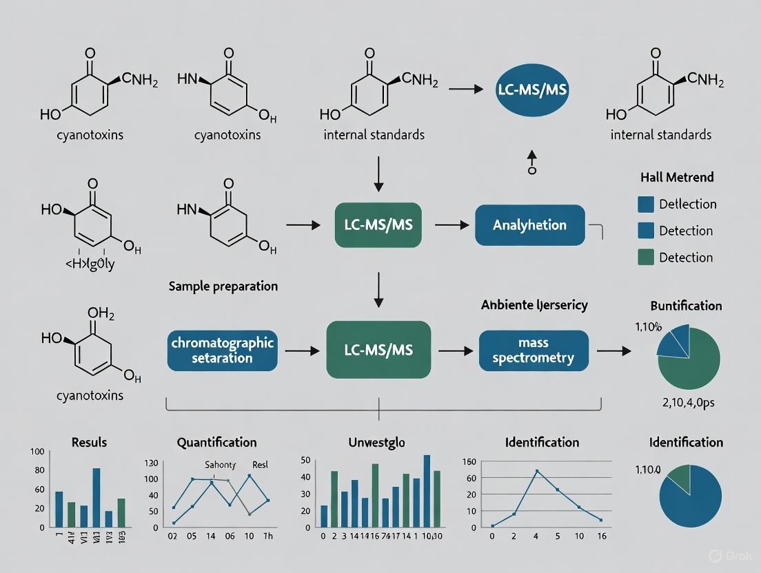
Figure 2: LC-MS/MS workflow for multi-class cyanotoxin analysis. The process from sample preparation to data acquisition is optimized for speed and specificity [5].
Quantification and Validation
- Calibration: Prepare a series of calibration standards (e.g., 0.1, 0.5, 1, 10, 50, 100, 500 μg/L) from certified stock solutions.
- Quality Control: Include procedural blanks and spiked samples (at low, mid, and high concentrations) with each batch of samples to monitor for contamination and assess accuracy/precision.
- Identification: Confirm analyte presence by matching the retention time and the relative abundance of the two MRM transitions with those of the calibration standard.
- Quantification: Use the primary MRM transition for quantification, based on the linear calibration curve.
The Scientist's Toolkit: Research Reagent Solutions
Table 4: Essential Reagents and Materials for Cyanotoxin Research
| Item | Function/Application | Examples / Notes |
|---|---|---|
| Certified Cyanotoxin Standards | Calibration and method validation for LC-MS/MS and other analytical techniques. | Microcystin-LR, Anatoxin-a, Cylindrospermopsin, Saxitoxin, Nodularin-R. Essential for accurate quantification [9] [5]. |
| Immunoassay Kits (ELISA) | High-throughput screening for specific toxin classes (e.g., MCs, ATX, CYN, STX). | ADDA-ELISA kits can detect over 100 microcystin congeners but cannot distinguish between them [9]. |
| LC-MS Grade Solvents | Mobile phase preparation and sample extraction to minimize background noise and ion suppression. | Water, methanol, and acetonitrile. Additives like formic acid are used to promote protonation in ESI+ [5]. |
| Solid Phase Extraction (SPE) Cartridges | Sample clean-up and pre-concentration of toxins from large water volumes. | C18 or polymeric sorbents. Note: The featured protocol uses a water-based extraction, simplifying biomass analysis [5]. |
| Chromatographic Columns | Analytical separation of cyanotoxin congeners prior to mass spectrometric detection. | Reversed-phase C18 columns (e.g., 100-150 mm length, sub-2 μm particles) for high-resolution separation [5]. |
The problem of cyanotoxins is intensifying, fueled by the synergistic effects of persistent nutrient pollution and the escalating impacts of climate change [6] [7] [4]. This expansion necessitates advanced analytical capabilities to accurately assess risk and guide mitigation efforts. The detailed LC-MS/MS protocol outlined herein provides researchers with a robust, rapid, and comprehensive method for monitoring a wide array of potent cyanotoxins in ambient freshwaters. By integrating such advanced analytical techniques with a deeper understanding of the environmental drivers, the scientific community can better inform public health protection, water resource management, and regulatory strategies in a changing global environment.
Cyanotoxins, toxic secondary metabolites produced by cyanobacteria, pose a significant and growing threat to water quality and public health worldwide. The proliferation of harmful cyanobacterial blooms (HCBs), driven by eutrophication and climate change, has increased the frequency and extent of human and animal exposure to these potent toxins [11]. Cyanotoxins exhibit remarkable structural and functional diversity, primarily classified into hepatotoxins and neurotoxins based on their target organs and mechanisms of action [12] [13]. This document provides detailed application notes and protocols for the analysis of four major cyanotoxin classes—microcystins, anatoxins, cylindrospermopsins, and saxitoxins—within the context of a broader thesis research project focusing on LC-MS/MS method development for cyanotoxin analysis in ambient freshwaters. The information is structured to assist researchers, scientists, and drug development professionals in implementing robust detection methodologies that address the complex analytical challenges presented by cyanotoxin diversity.
Cyanotoxin Classification and Chemical Diversity
Cyanotoxins are structurally diverse compounds that can be classified based on their chemical structures and primary toxicological mechanisms. Table 1 summarizes the major cyanotoxin classes, their chemical characteristics, and primary toxicity profiles.
Table 1: Structural Classification and Toxicity Profiles of Major Cyanotoxin Classes
| Cyanotoxin Class | Chemical Structure | Primary Toxicity | Common Variants | Key Structural Features |
|---|---|---|---|---|
| Microcystins (MCs) | Cyclic heptapeptide | Hepatotoxic [13] | 246+ identified; MC-LR, MC-RR, MC-YR most common [12] | Contain ADDA side chain; variable L-amino acids at positions 2 & 4 [12] |
| Anatoxins (ATXs) | Alkaloids [14] | Neurotoxic [13] | Anatoxin-a, Homoanatoxin-a, Dihydroanatoxin-a [15] | Low molecular weight; secondary amine structure; polar [15] |
| Cylindrospermopsins (CYNs) | Alkaloids [12] [14] | Hepatotoxic, Cytotoxic [13] | Cylindrospermopsin, 7-epi-CYL, 7-deoxy-CYL [15] | Tricyclic guanidine moiety coupled with hydroxymethyl uracil [12] |
| Saxitoxins (STXs) | Alkaloids [12] | Neurotoxic [13] | 57+ analogs [12] | Tetrahydropurine structure; carbamoyl, sulfamate, and hydroxyl substitutions |
The structural diversity within each class presents significant analytical challenges. Microcystins alone comprise over 246 structurally similar congeners that share the common ADDA side chain but differ in toxicity [12]. Anatoxins are particularly challenging due to their small molecular size, polarity, and potential for misidentification from interferences such as phenylalanine [15]. These variations necessitate highly selective analytical methods capable of distinguishing between congeners with different toxicological profiles.
Analytical Methodologies for Cyanotoxin Detection
Multiple analytical techniques are available for cyanotoxin detection, each with distinct advantages and limitations. While enzyme-linked immunosorbent assays (ELISA) provide rapid screening capabilities and do not require expensive equipment [9], they generally lack congener specificity and may exhibit variable cross-reactivities with different toxin variants [9]. Biochemical methods such as the protein phosphatase inhibition assay (PPIA) are well-suited for screening microcystins but cannot detect other toxin classes [9] [12]. Liquid chromatography coupled with mass spectrometry (LC-MS) has emerged as the gold standard for cyanotoxin analysis due to its high sensitivity, specificity, and ability to provide congener-specific information [9] [16].
LC-MS/MS as the Preferred Analytical Technique
Liquid chromatography tandem mass spectrometry (LC-MS/MS) offers distinct advantages for cyanotoxin monitoring in research and regulatory contexts. The technique provides high sensitivity with detection limits in the ng/L range, enabling compliance with stringent WHO guideline values (e.g., 1 μg/L for MC-LR in drinking water) [9] [11]. LC-MS/MS allows for multiplexed analysis of different cyanotoxin classes in a single run, though method development must account for their varying chemical and physical properties [15]. The structural confirmation capabilities through product ion scanning provide confidence in identifications, which is particularly important for distinguishing between congeners and avoiding false positives [15] [17].
Table 2 summarizes key performance metrics for LC-MS/MS methods across the four major cyanotoxin classes, demonstrating the technique's versatility for comprehensive cyanotoxin monitoring.
Table 2: Analytical Performance Metrics for LC-MS/MS Methods by Cyanotoxin Class
| Cyanotoxin Class | Representative Analytes | Limit of Detection (LOD) | Linear Range | Key Analytical Considerations |
|---|---|---|---|---|
| Microcystins | MC-LR, MC-RR, MC-YR [12] | 1-2.8 ng/g in biofilm matrices [15] | Wide dynamic range with R² >0.99 [17] | 246+ congeners; requires congener-specific standards; matrix effects significant |
| Anatoxins | Anatoxin-a, H₂ATX [15] | Sub-ng/g range in biofilm matrices [15] | Not specified in sources | Phenylalanine interference; polarity challenges; limited standards |
| Cylindrospermopsins | CYL, 7-epi-CYL [15] | 0.14 ng/g in biofilm matrices [15] | Not specified in sources | Multiple toxicity mechanisms; relatively few analogs |
| Saxitoxins | Various saxitoxin analogs | Not specified in sources | Not specified in sources | High polarity; HILIC chromatography preferred; marine origins |
Detailed LC-MS/MS Protocol for Multiclass Cyanotoxin Analysis
Sample Preparation and Extraction
Proper sample preparation is critical for accurate cyanotoxin quantification. For water samples, solid-phase extraction (SPE) effectively concentrates analytes and reduces matrix interference. Recent advancements include fully automated SPE workflows in 96-well plate formats that process 1-96 samples within an hour, reducing manual intervention by 90% while maintaining excellent accuracy [16]. This automated approach eliminates traditional drying and reconstitution steps, allowing direct LC-MS analysis of eluted samples and significantly improving throughput [16].
For complex matrices such as fish tissue, an optimized extraction protocol has been validated for multiple species. The method incorporates homogenization followed by extraction with methanolic solutions, cleanup steps, and analysis by UHPLC-MS/MS. This protocol has demonstrated limits of detection and quantification of 1 and 3 μg/kg respectively, with mean recovery of 70.0-120.0%, repeatability ≤12.6%, and intra-laboratory reproducibility ≤18.7% [17].
Instrumental Analysis Parameters
The following protocol, adapted from Zamlynny et al. [15], provides a validated approach for multiclass cyanotoxin analysis:
Chromatographic Conditions:
- Column: Reverse-phase C18 column (e.g., 100 × 2.1 mm, 1.7 μm)
- Mobile Phase A: Water with 0.1% formic acid
- Mobile Phase B: Acetonitrile with 0.1% formic acid
- Gradient Program: Optimized linear gradient from 5% B to 95% B over 15 minutes
- Flow Rate: 0.3 mL/min
- Column Temperature: 40°C
- Injection Volume: 5-10 μL
Mass Spectrometric Conditions:
- Ionization Mode: Electrospray ionization (ESI) positive mode for most cyanotoxins
- Source Temperature: 150°C
- Desolvation Temperature: 500°C
- Cone Gas Flow: 50 L/hr
- Desolvation Gas Flow: 1000 L/hr
- Collision Gas: Argon
- Data Acquisition: Multiple reaction monitoring (MRM) with optimized transitions for each analyte
Method Validation and Quality Assurance
Comprehensive method validation should include determination of limits of detection (LOD) and quantification (LOQ), linear range, accuracy, precision, and matrix effects. For multiclass methods, LODs typically range from 0.14 ng/g for cylindrospermopsin to 2.8 ng/g for certain microcystin congeners in wet biofilm matrices [15]. Accuracy should fall within 65-116% of reference values, demonstrating acceptable method performance across toxin classes [15]. Incorporation of internal standards, particularly stable isotope-labeled analogs when available, corrects for matrix effects and improves extraction recovery rates [16] [15]. Analytes should be identified based on both retention time and product ion ratio matching with available certified reference materials [15].
Experimental Workflow Visualization
The following diagram illustrates the comprehensive workflow for multiclass cyanotoxin analysis using LC-MS/MS, from sample collection to data interpretation:
Advanced Methodological Considerations
Matrix-Specific Analytical Challenges
Different sample matrices present unique challenges for cyanotoxin analysis. Benthic biofilm samples often contain high levels of organic matter that can cause significant matrix effects, requiring effective cleanup procedures and the use of matrix-matched calibration standards [15]. Biological tissues, such as fish liver and muscle, present additional complexities due to protein binding and the presence of co-extracted lipids, necessitating robust extraction and purification protocols [17]. For anatoxin analysis, particular attention must be paid to potential misidentification from phenylalanine, which shares identical nominal mass with anatoxin-a (both m/z 166) but can be distinguished by unique fragmentation patterns and retention times [15].
Emerging Cyanotoxins and Method Expansion
The continuous discovery of new cyanotoxins requires analytical methods to remain adaptable. Aetokthonotoxin (AETX), a recently identified cyanotoxin linked to avian vacuolar myelinopathy in bald eagles, represents an emerging analytical challenge [15]. Successful incorporation of AETX into multiclass methods requires synthesis of reference standards and characterization of its mass spectrometric behavior [15]. Similarly, the recent identification of 10-hydroxy anatoxin analogues necessitates method validation to ensure accurate detection and quantification of these emerging variants [15].
The Scientist's Toolkit: Essential Research Reagents
Table 3 catalogues essential reagents and reference materials required for implementing robust LC-MS/MS methods for cyanotoxin analysis.
Table 3: Essential Research Reagents for Cyanotoxin Analysis by LC-MS/MS
| Reagent/ Material | Function/Purpose | Specifications/Standards | Application Notes |
|---|---|---|---|
| Certified Reference Materials (CRMs) | Quantification and method validation | CRM-ATX, CRM-CYN, CRM-MCLR, CRM-NODR [15] | Essential for calibration; verify source and certification |
| Stable Isotope-Labeled Internal Standards | Correction for matrix effects and recovery | 13C4-(+)-anatoxin-a [15] | Improve accuracy and precision; use for each toxin class when available |
| LC-MS Grade Solvents | Mobile phase preparation | Optima LC-MS grade methanol/acetonitrile [15] | Minimize background noise and signal suppression |
| Acid Modifiers | Mobile phase additive | LC-MS grade formic acid (0.1%) [15] | Improves ionization efficiency in positive ESI mode |
| SPE Sorbents | Sample clean-up and concentration | C18, mixed-mode, polymeric sorbents [16] | Select based on toxin polarity and matrix complexity |
| Matrix Reference Materials | Quality control in complex matrices | RM-BGA (blue-green algal supplement) [15] | Verify method performance in relevant matrices |
Application in Environmental Monitoring and Public Health Protection
Effective cyanotoxin monitoring requires careful consideration of sampling strategies to accurately represent environmental contamination. Passive sampling devices offer time-integrated assessment of cyanotoxin presence and are particularly valuable for detecting trace-level contaminants that may be missed through grab sampling [11]. For public health protection, monitoring programs should prioritize water bodies used for drinking water sources and recreational activities, where human exposure risk is highest [13]. The established WHO guideline of 1 μg/L for microcystin-LR in drinking water provides a benchmark for method sensitivity requirements [11], though some agencies have implemented even stricter limits of 0.1–0.3 μg/L for sensitive populations [11].
Recent evidence of cyanotoxin bioaccumulation in aquatic organisms, particularly fish species used for human consumption, highlights the importance of extending monitoring beyond water samples to include biological tissues [17]. Studies have quantified microcystin concentrations up to 88.3 μg/kg in the liver and viscera of perch and sander, with smaller amounts (up to 6.1 μg/kg) detected in muscle tissue [17], indicating potential human dietary exposure routes that require further investigation.
The structural and functional diversity of cyanotoxin classes presents significant analytical challenges that can be effectively addressed through modern LC-MS/MS methodologies. This document has outlined comprehensive protocols and application notes for the detection and quantification of microcystins, anatoxins, cylindrospermopsins, and saxitoxins in ambient freshwater systems. The continued development and refinement of multiclass analytical methods remains essential for understanding cyanotoxin occurrence, environmental fate, and potential human health impacts. As cyanobacterial blooms increase in frequency and duration worldwide, robust analytical capabilities form the foundation for effective risk assessment and management strategies to protect public health and aquatic ecosystems.
Why LC-MS/MS? Overcoming the Limitations of Immunoassays and Biological Tests
The increasing frequency and magnitude of toxic cyanobacterial blooms in freshwater bodies constitute a major threat to public health and aquatic ecosystems globally [18]. These blooms produce a wide array of toxic metabolites known as cyanotoxins, which include hepatotoxic cyclic peptides (microcystins, nodularins) and neurotoxic alkaloids (cylindrospermopsins, anatoxins, saxitoxins) [18]. Accurate monitoring of these contaminants is essential for protecting public health through drinking water safety and recreational water quality management.
Traditional detection methods, particularly immunoassays and biological tests, have long served as primary tools for cyanotoxin analysis. However, these approaches harbor significant limitations that can compromise data accuracy and reliability. This application note demonstrates how Liquid Chromatography-Tandem Mass Spectrometry (LC-MS/MS) overcomes these constraints, providing unparalleled specificity, sensitivity, and comprehensiveness for cyanotoxin monitoring in ambient freshwaters.
Limitations of Traditional Cyanotoxin Detection Methods
Immunoassays: Selectivity and Cross-Reactivity Challenges
Enzyme-Linked Immunosorbent Assay (ELISA) represents one of the more commonly utilized cyanotoxin testing methods due to its operational simplicity and minimal equipment requirements [9]. However, this technique faces fundamental limitations:
Limited Selectivity: ELISA kits generally have limitations in selectivity and are not congener specific [9]. The microcystins/nodularins (ADDA) kit, for instance, is designed to detect over 100 microcystin congeners but cannot distinguish between them [9]. This lack of congener-specific data is problematic since the toxicity of individual microcystins is significantly affected by their amino acid constituents [19].
Variable Cross-Reactivity: The ability of ELISA to recognize different variants or congeners of cyanotoxins can vary quantitatively due to different cross-reactivities [9]. This variability can lead to either overestimation or underestimation of total toxin concentrations, potentially biasing risk assessments.
Antibody Dependency: The technique's reliance on antibodies introduces vulnerabilities like batch-to-batch variability and cross-reactivity with non-target compounds [20]. These drawbacks can impede accuracy, particularly in complex biological matrices where subtle differences between closely related molecules hold critical importance.
Biological Assays: Quantitative and Specificity Limitations
Biological tests, including Protein Phosphatase Inhibition Assays (PPIA) and mammalian toxicity assays, provide functional toxicity information but face significant analytical challenges:
Lack of Compound Specificity: These assays respond to classes of compounds with similar biological activity but cannot identify or quantify specific cyanotoxin congeners [9].
Matrix Interference: PPIA results can be biased by interfering substrates present in environmental samples [19], complicating accurate quantification.
Ethical and Practical Concerns: Mammalian toxicity assays (e.g., mouse bioassay) raise ethical concerns and provide limited quantitative data for risk assessment [9].
Table 1: Comparison of Cyanotoxin Detection Method Capabilities
| Feature | ELISA | Biological Tests | LC-MS/MS |
|---|---|---|---|
| Congener Specificity | Limited to non-existent | None | High for all toxin classes |
| Cross-reactivity Issues | Significant concern | Not applicable | Minimal |
| Multi-toxin Screening | Class-specific only | Varies by assay | Excellent (18+ toxins simultaneously) |
| Quantitative Accuracy | Moderate, affected by cross-reactivity | Semi-quantitative at best | High precision and accuracy |
| Structural Information | None | None | Detailed molecular characterization |
| Throughput | High | Low to moderate | Moderate to high |
| Method Development Time | Short | Short | Longer initial development |
The LC-MS/MS Advantage: Technical Foundations
Liquid Chromatography-Tandem Mass Spectrometry (LC-MS/MS) has emerged as the gold standard for cyanotoxin analysis, revolutionizing the field with its precision, sensitivity, and specificity [20]. This technique combines the physical separation capabilities of liquid chromatography with the mass analysis power of tandem mass spectrometry.
Principle of Operation
LC-MS/MS operates through a multi-stage process:
Chromatographic Separation: Toxins are first separated by liquid chromatography based on their chemical properties and interaction with the chromatographic stationary phase.
Ionization: Separated compounds are ionized (typically by electrospray ionization) to create gas-phase ions.
Mass Selection: The first quadrupole mass analyzer selects ions of a specific mass-to-charge ratio (precursor ions).
Fragmentation: Selected ions are fragmented in a collision cell using inert gas, creating product ions.
Product Analysis: The second mass analyzer filters the product ions for detection.
This two-stage mass analysis provides exceptional specificity by monitoring both the parent ion and unique fragment ions, creating a highly specific "mass fingerprint" for each compound [20] [21].
Key Advantages for Cyanotoxin Analysis
High Specificity: LC-MS/MS's ability to differentiate between molecular isoforms, modifications, and structurally similar compounds far exceeds the capabilities of immunoassays [20]. This precision is invaluable for distinguishing between cyanotoxin congeners with varying toxicities.
Multi-class Capability: Modern LC-MS/MS methods can simultaneously screen for multiple cyanotoxin classes. Recent research demonstrates methods capable of detecting 18 cyanotoxins simultaneously, including anatoxin-a, homoanatoxin-a, cylindrospermopsin, nodularin, guanitoxin, multiple microcystins, and saxitoxins in a short acquisition time of 8 minutes [5].
Enhanced Sensitivity: LC-MS/MS provides superior sensitivity allowing for detection and quantification of molecules at significantly lower concentrations compared to ELISA [20]. This enables monitoring at levels relevant for public health protection, with detection limits for microcystins reaching 1.3-23.7 ng/L in water samples [19].
Structural Elucidation: Advanced LC-MS/MS techniques can identify previously unknown cyanotoxin metabolites and variants, contributing to the discovery of new toxicologically relevant compounds [5] [19].
Experimental Protocol: Multi-class Cyanotoxin Analysis in Ambient Freshwaters
Sample Collection and Preparation
Materials:
- Hydrophilic polypropylene filtration membranes (avoid nylon and polyethersulfone to prevent microcystin loss) [19]
- Solid Phase Extraction (SPE) cartridges (Strata X or Oasis HLB) [19]
- Stable Isotope-Labeled (SIL) internal standards (MC-RR-15N13, MC-LR-15N10, MC-LA-15N7) [19]
- LC-MS grade solvents (methanol, acetonitrile, water with 0.1% formic acid)
Procedure:
- Sample Collection: Collect water samples from multiple depths and locations within the water body. Preserve samples at 4°C during transport and process within 24 hours.
Filtration: Filter water samples through hydrophilic polypropylene membranes (0.45 μm) to remove particulate matter while maintaining high microcystin recoveries [19].
Solid Phase Extraction:
- Condition SPE cartridges with 10 mL methanol followed by 10 mL Milli-Q water.
- Load 100-1000 mL filtered water sample (volume dependent on expected toxin concentrations).
- Wash with 10 mL 10% methanol in Milli-Q water.
- Elute toxins with 10 mL methanol containing 0.1% formic acid.
- Evaporate eluate to dryness under gentle nitrogen stream and reconstitute in 100 μL initial mobile phase.
Cell Lysis for Benthic Cyanobacteria:
- For benthic mat samples, implement mechanical disruption (bead beating) combined with repeated extraction with 75% aqueous methanol.
- Use SIL internal standards to evaluate lysis efficiency and correct for matrix effects [19].
LC-MS/MS Analysis
Instrumentation:
- LC System: Ultra-high performance liquid chromatography system with binary pump, autosampler, and temperature-controlled column compartment
- Mass Spectrometer: Triple quadrupole mass spectrometer with electrospray ionization (ESI) source
- Analytical Column: C8 or C18 reversed-phase column (100 × 2.1 mm, 1.7 μm)
Chromatographic Conditions:
- Mobile Phase A: Water with 0.1% formic acid
- Mobile Phase B: Acetonitrile with 0.1% formic acid
- Flow Rate: 0.3 mL/min
- Column Temperature: 40°C
- Injection Volume: 10 μL
- Gradient Program: Optimized for separation of 18 cyanotoxins within 8 minutes [5]
Mass Spectrometric Parameters:
- Ionization Mode: Positive electrospray ionization
- Ion Source Temperature: 500°C
- Ion Spray Voltage: 5500 V
- Nebulizer Gas: 50 psi
- Heater Gas: 50 psi
- Curtain Gas: 35 psi
- Collision Gas: Medium
- Detection: Multiple Reaction Monitoring (MRM) with optimized transitions for each cyanotoxin
Table 2: Example MRM Transitions for Key Cyanotoxin Classes
| Toxin Class | Specific Congener | Precursor Ion (m/z) | Product Ion 1 (m/z) | Product Ion 2 (m/z) |
|---|---|---|---|---|
| Microcystins | MC-LR | 995.5 | 135.1 | 213.1 |
| MC-RR | 520.0 | 135.1 | 213.1 | |
| MC-LA | 910.5 | 135.1 | 213.1 | |
| Anatoxins | Anatoxin-a | 166.1 | 131.1 | 149.0 |
| Homoanatoxin-a | 180.1 | 131.1 | 163.1 | |
| Other Toxins | Cylindrospermopsin | 416.1 | 194.1 | 176.1 |
| Saxitoxin | 300.1 | 204.1 | 282.1 | |
| Nodularin | 825.5 | 135.1 | 213.1 |
Method Validation
To ensure reliable results, LC-MS/MS methods require comprehensive validation addressing these essential parameters [22] [23]:
Accuracy and Precision: Assess by spiking toxin-free matrix with known toxin concentrations at multiple levels. Acceptance criteria typically require accuracy within ±15% and precision with CV ≤15%.
Specificity: Verify no interference from matrix components at the retention times of target analytes.
Linearity: Establish calibration curves across the analytical measurement range (typically 1-1000 μg/L for most cyanotoxins) with coefficient of determination (R²) ≥0.99.
Recovery: Evaluate extraction efficiency using stable isotope-labeled internal standards, with acceptable recoveries typically 70-120%.
Matrix Effects: Quantify suppression or enhancement of ionization using post-column infusion or post-extraction addition techniques.
Limit of Quantification (LOQ): Define as the lowest concentration measurable with acceptable accuracy and precision, typically with signal-to-noise ratio ≥10:1.
Essential Research Reagent Solutions
Table 3: Key Research Reagents for LC-MS/MS Cyanotoxin Analysis
| Reagent/Material | Function | Application Notes |
|---|---|---|
| Stable Isotope-Labeled Internal Standards | Correct for extraction losses and matrix effects | Essential for accurate quantification; should represent different toxin classes (e.g., MC-RR-15N13, MC-LR-15N10) [19] |
| Hydrophilic-Lipophilic Balanced SPE Sorbents | Toxin concentration and sample cleanup | Oasis HLB or Strata X provide broad-spectrum retention; mechanism differs between sorbents [19] |
| Chromatographic Columns | Compound separation | C8 or C18 columns (100 × 2.1 mm, 1.7 μm) provide optimal separation for cyanotoxin mixtures |
| Mass Spectrometry Calibrants | Instrument mass accuracy calibration | Required before each analysis sequence; ensures accurate mass assignment |
| Toxin Reference Standards | Method calibration and quantification | Commercially available for common toxins; necessary for semi-quantification of non-available congeners |
Analytical Workflow and Method Validation
The following diagram illustrates the complete LC-MS/MS analytical workflow for cyanotoxin detection in freshwater samples, from sample preparation to data analysis:
The validation process is critical for generating reliable data. Series validation should be characterized as a dynamic, ongoing process that monitors method performance throughout the method's life cycle [23]. Key validation parameters include:
Calibration Verification: Each analytical series should include calibration standards verifying the analytical measurement range, with predefined pass criteria for signal intensity at the lower limit of quantification, calibration curve slope, intercept, and coefficient of determination [23].
Quality Control Samples: Include blank samples, replicates, and quality control samples at low, medium, and high concentrations throughout the analytical sequence to monitor performance.
Stability Assessment: Evaluate analyte stability in the sample matrix under storage and processing conditions to ensure integrity of results [22].
LC-MS/MS represents the current gold standard for cyanotoxin analysis in ambient freshwaters, effectively overcoming the fundamental limitations of immunoassays and biological tests. While the technique requires more sophisticated instrumentation and expertise than traditional methods, its unparalleled specificity, multi-toxin capability, and quantitative accuracy make it indispensable for comprehensive risk assessment and regulatory compliance.
The method's ability to simultaneously identify and quantify numerous cyanotoxin congeners, including previously unknown variants, provides researchers and water quality managers with the detailed data necessary for accurate health risk assessment and informed decision-making. As cyanobacterial blooms continue to increase in frequency and magnitude globally [18], adopting robust LC-MS/MS methodologies becomes increasingly critical for protecting public health and aquatic ecosystems.
Cyanobacterial Harmful Algal Blooms (CyanoHABs) represent a significant threat to water security, public health, and aquatic ecosystems worldwide. These blooms, caused by the rapid proliferation of toxin-producing cyanobacteria, have shown a marked increase in frequency and geographic distribution in recent decades, a trend exacerbated by eutrophication and climate change [24] [25] [26]. The detection and quantification of their toxic metabolites, cyanotoxins, are critical for risk assessment and management. This application note details the integration of advanced satellite-based monitoring with cutting-edge liquid chromatography-tandem mass spectrometry (LC-MS/MS) methodologies to provide a comprehensive framework for understanding CyanoHAB prevalence and distribution on a broad scale, specifically tailored for research in ambient freshwaters.
National and Global Prevalence of CyanoHABs
The scale of the CyanoHAB challenge is substantial, with impacts documented across diverse aquatic environments.
United States Distribution
In the United States, CyanoHABs are a pervasive issue affecting all 50 states [24]. Coastal waters experience a range of harmful algal poisoning syndromes, including Paralytic Shellfish Poisoning (PSP), Neurotoxic Shellfish Poisoning (NSP), and Ciguatera Poisoning (CP), in addition to cyanobacterial blooms [24]. The U.S. Environmental Protection Agency (EPA) has established a forecasting and monitoring network for large waterbodies, which currently provides weekly cyanobacterial bloom forecasts for 2,192 lakes in the contiguous United States that are resolvable by satellite technology [27]. This extensive coverage highlights the national significance of freshwater CyanoHABs.
Global Occurrence and Emerging Trends
Globally, the occurrence of CyanoHABs is a growing concern. For instance, a study on the Swedish west coast reported the presence of the cyanotoxin nodularin in edible bivalves, a finding not typical for that region, indicating the expanding reach of toxic blooms [25]. Furthermore, the recent identification of a new cyanotoxin, aetokthonotoxin (AETX), linked to mass mortalities of bald eagles in the Eastern United States, underscores the continuous emergence of new threats and the critical need for versatile monitoring methods [26]. Reports of harmful algal blooms have "drastically increased" over the past forty years, a trend attributed to a combination of pollution, coastal development, and improved detection capabilities [24].
Table 1: Documented Spatial Scale of CyanoHABs
| Geographic Scale | Documented Prevalence | Key Evidence |
|---|---|---|
| United States | Impacts all 50 states [24] | EPA forecasting for 2,192 satellite-resolvable lakes and reservoirs [27] |
| Coastal Waters | Multiple HAB poisoning syndromes [24] | PSP, NSP, ASP, Ciguatera, fish kills, marine mammal mortalities [24] |
| Europe (Sweden) | Occurrence in non-typical regions [25] | First evidence of Nodularin in mussels/oysters from the west coast [25] |
| Global | Widespread and increasing [24] [25] | Increased reports over the past four decades [24]; Blooms linked to eutrophication and climate [25] |
Forecasting and Monitoring CyanoHABs at Scale
Large-scale monitoring relies on a combination of remote sensing and in-situ data to predict bloom events.
Satellite-Based Forecasting
The multi-agency Cyanobacteria Assessment Network (CyAN) project leverages satellite data to provide near-real-time monitoring and 7-day probabilistic forecasts for CyanoHABs [27]. The forecasting model, which employs a Bayesian hierarchical structure (INLA), outperforms various machine learning and neural network models, achieving a prediction accuracy of 90% with 88% sensitivity and 91% specificity [27] [28]. A bloom is defined using a threshold of median lake chlorophyll-a ≥12 µg/L with cyanobacteria dominance, corresponding to the World Health Organization's Recreation Alert Level 1 [27] [28]. These forecasts are generated weekly from April through November and are accessible via an EPA dashboard [27].
Community-Based and Aerosol Monitoring
Complementing federal programs, community-led research initiatives are addressing data gaps, particularly in underserved rural areas. One such study in northeastern North Carolina deployed a network of low-cost PurpleAir sensors to measure fine particulate matter (PM({2.5})) and investigate its potential correlation with aerosolized emissions from CyanoHABs [29] [30]. While this specific study found PM({2.5}) variation was more closely associated with criteria air pollutants than satellite-based CyanoHAB indicators, it demonstrates a scalable community science model for high-resolution environmental monitoring [29].
Figure 1: Integrated Workflow for Large-Scale CyanoHAB Analysis. This diagram outlines the process from satellite data acquisition and forecasting to ground-truthing via field sampling and sophisticated LC-MS/MS analysis.
Analytical Core: LC-MS/MS for Cyanotoxin Analysis
Accurate identification and quantification of cyanotoxins are paramount for confirming satellite data and assessing public health risk. LC-MS/MS has emerged as the gold standard for this purpose.
The Need for Multi-Toxin Methods
Cyanobacteria produce a vast array of toxins with varying chemical properties. While initial detection may rely on rapid techniques like Enzyme-Linked Immunosorbent Assays (ELISA), these methods are not congener-specific and can struggle with selectivity [9] [31]. The development of multi-class LC-MS/MS methods allows researchers to simultaneously screen for a wide spectrum of cyanotoxins in a single analysis, providing a comprehensive toxin profile that is essential for accurate risk assessment [32] [26].
Advanced Multi-Class LC-MS/MS Protocols
Recent methodological advances have significantly expanded the capabilities of LC-MS/MS. The following protocol is synthesized from current research for the analysis of ambient freshwaters.
Protocol: Multi-Class Identification and Quantification of Cyanotoxins in Ambient Freshwaters
1. Sample Collection and Preparation
- Collection: Collect water samples in amber glass containers to prevent toxin adsorption and photodegradation [31].
- Preservation: Immediately cool samples on ice. Quench residual disinfectants (e.g., chlorine) with sodium thiosulfate or ascorbic acid if present. For extended hold times, freezing is appropriate [31].
- Extraction: For biomass, a simplified extraction procedure using lyophilized material can be employed, eliminating the need for complex solid-phase extraction in many cases [32].
2. LC-MS/MS Analysis
- Instrumentation: Liquid chromatography system coupled to a triple quadrupole mass spectrometer (LC-MS/MS) operating in Multiple Reaction Monitoring (MRM) mode [32] [9] [31].
- Chromatography: Utilize reversed-phase liquid chromatography. The method should be optimized to separate a wide range of cyanotoxins, including:
- Acquisition Time: Methods can be optimized for short run times, with one reported method achieving analysis of 18 cyanotoxins in 8 minutes [32].
3. Identification and Quantification
- Identification: Confirm analyte identity by matching both the retention time and the product ion ratio against certified reference standards [25] [26].
- Quantification: Use a linear calibration curve across the expected concentration range. For toxins without available standards, semi-quantitative analysis using the closest structural analogue is possible [25].
Table 2: Performance Characteristics of a Representative Multi-Class LC-MS/MS Method [32] [26]
| Parameter | Method Performance | Analytical Scope |
|---|---|---|
| Target Cyanotoxins | 18 analytes in an 8-min method [32]; Up to 39 analytes in extended panels [26] | Anatoxins, Microcystins, Cylindrospermopsin, Nodularin, Saxitoxins, Guanitoxin, Aetokthonotoxin [32] [26] |
| Limits of Detection (LOD) | e.g., 0.14 ng/g for CYN to 2.8 ng/g for [Dha7]MC-LR in biofilm [26] | Varies by analyte, matrix, and instrument sensitivity |
| Linearity | Linear over the full calibration range for most analytes [32] [25] | Demonstrated for a wide concentration range (e.g., 3.12–200 µg/kg) [25] |
| Key Innovation | First method to include Guanitoxin [32]; Expanded coverage of ATX analogues and AETX [26] | Resolves MC-LR-[Dha7] and MC-LR-[Asp3] as separate signals [25] |
The Scientist's Toolkit: Research Reagent Solutions
Successful implementation of the aforementioned protocols requires a suite of high-quality reagents and materials.
Table 3: Essential Research Reagents and Materials for LC-MS/MS Analysis of Cyanotoxins
| Item | Function / Application | Specifications & Examples |
|---|---|---|
| Certified Reference Materials (CRMs) | Critical for method calibration, quantification, and ensuring accuracy. | CRM for Anatoxin-a (CRM-ATX), CRM for Microcystin-LR (CRM-MCLR), CRM for Cylindrospermopsin (CRM-CYN) [26] |
| LC-MS/MS Solvents | Mobile phase preparation for chromatography. | Optima LC-MS grade Methanol and Acetonitrile [26] |
| Internal Standards | Correct for matrix effects and variability in sample preparation/injection. | Stable isotope-labeled standards, e.g., 13C4-(+)-Anatoxin-a [26] |
| Passive Sampling Devices | Time-integrated sampling of water to capture episodic toxin release. | Used for extracting cyanotoxins for subsequent LC-MS/MS analysis [26] |
| Quenching Agents | Preserve sample integrity by neutralizing disinfectants in collected water. | Sodium thiosulfate or ascorbic acid [31] |
Figure 2: LC-MS/MS vs. Alternative Detection Methods. This diagram contrasts the high-specificity, multi-toxin capability of LC-MS/MS with the rapid but less specific nature of screening methods like ELISA and PPIA.
Understanding the prevalence and distribution of CyanoHABs on a national and global scale requires a synergistic approach. Satellite-based monitoring and forecasting provide the macroscopic view necessary for early warning and prioritization. This large-scale spatial data, however, must be grounded by precise, congener-specific chemical analysis. Advanced multi-class LC-MS/MS methods represent the pinnacle of cyanotoxin analysis, offering the sensitivity, specificity, and comprehensive profiling capability required to protect public health and aquatic ecosystems effectively. As the challenge of CyanoHABs continues to grow, the integration of these powerful tools will be indispensable for researchers and environmental managers worldwide.
Multiclass Method Development: Expanding the Scope of Cyanotoxin Detection
The analysis of cyanotoxins in ambient freshwater is critical for safeguarding public health and ecosystem integrity. Traditional sample preparation, particularly solid-phase extraction (SPE), has long been the standard for concentrating and purifying samples prior to liquid chromatography-tandem mass spectrometry (LC-MS/MS) analysis. While effective, SPE presents significant limitations including time-consuming procedures, high solvent consumption, requirements for specialized equipment, and potential analyte loss during the multiple transfer steps [33] [34].
Recent methodological advances demonstrate a paradigm shift toward streamlined techniques that maintain data quality while significantly improving analytical efficiency. This application note details two such approaches—direct injection and simplified extraction protocols—that effectively move beyond traditional SPE for cyanotoxin analysis in freshwater samples, enabling faster response times for water quality monitoring without compromising sensitivity or accuracy.
Current Landscape & Methodological Comparison
Limitations of Traditional SPE
Conventional SPE, while robust for cyanotoxin extraction, involves multiple steps including cartridge conditioning, sample loading, washing, and elution, typically requiring 30-60 minutes per sample [33]. Even with optimization using C18 phases or graphitized carbon, recovery rates can vary significantly—from 22% for cylindrospermopsin to 94% for anatoxin-a [34] [35]. The process also consumes considerable volumes of high-purity solvents, generating waste and increasing analytical costs.
Emerging Streamlined Approaches
Table 1: Comparison of Cyanotoxin Sample Preparation Methods
| Method Characteristic | Traditional SPE | Direct Injection | Simplified Extraction |
|---|---|---|---|
| Sample Processing Time | 30-60 minutes [33] | <30 minutes [36] | ~15 minutes [5] |
| Sample Volume Required | 50-1000 mL [34] | 1 mL [36] | Lyophilized biomass [5] |
| Solvent Consumption | High (tens of mL) [33] | Low (<2 mL) [36] | Minimal (water-based) [5] |
| Equipment Needs | SPE vacuum manifold, cartridges | Standard LC-MS/MS | Standard lab equipment |
| Limits of Quantification | pg/L range [34] | 0.0075-0.075 ng/mL [36] | Not specified |
| Key Advantages | High pre-concentration, clean-up | Simplicity, speed | No specialized equipment, rapid |
| Reported Applications | Drinking water, environmental [34] | Drinking water [36] | Cyanobacterial biomass [5] |
Protocols for Streamlined Cyanotoxin Analysis
Direct Injection with Limited Clean-up
Principle: This approach leverages the high sensitivity of modern triple quadrupole MS systems to eliminate extraction and concentration steps, injecting minimally processed water samples directly [36].
Experimental Protocol:
Sample Collection: Collect water samples in amber glass containers to prevent photodegradation. For finished drinking water samples, immediately quench residual disinfectants (e.g., chlorine) using sodium thiosulfate or ascorbic acid [31] [37].
Sample Preservation: Cool samples immediately after collection and maintain at 4°C during transport and storage. If extended holding times are anticipated, freeze samples at -20°C with precautions to avoid container breakage [31] [37].
Cell Lysis (for intracellular toxins):
- Aliquot 950 µL of water sample into culture tubes.
- Add 50 µL of appropriate internal standard solution (e.g., L-phenylalanine-d5 for anatoxin-a, uracil-d4 for cylindrospermopsin).
- Vortex for 1 minute.
- Store at -20°C for 1 hour, then thaw in a water bath for 15 minutes.
- Repeat freeze-thaw cycle twice more to ensure complete cell lysis [36].
Clarification: Filter samples through a PVDF syringe filter (0.22 µm, hydrophilic) to remove particulate matter.
Sample Dilution: Dilute filtered sample 1:1 by volume with LC-MS grade acetonitrile to match initial mobile phase composition.
LC-MS/MS Analysis:
- Instrumentation: SCIEX 7500 system or equivalent triple quadrupole MS
- Column: Phenomenex Synergi Polar-RP (100 Å, 100 × 3.0 mm, 2.5 µm)
- Mobile Phase: A) Water with 0.1% formic acid; B) Acetonitrile
- Gradient: 5% B to 95% B over 14 minutes
- Flow Rate: 0.400 mL/min
- Injection Volume: 10 µL
- Detection: Multiple reaction monitoring (MRM) in positive ESI mode [36]
Performance Characteristics: This method achieves LOQs of 0.0075-0.075 ng/mL for microcystins, anatoxin-a, and cylindrospermopsin, with accuracy of ±30% and precision <11% CV in drinking water matrices [36].
Simplified Extraction for Cyanobacterial Biomass
Principle: This method replaces organic solvent-based extraction with water-based extraction of lyophilized cyanobacterial biomass, eliminating the need for SPE entirely while maintaining comprehensive toxin coverage [5].
Experimental Protocol:
Sample Collection and Preparation:
- Collect cyanobacterial bloom material from surface waters using plankton nets or grab samples.
- Immediately freeze samples on dry ice or in liquid nitrogen.
- Lyophilize samples until completely dry (typically 24-48 hours).
Water-Based Extraction:
- Weigh 10-50 mg of lyophilized cyanobacterial biomass into a centrifuge tube.
- Add appropriate volume of ultrapure water (typically 1-5 mL) based on expected toxin concentrations.
- Vortex vigorously for 1 minute to suspend biomass.
- Sonicate in a water bath for 10 minutes at room temperature.
- Centrifuge at 10,000 × g for 10 minutes to pellet insoluble material.
- Transfer supernatant to a clean vial for analysis.
Alternative Extraction Optimization:
LC-MS/MS Analysis:
- Instrumentation: Standard LC-MS/MS system with multiple reaction monitoring capability
- Chromatography: Reversed-phase separation with rapid gradient (8 minutes total acquisition time)
- Analyte Coverage: 18 cyanotoxins including anatoxin-a, homoanatoxin-a, cylindrospermopsin, guanitoxin, seven microcystins, and five saxitoxins [5]
- Quality Control: Use isotopically labeled internal standards where available to correct for matrix effects
Performance Characteristics: This streamlined approach enables simultaneous detection of 18 cyanotoxins in 8 minutes, with guanitoxin inclusion representing the first such reported method [5]. The simplified extraction reduces sample handling and eliminates SPE cartridge variability.
The Scientist's Toolkit: Essential Research Reagents
Table 2: Key Research Reagent Solutions for Streamlined Cyanotoxin Analysis
| Reagent/ Material | Function & Importance | Application Notes |
|---|---|---|
| Amber Glass Containers | Prevents photodegradation of light-sensitive cyanotoxins during sample collection and storage [31] [37] | Required for all sampling; plastic containers may adsorb certain cyanotoxins |
| Sodium Thiosulfate | Quenches residual disinfectants in finished drinking water samples that could degrade cyanotoxins [31] [37] | Critical for accurate quantification in treated water; use immediately upon sampling |
| PVDF Syringe Filters (0.22 µm) | Removes particulate matter and cells from water samples prior to direct injection [36] | Hydrophilic version recommended for aqueous samples; prevents column clogging |
| LC-MS Grade Acetonitrile | Protein precipitation and mobile phase component for direct injection methods [36] | High purity essential to minimize background noise and system contamination |
| Deuterated Internal Standards | Corrects for matrix effects and extraction efficiency variations [36] [26] | L-phenylalanine-d5 for anatoxin-a; uracil-d4 for cylindrospermopsin |
| Lyophilization Equipment | Preserves cyanobacterial biomass and facilitates water-based extraction [5] | Maintains toxin integrity before analysis; enables dry weight normalization |
| Cyanotoxin Certified Reference Materials | Method validation, calibration curves, and quality control [26] | Essential for accurate quantification; available from National Research Council Canada |
Analytical Performance & Validation
Streamlined methods demonstrate performance comparable to traditional approaches while offering significant efficiency improvements. Direct injection achieves detection limits between 0.0075-0.075 ng/mL, sufficient to meet or exceed US EPA health advisory levels of 0.3 ng/mL for microcystins and 0.7 ng/mL for cylindrospermopsin in drinking water [36]. The simplified water-based extraction successfully detects 18 cyanotoxins in cyanobacterial biomass with high selectivity and a rapid 8-minute chromatographic separation [5].
Method validation should include assessment of specificity, linearity, accuracy, precision, limits of detection (LOD), and limits of quantification (LOQ). For multiclass methods, acceptable accuracy typically ranges from 65-116% with precision <15% RSD across different matrices including water, biofilm, and dietary supplements [26]. Ion ratio confirmation with tolerances of ±30% provides additional confidence in compound identification [36].
The movement beyond solid-phase extraction represents a significant advancement in cyanotoxin analysis, addressing the critical need for rapid, reliable methods in environmental monitoring and public health protection. Direct injection and simplified extraction protocols detailed in this application note demonstrate that efficient sample preparation need not compromise data quality. By adopting these streamlined approaches, researchers and water quality professionals can enhance monitoring capabilities, reduce analytical costs, and accelerate response times to potentially harmful cyanobacterial bloom events. As LC-MS/MS technology continues to evolve toward greater sensitivity, further simplifications in sample preparation will undoubtedly emerge, driving the field toward increasingly efficient and accessible cyanotoxin monitoring solutions.
Chromatographic Optimization for Diverse Polar and Non-Polar Toxins
The analysis of cyanotoxins in ambient freshwaters is critical for safeguarding public and environmental health. The primary analytical challenge lies in the simultaneous extraction and quantification of toxins with widely divergent chemical properties, ranging from non-polar cyclic peptides like microcystins to highly polar alkaloids such as anatoxins and saxitoxins [38] [39]. Liquid chromatography-tandem mass spectrometry (LC-MS/MS) has emerged as the preferred technique for such multiclass analysis. This application note details optimized protocols for the simultaneous determination of a broad spectrum of cyanotoxins, meeting the rigorous demands of research and regulatory science.
Optimized Chromatographic and MS Parameters
Method development focused on achieving robust retention and separation for 18 cyanotoxins, including guanitoxin, in a rapid 8-minute acquisition time [5]. Key to this success was the selection of a chromatographic strategy that could accommodate hydrophilic and lipophilic properties without requiring dimension-switching or compromised MS sensitivity.
Core Chromatographic Conditions
- Column: C18 reverse-phase column (e.g., 100 mm x 2.1 mm, 1.7 µm) [5].
- Mobile Phase A: Deionized water with 0.1% formic acid.
- Mobile Phase B: Methanol or acetonitrile with 0.1% formic acid [5] [39].
- Gradient Elution: A linear gradient from 5% B to 95% B over 5.5 minutes, followed by a 1.5-minute re-equilibration, has been demonstrated to effectively elute a wide range of toxins [5].
- Flow Rate: 0.3 mL/min.
- Injection Volume: 5-10 µL.
- Column Temperature: 40 °C.
Mass Spectrometric Detection
Detection was performed using a triple quadrupole mass spectrometer with Electrospray Ionization (ESI) in both positive and negative modes, switching as needed for specific analytes [39] [26]. The use of Multiple Reaction Monitoring (MRM) provides high selectivity and sensitivity. Key MS parameters are summarized in Table 1.
Table 1: Optimized Mass Spectrometric Parameters for Key Cyanotoxin Classes.
| Toxin Class | Example Toxin | Ionization Mode | Precursor Ion (m/z) | Product Ion 1 (m/z) | Product Ion 2 (m/z) |
|---|---|---|---|---|---|
| Microcystins | MC-LR | Positive | 995.5 | 135.2 | 213.2 |
| Nodularin | NOD-R | Positive | 825.5 | 135.2 | 163.2 |
| Anatoxins | Anatoxin-a (ATX) | Positive | 166.1 | 149.1 | 131.1 |
| Saxitoxins | GTX-2,3 | Positive | 396.2 | 316.2 | 298.2 |
| Cylindrospermopsin | CYN | Positive | 416.2 | 176.2 | 194.2 |
| Guanitoxin | Guanitoxin | Positive | 256.1 | 159.1 | 131.1 |
Detailed Experimental Protocol
Sample Preparation and Extraction
A simplified, efficient extraction procedure suitable for lyophilized cyanobacterial biomass or filter-feeding organisms is recommended.
- Step 1: Homogenization. Lyophilize water samples or tissue samples and homogenize into a fine powder using a ball mill.
- Step 2: Extraction. Weigh 100 mg of homogenized sample into a centrifuge tube. Add 1 mL of a 100% methanol extraction solvent. The use of water-based extraction or pure methanol can effectively replace more complex solid-phase extraction methods for biomass samples [5].
- Step 3: Mixing and Centrifugation. Vortex mix for 1 minute, then place the tube in an ultrasonic bath for 10 minutes. Centrifuge at 14,000 x g for 10 minutes at 4°C.
- Step 4: Dilution and Filtration. Transfer the supernatant to a new tube. Dilute 1:5 with ultrapure water to ensure compatibility with the LC gradient starting conditions. Filter the diluted extract through a 0.22 µm PVDF or nylon syringe filter into an LC vial for analysis.
LC-MS/MS Analysis and Quantification
- Step 5: Instrument Calibration. Prepare a mixed stock solution of all target cyanotoxin standards. Create a calibration curve with at least 5 concentration levels, analyzed in duplicate. The linear range typically spans from 3.12–200 µg/kg for toxins in tissue matrices, though some lipophilic microcystins (e.g., MC-LF, MC-LW) may show lower linearity (R² ≤ 0.98) [39] [25].
- Step 6: Sample Analysis. Inject the processed samples using the chromatographic and MS parameters detailed in Section 2.
- Step 7: Identification and Quantification. Identify toxins by matching the retention time and ion ratio of the qualifier/quantifier transitions with those of the calibration standard. Use the external standard method for quantification.
The following workflow diagram illustrates the complete analytical procedure:
Figure 1: Analytical Workflow for Cyanotoxin Analysis.
Critical Method Performance Data
The described method has been rigorously validated for performance in complex matrices. Table 2 summarizes key validation parameters for a selection of critical toxins, demonstrating the method's robustness despite the chemical diversity of the analytes.
Table 2: Method Validation Parameters for Selected Cyanotoxins in Biological Matrix.
| Toxin | Linear Range (µg/kg) | Limit of Detection (LOD) (ng/g) | Limit of Quantification (LOQ) (µg/kg) | Accuracy (%) | Recovery (%) |
|---|---|---|---|---|---|
| Microcystin-LR | 3.12–200 [25] | 0.14 - 2.8 [26] | 50 [40] | 65 - 116 [26] | <70 (but stable for some toxins) [25] |
| Nodularin-R | 3.12–200 [25] | - | 50 [40] | - | <70 (but stable) [25] |
| Anatoxin-a | 3.12–200 [25] | - | 50 [40] | - | <70 (but stable) [25] |
| Cylindrospermopsin | 3.12–200 (Quadratic) [25] | 0.14 [26] | 50 [40] | 116 [26] | <70 (but stable) [25] |
The Scientist's Toolkit: Essential Research Reagents and Materials
Successful implementation of this multiclass method requires specific, high-quality reagents and materials. This section details the essential components of the analytical toolkit.
Table 3: Essential Research Reagent Solutions for Cyanotoxin Analysis.
| Item | Function / Application | Specification / Notes |
|---|---|---|
| Certified Reference Materials (CRMs) | Quantification and method validation. | Critical for anatoxin-a, cylindrospermopsin, microcystins (e.g., MC-LR, MC-RR), and nodularin-R [26]. |
| LC-MS Grade Solvents | Mobile phase and sample extraction. | Methanol and acetonitrile with 0.1% formic acid; minimizes background noise and ion suppression [5] [26]. |
| C18 Reverse-Phase UHPLC Column | Chromatographic separation of analytes. | 100-150 mm length, 2.1 mm internal diameter, sub-2 µm particle size for high-resolution separation [5]. |
| Mass Spectrometer Tuning Solution | Instrument calibration and performance verification. | Ensures optimal sensitivity and mass accuracy for MRM transitions. |
| In-house Cyanotoxin Reference Material | Quality control for non-commercial analogues. | E.g., [Leu1]MC-LY, used when CRMs are unavailable [26]. |
| Solid Phase Extraction (SPE) Cartridges | Sample clean-up and concentration (if required). | Can be omitted for biomass extraction using the described methanolic protocol [5]. |
Application in Environmental Monitoring
This optimized protocol is particularly suited for monitoring programs in ambient freshwaters. The method's ability to detect guanitoxin, a potent neurotoxin, and resolve critical microcystin congeners like MC-LR-[Dha7] and MC-LR-[Asp3] as separate MRM signals, represents a significant advancement [5] [25]. Application to environmental samples, such as those from the Swedish coast, has successfully quantified nodularin in bivalves at levels up to 397 µg/kg, highlighting the transfer of toxins through the aquatic food web [25]. The inclusion of aetokthonotoxin (AETX), an emerging cyanotoxin, further ensures the method's relevance for addressing current ecological threats [26].
Liquid chromatography-tandem mass spectrometry (LC-MS/MS) has become the preferred technique for the sensitive and specific detection of cyanotoxins in ambient freshwater ecosystems [39] [41]. The analysis of these toxins is crucial for environmental and public health, as harmful algal blooms (HABs) can produce multiple classes of cyanotoxins, posing risks to wildlife and humans through recreational and drinking water exposure [42] [43]. Among LC-MS/MS techniques, Multiple Reaction Monitoring (MRM) is particularly powerful for quantitative analysis. MRM methods achieve high selectivity by monitoring specific precursor ion → product ion transitions, effectively isolating the target analyte from complex sample matrices [42]. This application note details protocols for MRM-based detection of major cyanotoxin classes, enabling researchers to accurately assess the full toxic load of cyanobacterial blooms.
Cyanotoxin Classes and Analytical Challenges
Cyanotoxins comprise several classes with diverse chemical properties, complicating their simultaneous analysis. Microcystins (MCs), potent hepatotoxins with over 279 known analogues, are cyclic peptides containing a characteristic Adda amino acid [9] [39]. Anatoxins (ATXs), such as anatoxin-a and homoanatoxin-a, are neurotoxic bicyclic secondary amines [42]. Saxitoxins (STXs), also neurotoxic, are highly polar alkaloids that cause paralytic shellfish poisoning [9] [42]. A primary analytical challenge is developing a single method that effectively retains and separates both hydrophilic toxins like STXs and lipophilic toxins like MCs [42]. Furthermore, many cyanobacterial species produce multiple toxin classes simultaneously, necessitating comprehensive multiclass methods for accurate risk assessment [42].
MRM Transitions and Mass Spectrometric Parameters
The following table summarizes optimized MRM transitions for major cyanotoxin congeners, providing a core panel for high-selectivity analysis. These parameters form the basis for sensitive detection and quantification in complex environmental samples.
Table 1: Optimized MRM Transitions for Key Cyanotoxin Analysis
| Toxin Class & Congener | Precursor Ion (m/z) | Product Ion 1 (m/z) | Product Ion 2 (m/z) | Key Fragmentor (V) | Collision Energy (eV) |
|---|---|---|---|---|---|
| Microcystins (MCs) | |||||
| MC-RR | 520.0 [42] | 135.0 [42] | 213.1 [42] | Specific data not available in search results | Specific data not available in search results |
| MC-YR | 1,045.5 [42] | 135.0 [42] | 213.1 [42] | Specific data not available in search results | Specific data not available in search results |
| MC-LR | 995.5 [42] | 135.0 [42] | 213.1 [42] | Specific data not available in search results | Specific data not available in search results |
| Anatoxins (ATXs) | |||||
| Anatoxin-a (ANA-a) | 166.1 [42] | 149.0 [42] | 131.0 [42] | Specific data not available in search results | Specific data not available in search results |
| Homoanatoxin-a (HATX) | 180.1 [42] | 163.0 [42] | 145.0 [42] | Specific data not available in search results | Specific data not available in search results |
| Saxitoxins (STXs) | |||||
| Saxitoxin (STX) | 300.2 [42] | 204.2 [42] | 282.2 [42] | Specific data not available in search results | Specific data not available in search results |
| Decarbamoylsaxitoxin (dcSTX) | 257.2 [42] | 239.2 [42] | 220.2 [42] | Specific data not available in search results | Specific data not available in search results |
| Gonyautoxin-5 (GTX-5) | 380.2 [42] | 300.2 [42] | 204.2 [42] | Specific data not available in search results | Specific data not available in search results |
A significant advancement in MRM analysis is the use of in-source fragmentation to create unique transitions for epimers that are chromatographically challenging to resolve. For example, this technique has been successfully applied to develop distinct MRMs for pairs of saxitoxin epimers, enhancing the specificity of the analysis without requiring complete baseline separation [42].
Detailed Experimental Protocol for Multiclass Cyanotoxin Analysis
Sample Collection and Preparation
Proper sample preparation is critical for accurate cyanotoxin quantification, which includes both intracellular and extracellular fractions [41].
- Sample Collection: Water samples should be collected from relevant depths and locations within the water body. Phytoplankton or scum samples can be collected using plankton nets or by grabbing surface scums [39]. Preserve samples on ice and process them within hours of collection.
- Cell Lysis: For total toxin analysis (intracellular + extracellular), efficient cell lysis is mandatory. Cyanobacterial cells have thick, cross-linked peptidoglycan layers, requiring robust lysis methods [41].
- Freeze-Thaw Method: Subject samples to three sequential freeze-thaw cycles at -80°C to lyse cells [42].
- Chemical Lysis: As an alternative, use a detergent-enzyme cocktail (e.g., containing lysozyme and proteinase K) for near 100% lysis efficiency [41].
- Sonication: Probe sonication for ~5 minutes can achieve approximately 80% lysis efficiency but may risk fragmenting biomolecules if over-applied [41].
- Solid-Phase Extraction (SPE): For water samples with low toxin levels, concentrate using SPE cartridges (e.g., C18 or graphitized carbon). Condition cartridges with methanol and equilibrate with water. Load samples, wash, and elute toxins with a solvent like methanol containing 0.1% formic acid. Evaporate eluent to dryness under a gentle nitrogen stream and reconstitute in initial mobile phase for LC-MS/MS analysis [39].
- Filtration and Extraction: For cyanobacterial cells or bloom material, after lysis, filter the sample through glass microfiber filters [42]. The optimal extraction solvent for a multiclass analysis of MCs, STXs, and ATXs from cyanobacterial samples has been determined to be 80:20 acetonitrile:water with 0.1% formic acid [42]. Sonicate the sample for 20 minutes, then centrifuge at 5000 rpm for 15 minutes at 17°C. Collect the filtrate for analysis [42].
LC-MS/MS Analysis Using HILIC Separation
Hydrophilic interaction liquid chromatography (HILIC) is ideal for multiclass analysis due to its ability to retain both hydrophilic and semi-hydrophilic toxins.
- Chromatography Conditions:
- Column: Waters BEH Amide (2.1 × 100 mm, 1.7 µm particle size) [42].
- Mobile Phase: A) 2 mM ammonium formate in ultrapure water (pH 3.5); B) Acetonitrile + 0.25% (v/v) formic acid [42].
- Gradient Elution:
- 0.00 min: 90% B
- 3.00 min: 90% B (hold)
- 3.01 min: Begin gradient to lower %B (specific gradient profile not fully detailed in search results)
- Flow Rate: 0.5 mL/min [42]
- Column Temperature: 40°C [42]
- Injection Volume: 1 µL [42]
- Mass Spectrometry Conditions:
- Ion Source: Electrospray Ionization (ESI), positive mode for most cyanotoxins [42] [39].
- Instrument Operation: Triple quadrupole mass spectrometer operated in MRM mode [42].
- Source Parameters:
- Drying Gas Temperature and Flow: Optimize for your specific instrument.
- Nebulizer Pressure: Optimize for your specific instrument.
- Capillary Voltage: Optimize for your specific instrument.
- MRM Monitoring: Use the transitions listed in Table 1. For each analyte, monitor at least two MRM transitions to ensure correct identification (primary for quantification, secondary for confirmation) [42].
Method Validation
For reliable results, the analytical method should be validated for the following parameters [42] [39]:
- Linearity: Prepare a calibration curve with at least 5 concentration levels. The method should demonstrate a coefficient of determination (R²) ≥ 0.991 for most analytes [42].
- Limit of Detection (LOD) and Quantification (LOQ): Determine the LOD (lowest detectable level) and LOQ (lowest quantifiable level with acceptable precision and accuracy). For the described HILIC-MS/MS method, LODs between 0.00770 and 9.75 µg L⁻¹ have been achieved [42].
- Precision: Evaluate repeatability by analyzing replicates (n≥5) within a day and intermediate precision over different days. Percentage relative standard deviations (%RSD) for peak area should ideally be below 15% [42].
- Accuracy/Recovery: Perform spike-and-recovery experiments at multiple concentrations. Recoveries for cyanotoxins in bivalve tissues, for instance, can range from 75.6% to 117%, though some lipophilic toxins like MC-LF may show lower but stable recoveries (<70%) [42] [39].
- Specificity: Verify that the method can unequivocally identify the analyte in the presence of matrix components, confirmed by monitoring two MRM transitions per analyte [39].
The Scientist's Toolkit: Essential Research Reagents and Materials
Table 2: Key Research Reagents and Materials for Cyanotoxin Analysis by LC-MS/MS
| Item | Function / Application | Example / Specification |
|---|---|---|
| Certified Reference Standards | Quantification and method calibration; essential for confirming retention times and MRM transitions. | MC-RR, MC-LR, MC-YR, Anatoxin-a, Homoanatoxin-a, Saxitoxin, GTXs [42] [39] |
| HILIC Column | Chromatographic separation of multiclass cyanotoxins with differing polarities. | Waters BEH Amide (2.1 × 100 mm, 1.7 µm) [42] |
| LC-MS Grade Solvents | Mobile phase preparation; minimizes background noise and ion suppression. | Acetonitrile, Water, Formic Acid [42] |
| Additives for Mobile Phase | Modifies pH and ionic strength to improve chromatography and ionization. | Ammonium Formate [42] |
| Sample Preparation Supplies | Cell lysis, filtration, and extraction. | Spin filters (0.22 µm), Glass microfiber filters, Sonication equipment [42] [41] |
| Solid-Phase Extraction (SPE) | Sample cleanup and concentration for low-level detection in water. | C18 or Graphitized Carbon Cartridges [39] |
Application in Environmental Monitoring
The developed multiclass HILIC-MS/MS method with MRM detection has been successfully applied to analyze cultured cyanobacteria and environmental field samples, with microcystins and saxitoxins detected [42]. Such methods are crucial for identifying the co-occurrence of multiple toxin classes, which is common in blooms formed by species capable of producing several toxins [42]. This comprehensive approach provides a more accurate assessment of the total toxic risk than methods targeting a single toxin class. Furthermore, applying these methods to filter-feeding organisms like bivalves has revealed the accumulation of cyanotoxins such as nodularin, highlighting a potential pathway for human exposure and the need for expanded monitoring beyond water samples alone [39].
The application of MRM transitions in LC-MS/MS analysis provides a robust and highly selective framework for the simultaneous quantification of multiple cyanotoxin classes in ambient freshwaters. The protocols outlined here—from optimized sample preparation that includes effective cell lysis to HILIC chromatography and specific MRM transitions—enable researchers to overcome the challenge posed by the diverse physicochemical properties of these toxins. By adopting such multiclass methods, scientists and public health professionals can better monitor recreational water bodies, conduct ecological risk assessments, and investigate poisoning events, ultimately contributing to the protection of public health and aquatic ecosystems.
The analysis of cyanotoxins in ambient freshwaters is a critical component of public health and environmental safety research. The recent discovery of aetokthonotoxin (AETX), a potent neurotoxin linked to mass wildlife mortalities, presents new challenges for analytical chemists [26] [44]. Simultaneously, the continuous identification of novel congeners across cyanotoxin classes necessitates advanced analytical approaches capable of detecting both known and emerging compounds [45] [46]. This application note details validated liquid chromatography-tandem mass spectrometry (LC-MS/MS) methodologies for incorporating AETX and novel cyanotoxin congeners into comprehensive monitoring programs, providing researchers with practical tools for addressing these emerging analytical challenges.
Background
The Aetokthonotoxin Challenge
Aetokthonotoxin is a pentabrominated biindole alkaloid produced by the epiphytic cyanobacterium Aetokthonos hydrillicola [44] [47]. Unlike many cyanotoxins associated with planktonic blooms, AETX originates from epiphytic cyanobacteria growing on aquatic vegetation, particularly the invasive water thyme (Hydrilla verticillata) [44]. This toxin has been implicated in vacuolar myelinopathy (VM), a fatal neurodegenerative disease that has caused significant mortality events in bald eagles and other aquatic birds in the southeastern United States [26] [44] [47].
The unique brominated structure of AETX and its epiphytic origin present distinct analytical challenges, as it is not covered by most existing multiclass cyanotoxin methods [26]. Furthermore, AETX production is dependent on bromide availability, which is linked to hyper-accumulation by the host plant, adding environmental complexity to toxin occurrence patterns [44] [47].
Proliferation of Novel Congeners
Beyond AETX, the expanding diversity of known cyanotoxin variants necessitates analytical approaches with broader coverage:
- Microcystins: Over 279 analogues identified, with continuous discovery of new variants [39] [46]
- Anatoxins: Recent identification of 10-hydroxy analogues and other variants [26]
- Structural Diversity: Novel chlorinated congeners and modified structures being reported in environmental samples [45]
The lack of commercial standards for most emerging congeners requires analytical approaches that can provide confident identification without reference materials [45] [46].
Materials and Methods
Research Reagent Solutions
Table 1: Essential Research Reagents for Cyanotoxin Analysis
| Reagent/Category | Specific Examples | Function/Application |
|---|---|---|
| Certified Reference Materials | CRM-ATX, CRM-CYN, CRM-MCLR, CRM-NODR [26] | Method calibration and quantification |
| Internal Standards | 13C4-(+)-anatoxin-a, H2ATX [26] | Compensation for matrix effects and recovery |
| Solvents | Optima LC-MS grade methanol/acetonitrile [26] | Sample extraction and mobile phase preparation |
| Specialized Reagents | Sodium borohydride, formic acid [26] | Derivatization and mobile phase modification |
| Synthesis Materials | THF-d8, benzoic acid PS1 qNMR standard [26] | AETX standard preparation and quantification |
Multiclass LC-MS/MS Method for AETX and Analogues
Instrumentation Parameters
- LC System: Ultra-high performance liquid chromatography system capable of binary gradient separation
- MS Detector: Triple quadrupole mass spectrometer with positive electrospray ionization (ESI+)
- Data Acquisition: Multiple Reaction Monitoring (MRM) mode for targeted quantification [26] [48]
Chromatographic Conditions
- Column: C18 reverse-phase column (100 × 2.1 mm, 1.7-1.8 μm)
- Mobile Phase A: Water with 0.1% formic acid
- Mobile Phase B: Methanol or acetonitrile with 0.1% formic acid
- Gradient Program: Optimized for simultaneous separation of hydrophilic and lipophilic cyanotoxins [26] [39]
- Flow Rate: 0.2-0.4 mL/min
- Column Temperature: 40°C
- Injection Volume: 1-10 μL
Mass Spectrometric Detection
Table 2: MS/MS Parameters for Key Emerging Cyanotoxins
| Toxin Class | Precursor Ion (m/z) | Product Ions (m/z) | Collision Energy (V) |
|---|---|---|---|
| Aetokthonotoxin | 849.7 [26] | 771.7, 693.6 (proposed) | Compound-specific optimization required |
| Anatoxin-a | 166.1 [48] | 149.1, 131.1 | 20-25 |
| Cylindrospermopsin | 416.1 [26] | 194.1, 176.1 | 25-30 |
| Microcystin-LR | 995.5 [48] | 135.1, 213.1 | 40-45 |
| Nodularin-R | 825.5 [48] | 135.1, 213.1 | 40-45 |
Sample Preparation Protocol
Environmental Sample Collection
- Water Samples: Collect in amber glass or polyethylene bottles, preserve at 4°C
- Biofilm/Benthic Samples: Collect using appropriate substrates or sampling devices
- Biological Tissues: Homogenize under controlled conditions [39]
- Preservation: Freeze immediately at -20°C or lower until extraction
Extraction Procedure
The following protocol is adapted from validated methods for multiclass cyanotoxin analysis [26] [48]:
- Homogenization: Weigh 10-100 mg of sample (dry weight) or measure 10-100 mL water sample
- Primary Extraction: Add 1-10 mL of 75% (v/v) aqueous methanol
- Sonication: Sonicate for 15 minutes in a controlled temperature sonication bath
- Centrifugation: Centrifuge at 4000 × g for 10 minutes
- Secondary Extraction: Re-extract pellet with 75% methanol and n-butanol (1:1 ratio)
- Combination: Pool supernatants from all extraction steps
- Concentration: Evaporate under nitrogen stream at 40°C
- Reconstitution: Dissolve residue in 0.5-1.0 mL of 5% (v/v) aqueous methanol
- Filtration: Pass through 0.2 μm membrane filter prior to LC-MS/MS analysis
AETX-Specific Workflow
AETX Analysis Workflow
Quality Assurance and Validation
- Identification Criteria: Match retention time (±0.1 min) and ion ratio (±30%) with standards [26]
- Calibration: External calibration with matrix-matched standards when possible
- Quality Controls: Include procedure blanks, duplicates, and spiked samples in each batch
- Confirmation: Use product ion ratio matching for confident identification [26]
Results and Discussion
Method Performance Characteristics
Table 3: Validation Data for Multiclass Cyanotoxin Method Including AETX [26]
| Performance Parameter | AETX | CYN | ATX | MC Congeners |
|---|---|---|---|---|
| Limit of Detection (ng/g) | Method in development | 0.14 | 0.5-1.0 | 0.1-2.8 |
| Linear Range | Under evaluation | 3 orders of magnitude | 3 orders of magnitude | 3 orders of magnitude |
| Accuracy (% Recovery) | Validation ongoing | 116% | 85-115% | 65-110% |
| Precision (% RSD) | Validation ongoing | <15% | <15% | <15% |
Complementary PCR Detection Protocol
For early warning of potential AETX contamination, a molecular detection method for the producer organism has been developed [44] [47]:
Primer Design
- Target Genes: Three loci of the AETX biosynthetic gene cluster (aetA, aetB, aetD)
- Specificity: Designed from unique halogenase and nitrile synthase sequences
- Amplicon Size: 200-500 bp for robust amplification
PCR Protocol
- DNA Extraction: Use commercial kit for environmental samples
- Reaction Setup: 25 μL reaction volume with standard PCR components
- Cycling Conditions:
- Initial denaturation: 95°C for 5 min
- 35 cycles: 95°C for 30s, 55-60°C for 30s, 72°C for 45s
- Final extension: 72°C for 7 min
- Analysis: Agarose gel electrophoresis for amplicon detection
High-Resolution MS for Novel Congener Identification
For comprehensive screening beyond targeted analysis, high-resolution mass spectrometry (HRMS) provides powerful capabilities for novel congener identification [45] [46]:
Untargeted Workflow
Novel Congener Identification
Data Analysis Approach
- Spectral Libraries: Utilize CyanoMetDB mass list containing 2000+ cyanobacterial metabolites [45]
- Diagnostic Fragments: Monitor characteristic microcystin fragments (135, 213 m/z) [46]
- Computational Tools: Implement Python-based algorithms for putative structural assignment [46]
- Confidence Levels: Apply Schymanski scale for identification confidence [45]
Application to Environmental Monitoring
Field Study Results
The developed multiclass method has been successfully applied to various environmental matrices [26] [44]:
- Benthic Biofilms: Detection of ATX analogues in Microcoleus-dominated communities
- Epiphytic Consortia: AETX detection in Hydrilla-associated cyanobacteria
- Passive Samplers: Demonstration of method applicability for time-integrated monitoring
- Drinking Water Sources: Identification of cyanotoxin profiles in source waters
Geographical Distribution
PCR-based monitoring has revealed that toxigenic Aetokthonos spp. can colonize a broader array of aquatic plants beyond Hydrilla verticillata, including American water-willow (Justicia americana) [44] [47]. However, AETX accumulation appears to be host-dependent, potentially linked to bromide hyper-accumulation in specific plants.
Troubleshooting and Optimization
Common Challenges
- Ion Suppression: Mitigate through sample dilution or improved clean-up
- Retention Shift: Maintain consistent mobile phase pH and column conditioning
- Sensitivity Issues: Optimize source parameters and collision energies
- False Positives: Confirm with ion ratio matching and secondary transitions
AETX-Specific Considerations
- Standard Availability: Quantification requires synthesized AETX characterized by NMR [26]
- Stability: Evaluate stability in various matrices under different storage conditions
- Extraction Efficiency: Optimize solvent composition for brominated indole structure
The incorporation of emerging toxins like aetokthonotoxin and novel congeners into routine monitoring programs requires carefully developed and validated LC-MS/MS methods. The protocols detailed herein provide researchers with robust analytical tools for comprehensive cyanotoxin assessment in ambient freshwaters. The combination of targeted MS/MS with confirmatory criteria, complemented by HRMS screening and molecular detection methods, offers a layered approach suitable for both regulatory monitoring and research applications. As cyanotoxin diversity continues to expand, these adaptable frameworks will remain essential for protecting water quality and public health.
The comprehensive monitoring of cyanotoxins in ambient freshwaters presents a significant analytical challenge due to the vast structural diversity of these toxins and their dynamic occurrence in the environment. Traditional targeted methods, while sensitive and quantitative, are limited to a predefined set of analytes, potentially missing novel or unexpected toxins [49]. This application note details integrated analytical workflows that combine targeted, suspect, and non-targeted screening with in silico toxicological modeling to create a comprehensive framework for cyanotoxin assessment. These workflows, developed within the context of advanced LC-MS/MS research, enable the identification of known cyanotoxins, the annotation of suspected compounds, and the discovery of previously overlooked toxicants, thereby providing a more complete picture of the contaminant landscape in freshwater ecosystems.
The protocols described herein leverage complementary mass spectrometry approaches—from highly sensitive triple quadrupole systems for quantification to high-resolution accurate mass instruments for identification—to address the complex challenges of cyanotoxin analysis. Furthermore, by incorporating in silico toxicology protocols, these workflows facilitate the rapid hazard assessment of identified compounds, even in the absence of experimental toxicity data, supporting a more proactive approach to water quality and risk assessment [50].
Integrated Screening Workflow for Cyanotoxins
The following workflow diagram illustrates the synergistic integration of targeted, suspect, and non-targeted screening strategies with in silico modeling for a comprehensive cyanotoxin analysis.
Figure 1: Integrated analytical workflow for comprehensive cyanotoxin analysis, combining targeted, suspect, and non-targeted screening approaches with in silico modeling for risk assessment.
Experimental Protocols
Sample Preparation and LC-MS/MS Analysis
Protocol 1: Simplified Extraction of Cyanotoxins from Environmental Samples
- Sample Homogenization: Lyophilize water samples (100-500 mL) or cyanobacterial biomass. Homogenize the dry material to a fine powder using a ball mill or similar device [5].
- Water-Based Extraction: Weigh 50 mg of lyophilized sample into a centrifuge tube. Add 10 mL of ultrapure water and vortex vigorously for 1 minute.
- Extraction and Cleanup: Sonicate the mixture for 10 minutes in a water bath sonicator, then centrifuge at 10,000 × g for 10 minutes. Transfer the supernatant to a clean vial. This simplified water-based extraction effectively replaces traditional solid-phase extraction methods for many applications, reducing time and cost [5].
- Sample Storage: Store extracts at -20°C until LC-MS/MS analysis. For long-term storage, maintain at -80°C.
Protocol 2: Rapid LC-MS/MS Analysis for Multi-Class Cyanotoxins
Chromatographic Separation:
- Column: C18 reversed-phase column (e.g., 100 × 2.1 mm, 1.8 μm)
- Mobile Phase A: Water with 0.1% formic acid
- Mobile Phase B: Acetonitrile with 0.1% formic acid
- Gradient: Optimize for linear increase from 5% B to 95% B over 6 minutes, followed by re-equilibration
- Flow Rate: 0.4 mL/min
- Injection Volume: 5 μL
- Total Run Time: 8 minutes [5]
Mass Spectrometric Detection:
- Ionization: Electrospray ionization (ESI) in positive mode
- Instrumentation: Triple quadrupole mass spectrometer operating in multiple reaction monitoring (MRM) mode
- Source Parameters: Optimize for cyanotoxin classes (capillary voltage: 3.5 kV; source temperature: 150°C; desolvation temperature: 500°C)
- Acquisition: Program MRM transitions for 18 cyanotoxins including anatoxin-a, homoanatoxin-a, cylindrospermopsin, guanitoxin, microcystin variants (RR, LA, LR, LY, LW, YR), and saxitoxin analogs [5]
suspect and Non-Targeted Screening Protocols
Protocol 3: Suspect Screening Workflow with HRMS
- Liquid Chromatography: Utilize UHPLC systems with C18 columns (100 × 2.1 mm, 1.7 μm) for high-resolution separations. Employ a 15-minute gradient from 5% to 100% organic phase (methanol or acetonitrile) with 0.1% formic acid [49].
- High-Resolution Mass Spectrometry:
- Instrument: Q-TOF or Orbitrap mass spectrometer
- Acquisition: Data-independent acquisition (DIA) or data-dependent acquisition (DDA) mode
- Mass Resolution: >25,000 FWHM for accurate mass measurements
- Mass Accuracy: <5 ppm for elemental composition assignment [51]
- Data Processing:
- Convert raw files to open formats (e.g., mzML)
- Perform peak picking, deconvolution, and alignment
- Screen against suspect lists (e.g., NORMAN Suspect List Exchange) using exact mass (±5 ppm), isotope patterns, and retention time indices when available [52]
- Annotate compounds using MS/MS spectral matching to databases
Protocol 4: Non-Targeted Screening with Prioritization Strategies
- Feature Detection: Use software (e.g., MS-DIAL, XCMS) to detect chromatographic features in the HRMS data. Apply blank subtraction and replicate filtering to remove artifacts [52].
- Prioritization Strategies: Implement a multi-criteria approach to manage data complexity:
- Chemistry-Driven Prioritization: Apply mass defect filtering for halogenated compounds or homologue series detection [52]
- Process-Driven Prioritization: Compare spatial/temporal samples (e.g., upstream vs. downstream) to highlight relevant features [52]
- Effect-Directed Prioritization: Correlate features with biological response data or use virtual effect-directed analysis [52]
- Prediction-Based Prioritization: Calculate risk quotients using predicted concentrations and toxicities (e.g., MS2Tox) [52]
- Structure Elucidation: Use molecular networking, fragmenter tools, and combinatorial spectroscopy to propose structures for high-priority unknowns.
In Silico Toxicology Assessment
Protocol 5: Computational Toxicology Evaluation
- Data Gap Filling: For cyanotoxins and transformation products lacking experimental toxicity data, employ quantitative structure-activity relationship (QSAR) models to predict toxicological endpoints [50].
- Model Application:
- Utilize documented in silico toxicology protocols for major endpoints of concern (genetic toxicity, carcinogenicity, acute toxicity) [50]
- Apply complementary QSAR methodologies (statistical-based and expert rule-based) per ICH M7 guidelines for impurity assessment [50]
- Implement the FAIR principles (Findable, Accessible, Interoperable, Reusable) for predictive models to ensure reproducibility and regulatory acceptance [53]
- Risk Characterization: Integrate exposure estimates from MS data with hazard predictions from in silico models to calculate risk quotients and prioritize compounds for further testing [52].
Results and Data Presentation
Cyanotoxin Occurrence in Environmental Samples
Table 1: Cyanotoxin concentrations (μg/L) detected in six eutrophic lakes in China during summer 2022, demonstrating the prevalence of microcystins and the need for comprehensive monitoring strategies [54].
| Lake Name | Microcystins (Mean) | Microcystins (Range) | Anatoxin-a | Cylindrospermopsin | Dominant Microcystin Variants |
|---|---|---|---|---|---|
| Hulun Lake | 3.61 μg/L | 0.89-7.02 μg/L | Detected (ng/L) | Detected (ng/L) | MC-LR, MC-RR, MC-YR |
| Wuliangsuhai Lake | 0.13 μg/L | 0.09-0.21 μg/L | Not Detected | Detected (ng/L) | MC-LR, MC-RR |
| Chaohu Lake | 3.60 μg/L | 1.15-8.74 μg/L | Detected (ng/L) | Detected (ng/L) | MC-LR, MC-RR, MC-YR |
| Taihu Lake | 2.18 μg/L | 0.42-5.11 μg/L | Detected (ng/L) | Detected (ng/L) | MC-LR, MC-RR, MC-YR |
| Xingyun Lake | 0.57 μg/L | 0.21-1.34 μg/L | Not Detected | Detected (ng/L) | MC-LR, MC-RR |
| Dianchi Lake | 2.56 μg/L | 0.78-6.92 μg/L | Detected (ng/L) | Detected (ng/L) | MC-LR, MC-RR, MC-YR |
Analytical Method Performance
Table 2: Validation data for UPLC-MS/MS analysis of microcystins in algal food supplements, demonstrating method reliability for cyanotoxin quantification [40].
| Toxin | Spiked Concentration (μg/kg) | Recovery (%) | Repeatability (% RSD) | Reproducibility (% RSD) | LOD (μg/kg) | LOQ (μg/kg) |
|---|---|---|---|---|---|---|
| MC-RR | 50 | 80.00 | 4.20 | 13.26 | 22.5 | 50 |
| MC-YR | 50 | 95.20 | 4.80 | 11.20 | 22.5 | 50 |
| MC-LR | 50 | 98.40 | 3.20 | 9.80 | 22.5 | 50 |
| Nodularin | 50 | 88.90 | 5.10 | 12.45 | 22.5 | 50 |
The Scientist's Toolkit: Essential Research Reagents and Equipment
Table 3: Key research reagent solutions and instrumentation for advanced cyanotoxin analysis workflows.
| Category | Item | Function/Application |
|---|---|---|
| Chromatography | C18 Reversed-Phase Columns | Separation of cyanotoxins based on hydrophobicity [5] |
| Vanquish UHPLC Systems | High-pressure chromatographic separation for complex environmental samples [55] | |
| Mass Spectrometry | TSQ Altis/Quantis Triple Quadrupole MS | Sensitive targeted quantification of known cyanotoxins in MRM mode [55] |
| Orbitrap Exploris HRMS Instruments | High-resolution accurate mass measurements for suspect and non-targeted screening [55] | |
| Sample Preparation | Lyophilization Equipment | Sample concentration and preservation prior to extraction [5] |
| QuEChERS Extraction Kits | Efficient extraction and clean-up for complex matrices [49] | |
| Data Analysis | Compound Discoverer Software | Platform for suspect and non-targeted screening data processing [52] |
| NORMAN Suspect List Exchange | Database of suspected environmental contaminants for screening [52] | |
| In Silico Toxicology | QSAR Toolboxes | Prediction of toxicological properties based on chemical structure [50] |
| CompTox Chemicals Dashboard | Database of chemical properties and predicted toxicities [52] |
Workflow Integration and Data Analysis
The integration of multiple screening approaches creates a powerful framework for comprehensive cyanotoxin assessment. The following diagram illustrates the data analysis pathway and how information flows between the different screening tiers and in silico modeling.
Figure 2: Data analysis workflow showing integration of results from targeted, suspect, and non-targeted screening with in silico modeling for comprehensive risk assessment and decision support.
The integrated workflows presented in this application note demonstrate a powerful paradigm for comprehensive cyanotoxin assessment in ambient freshwaters. By combining targeted quantification of known toxins, suspect screening for anticipated compounds, and non-targeted discovery for novel toxicants, researchers can overcome the limitations of single-method approaches. The incorporation of in silico toxicology models further strengthens these workflows by providing a means for rapid hazard assessment of identified compounds, supporting risk-based prioritization even for substances lacking experimental toxicity data.
These advanced protocols, framed within the context of LC-MS/MS methods for cyanotoxin research, provide researchers and drug development professionals with robust methodologies for comprehensive contaminant assessment. The structured workflows, detailed experimental protocols, and data analysis strategies outlined herein facilitate the implementation of these integrated approaches, ultimately contributing to more effective monitoring and management of cyanotoxin risks in freshwater environments.
Overcoming Analytical Challenges: Matrix Effects, Interferences, and Congener Complexity
Anatoxin-a (ATX) is a potent low molecular weight neurotoxin produced by several cyanobacterial genera, including Anabaena, Planktothrix, and Oscillatoria [56]. It acts as a potent nicotinic agonist that causes muscle fasciculation, convulsions, and rapid death due to respiratory failure in exposed organisms[cite:3]. Forensic investigations of suspected ATX poisonings are frequently hampered by difficulties in detecting this toxin in biological matrices due to its rapid decay and, more critically, by its misidentification for the amino acid phenylalanine (Phe) during liquid chromatography-mass spectrometry (LC-MS) analysis[cite:1][cite:3]. This misidentification occurs because these compounds are isobaric (same nominal mass of 165 Da) and exhibit similar retention times in reversed-phase liquid chromatography[cite:1][cite:3]. The consequence of this analytical challenge was starkly demonstrated in a high-profile investigation of a young adult's death in the USA in 2002, where ATX was mistakenly identified using single quadrupole LC-MS, with the signal actually originating from Phe[cite:3]. This application note presents robust strategies to prevent the misidentification of ATX in forensic and environmental investigations, with a focus on LC-MS/MS methodologies suitable for ambient freshwater analysis.
Table 1: Key Characteristics of Anatoxin-a and Phenylalanine
| Parameter | Anatoxin-a | Phenylalanine |
|---|---|---|
| Chemical Nature | Bicyclic secondary amine neurotoxin [56] | Proteinogenic amino acid [56] |
| Nominal Mass | 165 Da [57] [56] | 165 Da [57] [56] |
| Exact Mass | 165.11536 [57] [56] | 165.07898 [57] [56] |
| LC Retention Time | Similar elution in reversed-phase LC [56] | Similar elution in reversed-phase LC [56] |
| Primary Interference Issue | Misidentification in single quadrupole MS due to isobaric nature and co-elution [56] | Signal misinterpreted as ATX, leading to false positives [56] |
The Analytical Challenge: Mechanisms of Interference
The core of the misidentification problem lies in the insufficient mass resolution of commonly used single quadrupole mass spectrometers. These instruments cannot distinguish between compounds sharing the same nominal mass, such as ATX and Phe. While high-resolution mass spectrometry (HRMS) can differentiate them based on their exact mass difference of approximately 0.03638 Da [57] [56], this capability is not inherent to many routine analytical labs. Furthermore, even with HRMS, co-elution can cause ion suppression and affect quantification. The fragmentation patterns of these molecules, while distinct, can appear superficially similar in product ion scans without proper optimization and reference standards, potentially leading to confirmation errors. This challenge is particularly acute in complex matrices like benthic cyanobacteria, stomach contents of intoxicated animals, or organic-rich freshwater, where Phe is often abundant and can dominate the signal at m/z 165 [56].
Resolving Strategies and Experimental Protocols
Several established and emerging LC-MS/MS strategies effectively resolve ATX from Phe interference. The following sections outline detailed protocols for the most robust and commonly applied techniques.
High-Resolution Accurate Mass Spectrometry (HRMS)
Hybrid quadrupole time-of-flight (QqTOF) mass spectrometry leverages the exact mass difference between ATX and Phe to provide unambiguous differentiation [57] [56].
Protocol: QqTOF-Based Differentiation of ATX and Phe
- Instrumentation: Liquid chromatography system coupled to a hybrid quadrupole time-of-flight mass spectrometer.
- Chromatography:
- Column: C18 reversed-phase column (e.g., 100 mm x 2.1 mm, 1.8 µm).
- Mobile Phase A: Water with 0.1% formic acid.
- Mobile Phase B: Acetonitrile with 0.1% formic acid.
- Gradient: Begin at 5% B, increase to 40% B over 8 minutes, followed by a wash and re-equilibration step [5].
- Flow Rate: 0.4 mL/min.
- Column Temperature: 40°C.
- Injection Volume: 1-10 µL.
- Mass Spectrometry (QqTOF):
- Ionization: Electrospray Ionization (ESI), positive mode.
- Source Conditions: Optimize for capillary voltage, desolvation temperature, and gas flows for maximum sensitivity for m/z 165.
- MS1 Acquisition: Acquire data in high-resolution mode (Resolving Power >20,000 FWHM). Center the acquisition on m/z 165 with a span sufficient to monitor both compounds.
- Data Processing: Extract ion chromatograms using a narrow mass extraction window (e.g., 5-10 ppm). Anatoxin-a is identified by its accurate mass of 165.11536 and phenylalanine by 165.07898 [57] [56].
- Confirmation: For definitive confirmation, use data-dependent or targeted MS/MS acquisition. Compare the high-resolution product ion spectrum of the analyte against a certified reference standard.
Tandem Mass Spectrometry (MS/MS and MS³)
The use of multiple stages of mass spectrometry provides characteristic fragmentation information that clearly distinguishes between ATX and Phe [57] [56]. This is the most accessible and robust method for triple quadrupole and ion trap instruments.
Protocol: LC-MS³ Method for ATX Using an Ion Trap Mass Spectrometer
- Sample Preparation: Extracellular toxins from water samples can be analyzed directly or after solid-phase extraction. Intracellular toxins from cyanobacterial biomass require extraction. A simplified water-based extraction of lyophilized cyanobacterial biomass can be used: homogenize ~10 mg of dry weight biomass with 1 mL of water, vortex, sonicate for 5-10 minutes, and centrifuge. Dilute the supernatant as needed [5].
- Instrumentation: Liquid chromatography system coupled to a quadrupole ion trap mass spectrometer.
- Chromatography: Use the chromatographic conditions described in Section 3.1, Protocol Step 2.
- Mass Spectrometry (Ion Trap):
- Ionization: ESI, positive mode.
- MS2 Method: For the precursor ion m/z 165, use an isolation width of 2 m/z and collision energy optimized to generate abundant product ions.
- MS3 Method: For unambiguous identification, select the primary fragment ion of ATX at m/z 147 for further fragmentation.
Table 2: Characteristic Fragmentation Patterns of ATX and Phe
| Compound | MS1 (Precursor) | MS2 Primary Product Ions | MS3 (from m/z 147) |
|---|---|---|---|
| Anatoxin-a | m/z 165 | m/z 147 (loss of H₂O), m/z 132, m/z 107 [57] [56] | m/z 130, and other characteristic ions [57] [56] |
| Phenylalanine | m/z 165 | m/z 120 (immonium ion) [56] | Not Applicable |
Chemical Derivatization
Chemical modification of the anatoxin-a molecule prior to analysis is an effective strategy to shift its mass and chromatographic properties away from those of phenylalanine.
Protocol: Fluorimetric Detection via NBD-F Derivatization
- Principle: Anatoxin-a, being a secondary amine, reacts with the fluorogenic reagent 4-fluoro-7-nitro-2,1,3-benzoxadiazole (NBD-F) to form a fluorescent derivative. Phenylalanine, a primary amine, reacts differently and does not interfere in the specific detection of the ATX derivative [56].
- Derivatization Procedure:
- To a dried-down sample or standard, add 50 µL of a 10 mM NBD-F solution in methanol and 50 µL of a 0.1 M sodium borate buffer (pH 8.0).
- Heat the mixture at 60°C for 2 minutes.
- Cool the reaction vial on ice and then add 100 µL of 0.1 M HCl to stop the reaction.
- Dilute the mixture with the LC mobile phase and inject [56].
- LC-Fluorescence Analysis:
- Column: C18 reversed-phase column.
- Mobile Phase: Appropriate gradient of water and acetonitrile or methanol.
- Detection: Fluorescence detection with excitation/emission wavelengths optimized for the NBD-ATX derivative (e.g., Ex ~470 nm, Em ~530 nm).
The Scientist's Toolkit: Essential Research Reagents
Table 3: Key Research Reagents and Materials for ATX Analysis
| Reagent / Material | Function / Application | Notes |
|---|---|---|
| Anatoxin-a Certified Reference Material (CRM) | Quantification and method calibration | Essential for accurate identification and quantification; available from suppliers like the National Research Council Canada (NRC) [15] [26]. |
| Phenylalanine Standard | Interference check and method validation | Used to confirm separation from ATX and validate method selectivity. |
| 13C4-(+)-Anatoxin-a Internal Standard | Compensates for matrix effects and losses | Improves quantitative accuracy in complex matrices [26]. |
| 4-Fluoro-7-nitro-2,1,3-benzoxadiazole (NBD-F) | Derivatizing agent for fluorimetric LC | Converts ATX into a fluorescent derivative to avoid Phe interference [56]. |
| C18 Reversed-Phase LC Columns | Chromatographic separation | Standard for cyanotoxin analysis; provides the initial separation of analytes. |
| Lyophilizer | Sample preparation | Preserves cyanobacterial biomass and concentrates samples for analysis [5]. |
The misidentification of anatoxin-a due to phenylalanine interference is a critical analytical pitfall in cyanotoxin research and forensic toxicology. Over-reliance on low-resolution MS without MS/MS data is a known risk factor [56]. The methodologies detailed in this application note provide a multi-layered defense. While high-resolution QqTOF MS delivers definitive exact mass discrimination, the more widely available LC-MS³ approach on an ion trap instrument offers a highly robust and accessible solution for unambiguous identification. Derivatization remains a valid orthogonal technique. For modern environmental monitoring, incorporating these resolved ATX analysis techniques into comprehensive multiclass LC-MS/MS methods is the most effective strategy to ensure public and ecological health protection against this potent neurotoxin.
Managing Complex Congener Profiles with Limited Commercial Standards
The analysis of cyanotoxins in ambient freshwaters is critical for protecting public and ecological health. A significant challenge in this field is accurately identifying and quantifying a vast array of toxin variants, known as congeners, when commercial analytical standards are available for only a limited subset. Cyanotoxins like microcystins can have over 100 known congeners [9], yet standards are often scarce. This application note details robust LC-MS/MS methods and strategic approaches to overcome this limitation, enabling reliable quantification and identification of complex congener profiles in freshwater research.
The Cyanotoxin Congener Challenge
Cyanotoxins are not single compounds but are comprised of multiple congeners with varying structures and toxicities. The microcystins/nodularins (ADDA) ELISA kit, for example, is designed to detect over 100 microcystin congeners but cannot distinguish between them [9]. This lack of congener-specificity can be a critical drawback for risk assessments, as congeners exhibit differing toxicological properties.
While Liquid Chromatography coupled with Mass Spectrometry (LC-MS) can precisely identify specific congeners, its effectiveness is constrained by the availability of purified standards [9]. Methods that utilize liquid chromatography combined with mass spectrometry (LC-MS) can precisely and accurately identify specific microcystin congeners for which standards are available. At this time there are only standards for a limited number of the known microcystin congeners [9]. This application note provides protocols to navigate this analytical gap.
Methodologies for Comprehensive Analysis
Standard-Dependent Quantitative Analysis
For congeners where standards are available, LC-MS/MS operated in Multiple Reaction Monitoring (MRM) mode offers the gold standard for sensitivity and specificity. The development of a multiclass LC-MS/MS method capable of detecting analytes from several toxin classes, including 17 microcystins, nodularin-R, three cylindrospermopsins, and 17 anatoxins, has been demonstrated [26].
Sample Preparation Workflow:
- Enzymatic Digestion: For complex matrices like fish tissue, enzymatic digestion can aid in extraction. A single-day protocol using this method showed recovery rates of 35–88% for various ciguatoxins (structurally analogous marine toxins) as determined by LC-MS/MS [58].
- Solid-Phase Extraction (SPE): Extracts are cleaned up by defatting and two successive SPE steps to remove matrix interferents [58].
- Concentration and Reconstitution: The purified extract is concentrated under a gentle stream of nitrogen and reconstituted in an injection-compatible solvent [26].
Table 1: Validation Parameters for a Multiclass LC-MS/MS Cyanotoxin Method in Cyanobacterial Biofilms
| Parameter | Performance Data | Toxins Exemplified |
|---|---|---|
| Limits of Detection (LOD) | 0.14 ng/g for CYN to 2.8 ng/g for [Dha7]MC-LR | Cylindrospermopsin (CYN), Microcystins (MCs) [26] |
| Accuracy | 65% for [Leu1]MC-LY to 116% for CYN | Microcystins, Cylindrospermopsin [26] |
| Linear Range | Evaluated using neat and matrix-matched standards | Multiple classes [26] |
Strategies for Congeners Lacking Commercial Standards
When standards are unavailable, the following strategies can be employed:
- Surrogate Standard Quantification: Use a available standard from the same toxin class (e.g., MC-LR) to quantify other congeners in that class (e.g., MC-LA) [26]. This provides a semi-quantitative estimate but must be interpreted with caution due to potential differences in ionization efficiency.
- High-Resolution Mass Spectrometry (HRMS): Techniques like LC/TOF-MS can determine the exact mass of an unknown ion, allowing for the tentative identification of congeners based on their molecular formula [9] [26].
- Tiered Identification Approach: A two-tier LC-MS/MS approach can be implemented for confident identification:
- Quantitative NMR (qNMR) for Novel Toxins: For newly identified cyanotoxins like Aetokthonotoxin (AETX), a calibrated solution can be prepared and quantified using 1H-NMR with an external calibrant like benzoic acid. This provides a primary standard for subsequent LC-MS analysis [26].
The Scientist's Toolkit: Research Reagent Solutions
Table 2: Essential Research Reagents for Cyanotoxin LC-MS/MS Analysis
| Reagent / Material | Function | Example & Note |
|---|---|---|
| Certified Reference Materials (CRMs) | Provides definitive identity and concentration for instrument calibration and quality control. | Available for common toxins (e.g., MC-LR, ATX, CYN) from suppliers like the National Research Council of Canada (NRC) [26]. |
| Stable Isotope-Labeled Internal Standards | Corrects for matrix effects and losses during sample preparation; essential for high-accuracy quantification. | e.g., 13C4-(+)-anatoxin-a; should be added to the sample at the earliest possible step [59] [26]. |
| Matrix-Matched Calibration Standards | Compensates for matrix-induced suppression or enhancement of the MS signal (matrix effects). | Prepared by spiking the analyte into a representative blank matrix (e.g., toxin-free biofilm extract) [60]. |
| Quality Control Materials | Monures the accuracy and precision of the analytical run over time. | In-house reference materials (e.g., RM-BGA) or characterized field samples [26]. |
Workflow and Identification Pathways
The following diagrams illustrate the core logical workflows for managing congener analysis with limited standards.
Analytical decision pathway for congener identification and quantification.
Process for developing a quantitative method for a novel cyanotoxin without a commercial standard.
Method Validation and Quality Assurance
Validating methods intended for use with limited standards requires tailored approaches. Key parameters and acceptance criteria, synthesized from general bioanalytical guidance for protein LC-MS/MS [59], are suggested below.
Table 3: Key Validation Parameters for LC-MS/MS Methods with Limited Standards
| Parameter | Recommended Approach / Acceptance Criteria |
|---|---|
| Lower Limit of Quantification (LLOQ) | Accuracy and precision within ±25% [59] |
| Calibration Standards | Accuracy within ±20% for all points except LLOQ [59] |
| Accuracy & Precision | Within 20% (LLOQ QC within 25%) across a minimum of 3 runs [59] |
| Selectivity/Specificity | Demonstrate no significant interference in 6-10 individual matrix lots; LLOQ accuracy within ±25% for ≥80% of lots [59] |
| Matrix Effect | IS-normalized matrix factor CV within 20% across 6-10 matrix lots [59] |
| Stability | Within 20% of nominal concentration [59] |
Effectively managing complex cyanotoxin congener profiles in ambient freshwaters is analytically demanding but achievable. By leveraging a combination of standard-dependent quantification, strategic surrogate approaches, and advanced HRMS techniques, researchers can generate high-quality data even with limited commercial standards. Robust sample preparation, method validation, and the use of appropriate reagent solutions form the foundation of a reliable LC-MS/MS protocol, ultimately supporting accurate risk assessment and protection of water resources.
Strategies for Matrix Effect Reduction in Diverse Water Samples
Matrix effects represent a significant challenge in liquid chromatography-tandem mass spectrometry (LC-MS/MS) analysis of cyanotoxins in ambient freshwater samples. These effects occur when co-eluting compounds from the sample matrix interfere with the ionization process of target analytes in the mass spectrometer interface, leading to either signal suppression or enhancement [61]. In environmental water analysis, matrix effects can originate from diverse sources including dissolved organic matter, humic substances, inorganic salts, phytoplankton pigments, and degradation products from biological activity [62] [63]. The variable composition of ambient freshwater matrices across different geographical locations and seasonal periods further complicates the development of robust analytical methods, necessitating comprehensive strategies to identify, quantify, and mitigate these effects to ensure accurate quantification of cyanotoxins at trace levels.
The analytical signal alteration caused by matrix effects can significantly impact method accuracy, precision, and sensitivity, potentially leading to both false positives and false negatives in regulatory monitoring [64] [61]. For cyanotoxin analysis, where compounds like microcystins pose significant human health risks at concentrations as low as 1 μg/L (the WHO provisional guideline value for MC-LR in drinking water), uncontrolled matrix effects can compromise data quality and subsequent risk assessment decisions [62] [65]. This application note presents validated protocols for assessing and minimizing matrix effects specifically within the context of multi-class cyanotoxin analysis in diverse freshwater environments, supporting the development of reliable monitoring approaches for researchers and analytical scientists.
Matrix Effect Assessment Protocols
Post-Extraction Spiking Method
The post-extraction spiking method provides a quantitative assessment of matrix effects by comparing analyte responses in neat solvent versus matrix-matched samples [61]. This approach directly measures the extent of ionization suppression or enhancement occurring during MS analysis.
Materials:
- Blank ambient water samples from monitoring sites (pre-screened for target cyanotoxins)
- Cyanotoxin analytical standards (minimum: MC-LR, CYL, ATX-a, NOD)
- LC-MS grade methanol and acetonitrile
- Formic acid (≥98% purity) and ammonium hydroxide (≥25% NH₃ basis)
- UPLC system with tandem mass spectrometer (preferably triple quadrupole)
Procedure:
- Prepare a mixed cyanotoxin standard solution in LC-MS grade methanol at a concentration that will yield appropriate detector response when spiked (typically 1-10 μg/L final concentration)
- Extract blank water samples using the intended extraction protocol (see Section 4.1)
- Divide each extracted sample into two aliquots:
- Matrix-matched standard: Spike with cyanotoxin mixture post-extraction
- Control extract: No spike added (matrix blank)
- Prepare equivalent concentration standards in pure mobile phase (neat standards)
- Analyze all samples using the LC-MS/MS method and record peak areas for each cyanotoxin
- Calculate matrix effect (ME) using the following equation: ME (%) = [(Peak area of matrix-matched standard - Peak area of control extract) / Peak area of neat standard] × 100
Interpretation:
- ME = 100% indicates no matrix effect
- ME < 100% indicates signal suppression
- ME > 100% indicates signal enhancement
- ME values outside 80-120% range typically require mitigation strategies [61]
Post-Column Infusion Analysis
The post-column infusion method provides a qualitative, chromatographic profile of matrix effects throughout the analytical run, identifying regions of ionization interference [61]. This approach is particularly valuable for method development as it guides chromatographic optimization to shift analyte retention away from problematic regions.
Materials:
- Cyanotoxin standard solution (2.5 μg/mL in methanol)
- Syringe pump for constant infusion
- Zero-dead-volume PEEK mixing tee
- Blank water extracts from various sampling sites
Procedure:
- Connect the syringe pump containing cyanotoxin standard solution to a mixing tee installed post-column but pre-ESI source
- Infuse the cyanotoxin standard at a constant flow rate (typically 5-10 μL/min)
- Program the LC system with the intended gradient method and inject a blank water extract
- Monitor multiple reaction monitoring (MRM) transitions for all target cyanotoxins throughout the chromatographic run
- Document regions of signal suppression/enhancement observed in the baseline
- Repeat with blank extracts from different water sources to assess matrix variability
Data Analysis: The resulting chromatogram shows a stable baseline in the absence of matrix effects. Signal depression indicates regions where co-eluting matrix components suppress ionization, while signal elevation indicates enhancement regions. Method optimization should focus on shifting analyte retention times away from these identified interference regions through gradient adjustment or chromatographic selectivity changes [61].
Table 1: Comparison of Matrix Effect Assessment Methods
| Method | Detection Principle | Quantitative/Qualitative | Throughput | Key Applications |
|---|---|---|---|---|
| Post-Extraction Spiking | Comparison of analyte response in matrix vs. neat solution | Quantitative | Medium | Method validation, accuracy assessment |
| Post-Column Infusion | Monitoring signal changes during blank extract injection | Qualitative | Low | Method development, retention time optimization |
| Standard Addition | Response comparison in sample vs. sample with standard spikes | Quantitative | High | Routine analysis when matrix effects are unavoidable |
Comprehensive Matrix Effect Reduction Strategies
Sample Preparation and Cleanup Techniques
Solid-Phase Extraction (SPE) SPE effectively reduces matrix effects by selectively retaining target analytes while excluding interfering compounds. For cyanotoxins in freshwater, several sorbent chemistries have demonstrated efficacy:
- Hydrophilic-Lipophilic Balance (HLB) Polymers: Effective for a broad range of cyanotoxins from polar CYL to moderately non-polar MCs [64]. HLB cartridges (60 mg/3 mL) typically show >85% recovery for most cyanotoxins while significantly reducing matrix effects from humic substances.
- Graphitized Carbon Black (GCB): Particularly effective for removing pigment interferences from chlorophyll-rich samples [64]. Note: GCB may strongly retain planar cyanotoxin molecules, requiring optimization to prevent analyte loss.
- Primary Secondary Amine (PSA): Excellent for removing fatty acids and sugars from eutrophic water samples [64].
Protocol:
- Condition HLB cartridge with 3 mL methanol followed by 3 mL Milli-Q water
- Load 100-500 mL water sample (pH adjusted to 7.0 ± 0.5) at 5-10 mL/min flow rate
- Wash with 3 mL 5% methanol in water to remove polar interferents
- Elute cyanotoxins with 3 mL methanol containing 0.1% formic acid
- Evaporate eluent under gentle nitrogen stream at 40°C and reconstitute in 1 mL initial mobile phase
Dispersive-SPE (d-SPE) d-SPE provides a rapid cleanup approach suitable for high-throughput analysis, particularly when combined with QuEChERS extraction methodologies:
- Transfer 1 mL sample extract to 2 mL d-SPE tube containing 50 mg PSA + 50 mg C18 + 150 mg MgSO₄
- Vortex vigorously for 30 seconds
- Centrifuge at 10,000 × g for 2 minutes
- Transfer supernatant to autosampler vial for analysis
d-SPE with PSA/C18 combination typically reduces matrix effects by 60-80% for most microcystin congeners in complex freshwater samples [64].
Chromatographic Optimization Strategies
Alkaline Mobile Phase Conditions Implementation of alkaline chromatographic conditions (pH ~11) using ammonium hydroxide as mobile phase modifier has demonstrated significant reduction in matrix effects for lipophilic toxins compared to acidic conditions [66]. The alkaline environment improves separation of acidic cyanotoxins and reduces co-elution of interfering compounds.
Protocol for Alkaline Chromatography:
- Column: BEH C18, 100 × 2.1 mm, 1.7 μm particles
- Mobile Phase A: 2 mM ammonium hydroxide in water (pH ~10.5)
- Mobile Phase B: 2 mM ammonium hydroxide in acetonitrile
- Gradient: 10% B to 90% B over 7.5 minutes
- Flow rate: 0.4 mL/min
- Column temperature: 50°C
This approach reduced matrix effects from 55-76% signal suppression under acidic conditions to negligible levels (<10%) for okadaic acid and dinophysistoxin-1 in complex shellfish matrices, with similar benefits observed for cyanotoxins in freshwater [66].
Chromatographic Dilution Strategic sample dilution before injection represents a straightforward approach to reduce matrix effects when method sensitivity permits:
- Prepare sample extracts following standard protocol
- Serially dilute with initial mobile phase (1:2, 1:5, 1:10)
- Analyze diluted samples and monitor matrix effects
- Select the maximum dilution factor that maintains acceptable signal-to-noise ratio (>10:1 for quantitation)
Sample dilution of 1:5 to 1:10 typically reduces matrix effects by 40-70% while maintaining adequate sensitivity for cyanotoxins at regulatory limits [66] [61].
Advanced Calibration Techniques
Standard Addition Method The standard addition approach effectively compensates for matrix effects by preparing calibration standards in the actual sample matrix, making it particularly valuable for samples with highly variable or unpredictable matrix composition [62] [61].
Protocol:
- Divide the final sample extract into four equal aliquots (minimum 200 μL each)
- Spike three aliquots with increasing concentrations of cyanotoxin standard mixture
- Leave one aliquot unspiked (representing native analyte concentration)
- Analyze all aliquots and plot peak area versus spiked concentration
- Extrapolate the calibration line to determine native concentration in the unspiked sample
Standard addition provides accurate quantification even with matrix effects up to 80% suppression, though it requires additional analysis time and careful execution [62].
Matrix-Matched Calibration When sample volume is insufficient for standard addition, matrix-matched calibration using blank matrix from a different location can provide partial compensation:
- Identify and pool blank water samples from multiple sources
- Prepare calibration standards in the pooled blank matrix across the required concentration range
- Process and analyze calibration standards alongside samples using identical protocols
While not as effective as standard addition, matrix-matched calibration typically improves accuracy by 30-50% compared to solvent-based calibration in matrix-affected samples [64] [61].
Application to Cyanotoxin Analysis in Ambient Freshwaters
Comprehensive Multi-Toxin LC-MS/MS Protocol
Sample Collection and Preparation
- Collect 500 mL water samples in amber glass bottles, preserving with 1% (v/v) formic acid for microcystins or 0.1% (v/v) acetic acid for cylindrospermopsin
- Filter through GF/F filters (0.7 μm nominal pore size) to separate particulate and dissolved fractions
- Extract filters by sonication in 10 mL methanol for intracellular toxins
- Concentrate dissolved fraction using solid-phase extraction (Oasis HLB cartridges)
- Combine particulate and dissolved fractions for comprehensive toxin profiling
LC-MS/MS Analysis Instrumentation:
- UPLC system with binary pump and temperature-controlled autosampler
- Tandem quadrupole mass spectrometer with electrospray ionization
- Analytical column: ACQUITY UPLC BEH C18 (1.7 μm, 2.1 × 100 mm)
- Column temperature: 50°C
- Injection volume: 10-50 μL
Mobile Phase:
- A: 0.1% formic acid in water
- B: 0.1% formic acid in acetonitrile
- Gradient: 0 min (10% B), 1 min (10% B), 6 min (90% B), 7 min (90% B), 7.5 min (10% B)
- Flow rate: 0.4 mL/min
MS Conditions:
- Ionization mode: Positive ESI for most cyanotoxins (negative for CYN)
- Source temperature: 150°C
- Desolvation temperature: 600°C
- Desolvation gas: 1000 L/hr
- Capillary voltage: 3.0 kV
- Data acquisition: MRM mode with minimum 12 points per peak
This method enables simultaneous detection of 18 cyanotoxins including ATX-a, CYN, NOD, and multiple MC congeners in a short 8-minute acquisition time [5].
Method Validation Data
Table 2: Method Performance Characteristics for Selected Cyanotoxins in Freshwater
| Cyanotoxin | Retention Time (min) | LOD (ng/L) | LOQ (ng/L) | ME (%) Tap Water | ME (%) Eutrophic Lake | Recovery (%) |
|---|---|---|---|---|---|---|
| MC-LR | 4.2 | 2.5 | 8.5 | 95 | 65 | 92 |
| MC-RR | 3.8 | 3.1 | 10.3 | 98 | 71 | 88 |
| CYL | 2.1 | 5.2 | 17.3 | 102 | 89 | 85 |
| ATX-a | 2.8 | 1.8 | 6.0 | 97 | 78 | 90 |
| NOD | 4.5 | 2.2 | 7.3 | 94 | 62 | 86 |
Matrix effects (ME) were more pronounced in eutrophic lake water compared to tap water, necessitating implementation of the described mitigation strategies, particularly for microcystins and nodularin [62] [5] [39].
The Scientist's Toolkit: Essential Research Reagents
Table 3: Key Research Reagent Solutions for Cyanotoxin Analysis
| Reagent/ Material | Function | Application Notes | Alternative Options |
|---|---|---|---|
| Oasis HLB Cartridges | Broad-spectrum SPE cleanup | Optimal for simultaneous extraction of polar & non-polar cyanotoxins | Strata-X, Bond Elut PPL |
| Primary Secondary Amine (PSA) | d-SPE sorbent for pigment removal | Effective for chlorophyll-rich samples; may retain planar molecules | Z-Sep, C18 for lipophilic interferences |
| Ammonium Hydroxide | Alkaline mobile phase modifier | Reduces matrix effects for acidic compounds; use with compatible columns | Ammonium acetate, ammonium formate |
| Formic Acid | Acidic mobile phase modifier | Enhances [M+H]+ ionization; improves chromatography for basic compounds | Trifluoroacetic acid (may cause signal suppression) |
| Cyanotoxin Isotope-Labeled IS | Internal standards for quantification | Ideal for compensation of matrix effects; limited commercial availability | Structural analogs as surrogate standards |
Workflow Visualization
Figure 1: Comprehensive workflow for matrix effect management in cyanotoxin analysis, illustrating decision points for implementing mitigation strategies based on initial assessment results.
Figure 2: Matrix effect reduction strategies categorized by approach type, showing the relationships between major technique categories and specific methodologies.
Effective management of matrix effects is essential for generating reliable cyanotoxin data in diverse freshwater matrices. The combination of sample cleanup techniques, chromatographic optimization, and appropriate calibration strategies provides a comprehensive approach to address this challenge. For routine monitoring of known cyanotoxins, SPE cleanup with alkaline chromatography and matrix-matched calibration delivers robust performance. For exploratory research or highly variable samples, standard addition or isotope-labeled internal standards provide superior accuracy despite increased analytical time and cost. Implementation of these protocols will enhance data quality in cyanotoxin monitoring programs and support evidence-based risk assessment and management decisions for freshwater resources.
Passive Sampling Techniques for Time-Integrated Monitoring and Low-Level Detection
The increasing global prevalence of cyanobacterial harmful algal blooms (cyanoHABs) presents significant risks to aquatic ecosystems, drinking water supplies, and public health. Conventional water monitoring primarily relies on discrete "grab" sampling, which captures only a single moment in time and can easily miss ephemeral or transient toxic events [67]. Passive sampling techniques have emerged as a powerful complementary approach, providing time-integrated data capable of adsorbing dissolved contaminants over time and detecting low concentrations of cyanotoxins that traditional methods may overlook [68] [11]. When framed within research on LC-MS/MS methods for cyanotoxin analysis, these techniques enable more comprehensive contaminant profiling and more accurate risk assessment in ambient freshwater systems.
Principles and Advantages of Passive Sampling
Passive sampling devices, such as Solid Phase Adsorption Toxin Tracking (SPATT) samplers, operate on the principle of continuous adsorption of dissolved cyanotoxins from the water column over a defined deployment period [68]. This approach fundamentally differs from grab sampling by providing a cumulative measure of contaminant presence rather than a single snapshot.
The key advantages of this methodology include:
- Enhanced Detection Sensitivity: SPATT samplers can passively adsorb dissolved cyanotoxins over time, providing time-integrated data capable of detecting low concentrations of cyanotoxins that traditional discrete sampling may miss [68].
- Capturing Transient Events: Passive sampling captures episodic and transient bloom events that traditional discrete monitoring can easily miss, especially in large, dynamic systems like estuaries [67].
- Matrix Versatility: Passive samplers have been successfully deployed in various aquatic environments, including lakes, reservoirs, rivers, and estuaries, demonstrating broad applicability across freshwater systems [68] [69].
Table 1: Comparison of Cyanotoxin Monitoring Approaches
| Parameter | Grab Sampling | Passive Sampling |
|---|---|---|
| Temporal Resolution | Single point in time | Time-integrated (days to weeks) |
| Detection Sensitivity | May miss low-level or transient contamination | Capable of detecting low concentrations through analyte accumulation |
| Ephemeral Bloom Detection | Limited, unless sampling coincides with event | Effective at capturing transient events |
| Cost and Labor | Repeated sampling required for temporal coverage | Lower frequency of retrieval and analysis |
| Data Interpretation | Direct concentration measurement | Requires calibration for quantitative analysis |
Passive Sampling Protocols for Cyanotoxin Analysis
SPATT Sampler Deployment and Retrieval
The following protocol details the deployment of SPATT samplers for cyanotoxin monitoring, based on methodologies successfully implemented by the U.S. Geological Survey in collaboration with state environmental agencies [68].
Materials and Reagents:
- SPATT samplers containing appropriate adsorbent resin (e.g., HP700 for broad-spectrum cyanotoxin capture)
- Amber glass containers for sample transport
- Sodium thiosulfate or ascorbic acid (for quenching residual disinfectants if present)
- Coolers with ice packs for sample transport
- Field data loggers for recording temperature and environmental parameters
Procedure:
- Pre-deployment Preparation: Condition SPATT samplers according to manufacturer specifications, typically involving rinsing with methanol and ultrapure water.
- Field Deployment: Secure samplers in the water column at predetermined locations and depths, ensuring continuous exposure to flowing water while avoiding direct sediment contact.
- Deployment Duration: Typical deployment periods range from 7 to 14 days, though this may be adjusted based on site-specific conditions and monitoring objectives.
- Sample Retrieval: Carefully retrieve samplers and place them immediately in amber glass containers to minimize photodegradation of cyanotoxins.
- Preservation and Transport: Add appropriate preservatives if subsequent ELISA analysis is planned [68], chill samples immediately, and transport to the laboratory under cooled conditions (4°C).
Sample Extraction and Preparation for LC-MS/MS Analysis
Effective extraction of cyanotoxins from passive samplers is critical for accurate LC-MS/MS quantification. The following protocol has been validated for multi-class cyanotoxin analysis [5] [26].
Materials and Reagents:
- LC-MS grade methanol and acetonitrile
- LC-MS grade formic acid
- Ultrapure water (18 MΩ·cm)
- Certified reference standards for target cyanotoxins
- Internal standards (e.g., 13C4-(+)-anatoxin-a for anatoxin quantification)
Extraction Procedure:
- Extract Preparation: Remove adsorbent material from SPATT housings and transfer to appropriate extraction vessels.
- Solvent Extraction: Add 50 mL of 100% methanol to each sample and agitate for 60 minutes on an orbital shaker.
- Extract Concentration: Evaporate extracts under a gentle nitrogen stream at 40°C until near dryness.
- Reconstitution: Reconstitute samples in 1 mL of 50:50 methanol:water with 0.1% formic acid for LC-MS/MS analysis.
- Centrifugation: Clarify extracts by centrifugation at 14,000 × g for 10 minutes before LC-MS/MS analysis.
Figure 1: Workflow for passive sampling of cyanotoxins using SPATT samplers, illustrating the comprehensive process from deployment to data interpretation.
LC-MS/MS Analysis of Cyanotoxins from Passive Samplers
Method Development for Multi-Toxin Detection
Advanced LC-MS/MS methods enable simultaneous detection of multiple cyanotoxin classes from passive sampler extracts. Recent methodological advances have addressed the challenge of analyzing cyanotoxins with varying chemical properties within a single analytical run [5] [26].
Chromatographic Conditions:
- Column: C18 reversed-phase column (e.g., 100 × 2.1 mm, 1.7 μm)
- Mobile Phase A: Water with 0.1% formic acid
- Mobile Phase B: Acetonitrile with 0.1% formic acid
- Gradient Program: 5% B to 95% B over 8 minutes [5]
- Flow Rate: 0.4 mL/min
- Injection Volume: 5 μL
Mass Spectrometric Parameters:
- Ionization Mode: Electrospray ionization (ESI) positive and negative mode switching
- Source Temperature: 150°C
- Desolvation Temperature: 500°C
- Data Acquisition: Multiple reaction monitoring (MRM) for maximum selectivity
This optimized method can detect 18 cyanotoxins simultaneously, including microcystin congeners, anatoxin-a, cylindrospermopsin, saxitoxins, and emerging toxins like guanitoxin, in a short acquisition time of 8 minutes [5].
Quantitative Performance and Validation
Comprehensive method validation is essential for reliable cyanotoxin quantification from passive samplers. The following performance characteristics have been reported for multi-class cyanotoxin analysis:
Table 2: Performance Characteristics of LC-MS/MS Methods for Cyanotoxin Analysis
| Analyte Class | Representative Analytes | Limits of Detection (ng/g) | Linear Range | Accuracy (%) |
|---|---|---|---|---|
| Microcystins | [Dha7]MC-LR | 2.8 | 1-1000 μg/L | 85-115 |
| Cylindrospermopsins | CYN | 0.14 | 0.5-500 μg/L | 90-116 |
| Anatoxins | Anatoxin-a | 0.25 | 0.5-500 μg/L | 80-110 |
| Emerging Toxins | AETX | Method developed | Varies by analyte | Under validation |
Comparative Methodologies and Integrated Monitoring Approaches
Passive vs. Grab Sampling: Performance Comparison
Field studies demonstrate the superior performance of passive sampling for detecting cyanotoxins compared to traditional grab sampling. A comprehensive four-year monitoring study in the Sacramento-San Joaquin Delta highlighted these advantages under different hydrologic regimes [67].
Table 3: Field Comparison of SPATT versus Grab Sampling for Cyanotoxin Detection
| Monitoring Context | SPATT Sampling Results | Grab Sampling Results | Interpretation |
|---|---|---|---|
| Seneca Lake, NY | Consistent microcystin and anatoxin detection | Less frequent detection | SPATT provided more continuous contaminant profile [68] |
| Drought Conditions | Record microcystins (>1000 μg/L) | Detected contamination | Both methods effective during sustained blooms [67] |
| Wet Year Conditions | Microcystins detected | No microcystins detected at primary sites | SPATT captured transient events missed by grab sampling [67] |
Complementary Molecular Approaches
Passive sampling can be extended beyond cyanotoxin capture to include genetic markers of toxin-producing cyanobacteria. Recent research demonstrates that passive sampling of cyanobacterial DNA can detect toxin-producing genes not identified in concurrent grab samples [70].
Protocol for Genetic Passive Sampling:
- Deploy DNA-compatible passive samplers alongside SPATT devices
- Extract DNA from passive samplers using commercial kits
- Analyze via quantitative PCR (qPCR) for cyanotoxin gene targets (e.g., mcyE/ndaF for microcystin production)
- Correlate genetic results with chemical analysis from SPATT
This integrated approach provides early warning of cyanotoxin risk by identifying the potential for toxin production before toxins reach detectable concentrations [70] [37].
The Researcher's Toolkit: Essential Reagents and Materials
Successful implementation of passive sampling for cyanotoxin monitoring requires specific materials and reagents optimized for this application.
Table 4: Essential Research Reagents and Materials for Passive Sampling of Cyanotoxins
| Item | Function | Application Notes |
|---|---|---|
| SPATT Samplers | Passive adsorption of dissolved cyanotoxins | HP700 resin effective for broad-spectrum cyanotoxin capture [68] |
| Certified Reference Materials | Quantification and method validation | Essential for ATX, CYN, MC variants; limited availability for emerging toxins [26] |
| LC-MS Grade Solvents | Sample extraction and mobile phase preparation | Minimize background interference in mass spectrometric detection [5] |
| Stable Isotope Internal Standards | Quantitation accuracy and matrix effect compensation | 13C4-ATX corrects for signal suppression/enhancement [26] |
| DNA Extraction Kits | Genetic analysis of passive samples | Enables detection of toxin-producing genes as early warning tool [70] |
Figure 2: Integrated monitoring framework combining complementary approaches for comprehensive cyanotoxin risk assessment.
Application Notes and Implementation Guidelines
Case Study: Large-Scale Implementation
California's Freshwater and Estuarine HAB Program (FHAB) provides a successful model for implementing passive sampling in comprehensive monitoring strategies. The program incorporates three key recommendations particularly relevant to researchers [69]:
Cohesive Program Design: Monitoring programs should be cohesive across connected waterbodies and organizational boundaries, as demonstrated by the Klamath Basin Monitoring Program which provides basin-wide insights into HAB origin and transport dynamics.
Multiple Sampling Approaches: Combining water grab samples, passive sampling devices, and tissue samples (e.g., Asian clams) provides a more accurate assessment of contamination than any single approach.
Toxin Mixture Assessment: Monitoring should assess mixtures of both cyanotoxins and marine algal toxins in estuarine and coastal environments, as multiple toxins are frequently detected simultaneously.
Data Interpretation and Quality Control
Effective interpretation of passive sampling data requires consideration of several factors:
- Environmental Variables: Flow rates, temperature, and biofouling can influence sampling rates and require documentation.
- Quantitative Conversion: While excellent for presence/absence and temporal trend analysis, converting SPATT concentrations to water concentrations requires site-specific calibration.
- Comparative Analysis: ELISA results from passive sampler extracts often show higher cyanotoxin concentrations than LC-MS/MS, likely due to interference from dissolved organic matter and ELISA's ability to detect a broader range of congeners [68].
- Method Correlation: Establish correlation factors between passive sampling results and traditional water concentration measurements for your specific monitoring environment.
Passive sampling techniques represent a significant advancement in cyanotoxin monitoring, providing time-integrated data that captures the dynamic nature of cyanobacterial blooms and toxin production. When coupled with sophisticated LC-MS/MS analytical methods, these approaches enable comprehensive assessment of cyanotoxin occurrence, distribution, and temporal trends at sensitivity levels unattainable through discrete grab sampling alone. The protocols and application notes presented here provide researchers with a framework for implementing these powerful monitoring tools in diverse freshwater environments, ultimately supporting more effective water resource management and public health protection.
The increasing global prevalence of harmful cyanobacterial blooms (HCBs) poses significant risks to public health, ecosystems, and water security. Effective risk management requires monitoring programs capable of detecting low concentrations of cyanotoxins across diverse freshwater environments [71]. Traditional analytical methods often involve time-consuming sample preparation and extended chromatographic run times, creating bottlenecks for large-scale monitoring efforts. This application note details the development and implementation of high-throughput liquid chromatography-tandem mass spectrometry (LC-MS/MS) methods for the rapid, multi-class analysis of cyanotoxins in ambient freshwater. We present a streamlined workflow that reduces analysis time from conventional 20-minute methods to under 9 minutes while maintaining robust performance characteristics, enabling researchers to achieve greater analytical throughput for routine monitoring applications [5] [72].
Current Methodological Landscape
The United States Environmental Protection Agency (EPA) recognizes several analytical techniques for cyanotoxin detection, including enzyme-linked immunosorbent assays (ELISA), protein phosphatase inhibition assays (PPIA), and various chromatographic methods coupled with different detectors [9]. While immunological methods like ELISA offer rapid results without requiring expensive equipment, they generally lack congener specificity and may exhibit variable cross-reactivities with different toxin variants [9]. Liquid chromatography combined with mass spectrometry (LC-MS, LC-MS/MS) has emerged as the gold standard for cyanotoxin analysis due to its high sensitivity, specificity, and ability to identify and quantify multiple toxin classes simultaneously [9] [26]. Recent methodological advances have focused on expanding toxin coverage, simplifying sample preparation, and reducing analysis time to support more effective monitoring programs [5] [26] [16].
High-Throughput Workflow Implementation
Automated Sample Preparation
Efficient sample preparation is crucial for high-throughput analysis. Automation significantly enhances throughput while reducing manual intervention and potential errors.
Solid-Phase Extraction (SPE) Automation: A fully automated SPE workflow in a 96-well plate format enables preparation of up to 96 water samples within approximately one hour, representing a 90% reduction in manual intervention compared to traditional methods [16]. This approach:
- Eliminates drying and reconstitution steps typically required in traditional SPE protocols
- Utilizes only 5 mL of sample for triplicate analysis, minimizing solvent consumption
- Incorporates internal standards to correct for matrix effects and improve extraction recovery rates
- Has been validated for eight major microcystins and nodularin-R in various water matrices (river, lake, pond, and HPLC-grade water) [16]
Dual-SPE Cartridge Configuration: For comprehensive multi-class cyanotoxin analysis, a dual-SPE approach using Oasis HLB combined with Supelclean ENVI-Carb cartridges provides effective retention of cyanotoxins with diverse physicochemical properties [34]. Optimization studies demonstrate that Oasis HLB effectively retains microcystins and nodularin, while Supelclean ENVI-Carb shows superior retention for cylindrospermopsin and anatoxin-a [34].
Rapid LC-MS/MS Analysis
Chromatographic Acceleration: A rapid LC-MS/MS method enables simultaneous analysis of eighteen cyanotoxins in a shortened acquisition time of 8 minutes [5]. The method covers:
- Neurotoxins: anatoxin-a, homoanatoxin-a, guanitoxin (first reported detection), and five saxitoxins
- Hepatotoxins: cylindrospermopsin, deoxy-cylindrospermopsin, nodularin, and seven microcystins (RR, [D-Asp3]RR, LA, LR, LY, LW, YR)
Method Simplification: A key innovation involves replacing traditional solid-phase extraction with water-based extraction for lyophilized cyanobacterial biomass samples, significantly simplifying the analytical workflow while maintaining performance [5].
Further Throughput Enhancement: Building on this approach, method runtime can be reduced to just 9 minutes while maintaining coverage of 220+ compounds, representing a 50% reduction compared to previously established 20-minute methods [72]. This accelerated chromatography employs:
- Reversed-phase separation with sub-2µm particles for high efficiency
- Optimized gradient profiles for rapid elution
- Maintained chromatographic resolution for isobaric compound pairs (isoleucine/leucine)
Table 1: Performance Characteristics of Rapid LC-MS/MS Cyanotoxin Methods
| Parameter | 8-minute Method [5] | 9-minute Method [72] | Extended Multiclass Method [26] |
|---|---|---|---|
| Analysis Time | 8 minutes | 9 minutes | Not specified |
| Toxin Coverage | 18 cyanotoxins | 220+ compounds (broader metabolomics) | 17 microcystins, nodularin-R, 3 cylindrospermopsins, AETX, 17 anatoxins |
| Key Innovations | First guanitoxin detection; water-based extraction | Direct amino acid detection without derivatization; automated sample preparation | Includes emerging cyanotoxin AETX; 10-hydroxy anatoxin analogues |
| LOD in Matrix | Not specified | Not specified | 0.14 ng/g (CYN) to 2.8 ng/g ([Dha7]MC-LR) in wet biofilms |
| Application | Cyanobacterial biomass | Cell culture media (concept applicable) | Benthic/epiphytic biofilm, dietary supplements, passive samplers |
Experimental Protocol: High-Throughput Cyanotoxin Analysis
Sample Preparation (Automated SPE)
Materials:
- Oasis HLB cartridges (500 mg, 6cc) or 96-well plates
- Supelclean ENVI-Carb cartridges (500 mg, 6cc) for dual-SPE approach
- Automated liquid handling system (e.g., Andrew+ Pipetting Robot)
- Internal standards: 13C4-(+)-anatoxin-a, or other isotope-labeled cyanotoxins
Procedure:
- Sample Pre-treatment: Filter water samples through GF/F filters (0.7 µm) to remove particulate matter.
- SPE Conditioning: Condition Oasis HLB sorbents with 10 mL methanol followed by 10 mL Milli-Q water.
- Sample Loading: Load 100-500 mL of water sample at neutral pH at a flow rate of 5-10 mL/min.
- Cartridge Washing: Wash with 10 mL of 10% methanol in Milli-Q water.
- Analyte Elution: Elute with 10 mL of methanol heated to 50°C, or 5 mL of basified methanol (0.1% NH₄OH) for specific microcystins (MC-LW, MC-LF) [34].
- Evaporation and Reconstitution: Evaporate eluate to dryness under nitrogen stream and reconstitute in 100-200 µL of initial mobile phase.
LC-MS/MS Analysis
Chromatographic Conditions:
- Column: ACQUITY Premier HSS T3 (2.1 × 100 mm, 1.8 µm) or equivalent
- Mobile Phase A: 0.1% formic acid in water
- Mobile Phase B: 0.1% formic acid in acetonitrile or methanol
- Gradient Program:
- 0-1 min: 5% B
- 1-6 min: 5-95% B (linear gradient)
- 6-7 min: 95% B (hold)
- 7-7.1 min: 95-5% B
- 7.1-9 min: 5% B (re-equilibration)
- Flow Rate: 0.4-0.6 mL/min
- Injection Volume: 5-10 µL
- Column Temperature: 40°C
Mass Spectrometric Conditions:
- Ionization Mode: Electrospray ionization (ESI), positive mode
- Source Temperature: 150°C
- Desolvation Temperature: 500°C
- Cone Gas Flow: 50 L/hr
- Desolvation Gas Flow: 1000 L/hr
- Data Acquisition: Multiple reaction monitoring (MRM)
- Collision Gas: Argon
Table 2: Representative MRM Transitions for Key Cyanotoxin Classes
| Cyanotoxin Class | Example Compounds | Precursor Ion (m/z) | Product Ions (m/z) | Cone Voltage (V) | Collision Energy (eV) |
|---|---|---|---|---|---|
| Anatoxins | Anatoxin-a | 166.1 | 149.1, 131.1 | 30 | 15, 20 |
| Homoanatoxin-a | 180.1 | 163.1, 135.1 | 30 | 16, 22 | |
| Microcystins | MC-LR | 995.5 | 135.1, 213.1 | 60 | 40, 55 |
| MC-RR | 520.0 | 135.1, 213.1 | 70 | 60, 65 | |
| Cylindrospermopsins | CYL | 416.1 | 194.1, 176.1 | 50 | 30, 35 |
| Saxitoxins | GTX-2,3 | 396.1 | 316.1, 298.1 | 40 | 25, 30 |
The Scientist's Toolkit: Essential Research Reagents and Materials
Table 3: Key Research Reagent Solutions for Cyanotoxin Analysis
| Item | Function | Example Products/Specifications |
|---|---|---|
| SPE Sorbents | Extract and concentrate cyanotoxins from water samples | Oasis HLB (hydrophilic-lipophilic balanced copolymer); Supelclean ENVI-Carb (graphitized non-porous carbon) [34] |
| LC Columns | Chromatographic separation of cyanotoxin congeners | ACQUITY Premier HSS T3 (2.1 × 100 mm, 1.8 µm); C18 reverse-phase columns with high retention of polar compounds [72] |
| Certified Reference Materials | Method validation, calibration standards, quality control | Certified reference materials for anatoxin-a, cylindrospermopsin, microcystin variants, nodularin-R (available from National Research Council of Canada) [26] |
| Isotope-Labeled Internal Standards | Quantification correction, monitor analytical variability | 13C4-(+)-anatoxin-a; other deuterated or 13C-labeled cyanotoxin analogues [26] |
| Automated Liquid Handling Systems | High-throughput sample preparation, improve reproducibility | Andrew+ Pipetting Robot; systems compatible with 96-well plate formats [72] [16] |
Workflow Visualization
The following diagram illustrates the integrated high-throughput workflow for cyanotoxin analysis, highlighting parallel processing pathways and critical decision points:
High-Throughput Cyanotoxin Analysis Workflow
This integrated workflow demonstrates how automated sample preparation coupled with rapid LC-MS/MS analysis creates an efficient pipeline for routine cyanotoxin monitoring. The parallel processing options allow laboratories to select approaches based on available equipment and throughput requirements.
Method Validation and Application
The described high-throughput methods have been validated according to accepted guidelines, demonstrating excellent performance characteristics:
Precision and Accuracy: Intraday and interday precision for automated methods typically show relative standard deviations (RSD) below 15%, with accuracy values ranging from 85-115% across most analytes [73]. For multiclass cyanotoxin analysis, accuracy values from 65% for [Leu1]MC-LY to 116% for CYN have been reported in complex biofilm matrices [26].
Linearity and Sensitivity: Calibration curves typically exhibit excellent linearity (R² > 0.99) across relevant concentration ranges. Method detection limits for cyanotoxins in water matrices can reach sub-ng/L levels using dual-SPE preconcentration (4-150 pg/L) [34], while methods for biological matrices demonstrate LODs in the low ng/g range [26].
Application to Environmental Monitoring: These high-throughput approaches have been successfully applied to:
- Drinking water reservoirs and recreational waters [34] [71]
- Benthic and epiphytic cyanobacterial biofilms [26]
- Dietary supplement analysis [26]
- Passive sampler extracts for temporal monitoring [26]
The implementation of high-throughput LC-MS/MS methods for cyanotoxin analysis significantly advances our capacity for routine monitoring of ambient freshwaters. By reducing sample preparation time through automation and shortening chromatographic run times to under 9 minutes while maintaining analytical performance, these approaches enable more comprehensive spatial and temporal monitoring of HCBs. The ability to simultaneously detect multiple cyanotoxin classes, including emerging toxins like guanitoxin [5] and aetokthonotoxin [26], provides environmental managers and public health officials with critical data for risk assessment and mitigation. These methodological advances support more responsive monitoring programs capable of addressing the increasing challenges posed by cyanobacterial blooms in a changing climate.
Ensuring Data Reliability: Method Validation, Comparative Performance, and Quality Assurance
The reliable monitoring of cyanobacterial toxins in ambient freshwaters is a critical public health issue due to the increasing global prevalence of harmful algal blooms (CyanoHABs). For researchers and drug development professionals, the liquid chromatography-tandem mass spectrometry (LC-MS/MS) platform has emerged as the technique of choice for the simultaneous, sensitive, and confirmatory analysis of multiple cyanotoxin classes. The credibility of data generated by these methods, however, is contingent upon a rigorous validation process. This document details the essential validation parameters—Limit of Detection (LOD), Limit of Quantification (LOQ), Linearity, Accuracy, and Precision—within the context of developing robust LC-MS/MS methods for cyanotoxin analysis in freshwater environments. Adherence to these parameters ensures that analytical methods are fit-for-purpose, providing the reliable data necessary for risk assessment and environmental management [74] [75].
Key Validation Parameters and Typical Performance Data
The following table summarizes typical acceptance criteria and performance data for key validation parameters, as evidenced by recent research on LC-MS/MS methods for cyanotoxins.
Table 1: Key Validation Parameters for LC-MS/MS Analysis of Cyanotoxins
| Validation Parameter | Definition | Typical Performance in Cyanotoxin Analysis | Reference Method / Context |
|---|---|---|---|
| Limit of Detection (LOD) | The lowest concentration at which the analyte can be reliably detected. | Intracellular toxins: 1.0–22 pg on column.Extracellular (dissolved) toxins: 0.05–0.81 ng/mL.Water analysis: As low as 4 pg/L for some toxins using UHPLC-HRMS. | LC-MS/MS of nine MCs and NOD in (salt)water [74]. SPE-UHPLC-HRMS in water [34]. |
| Limit of Quantification (LOQ) | The lowest concentration at which the analyte can be reliably quantified with acceptable accuracy and precision. | Intracellular toxins: 5.5–124 pg on column.Extracellular toxins: 0.13–2.4 ng/mL.Water analysis: 0.1–0.5 µg/L for multi-toxin groups in raw/drinking water. | LC-MS/MS of nine MCs and NOD [74]. Multi-toxin group UPLC-MS/MS [76]. |
| Linearity | The ability of the method to obtain test results proportional to the concentration of the analyte. | Wide linear range: Demonstrated from low ng/L to high µg/L.Coefficient of determination (R²): >0.99 is typically achieved, e.g., R² = 0.9984 for Nodularin. | Validated HPLC method for Nodularin (50–5000 µg/L) [77]. |
| Accuracy | The closeness of agreement between a measured value and a known reference value. | Intracellular toxins: 73–117%.Extracellular toxins: 81–139%.Often reported as extraction recovery: 89–121% (intracellular), 73–102% (extracellular). | LC-MS/MS method using a corrective factor [74]. |
| Precision | The closeness of agreement between a series of measurements under specified conditions. | Intra- and inter-day variability: Typically <11%.Repeatability (Precision): Can range from 0.64% to 3.42% RSD. | LC-MS/MS method for MCs and NOD [74]. HPLC method for Nodularin [77]. |
Experimental Workflow for Method Validation
A typical workflow for the development and validation of an LC-MS/MS method for cyanotoxins involves several interconnected stages, from sample preparation to data analysis.
Diagram 1: Experimental Workflow for LC-MS/MS Method Validation.
Detailed Protocols
Sample Preparation and Toxin Extraction
A. Separation of Intra- and Extracellular Toxins
- Filtration: Filter a known volume of freshwater sample (e.g., 50-1000 mL) through a glass fiber filter (e.g., GF/F, 0.7 µm nominal pore size) under low vacuum pressure [74].
- Extracellular Fraction: The filtered water, which contains dissolved (extracellular) toxins, is collected for subsequent solid-phase extraction (SPE). Adjust the pH to ~7 before SPE if necessary [74] [34].
- Intracellular Fraction: The filter paper, containing the cyanobacterial biomass, is processed to extract intracellular toxins. This typically involves transferring the filter to a tube and subjecting it to cell lysis.
B. Cell Lysis for Intracellular Toxins
- Freeze-Thaw: Subject the filter/biomass to multiple (e.g., three) cycles of freezing (at -20°C or lower) and thawing (at room temperature) [75].
- Sonication: Immerse the tube in an ultrasonic bath for 10-15 minutes to disrupt cell walls and facilitate the release of intracellular toxins [76] [75].
- Centrifugation: Centrifuge the lysate at high speed (e.g., 10,000-15,000 × g) for 10-15 minutes. Collect the supernatant containing the extracted intracellular toxins for analysis and/or SPE clean-up [74].
Solid-Phase Extraction (SPE) for Water Samples
The following dual-SPE protocol is optimized for multi-class cyanotoxin recovery from freshwater [34]:
Cartridge Conditioning:
- Oasis HLB Cartridge: Condition with 10 mL methanol followed by 10 mL ultrapure water at a flow rate of ~5-10 mL/min. Do not let the sorbent dry out.
- Supelclean ENVI-Carb Cartridge: Condition with 10 mL methanol containing 0.1% formic acid followed by 10 mL ultrapure water.
Sample Loading: Load the filtered water sample (extracellular fraction) onto the conditioned Oasis HLB cartridge at a steady flow rate of 5-10 mL/min.
Cartridge Washing: After loading, wash the Oasis HLB cartridge with 10-20 mL of ultrapure water to remove salts and polar interferents. Dry the cartridge under vacuum for 15-30 minutes.
Elution:
- Elute Oasis HLB: Pass 10 mL of methanol, heated to 50°C, through the Oasis HLB cartridge to elute microcystins and nodularin. Collect the eluate.
- Elute ENVI-Carb: The water sample effluent from the Oasis HLB step (which contains more polar toxins like cylindrospermopsin and anatoxin-a) is loaded onto the conditioned ENVI-Carb cartridge. Elute with 5 mL of methanol acidified with 0.5% formic acid [34].
Evaporation and Reconstitution: Combine the eluates as appropriate and evaporate to dryness under a gentle stream of nitrogen at 40°C. Reconstitute the dry residue in an appropriate initial LC mobile phase (e.g., 100-200 µL of water with 0.1% formic acid) for analysis.
LC-MS/MS Analysis Conditions
Chromatography:
- Column: Reversed-phase C18 column (e.g., 100 mm x 2.1 mm, 1.7-1.8 µm particle size).
- Mobile Phase A: Water with 0.1% Formic Acid.
- Mobile Phase B: Acetonitrile or Methanol with 0.1% Formic Acid.
- Gradient: Use a fast, linear gradient (e.g., 5% B to 95% B over 5-10 minutes) for high-throughput analysis [78] [5].
- Flow Rate: 0.3-0.4 mL/min.
- Column Temperature: 40-50°C.
- Injection Volume: 5-20 µL.
Mass Spectrometry:
- Ionization: Electrospray Ionization (ESI) in positive mode.
- Scan Mode: Multiple Reaction Monitoring (MRM) for targeted quantification.
- Source Parameters: Optimize desolvation temperature, capillary voltage, and gas flows for maximum sensitivity.
- MRM Transitions: For each cyanotoxin, optimize two specific precursor-product ion transitions for quantification and confirmation. Example for MC-LR: Precursor ion m/z 995.5 > Product ions 135.0 (quantifier) and 213.1 (qualifier) [76] [39].
The Scientist's Toolkit: Research Reagent Solutions
Table 2: Essential Materials and Reagents for Cyanotoxin Analysis by LC-MS/MS
| Item | Function / Application | Example(s) |
|---|---|---|
| Cyanotoxin Standards | Calibration, quantification, and method validation. | Certified reference materials for MC-LR, MC-RR, Nodularin, Anatoxin-a, Cylindrospermopsin, etc. |
| Solid-Phase Extraction Cartridges | Pre-concentration and clean-up of water samples. | Oasis HLB: For microcystins, nodularin.Supelclean ENVI-Carb: For cylindrospermopsin, anatoxin-a. |
| LC-MS/MS System | Separation, detection, and quantification of analytes. | UHPLC system coupled to a triple quadrupole mass spectrometer. |
| Chromatography Column | Separation of cyanotoxin variants. | Reversed-phase C18 column (e.g., 100mm x 2.1mm, 1.7µm). |
| Solvents & Additives | Mobile phase preparation and sample reconstitution. | LC-MS grade Water, Acetonitrile, Methanol, Formic Acid, Ammonium Hydroxide. |
| Sample Filtration Equipment | Separation of intracellular and extracellular toxin fractions. | Glass Fiber Filters (0.7-1.2 µm pore size), filtration apparatus. |
Troubleshooting and Methodological Considerations
- Matrix Effects: Salinity and dissolved organic matter can suppress or enhance ionization, leading to inaccurate results [74]. To compensate:
- Use isotope-labeled internal standards where available.
- Perform standard addition or use matrix-matched calibration curves.
- Apply a corrective factor determined from recovery experiments in the specific matrix [74].
- Specificity: Confirm analyte identity by monitoring two MRM transitions and matching the ion ratio and retention time with those of the reference standard [15].
- Carryover: Include blank solvent injections after analyzing high-concentration samples to monitor and prevent carryover. Ensure a sufficient wash step in the LC gradient.
- Stability of Analytes: Cyanotoxins can degrade in solution. Store standard stock solutions and samples at -20°C and monitor stability over time.
Liquid chromatography-tandem mass spectrometry (LC-MS/MS) and enzyme-linked immunosorbent assay (ELISA) represent two fundamentally different analytical approaches for cyanotoxin detection in ambient freshwaters. The choice between these methods directly impacts data reliability, interpretive scope, and operational efficiency in research and monitoring programs. ELISA offers rapid, high-throughput screening through antibody-antigen recognition, while LC-MS/MS provides confirmatory, congener-specific quantification via mass-based detection [79] [80]. Within cyanotoxin research, particularly for public health protection and ecological study, understanding the capabilities and limitations of each technique is paramount. This application note examines the critical analytical parameters of cross-reactivity and specificity, providing structured protocols and data interpretation frameworks to guide method selection and implementation within a research context focused on LC-MS/MS methods for ambient freshwater analysis.
The fundamental differences between ELISA and LC-MS/MS originate from their distinct detection principles, which directly dictate their application strengths and weaknesses.
ELISA: Immunoassay-Based Screening
ELISA operates on the principle of antibody-antigen binding. In the common competitive format used for cyanotoxin detection, toxins in a sample compete with an enzyme-conjugated toxin for a limited number of antibody binding sites on a pre-coated plate [81]. The subsequent enzymatic reaction yields a colorimetric signal inversely proportional to the toxin concentration. The primary advantage of this format is its broad reactivity against a class of toxins; for microcystins, the antibody typically targets the common ADDA moiety, enabling detection of numerous congeners without separate identification [40] [80]. This makes ELISA an excellent high-throughput screening tool.
LC-MS/MS: Chromatographic and Mass-Based Quantification
LC-MS/MS separates analytes chromatographically before definitive identification and quantification via mass spectrometry. The process involves (1) chromatographic separation of toxins using reversed-phase or hydrophilic interaction liquid chromatography, (2) ionization of eluted compounds, (3) mass filtering in the first quadrupole to select precursor ions, (4) fragmentation of precursor ions in a collision cell, and (5) mass filtering in the second quadrupole to select characteristic product ions [5] [26]. This multiple reaction monitoring (MRM) approach provides high specificity based on both retention time and mass spectral data, enabling unambiguous identification and quantification of individual toxin congeners [5].
Critical Analytical Parameters: Cross-Reactivity vs. Specificity
Cross-Reactivity in ELISA: Challenge and Utility
Cross-reactivity describes an antibody's ability to bind structurally similar analogues beyond the primary target. In cyanotoxin analysis, this is both a key advantage and a significant interpretive challenge. For microcystin ELISA kits, antibodies targeting the common ADDA moiety detect a wide range of MC congeners, providing a "total microcystins" value [40] [80]. However, different congeners exhibit varying binding affinities, meaning the overall signal represents a weighted response rather than a true molar sum [82]. This can lead to discrepancies when comparing directly with LC-MS/MS data, as the measured "total" concentration depends on the specific congener mixture present. A sample rich in congeners with high cross-reactivity will overestimate the true total relative to a sample dominated by low-cross-reactivity congeners, even if the absolute toxin content is identical [82].
Specificity of LC-MS/MS: Congener-Resolved Data
LC-MS/MS achieves high specificity by combining chromatographic separation with mass-based detection. The MRM mode specifically monitors transitions from unique precursor ions to characteristic product ions for each analyte [5]. This allows researchers to distinguish and individually quantify specific cyanotoxin congeners, such as separating MC-LR from MC-RR, [D-Asp3]RR, and MC-LA, among others [5] [26]. This congener-specific data is critical for accurate risk assessments, as different congeners exhibit markedly different toxicities [40]. Furthermore, LC-MS/MS avoids false positives from matrix interferences that can plague immunoassays, a particular advantage in complex environmental samples like cyanobacterial blooms [26].
Table 1: Method Comparison for Cyanotoxin Analysis
| Parameter | ELISA | LC-MS/MS |
|---|---|---|
| Principle | Antibody-Antigen Binding | Mass-to-Charge Ratio Detection |
| Throughput | High (suitable for batch screening) | Moderate to Low |
| Specificity | Class-level (e.g., total microcystins) | Congener-level (e.g., MC-LR, MC-RR) |
| Cross-Reactivity | High (can be an advantage for class-level screening) | Negligible |
| Sample Prep Complexity | Low to Moderate (often dilution) | High (requires extraction, clean-up) |
| Data Output | Total class concentration | Individual congener concentration |
| Best Application | Rapid screening, early warning systems | Confirmatory analysis, congener profiling, research |
Experimental Protocols
Protocol: Multi-class Cyanotoxin Analysis via LC-MS/MS
Scope: This protocol describes the simultaneous extraction and analysis of 18 cyanotoxins (including anatoxin-a, cylindrospermopsin, nodularin, guanitoxin, multiple microcystins, and saxitoxins) from lyophilized cyanobacterial biomass using LC-MS/MS, adapted from a published rapid method [5].
Reagents and Materials:
- Lyophilized cyanobacterial biomass samples
- LC-MS grade methanol, acetonitrile, and water
- Formic acid (LC-MS grade)
- Certified reference standards for target cyanotoxins
- Internal standards (e.g., isotope-labeled cyanotoxins, if available)
- 0.22 μm nylon or PVDF syringe filters
Instrumentation:
- UHPLC system coupled to a triple quadrupole mass spectrometer
- Chromatographic column: C18 column (e.g., 100 mm x 2.1 mm, 1.7-1.8 μm)
- Centrifuge capable of >10,000 × g
- Vortex mixer
- Ultrasonic bath
Procedure:
- Sample Preparation: Homogenize lyophilized biomass. Weigh 50 ± 1 mg into a 15-mL centrifuge tube.
- Extraction: Add 5 mL of 100% methanol. Vortex for 1 minute. Sonicate in an ice bath for 10 minutes. Centrifuge at 10,000 × g for 10 minutes.
- Clean-up: Transfer the supernatant to a new tube. For complex matrices, a solid-phase extraction clean-up may be incorporated, though the referenced method simplifies this step [5].
- Dilution: Dilute an aliquot of the extract 1:10 with mobile phase A (water + 0.1% formic acid). Filter through a 0.22 μm syringe filter into an LC vial.
- LC-MS/MS Analysis:
- Chromatography: Use a binary gradient. Mobile Phase A: Water + 0.1% formic acid; Mobile Phase B: Methanol:Acetonitrile (90:10, v/v) + 0.1% formic acid. Use a gradient from 10% B to 95% B over 6 minutes, with a total run time of 8 minutes [5].
- Mass Spectrometry: Operate in positive electrospray ionization (ESI+) mode with multiple reaction monitoring (MRM). Optimize MRM transitions for each cyanotoxin and their corresponding internal standards.
Data Analysis: Quantify using an internal standard method with a 6-point calibration curve. Confirm analyte identity by matching retention times and ion ratios (qualifier/quantifier) with those of the calibration standards within specified tolerances (typically ± 0.1 min and ± 20-25%, respectively).
Protocol: Total Microcystins Analysis via ELISA
Scope: This protocol outlines the procedure for quantifying total microcystins and nodularin in freshwater samples using an EPA-approved ELISA kit, which can be automated for higher throughput [80].
Reagents and Materials:
- Commercial ELISA kit for total microcystins (ADDA-based)
- Deionized water
- Microplates, pipettes, and disposable tips
- Microplate reader capable of measuring absorbance at 450 nm
Instrumentation:
- Microplate washer (optional but recommended)
- Automated plate reader (450 nm filter)
Procedure:
- Sample Preparation: Filter or centrifuge water samples to remove particulate matter if measuring dissolved toxins. For total toxins, perform cell lysis (e.g., freeze-thaw cycles or sonication) prior to removal of biomass.
- Kit Reconstitution: Prepare all standards, controls, and reagents as per the manufacturer's instructions.
- Assay Setup: Add standards, controls, and samples to the appropriate wells of the antibody-coated microplate. Add the enzyme conjugate. Incubate to allow competitive binding. The newer Streptavidin-enhanced Sensitivity (SAES) assay may involve additional steps for enhanced sensitivity [80].
- Washing: Wash the plate thoroughly to remove unbound materials.
- Detection: Add substrate solution and incubate for the specified time to develop color. Add stop solution.
- Reading: Measure the absorbance at 450 nm using a microplate reader.
Data Analysis: Generate a standard curve by plotting the absorbance (or B/B0, where B is the absorbance of the standard and B0 is the absorbance of the zero standard) against the logarithm of the standard concentration. Interpolate sample concentrations from the standard curve. Report results as "total microcystins" in µg/L.
Quantitative Performance Data
Validation data from recent studies demonstrates the operational characteristics of both techniques. The following table compiles key performance metrics for each method.
Table 2: Quantitative Performance Metrics for Cyanotoxin Analysis
| Method | Analyte Coverage | Reported LOQ/LOD | Precision (CV%) | Recovery (%) | Key Limitations |
|---|---|---|---|---|---|
| ELISA (ADDA) | Total Microcystins & Nodularin | MRL: 0.3 µg/L (EPA 546) [80]; New SAES kit MRL: 0.1 µg/L [80] | Not specified in sources | Not specified in sources | Provides class-level data only; Results are antibody-dependent |
| LC-MS/MS (Multi-class) | Up to 18 cyanotoxins in one run [5] | LOQ: 50 µg/kg in food supplements [40]; LODs: 0.14-2.8 ng/g in biofilms [26] | CV < 8% for automated systems [83] | 65-116% in cyanobacterial biofilms [26]; Can be lower (<70%) for some toxins in complex matrices [25] | High equipment cost; Requires technical expertise; Complex sample preparation |
The Scientist's Toolkit: Essential Research Reagents and Materials
Table 3: Essential Research Reagents and Materials for Cyanotoxin Analysis
| Item | Function/Application | Examples/Notes |
|---|---|---|
| Cyanotoxin Certified Reference Materials (CRMs) | Method calibration, quantification, and quality control | Certified reference materials for ATX, CYN, MC-LR, NOD-R, etc.; Available from National Research Council of Canada (NRC) [26] |
| Stable Isotope-Labeled Internal Standards | Compensate for matrix effects and losses in sample preparation in LC-MS/MS | e.g., 13C4-(+)-anatoxin-a; Essential for achieving high accuracy in quantitative LC-MS/MS [26] |
| LC-MS Grade Solvents | Mobile phase preparation and sample extraction | Optima LC-MS grade methanol/acetonitrile; Minimize background noise and ion suppression [26] |
| ADDA-Based ELISA Kits | High-throughput screening of total microcystins | Commercial kits (e.g., Abraxis, Eurofins); New Streptavidin-enhanced Sensitivity (SAES) kits offer lower detection limits [80] |
| Solid-Phase Extraction (SPE) Cartridges | Sample clean-up and pre-concentration for LC-MS/MS | Used for complex matrices; Can be omitted in simplified methods for biomass [5] |
| Passive Sampling Devices | Time-integrated field monitoring of cyanotoxins | Used for environmental sampling; Extracts require analysis by LC-MS/MS [26] |
Data Interpretation and Reconciliation Framework
Discrepancies between ELISA and LC-MS/MS results are common and expected, primarily due to antibody cross-reactivity. An "effective concentration-equivalent concentration" (EC-EQ) approach has been proposed to reconcile data from these techniques [82]. This method involves:
- Improving ELISA Precision: Reporting sample effective concentrations (ECs) and equivalent concentrations (EQs) derived directly from the ELISA dose-response curve.
- Calculating Theoretical ELISA Response: Using LC-MS/MS quantified concentrations of individual congeners and their known ELISA cross-reactivities to calculate the theoretical combined ELISA response.
- Reporting via a Common Reference: Converting all concentrations into an equivalent concentration of a single reference toxin (e.g., MC-LR) for both datasets, enabling a more valid comparison [82].
This framework acknowledges that ELISA measures a weighted, overall reactivity, while LC-MS/MS measures the absolute concentration of individual congeners. Interpreting data within this context allows researchers to leverage the high throughput of ELISA for screening while relying on the specificity of LC-MS/MS for confirmatory analysis and detailed risk assessment.
The proliferation of toxin-producing cyanobacteria in freshwater systems poses a significant risk to public health, wildlife, and aquatic ecosystems [26] [84]. The analytical challenge lies in the diverse chemical properties of cyanotoxins, which include cyclic peptides (microcystins, nodularins), alkaloids (anatoxins, cylindrospermopsins, saxitoxins), and emerging toxins like aetokthonotoxin [84] [15]. Traditional methods often focus on a single toxin class, creating an critical need for comprehensive multiclass methods that can provide a complete contaminant profile from limited sample material.
Liquid chromatography coupled with tandem mass spectrometry (LC-MS/MS) has emerged as the premier technique for multiclass cyanotoxin analysis, capable of achieving the required sensitivity, selectivity, and broad analyte coverage [9] [31]. This review benchmarks the performance of recent advanced multiclass methods, detailing their analytical capabilities, validation parameters, and practical applications for ambient freshwater research.
Performance Benchmarking of Recent Multiclass Methods
Recent methodological advances have significantly expanded the scope of simultaneous cyanotoxin analysis. The tables below summarize the key performance characteristics of contemporary approaches.
Table 1: Analytical Scope and Limits of Detection of Recent Multiclass Methods
| Method Description | Toxin Classes Covered (Number of Analytes) | Representative Analytes | Matrix | Limits of Detection (LOD) | Citation |
|---|---|---|---|---|---|
| Comprehensive LC-MS/MS for benthic/epiphytic biofilms | MCs (17), ATXs (17), CYNs (3), AETX, NOD-R | Aetokthonotoxin, [Dha7]MC-LR, Anatoxin-a, CYN | Wet cyanobacterial biofilms | 0.14 ng/g (CYN) to 2.8 ng/g ([Dha7]MC-LR) | [26] [15] |
| Rapid LC-MS/MS with simplified extraction | MCs (7), STXs (5), ATX, hATX, CYN, dcCYN, NOD, GNT | Guanitoxin, MC-LR, Saxitoxins | Lyophilized cyanobacterial biomass | Not specified (8 min acquisition) | [5] |
| HILIC-MS/MS for broad polarity range | MCs, NOD, ATXs, CYNs, STXs | Microcystin-LA, Gonyautoxin-2, Neosaxitoxin | Freeze-dried cyanobacteria | 0.01 µg/g (CYN) to 0.99 µg/g (GTX-3) | [85] |
| HILIC-MS/MS for dietary supplements | MCs (2), NOD, ATX, NPAs (3) | MC-LR, MC-RR, Anatoxin-a, BMAA | Blue-Green Algal (BGA) supplements | 60 to 300 µg·kg⁻¹ | [86] |
Table 2: Method Validation and Performance Metrics
| Method Description | Accuracy Range (%) | Precision (% RSD) | Linear Range | Key Validation Findings | Citation |
|---|---|---|---|---|---|
| Comprehensive LC-MS/MS for benthic/epiphytic biofilms | 65 to 116 | Not specified | Not specified | Accuracy of 65% for [Leu1]MC-LY to 116% for CYN | [26] [15] |
| HILIC-MS/MS for broad polarity range | 83 to 107 | 1.5 to 5.8 | Not specified | Good performance in cyanobacterial samples (83-107% recovery) | [85] |
| HILIC-MS/MS for dietary supplements | Not specified | Not specified | Not specified | Quantification limits from 60 to 300 µg·kg⁻¹ | [86] |
The data reveal a trend towards expanding analyte coverage, particularly for challenging anatoxin analogues and emerging toxins like AETX and guanitoxin. The HILIC-based methods demonstrate a significant advantage in retaining highly polar saxitoxins, which elute in the void volume of reverse-phase methods [85]. The reported detection limits are fit-for-purpose, being sufficient for monitoring in both environmental blooms and regulated products like dietary supplements.
Detailed Experimental Protocols
Sample Preparation and Extraction
Efficient extraction is critical for multiclass analysis due to the varying physicochemical properties of the target toxins.
Protocol for Cyanobacterial Biomass/Biofilms [26] [85]
- Homogenization: If dealing with a wet biofilm, homogenize the sample prior to sub-sampling.
- Weighing: Accurately weigh a representative portion of the sample into a suitable extraction vessel.
- Liquid-Solid Extraction: Add 75% acetonitrile in water with 0.1% formic acid (e.g., 10 mL solvent per 1 g of sample).
- Agitation: Agitate vigorously for 30-60 minutes using a vortex mixer or orbital shaker.
- Centrifugation: Centrifuge the extract at >10,000 x g for 10 minutes to pellet insoluble material.
- Re-extraction: Repeat the extraction steps twice more on the pellet and combine the supernatants.
- Dilution and Filtration: Dilute the combined extract with water as needed to match the initial mobile phase composition. Filter through a 0.22 µm syringe filter (nylon or PTFE) prior to LC-MS/MS analysis.
Simplified Protocol for Rapid Analysis [5]
- Lyophilization: Freeze-dry the cyanobacterial sample to a constant weight.
- Weighing: Weigh the lyophilized powder.
- Aqueous-Based Extraction: Perform a single-step extraction using a water-based solvent, eliminating the need for solid-phase extraction (SPE).
- Clarification: Centrifuge and filter the extract for direct analysis.
Instrumental Analysis: LC-MS/MS Conditions
The core of these methods is the chromatographic separation coupled to tandem mass spectrometry.
HILIC-MS/MS Method for Comprehensive Coverage [85]
- Chromatography:
- Column: HILIC stationary phase (e.g., SeQuant ZIC-HILIC).
- Mobile Phase A: Aqueous 50 mM ammonium formate, pH 3.5.
- Mobile Phase B: Acetonitrile with 0.1% formic acid.
- Gradient: Start at 85% B, linearly decrease to 50% B over 10 minutes, hold, then re-equilibrate.
- Flow Rate: 0.4 mL/min.
- Column Temperature: 30 °C.
- Injection Volume: 1-5 µL.
- Mass Spectrometry:
- Ionization: Electrospray Ionization (ESI) with positive/negative polarity switching.
- Detection: Multiple Reaction Monitoring (MRM).
- Source Conditions: Optimize for desolvation temperature, capillary voltage, and gas flows for maximum sensitivity.
- Chromatography:
Rapid Reverse-Phase LC-MS/MS Method [5]
- Chromatography: Uses a fast reverse-phase gradient achieving separation in 8 minutes.
- Detection: MRM mode for 18 cyanotoxins.
The following workflow diagram illustrates the complete process from sample to result for a comprehensive multiclass analysis.
The Scientist's Toolkit: Essential Research Reagents and Materials
Successful implementation of multiclass cyanotoxin methods requires specific, high-quality reagents and reference materials.
Table 3: Key Research Reagent Solutions for Multiclass Cyanotoxin Analysis
| Reagent/Material | Function/Purpose | Specification/Notes | Citation |
|---|---|---|---|
| Certified Reference Materials (CRMs) | Quantification and method validation | CRMs for ATX, CYN, MCs (e.g., MC-LR, MC-RR), NOD-R, hATX from National Research Council Canada (NRC). | [26] |
| Aetokthonotoxin (AETX) Standard | Quantification of emerging cyanotoxin | Synthesized standard quantified via ¹H-NMR with benzoic acid as external standard. | [26] [15] |
| LC-MS Grade Solvents | Extraction and mobile phase preparation | Optima LC-MS grade Methanol, Acetonitrile; reduces background noise and ion suppression. | [26] [85] |
| Volatile Mobile Phase Additives | Chromatographic separation and ionization | Formic Acid (~98% LC-MS grade), Ammonium Formate (LC-MS grade); for HILIC and reverse-phase methods. | [85] |
| HILIC Chromatography Column | Retention of polar analytes | e.g., SeQuant ZIC-HILIC column; crucial for retaining STXs and other polar toxins. | [86] [85] |
| SPE Cartridges for Clean-up | Sample extract purification and concentration | Strata-X, Mixed-mode Cation-Exchange (MCX); used in tandem-SPE for complex matrices like supplements. | [86] |
Application in Ambient Freshwater Research
The developed multiclass methods have proven versatile for diverse sample types relevant to ambient freshwater monitoring. Key applications from the literature include:
- Analysis of Benthic and Epiphytic Cyanobacteria: The method from Zamlynny et al. was successfully applied to field samples of cyanobacterial biofilms, which are increasingly recognized as significant toxin sources [26] [15].
- Dietary Supplement Safety: Several methods have been used to screen Blue-Green Algae (BGA) dietary supplements, detecting MCs and non-protein amino acids like DAB and AEG, highlighting a direct human exposure pathway [86].
- Passive Sampler Extracts: Multiclass methods facilitate the analysis of toxins accumulated on passive sampling devices, enabling time-weighted average concentration measurements in water bodies [26].
- Shellfish and Biota Monitoring: The HILIC-MS/MS method has been demonstrated for analyzing toxin profiles in shellfish tissues, indicating its utility for assessing bioaccumulation in aquatic food webs [85].
The continuous refinement of multiclass LC-MS/MS methods marks significant progress in environmental analytical chemistry. The ability to simultaneously screen for dozens of cyanotoxins across multiple classes with high sensitivity and reliability provides researchers and regulators with a powerful tool for comprehensive risk assessment. Future developments will likely focus on further expanding the number of monitored analogues, improving high-throughput capabilities, and standardizing methods for complex matrices to better protect public health and aquatic ecosystems.
Liquid chromatography-tandem mass spectrometry (LC-MS/MS) has become the cornerstone technique for the precise identification and quantification of cyanotoxins in ambient freshwater ecosystems [9] [26]. However, the reliability of this advanced analysis is fundamentally dependent on the quality control measures integrated into the analytical workflow. The inherent chemical diversity of cyanotoxins—including microcystins (MCs), anatoxins (ATXs), cylindrospermopsins (CYNs), and saxitoxins (STXs)—coupled with the complex matrices of environmental samples, presents significant analytical challenges [87] [26]. Variations in the accuracy of commercial toxin standards, as highlighted by a study where the measured mass of microcystin-LR (MCLR) deviated from the vendor's stated mass by over 35% in two of seven cases, can directly impact quantitative results and toxicological interpretations [88]. This application note details the essential protocols for employing Certified Reference Materials (CRMs) and internal standards to ensure data accuracy, precision, and traceability in cyanotoxin analysis within a research setting focused on ambient freshwaters.
The Scientist's Toolkit: Essential Research Reagents
The following table catalogs the key reagents and materials critical for implementing robust quality control in cyanotoxin analysis.
Table 1: Key Research Reagents for Quality Control in Cyanotoxin LC-MS/MS Analysis
| Reagent/Material | Function and Importance in Quality Control |
|---|---|
| Certified Reference Materials (CRMs) | Provides the metrological traceability and accuracy benchmark for quantification. CRM solutions with concentrations verified by multiple methods (e.g., LC-MS, NMR) are the gold standard for calibrating analytical instruments [88] [26]. |
| Stable Isotope-Labeled Internal Standards (e.g., ¹³C₄-ATX) | Corrects for analyte loss during sample preparation, matrix effects, and instrument variability. They are added to the sample at the beginning of extraction and are structurally identical to the analyte but with a different mass [26]. |
| Chemical Standards (Non-Certified) | Used for method development and routine calibration. Their concentration and purity must be verified against a CRM before use, as significant variability between vendors and lots has been documented [88]. |
| Matrix Reference Materials (e.g., RM-BGA) | A material with a known and homogenous matrix (e.g., cyanobacterial biofilm, dietary supplement) with assigned analyte values. Used to validate method accuracy and assess matrix effects in specific sample types [26] [40]. |
| LC-MS/MS Grade Solvents | High-purity solvents (methanol, acetonitrile, water) minimize background contamination and ion suppression, ensuring optimal instrument sensitivity and stability. |
Experimental Protocols for Quality Control
Protocol 1: Verification of Commercial Chemical Standards
Objective: To confirm the concentration and purity of non-certified cyanotoxin standards against a CRM before their use in quantitative analysis.
- Materials: Commercial cyanotoxin standard (e.g., dry powder or solution), appropriate CRM (e.g., CRM-MCLR from the National Research Council Canada), LC-MS/MS grade methanol and water.
- Standard Reconstitution: Precisely reconstitute the commercial standard and the CRM according to the vendors' instructions, typically using a defined volume of methanol or methanol-water mixture.
- Calibration Curve Preparation: Using the CRM, prepare a calibration curve (e.g., six points) in the expected concentration range of the samples.
- Sample Preparation: Dilute the reconstituted commercial standard to a concentration that falls within the middle of the CRM calibration curve.
- LC-MS/MS Analysis: Analyze the calibration standards and the commercial standard sample using the validated multiclass LC-MS/MS method.
- Calculation and Acceptance Criteria: Quantify the commercial standard against the CRM calibration curve. The measured concentration should be within ±15% of the vendor's stated value. Investigate and correct for any significant deviations before proceeding with sample analysis [88].
Protocol 2: Implementation of Internal Standard Quantification
Objective: To account for procedural losses and matrix effects, thereby improving the precision and accuracy of quantification.
- Internal Standard Selection: Select a stable isotope-labeled internal standard for each analyte or analyte class where available (e.g., ¹³C₄-ATX for anatoxin-a). For cyanotoxins without a commercially available labeled analog, use a structurally similar compound as a surrogate internal standard [26].
- Addition to Sample: At the very beginning of the sample preparation, add a known, consistent amount of the internal standard solution to every sample, calibration standard, and quality control (QC) sample. The volume added should result in a concentration near the mid-point of the calibration curve.
- Sample Processing: Proceed with the normal sample extraction, clean-up (e.g., using QuEChERS or solid-phase extraction), and concentration steps [87] [40].
- LC-MS/MS Analysis and Data Processing: Analyze the samples. For quantification, plot the calibration curve using the ratio of the analyte peak area to the internal standard peak area against the analyte concentration. The internal standard corrects for variations in final extract volume and instrument response.
Protocol 3: Method Validation for Cyanotoxin Analysis
Objective: To establish and document the key performance characteristics of the LC-MS/MS method for cyanotoxins.
- Linearity and Calibration: Prepare and analyze at least a six-point calibration curve using CRMs. The coefficient of determination (R²) should be ≥0.99 [40].
- Limit of Detection (LOD) and Quantification (LOQ): The LOD is the lowest concentration that can be reliably detected, while the LOQ is the lowest concentration that can be quantified with acceptable accuracy and precision. These can be determined by spiking a blank matrix with decreasing toxin concentrations and analyzing replicates. The LOD is typically the concentration with a signal-to-noise ratio ≥3, and the LOQ ≥10 with an accuracy of 70-120% and precision ≤20% RSD [89]. Table 2 provides an example from a validated multiclass method.
- Accuracy and Precision: Assess accuracy (as % recovery) and precision (as % relative standard deviation, RSD) by analyzing QC samples spiked into a blank or reference matrix at low, medium, and high concentrations, with multiple replicates (n≥5) within a day (repeatability) and across different days (intermediate precision). Acceptable recovery is typically 70-120%, with an RSD ≤15% [26] [40].
- Matrix Effects: Evaluate the suppression or enhancement of ionization by comparing the analytical response of a toxin standard in pure solvent versus the same standard spiked post-extraction into a blank sample matrix extract. Matrix effects can be compensated for by using the appropriate internal standards [87] [40].
Table 2: Example Validation Parameters for a Multiclass LC-MS/MS Cyanotoxin Method in Biofilm Matrix [26]
| Analyte Class | Example Analyte | Limit of Detection (LOD) in wet biofilm (ng/g) | Accuracy Range (%) |
|---|---|---|---|
| Cylindrospermopsins | CYN | 0.14 | 88 - 116 |
| Microcystins | [Dha7]MC-LR | 2.8 | 65 - 115 |
| Anatoxins | ATX | Method reported | Method reported |
Workflow and Data Interpretation
Comprehensive Quality Control Workflow
The following diagram illustrates the integrated workflow for sample analysis, highlighting the critical points for the application of CRMs and internal standards.
Diagram Title: Integrated QC Workflow for Cyanotoxin Analysis
Troubleshooting and Data Quality Assessment
A systematic review of quality control data is essential before reporting sample results. The table below outlines common issues and their respective investigative actions.
Table 3: Troubleshooting Guide for Quality Control Data
| Quality Control Observation | Potential Cause | Investigative and Corrective Actions |
|---|---|---|
| Poor recovery of internal standard in a sample | Incomplete extraction, chemical degradation, or pipetting error. | Check internal standard preparation and pipettes. Re-extract the sample if necessary. |
| Calibration curve shows poor linearity (R² < 0.99) | Instrument contamination, degraded standards, or incorrect standard preparation. | Prepare fresh standards from CRMs, check and clean the LC-MS/MS instrument (e.g., source, cone). |
| High variability in replicate QC samples | Inconsistent sample processing, instrument instability, or matrix effects not adequately corrected. | Review sample preparation steps for consistency. Ensure internal standards are appropriate and added correctly. |
| Sample concentration exceeds the calibration range | Toxin level in the sample is too high. | Dilute the sample extract and re-analyze. Ensure the dilution is accounted for in the final calculation. |
The integration of Certified Reference Materials and internal standards is a non-negotiable practice for generating reliable, defensible, and traceable data in cyanotoxin research. The protocols outlined herein provide a framework for researchers to validate their analytical methods, verify the quality of commercial standards, and correct for the analytical variabilities inherent in LC-MS/MS analysis of complex environmental samples. By adhering to these quality control practices, scientists can ensure that their findings on cyanotoxin occurrence and concentration in ambient freshwaters accurately reflect environmental conditions, thereby strengthening the scientific foundation for risk assessment and public health protection.
The analysis of cyanotoxins in environmental freshwater samples is a critical component of public health and ecological risk assessment. The application of Liquid Chromatography-Tandem Mass Spectrometry (LC-MS/MS) provides the specificity, sensitivity, and multi-toxin capability required for effective monitoring programs [15]. This protocol details the comprehensive methodology for collecting, processing, and analyzing cyanotoxins in lakes and rivers, framed within a broader research thesis on advancing LC-MS/MS applications for ambient freshwater quality assessment. The procedures outlined are designed to support researchers and scientists in generating reliable, reproducible field data for cyanotoxin occurrence and concentration.
Experimental Protocol
Sample Collection and Handling
Proper sample collection and handling are paramount to ensuring the integrity of analytical results. The following procedures must be adhered to strictly [37].
- Collection Vessels: Use amber glass containers to prevent analyte adsorption onto plastic surfaces and to minimize photodegradation of cyanotoxins. The required sample volume depends on the laboratory's specified requirements and the target toxins.
- Quenching Disinfectant: For samples containing residual disinfectants (e.g., chlorine from treated drinking water), immediately quench the disinfectant upon collection. Appropriate quenching agents include sodium thiosulfate or ascorbic acid. The choice of agent should be consistent with the selected analytical method's data quality objectives.
- Preservation and Transportation: Cool samples to 4°C immediately after collection. Maintain this temperature during shipping and storage pending analysis. For extended holding times, freezing at -20°C is acceptable, with precautions taken to avoid container breakage.
LC-MS/MS Analysis of Cyanotoxins
This multiclass method is capable of detecting a wide range of cyanotoxins, including microcystins (MCs), anatoxins (ATXs), cylindrospermopsins (CYNs), nodularin (NOD-R), and the emerging cyanotoxin aetokthonotoxin (AETX) [15] [26].
Instrumentation and Reagents
- Liquid Chromatography System: Ultra-High Performance Liquid Chromatography (UHPLC) system.
- Mass Spectrometer: Triple quadrupole mass spectrometer equipped with an electrospray ionization (ESI) source.
- Chemicals and Reagents: LC-MS grade methanol, acetonitrile, and formic acid. Deionized water (e.g., 18.2 MΩ·cm from a Milli-Q system).
- Analytical Standards: Certified reference materials (CRMs) for target cyanotoxins should be acquired from recognized providers, such as the National Research Council of Canada (NRC). The method described by Zamlynny et al. utilized CRMs for (+)-anatoxin-a, cylindrospermopsin, multiple microcystin congeners (e.g., [Dha7]MC-LR, MC-LR, MC-RR), and nodularin-R [26].
Chromatographic Conditions
- Analytical Column: Reversed-phase C18 column (e.g., 100 mm x 2.1 mm, 1.7 µm particle size).
- Mobile Phase A: 0.1% Formic acid in water.
- Mobile Phase B: 0.1% Formic acid in acetonitrile.
- Gradient Elution: Employ a gradient program. An example from a validated method starts at 5% B, increases to 95% B over a defined period, holds, and then re-equilibrates to initial conditions [15].
- Flow Rate: 0.3 mL/min.
- Column Temperature: 40°C.
- Injection Volume: 5-10 µL.
Mass Spectrometric Detection
- Ionization Mode: Electrospray Ionization (ESI), positive mode.
- Detection Mode: Multiple Reaction Monitoring (MRM). For each analyte, the precursor ion and at least two characteristic product ions are monitored. The most abundant product ion is used for quantification, and the second is used for confirmation via ion ratio matching [15] [26].
- Source Parameters: Optimize parameters like capillary voltage, desolvation temperature, and gas flows for maximum sensitivity.
Table 1: Example MRM Transitions for Key Cyanotoxin Classes [15] [26]
| Cyanotoxin Class | Example Congener | Precursor Ion (m/z) | Product Ion 1 (m/z) (Quantifier) | Product Ion 2 (m/z) (Qualifier) |
|---|---|---|---|---|
| Microcystin | MC-LR | 995.5 | 135.0 | 213.1 |
| Anatoxin | Anatoxin-a (ATX) | 166.1 | 131.1 | 149.0 |
| Cylindrospermopsin | CYN | 416.1 | 194.1 | 176.1 |
| Other | Aetokthonotoxin (AETX) | 401.0 | 237.0 | 355.0 |
Quality Assurance and Validation
The method should be rigorously validated for application to real-world samples [40] [15].
- Calibration and Linearity: Prepare a calibration curve using matrix-matched standards. The curve should demonstrate a coefficient of determination (R²) > 0.99 over the working range.
- Limits of Detection (LOD) and Quantification (LOQ): Determine the LOD and LOQ for each analyte. In a validated multiclass method, LODs can range from 0.14 ng/g for CYN to 2.8 ng/g for [Dha7]MC-LR in wet biofilm samples [15].
- Accuracy and Precision: Assess accuracy through spike-recovery experiments, with acceptable recoveries typically between 70-120%. Evaluate precision (repeatability and reproducibility) with a relative standard deviation (RSD) of < 15% [40].
- Specificity: Confirm the identity of detected toxins by matching both the retention time and the product ion ratio of the sample analyte with those of the certified standard, within specified tolerances [40].
Application to Field Samples
Data from Environmental Monitoring
The application of this LC-MS/MS protocol to field samples provides critical data on cyanotoxin prevalence and concentration. A study analyzing algal food supplements, which are derived from environmental cyanobacterial biomass, found microcystin congeners in 9 out of 35 samples, with three samples exceeding a proposed guideline value of 1 µg/g [40]. This highlights the transfer of cyanotoxins from natural blooms into products and underscores the importance of environmental monitoring.
Complementary Monitoring Techniques
While LC-MS/MS is the gold standard for confirmatory, congener-specific analysis, other methods play a role in comprehensive field monitoring [9] [37].
- Enzyme-Linked Immunosorbent Assay (ELISA): Useful as a rapid, high-throughput screening tool. ELISA kits are available for major cyanotoxin classes but are not congener-specific and may exhibit cross-reactivity [9].
- Molecular Methods (qPCR): Quantitative Polymerase Chain Reaction (qPCR) can detect and quantify genes responsible for cyanotoxin production (e.g., the mcyE gene for microcystin). This does not measure the toxin itself but can predict the potential for toxin production in a water body weeks in advance, providing an early warning for managers [37].
- Satellite Monitoring: The EPA's Cyanobacteria Assessment Network (CyAN) app uses satellite data for the early detection of algal blooms in large water bodies, helping to target subsequent sampling and chemical analysis efforts [37].
Table 2: Comparison of Cyanotoxin Detection Methods for Field Application [9] [37]
| Method | Key Advantages | Key Limitations | Best Use in Field Monitoring |
|---|---|---|---|
| LC-MS/MS | Congener-specific, high sensitivity and accuracy, multiclass capability | High equipment cost, requires skilled operators, extensive sample preparation | Confirmatory analysis and precise quantification for risk assessment |
| ELISA | Rapid, cost-effective, minimal training required, high-throughput | Not congener-specific, potential for cross-reactivity, semi-quantitative | Initial screening and presence/absence testing |
| qPCR | High sensitivity, early warning of toxin potential, identifies toxin producers | Does not measure actual toxin concentration, requires genetic expertise | Forecasting bloom toxicity and guiding proactive management |
The Scientist's Toolkit: Research Reagent Solutions
Table 3: Essential Materials and Reagents for Cyanotoxin Analysis via LC-MS/MS
| Item | Function/Description |
|---|---|
| Amber Glass Sampling Vials | Prevents photodegradation of cyanotoxins during sample collection and storage. |
| Certified Reference Materials (CRMs) | Pure analyte standards for instrument calibration, quantification, and confirmation of analyte identity. |
| LC-MS Grade Solvents | High-purity methanol, acetonitrile, and water to minimize background noise and ion suppression. |
| Formic Acid | Mobile phase additive that promotes protonation of analytes for improved detection in positive ESI mode. |
| Solid Phase Extraction (SPE) Cartridges | Used for sample clean-up and pre-concentration of toxins from large water volumes to achieve lower detection limits. |
| Sodium Thiosulfate | Quenching agent used to neutralize residual disinfectants in water samples (e.g., from drinking water facilities). |
Experimental Workflow Diagram
The following diagram illustrates the comprehensive workflow for the analysis of cyanotoxins in field samples, from planning to data reporting.
Diagram 1: Workflow for Cyanotoxin Analysis in Field Samples. This chart outlines the sequential steps from initial project planning through to final data reporting, highlighting the critical stages of field sampling, laboratory analysis, and data handling.
Conclusion
LC-MS/MS has firmly established itself as the cornerstone technique for comprehensive cyanotoxin analysis, capable of sensitive, congener-specific, and multiclass determination essential for accurate risk assessment. The field is rapidly advancing beyond the quantification of known toxins, moving towards sophisticated suspect and non-targeted screening workflows that leverage high-resolution mass spectrometry and predictive in silico tools to discover novel cyanopeptides and transformation products. Future directions must focus on the development of more certified standards, the standardization of non-targeted approaches, and the expansion of monitoring to understand the fate of cyanotoxins in freshwater-marine continua. For biomedical and clinical research, these analytical advancements are pivotal in establishing clearer exposure pathways and health-based guideline values, ultimately strengthening public health protection against the emerging threat of cyanotoxins.
References
- 1. Health and Environmental Impacts of Cyanobacteria and ... [https://pmc.ncbi.nlm.nih.gov/articles/PMC11945519/]
- 2. Learn about Harmful Algae, Cyanobacteria and Cyanotoxins [https://www.epa.gov/habs/learn-about-harmful-algae-cyanobacteria-and-cyanotoxins]
- 3. Lesser-Known Cyanotoxins: A Comprehensive Review of ... [https://pmc.ncbi.nlm.nih.gov/articles/PMC11680425/]
- 4. Response of cyanobacterial blooms to climate warming [https://www.nature.com/articles/s41598-025-22633-8]
- 5. A rapid LC-MS/MS method for multi-class identification and ... [https://www.sciencedirect.com/science/article/pii/S0041010123002684]
- 6. Cyanotoxin production in shallow subtropical lakes is ... [https://www.sciencedirect.com/science/article/abs/pii/S1568988325002069]
- 7. Climate Change and Freshwater Harmful Algal Blooms [https://www.epa.gov/habs/climate-change-and-freshwater-harmful-algal-blooms]
- 8. Cyanobacteria in recreational freshwaters: Understanding ... [https://ncceh.ca/resources/evidence-reviews/cyanobacteria-recreational-freshwaters-understanding-exposures-and]
- 9. Detection Methods for Cyanotoxins [https://www.epa.gov/ground-water-and-drinking-water/detection-methods-cyanotoxins]
- 10. Cyanobacterial toxin biosensors for environmental ... [https://www.sciencedirect.com/science/article/pii/S2590093521000035]
- 11. Cyanotoxin Monitoring and Detection Using Passive ... [https://link.springer.com/article/10.1007/s11270-024-07195-1]
- 12. The Diversity of Cyanobacterial Toxins on Structural ... [https://pmc.ncbi.nlm.nih.gov/articles/PMC6784007/]
- 13. 2. Cyanotoxins - HCB-2 - ITRC [https://hcb-2.itrcweb.org/cyanotoxins/]
- 14. Harmful and beneficial properties of cyanotoxins [https://www.sciencedirect.com/science/article/pii/S0734975023001428]
- 15. Development and validation of a multiclass LC-MS ... [https://pubmed.ncbi.nlm.nih.gov/40146327/]
- 16. High-Throughput Analysis of Cyanotoxins Using ... [https://www.chromatographyonline.com/view/high-throughput-analysis-of-cyanotoxins-using-automated-sample-preparation-and-spe]
- 17. Development and validation of a LC-MS method for the ... [https://www.sciencedirect.com/science/article/pii/S2666154324004113]
- 18. Cyanotoxins in Bloom: Ever-Increasing Occurrence and ... [https://pmc.ncbi.nlm.nih.gov/articles/PMC9029585/]
- 19. Determination of microcystins and nodularins in ambient ... [https://www.sciencedirect.com/science/article/abs/pii/S0003267024012509]
- 20. The advantages and the limitations of LC-MS/MS and Elisa ... [https://www.biotrial.com/bioanalysis-lc-ms-ms-vs-elisa/]
- 21. 4. Advantages of LC–MS for the bioanalysis of biologics [https://www.bioanalysis-zone.com/advantages-lc-ms-biologics-4_ate_covaa/]
- 22. 8 Essential Characteristics of LC-MS/MS Method Validation [https://www.resolian.com/bioanalytics/8-essential-characteristics-of-lc-ms-ms-method-validation/]
- 23. A suggested standard for validation of LC-MS/MS based ... [https://pmc.ncbi.nlm.nih.gov/articles/PMC8600988/]
- 24. Distribution of HABs in the US - Harmful Algal Blooms [https://hab.whoi.edu/maps/regions-us-distribution/]
- 25. LC-MS/MS Analysis of Cyanotoxins in Bivalve Mollusks— ... [https://www.mdpi.com/2072-6651/15/5/329]
- 26. Development and validation of a multiclass LC–MS ... [https://link.springer.com/article/10.1007/s00216-025-05829-9]
- 27. Cyanobacterial Harmful Algal Blooms Forecasting Research [https://www.epa.gov/water-research/cyanobacterial-harmful-algal-blooms-forecasting-research]
- 28. CyanoHABs Forecasting in Satellite Monitored Lakes [https://www.lakescientist.com/research-brief-cyanohabs-forecasting-in-satellite-monitored-lakes/]
- 29. CyanoHABs and CAPs - RSC Publishing [https://pubs.rsc.org/en/content/articlehtml/2025/ea/d5ea00020c]
- 30. NASA GISS: Plaas et al. 2025: CyanoHABs and CAPs ... [https://www.giss.nasa.gov/pubs/abs/pl02000c.html]
- 31. Determination of Cyanotoxins in Drinking and Ambient ... [https://19january2021snapshot.epa.gov/cyanohabs/determination-cyanotoxins-drinking-and-ambient-freshwaters_.html]
- 32. A rapid LC-MS/MS method for multi-class identification and ... [https://pubmed.ncbi.nlm.nih.gov/37678578/]
- 33. Automation and miniaturization of solid-phase extraction ... [https://www.sciencedirect.com/science/article/pii/S0021967325001761]
- 34. Ultra-Trace Analysis of Cyanotoxins by Liquid ... [https://pmc.ncbi.nlm.nih.gov/articles/PMC7232229/]
- 35. Optimization of isolation and concentration of the common ... [https://www.sciencedirect.com/science/article/abs/pii/S0041010124007098]
- 36. A single direct injection method for the quantitation of ... [https://sciex.com/tech-notes/environmental-industrial/water-and-soil/a-single-direct-injection-method-for-the-quantitation-of-cyanoto]
- 37. HAB Methods | US EPA [https://www.epa.gov/habs/hab-methods]
- 38. Cyanotoxins on the move - Freshwater origins with marine ... [https://www.sciencedirect.com/science/article/abs/pii/S0025326X25004928]
- 39. LC-MS/MS Analysis of Cyanotoxins in Bivalve Mollusks ... [https://pmc.ncbi.nlm.nih.gov/articles/PMC10221150/]
- 40. LC-MS/MS Validation and Quantification of Cyanotoxins in ... [https://pmc.ncbi.nlm.nih.gov/articles/PMC9415669/]
- 41. Cyanobacterial Algal Bloom Monitoring: Molecular ... [https://pmc.ncbi.nlm.nih.gov/articles/PMC10144707/]
- 42. Simultaneous Multiclass Analysis of Cyanotoxins in ... [https://pmc.ncbi.nlm.nih.gov/articles/PMC11923511/]
- 43. Guidelines for Cyanobacteria at Recreational Freshwater ... [https://www.mass.gov/info-details/guidelines-for-cyanobacteria-at-recreational-freshwater-locations]
- 44. Occurrence of aetokthonotoxin producer in natural samples [https://pmc.ncbi.nlm.nih.gov/articles/PMC10206276/]
- 45. Detection of secondary cyanobacterial metabolites using ... [https://www.sciencedirect.com/science/article/abs/pii/S004896972303348X]
- 46. Identification of Novel Microcystins Using High-Resolution ... [https://www.osti.gov/pages/biblio/1876976]
- 47. Occurrence of aetokthonotoxin producer in natural samples [https://www.sciencedirect.com/science/article/abs/pii/S1568988323000513]
- 48. 5.4. Cyanotoxin Analysis Using LC-MS/MS [https://bio-protocol.org/exchange/minidetail?id=7413456&type=30]
- 49. Suspect and non-targeted screening methodologies for the ... [https://www.sciencedirect.com/science/article/abs/pii/S0308814625016255]
- 50. In silico toxicology protocols - PMC [https://pmc.ncbi.nlm.nih.gov/articles/PMC6026539/]
- 51. Mass Spectrometry Applications for Toxicology - PMC [https://pmc.ncbi.nlm.nih.gov/articles/PMC5282913/]
- 52. HPLC 2025 Preview: Prioritization Strategies in Non-Target ... [https://www.chromatographyonline.com/view/hplc-2025-preview-prioritization-strategies-in-non-target-screening-of-environmental-samples-by-chromatography-with-high-resolution-mass-spectrometry]
- 53. Making in silico predictive models for toxicology FAIR [https://www.sciencedirect.com/science/article/pii/S0273230023000533]
- 54. Occurrence and risk assessment of different cyanotoxins ... [https://www.sciencedirect.com/science/article/abs/pii/S0013935125004359]
- 55. Chromatography and MS Instruments for Clinical Research ... [https://www.thermofisher.com/us/en/home/clinical/clinical-translational-research/mass-spectrometry-applications-clinical-research/chromatography-mass-spectrometry-instruments.html]
- 56. Strategies to avoid the mis-identification of anatoxin-a ... [https://www.sciencedirect.com/science/article/abs/pii/S0021967305010174]
- 57. Strategies to avoid the mis-identification of anatoxin-a ... [https://pubmed.ncbi.nlm.nih.gov/16038198/]
- 58. Extraction and LC-MS/MS Analysis of Ciguatoxins [https://pmc.ncbi.nlm.nih.gov/articles/PMC8473320/]
- 59. Recommendations for Validation of LC-MS ... [https://pmc.ncbi.nlm.nih.gov/articles/PMC4287296/]
- 60. Development and validation of a reliable LC–MS/MS method ... [https://bmcresnotes.biomedcentral.com/articles/10.1186/s13104-019-4580-x]
- 61. Strategies for the Detection and Elimination of Matrix ... [https://www.chromatographyonline.com/view/strategies-detection-and-elimination-matrix-effects-quantitative-lc-ms-analysis-0]
- 62. Analysis of Cyanotoxins, Including Microcystins, in Drinking ... [https://www.waters.com/nextgen/us/en/library/application-notes/2017/analysis-of-cyanotoxins-in-drinking-and-surface-waters.html?srsltid=AfmBOopbkwJCWh2_YeklRftylRGbk1a0R7TJ62q_6r2zvPpTkwH6dMKo]
- 63. Evaluation of targeted and high-resolution mass ... [https://www.sciencedirect.com/science/article/pii/S2215016125005102]
- 64. LC-MS/MS Method Minimizing Matrix Effect for the Analysis ... [https://www.mdpi.com/2304-8158/12/8/1683]
- 65. Current approaches to cyanotoxin risk assessment and risk ... [https://pmc.ncbi.nlm.nih.gov/articles/PMC4587991/]
- 66. Evaluation of different strategies to minimize the matrix ... [https://www.sciencedirect.com/science/article/abs/pii/S0041010120304062]
- 67. Use of multiple sampling techniques for cyanobacteria and ... [https://www.sciencedirect.com/science/article/pii/S0025326X24005629]
- 68. Evaluation of passive samplers for cyanotoxin detection by ... [https://ui.adsabs.harvard.edu/abs/2025sir..rept55046J/abstract]
- 69. Integrative Harmful Algae Monitoring Strategy Developed for ... [https://coastalscience.noaa.gov/news/integrative-harmful-algae-monitoring-strategy-developed-for-californias-freshwater-to-marine-continuum/]
- 70. An innovative passive sampling approach for the detection ... [https://www.sciencedirect.com/science/article/pii/S004896972303214X]
- 71. 4. Monitoring - HCB-1 - ITRC [https://hcb-1.itrcweb.org/monitoring/]
- 72. Introducing a Rapid Throughput LC-MS Method for Cell ... [https://www.waters.com/nextgen/us/en/library/application-notes/2024/introducing-a-rapid-throughput-lc-ms-method-for-cell-culture-media-nutrient-and-metabolite-analysis-supporting-upstream-bioprocessing.html?srsltid=AfmBOoqqFNRC_l-0ujq4rUDKZ3oWH4rbhgNiXcq9NFzNHlDMuSZtSDfm]
- 73. Automated Workflow for High-Throughput LC–MS ... [https://www.mdpi.com/1422-0067/26/14/6999]
- 74. Extraction and analysis by liquid chromatography – tandem ... [https://www.sciencedirect.com/science/article/abs/pii/S0041010123003628]
- 75. A Mini-Review on Detection Methods of Microcystins - PMC [https://pmc.ncbi.nlm.nih.gov/articles/PMC7601875/]
- 76. Fast, rugged and sensitive ultra high pressure liquid ... [https://www.sciencedirect.com/science/article/pii/S0021967315018452]
- 77. Occurrence of Nodularin in South African Waters by High- ... [https://www.sciencedirect.com/science/article/abs/pii/S0041010125004003]
- 78. Comparative Analysis of Microcystin Prevalence in Michigan ... [https://pmc.ncbi.nlm.nih.gov/articles/PMC6356304/]
- 79. Interlaboratory comparisons of cyanotoxin analysis by ... [https://ui.adsabs.harvard.edu/abs/2023AWWWS...5E1342P/abstract]
- 80. The Evaluation of a New ELISA-Based Kit for Total ... [https://pmc.ncbi.nlm.nih.gov/articles/PMC11861646/]
- 81. Tox Tuesday: The advantages of ELISA drug testing [https://www.neogen.com/en/usac/neocenter/blog/tox-tuesday-the-advantages-of-elisa-drug-testing/]
- 82. Improving the Quantification of Cyanotoxins Using a Mass ... [https://pubmed.ncbi.nlm.nih.gov/36194659/]
- 83. Automated LC-MS/MS: Ready for the clinical routine ... [https://pmc.ncbi.nlm.nih.gov/articles/PMC10423925/]
- 84. Determination of Cyanotoxins and Prymnesins in Water, Fish ... [https://pmc.ncbi.nlm.nih.gov/articles/PMC8949488/]
- 85. Multi–class cyanobacterial toxin analysis using hydrophilic ... [https://www.sciencedirect.com/science/article/abs/pii/S0021967324008574]
- 86. Determination of Multiclass Cyanotoxins in Blue-Green ... [https://pmc.ncbi.nlm.nih.gov/articles/PMC9960112/]
- 87. Introduction to the Toxins Special Issue on LC-MS ... [https://pmc.ncbi.nlm.nih.gov/articles/PMC5666372/]
- 88. Variability of Microcystin-LR Standards Available from ... [https://pmc.ncbi.nlm.nih.gov/articles/PMC9611723/]
- 89. Tutorial on estimating the limit of detection using LC-MS ... [https://www.sciencedirect.com/science/article/abs/pii/S0003267016310364]