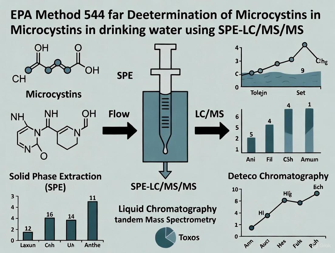
EPA Method 544: Mastering Microcystin Analysis in Drinking Water with SPE-LC/MS/MS
This article provides a comprehensive overview of EPA Method 544 for the detection and quantification of microcystins in drinking water using Solid-Phase Extraction (SPE) coupled with Liquid Chromatography-Tandem Mass Spectrometry...
EPA Method 544: Mastering Microcystin Analysis in Drinking Water with SPE-LC/MS/MS
Abstract
This article provides a comprehensive overview of EPA Method 544 for the detection and quantification of microcystins in drinking water using Solid-Phase Extraction (SPE) coupled with Liquid Chromatography-Tandem Mass Spectrometry (LC/MS/MS). Tailored for researchers, scientists, and drug development professionals, it covers foundational principles, step-by-step methodology, common troubleshooting strategies, and validation protocols to ensure accurate, reliable results in environmental monitoring and biomedical applications.
Understanding EPA Method 544: Fundamentals and Significance in Water Safety
Microcystins (MCs) are a class of potent hepatotoxins produced by certain species of freshwater cyanobacteria (commonly known as blue-green algae) and represent a significant global challenge to water quality, ecosystem health, and public safety [1] [2]. These cyclic heptapeptides are primarily associated with genera such as Microcystis, Planktothrix, Anabaena, and Oscillatoria [1] [3]. The incidence and severity of cyanobacterial harmful algal blooms (cyanoHABs), which release these toxins, are increasing worldwide, driven by eutrophication and climate change [4] [5]. This document provides a comprehensive introduction to microcystins, detailing their chemical diversity, environmental sources, mechanisms of toxicity, and public health impacts, thereby framing the critical need for robust analytical methods like EPA Method 544 for monitoring these toxins in drinking water.
Chemical Diversity and Structure of Microcystins
Fundamental Chemistry
Microcystins are characterized by a stable cyclic heptapeptide structure with a general framework of D-Ala1-X2-D-Masp3-Z4-Adda5-D-γ-Glu6-Mdha7 [1] [6]. Within this structure, X and Z are variable L-amino acids, and the systematic name of a microcystin congener (e.g., MC-LR) is derived from the one-letter codes of these variable amino acids [1]. The structure also incorporates several unique non-proteinogenic amino acids:
- Adda: (all-S,all-E)-3-amino-9-methoxy-2,6,8-trimethyl-10-phenyldeca-4,6-dienoic acid. This side chain is essential for the toxin's biological activity [1] [2].
- D-Masp: D-erythro-β-methyl-isoaspartic acid.
- Mdha: N-methyldehydroalanine [1].
The combination of the cyclic structure and these unique amino acids makes microcystins resistant to breakdown by common proteases and stable over a wide range of temperatures and pH levels [1] [2].
Structural Variants
The structural diversity of microcystins is vast, with over 250 different congeners identified to date [1] [4] [6]. This variation arises primarily from substitutions at the variable amino acid positions, though methylation, demethylation, and other post-biosynthetic modifications also contribute to the diversity [6]. The most common and extensively studied congener is Microcystin-LR (MC-LR), which contains Leucine (L) and Arginine (R) in the variable positions [1] [4]. Different congeners exhibit varying levels of toxicity, influenced by their hydrophobicity and specific interactions with cellular targets [4].
Table 1: Common Microcystin Congeners and Their Characteristics
| Congener Name | Variable Amino Acids (X, Z) | Relative Toxicity | Prevalence Notes |
|---|---|---|---|
| MC-LR | Leucine, Arginine | High (reference toxin) | Most common and widely studied variant [1] |
| MC-RR | Arginine, Arginine | Moderate | Often dominant in low-rainfall regions [7] |
| MC-YR | Tyrosine, Arginine | High | Less common than MC-LR and MC-RR |
| MC-LA | Leucine, Alanine | Moderate to High | Frequently detected in environmental blooms |
Producing Organisms and Bloom Formation
Microcystins are primarily produced by colonial cyanobacteria in freshwater ecosystems. The most notorious producer is Microcystis aeruginosa, but many other species within the genera Anabaena, Planktothrix, Oscillatoria, and Nostoc are also known producers [1] [3]. These organisms can form dense blooms, often visible as green scum or paint-like slicks on the water surface, a phenomenon exacerbated by eutrophication (excess nutrient loading, particularly phosphorus and nitrogen) and warming water temperatures [1] [3] [4].
Microcystis species possess gas vesicles that allow them to regulate buoyancy, enabling them to dominate the water surface and outcompete other microorganisms for light [3]. The production of microcystins is influenced by a complex interplay of environmental factors, including:
- Nutrient Availability: Phosphorus is a key driver of bloom formation, while the source and availability of nitrogen (e.g., nitrate, ammonium, urea) can influence the specific congeners produced and overall toxin yield [1] [4].
- Iron Supply: Microcystin production is upregulated under low iron conditions, as the molecule can bind iron, suggesting a potential evolutionary role in iron acquisition [1].
- Light and Temperature: Bright and red light can increase toxin production, whereas blue light reduces it. Higher temperatures are positively correlated with microcystin production [1] [3].
It is critical to note that not all cyanobacterial blooms are toxic, as toxic and non-toxic strains can co-exist, and toxicity can vary over the course of a bloom [3] [7]. Therefore, cell density alone is not a reliable indicator of risk, necessitating direct toxin measurement.
Emerging Concern: Transfer to Marine Ecosystems
Traditionally considered a freshwater problem, microcystins are increasingly detected in transitional and marine ecosystems, such as estuaries, brackish waters, and coastal areas [8]. The primary vector for this transfer is the transport of toxin-producing cells and dissolved toxins from rivers and freshwater runoff into marine environments. Some cyanobacteria can also survive and experience secondary outbreaks in these saline environments [8]. This expansion poses a new threat to marine food webs, fisheries, and human exposure through contaminated seafood.
Mechanisms of Toxicity and Human Health Impacts
Molecular Mechanism of Action
The primary mechanism of microcystin toxicity is the potent inhibition of crucial eukaryotic enzymes protein phosphatase 1 (PP1) and 2A (PP2A) [1] [4] [2]. The ADDA side-chain is essential for this inhibitory activity [1].
Upon exposure, microcystins are actively transported into hepatocytes (liver cells) via membrane transporters called organic anion transporting polypeptides (OATPs) [4]. The liver is the primary target organ due to its high expression of these transporters [4]. Once inside the cell, microcystins bind covalently to the catalytic subunits of PP1 and PP2A, effectively inhibiting their activity [2]. This inhibition leads to a catastrophic disruption of the critical balance of protein phosphorylation, causing:
- Hyperphosphorylation of cytoskeletal proteins (e.g., keratins), leading to loss of cellular structure, cytoskeletal collapse, and hepatocyte hemorrhage [4] [2].
- Generation of oxidative stress through the production of reactive oxygen species (ROS), triggering apoptosis (programmed cell death) and inflammation [4] [2].
- Promotion of tumorigenesis. The International Agency for Research on Cancer (IARC) has classified MC-LR as a Group 2B agent (possibly carcinogenic to humans) [4]. The disruption of PP2A, a known tumor suppressor, is a key mechanism in this process.
The following diagram illustrates this key molecular mechanism of action:
Human Health Effects
Human exposure occurs through ingestion of contaminated drinking water or food, inhalation of aerosols during recreational activities, and dermal contact with bloom-affected water [4] [9]. Health effects are dose-dependent and can be acute or chronic.
Table 2: Human Health Effects of Microcystin Exposure
| Category | Route of Exposure | Acute Symptoms/Effects | Potential Chronic Effects |
|---|---|---|---|
| Acute Recreational | Ingestion, Inhalation, Dermal | Abdominal pain, vomiting, diarrhea, headache, sore throat, dry cough, pneumonia, blistering around mouth, skin rashes/hives [1] [3] [9]. | - |
| Acute Drinking Water | Ingestion | Gastroenteritis, elevated liver enzymes (GGT), painful hepatomegaly (liver enlargement), kidney damage [4] [9]. | - |
| Chronic/Low-Dose | All routes | - | Potential promotion of liver and colorectal cancers; increased risk in populations with pre-existing liver or gastrointestinal disease [1] [4]. |
| Notable Incident | Intravenous (Dialysis) | Liver failure and death (e.g., Caruaru, Brazil, 1996) [1] [4]. | - |
Public Health Protection and Risk Assessment
Monitoring and managing the risk from microcystins is a multi-faceted process. A common public health approach involves using tiered alert level frameworks based on cyanobacterial cell counts or biomass, which trigger specific management actions [7] [5]. However, for the protection of drinking water, direct measurement of toxin concentration is paramount.
Regulatory Guidelines and Standards
Several national and international bodies have established guidelines or standards for microcystins in drinking water to limit public exposure.
Table 3: Microcystin Drinking Water Guidelines and Standards
| Agency/Guideline | Value | Notes |
|---|---|---|
| World Health Organization (WHO) | 1 μg/L (for MC-LR) | Guideline value for lifetime exposure [7] [5]. |
| U.S. Environmental Protection Agency (EPA) | 0.3 μg/L (infants/children) 1.6 μg/L (school-age/adults) | 10-day Health Advisories (HAs) for total microcystins [10]. Not legally enforceable federal standards. |
| Various U.S. States | Varies | Some states have implemented their own enforceable standards or guidelines based on EPA HAs [10]. |
The Scientist's Toolkit: Key Reagents for Microcystin Analysis
The accurate detection and quantification of microcystins, as mandated by methods like EPA 544, rely on a suite of specific research reagents and materials.
Table 4: Essential Research Reagents for Microcystin Analysis
| Reagent/Material | Function in Analysis | Application Notes |
|---|---|---|
| Solid Phase Extraction (SPE) Cartridges (e.g., C18) | Concentration and cleanup of microcystins from large water samples prior to analysis. | Essential for achieving low detection limits required for drinking water compliance [7]. |
| LC-MS/MS Grade Solvents (e.g., Methanol, Acetonitrile, Water) | Mobile phase for liquid chromatography (LC). Critical for separating different microcystin congeners. | High purity is required to minimize background noise and ion suppression. |
| Certified Reference Materials (CRMs) | Analytical standards for congener identification and quantification (e.g., MC-LR, -RR, -YR). | Used for instrument calibration and quality control. Necessary for definitive congener-specific analysis [6] [7]. |
| Protein Phosphatase Inhibition Assay Kits | Functional assessment of total toxic potential in a sample. | Measures the combined biological activity of all PP-inhibiting congeners; useful as a screening tool [2]. |
| Immunoaffinity Columns | Selective clean-up and enrichment of specific microcystin congeners from complex sample matrices. | Can improve selectivity and reduce matrix effects in challenging samples. |
| Stable Isotope-Labeled Internal Standards (e.g., ( ^{15}N_5 )-MC-LR) | Added to the sample prior to analysis to correct for analyte loss and matrix effects. | Crucial for achieving high accuracy and precision in quantitative LC-MS/MS methods. |
Experimental Protocol: Solid Phase Extraction (SPE) of Microcystins from Water
This protocol outlines a standard procedure for concentrating microcystins from drinking water samples, a critical pre-analysis step.
Principle: Microcystins are extracted from a large volume of water (100 mL to 1 L) using a reversed-phase C18 SPE cartridge. The toxins are adsorbed onto the hydrophobic sorbent, rinsed of impurities, and then eluted with a strong organic solvent for subsequent LC-MS/MS analysis.
Materials:
- Water sample (filtered through GF/F filter if particulate analysis is not required)
- C18 SPE cartridges (e.g., 500 mg sorbent)
- Vacuum manifold for SPE
- LC-MS grade Methanol
- LC-MS grade Water
- Aqueous formic or acetic acid (e.g., 0.1%)
- Elution solvent (e.g., 90% Methanol with 0.1% Formic Acid)
- Collection tubes
Procedure:
- Conditioning: Pass 10 mL of methanol through the C18 cartridge at a steady flow rate (~5 mL/min), followed by 10 mL of pure water. Do not allow the sorbent bed to run dry.
- Sample Loading: Acidify the known volume of water sample to pH ~4 with formic or acetic acid. Load the sample onto the conditioned cartridge under vacuum at a flow rate not exceeding 10 mL/min.
- Washing: After sample loading, wash the cartridge with 10-20 mL of a weak aqueous wash solution (e.g., 10-20% methanol in acidified water) to remove salts and other polar interferents.
- Drying: Draw air through the cartridge for 10-15 minutes under full vacuum to remove residual water and wash solvent.
- Elution: Elute the adsorbed microcystins into a clean collection tube using 10-20 mL of elution solvent (e.g., 90% Methanol with 0.1% Formic Acid). Allow the solvent to gravity-drip or use a very slow vacuum.
- Reconstitution: Evaporate the eluate to dryness under a gentle stream of nitrogen at 40°C. Reconstitute the dry residue in a known, small volume (e.g., 1.0 mL) of initial LC-MS mobile phase (e.g., 20% methanol) for analysis. Vortex thoroughly.
Notes: The specific volumes, acidification, and solvent strengths may require optimization based on the specific SPE sorbent and sample matrix. The use of stable isotope-labeled internal standards added prior to extraction is highly recommended for quantitative accuracy.
Microcystins pose a persistent and evolving threat to water security and public health globally. Their structural diversity, environmental stability, and potent mechanism of toxicity necessitate vigilant monitoring and management. The foundation of this protection lies in robust, sensitive, and congener-specific analytical methods. The subsequent development and validation of EPA Method 544 for the analysis of microcystins in drinking water using Solid Phase Extraction and Liquid Chromatography Tandem Mass Spectrometry (SPE-LC-MS/MS) represents a critical advancement in our ability to quantify these toxins at the stringent levels required by public health guidelines, ensuring the safety of drinking water supplies for communities worldwide.
Historical Development and Regulatory Context
EPA Method 544, titled "Determination of Microcystins and Nodularin in Drinking Water by Solid Phase Extraction and Liquid Chromatography/Tandem Mass Spectrometry (LC/MS/MS)," was developed by the U.S. Environmental Protection Agency and published in 2015 [11]. This method emerged in response to growing concerns about the health risks posed by cyanobacterial harmful algal blooms (HABs) in drinking water sources. Microcystins are toxic cyclic heptapeptides produced by cyanobacteria that present significant health risks to humans and animals, primarily as potent liver toxins [12].
The method gained significant regulatory importance when it was incorporated into the fourth Unregulated Contaminant Monitoring Rule (UCMR 4), which was finalized in December 2016 and became effective on January 19, 2017 [13]. Under UCMR 4, EPA implemented a phased monitoring approach for public water systems. This framework specifies that EPA Method 546 ("Determination of Total Microcystins and Nodularins in Drinking Water and Ambient Water by ADDA Enzyme-Linked Immunosorbent Assay") is used first as a screening tool. If total microcystins are detected at or above 0.3 µg/L by Method 546, then follow-up analysis is required using the more specific EPA Method 544 [14]. This strategic approach balances comprehensive monitoring with cost-effectiveness by reserving the more resource-intensive LC/MS/MS analysis for confirmed positive samples.
Analytical Scope and Target Analytes
EPA Method 544 is designed to specifically measure six individual microcystin congeners and nodularin-R in drinking water [14]. The table below details the complete panel of target analytes:
Table 1: Target Analytes and Minimum Reporting Levels in EPA Method 544
| Analyte | UCMR 4 Minimum Reporting Level (MRL) |
|---|---|
| Microcystin-RR | 0.005 µg/L (5 ng/L) |
| Microcystin-LR | 0.005 µg/L (5 ng/L) |
| Microcystin-YR | 0.005 µg/L (5 ng/L) |
| Microcystin-LA | 0.02 µg/L (20 ng/L) |
| Microcystin-LF | 0.02 µg/L (20 ng/L) |
| Microcystin-LY | 0.02 µg/L (20 ng/L) |
| Nodularin-R | 0.005 µg/L (5 ng/L) |
A key characteristic of Method 544 is its congener-specific nature, which simultaneously provides both its greatest strength and a notable limitation. While the method delivers highly specific identification and quantification of the listed analytes, it does not detect other microcystin congeners that may be present in a sample [14]. When the method was developed, the target list was limited to those congeners for which commercially available analytical standards existed [14].
Method Principles and Workflow
EPA Method 544 combines solid-phase extraction (SPE) for sample preparation with liquid chromatography tandem mass spectrometry (LC/MS/MS) for separation and detection. The workflow can be visualized as follows:
The method employs a surrogate standard approach using ethylated D5 microcystin-LR (MC-LR-C2D5) for quality control [12]. Samples are fortified with this surrogate before processing, allowing analysts to monitor method performance and correct for recovery variations throughout the analytical procedure.
Detailed Experimental Protocol
The standard methodology for EPA Method 544 involves the following specific procedures [12]:
Sample Preservation and Preparation: Collect a 250 mL water sample. The method permits reducing sample volume from the traditional 1 L to 250 mL, which decreases sample preparation time while maintaining adequate sensitivity. Spike the sample with the surrogate standard (ethylated D5 microcystin-LR).
Filtration and Extraction: Filter the sample through an Isopore hydrophilic polycarbonate membrane filter (0.4 μm pore size, 47 mm diameter). Incubate the filter with 80:20 (v/v) methanol/water for 1 hour at -20°C. Combine the filtrate and incubation solution.
Solid-Phase Extraction: Pass the combined extract through a solid-phase extraction cartridge (Waters Oasis HLB, 6 cc, 150 mg). Elute retained analytes with 90:10 (v/v) methanol/water.
Concentration and Reconstitution: Evaporate the eluate to dryness and reconstitute with 90:10 (v/v) methanol/water to a final volume of 500 μL, achieving a 500-fold concentration factor.
Liquid Chromatography: Inject 10 μL onto a Phenomenex Kinetex C18 column (2.6 μm, 100 Å, 100 mm × 2.1 mm) maintained at 40°C. Use a mobile phase gradient (Table 2) at a flow rate of 0.40 mL/min with a total run time of 10 minutes.
Table 2: Chromatographic Gradient Conditions for EPA Method 544
| Time (min) | % Mobile Phase A (Water with 0.1% Formic Acid) | % Mobile Phase B (Methanol with 0.1% Formic Acid) |
|---|---|---|
| 0.0 | 90 | 10 |
| 1.0 | 90 | 10 |
| 6.0 | 5 | 95 |
| 8.0 | 5 | 95 |
| 8.1 | 90 | 10 |
| 10.0 | 90 | 10 |
- Mass Spectrometric Detection: Operate the triple quadrupole mass spectrometer in multiple reaction monitoring (MRM) mode with electrospray ionization in positive ion mode. Monitor two MRM transitions for each analyte: one for quantitation and one for confirmation based on ion ratio.
Performance Characteristics and Quality Control
EPA Method 544 exhibits exceptional sensitivity, with demonstrated Minimum Reporting Levels (MRLs) ranging from 0.005 to 0.02 µg/L (5-20 ng/L) for the target analytes [12]. The method requires laboratories to demonstrate proficiency through rigorous Initial Demonstration of Capability (IDC) experiments, which include specific performance criteria:
Table 3: Initial Demonstration of Capability Requirements and Performance
| IDC Component | Performance Requirement | Demonstrated Performance |
|---|---|---|
| Minimum Reporting Level (MRL) Confirmation | 50-150% recovery in 7 replicates | MRLs of 0.005-0.02 µg/L confirmed |
| Laboratory Fortified Blank (LFB) Accuracy | ±30% of true value | 75-96% recovery achieved |
| Laboratory Fortified Blank (LFB) Precision | <30% RSD | 2.3-8.7% RSD achieved |
| Laboratory Reagent Blank (LRB) | <33% of MRL | Criteria met for all analytes |
The method validation data shows excellent linearity with correlation coefficients (r) >0.99 across calibration ranges, and limits of quantitation (LOQ) for solvent-based standards ranging from 0.5 to 2.5 ng/mL [12]. When back-calculated to the original sample considering the 500-fold concentration factor, this equates to method LOQs of 1-5 ng/L.
Comparative Method Analysis
Within the landscape of cyanotoxin detection methods, EPA Method 544 occupies a specific niche that complements other available approaches. The following table compares its characteristics with the commonly used ADDA-ELISA method (EPA Method 546):
Table 4: Comparison of EPA Method 544 and ADDA-ELISA (Method 546)
| Characteristic | EPA Method 544 (LC/MS/MS) | EPA Method 546 (ADDA-ELISA) |
|---|---|---|
| Target | 6 specific microcystin congeners + nodularin-R | Total microcystins (all ADDA-containing congeners) |
| Specificity | Congener-specific | Congener-generic (measures class) |
| Sensitivity | 0.005-0.02 µg/L MRL | 0.3 µg/L MRL |
| Quantitation | Absolute for specific congeners | Relative to microcystin-LR standard |
| Instrumentation | Sophisticated LC/MS/MS system | Simple plate reader |
| Throughput | Lower | Higher |
| Cost | Higher equipment and operational costs | Lower cost per sample |
| Information Content | Identifies and quantifies specific congeners | Provides aggregate measure |
The ADDA-ELISA method detects the ADDA side chain common to most microcystin congeners and nodularins, potentially measuring over 100 different variants but without distinguishing between them [15]. In contrast, Method 544 provides specific congener identification but only for the limited set of targeted analytes [14]. When samples contain only the congeners measurable by LC/MS/MS, both methods should provide comparable results above their quantitation limits. However, when samples contain additional microcystins not targeted by Method 544, the ADDA-ELISA result will typically be higher than the sum of congener concentrations measured by LC/MS/MS [14].
The Researcher's Toolkit: Essential Materials and Reagents
Successful implementation of EPA Method 544 requires specific research reagents and materials optimized for the SPE-LC/MS/MS workflow:
Table 5: Essential Research Reagent Solutions for EPA Method 544
| Reagent/Material | Specification | Function in Method |
|---|---|---|
| Analytical Standards | Individual microcystin (RR, LR, YR, LF, LW, LY, LA) and nodularin R | Quantitation and identification reference |
| Surrogate Standard | Ethylated D5 microcystin-LR (MC-LR-C2D5) | Quality control and recovery correction |
| SPE Cartridge | Waters Oasis HLB (6 cc, 150 mg) | Extraction and concentration of analytes |
| Chromatography Column | Phenomenex Kinetex C18 (2.6 µm, 100 Å, 100 mm × 2.1 mm) | LC separation of target analytes |
| Mobile Phase A | Water with 0.1% formic acid | LC mobile phase for hydrophilic interaction |
| Mobile Phase B | Methanol with 0.1% formic acid | LC mobile phase for hydrophobic interaction |
| Filtration Membrane | Isopore hydrophilic polycarbonate (0.4 μm pore size, 47 mm diameter) | Particle removal and cell lysis |
The technological foundation of Method 544 relies on the selectivity of tandem mass spectrometry operated in MRM mode, which provides definitive confirmation of analyte identity through transition ion ratios, significantly reducing false positives compared to less specific detection techniques [12]. The combination of efficient solid-phase extraction with high-resolution chromatographic separation and mass spectrometric detection creates a robust analytical system capable of reliably quantifying trace levels of these potent cyanotoxins in complex drinking water matrices.
Key Principles of SPE-LC/MS/MS Technology in Environmental Analysis
Solid Phase Extraction coupled to Liquid Chromatography/Tandem Mass Spectrometry (SPE-LC/MS/MS) represents a cornerstone technology for the sensitive and selective quantification of trace-level contaminants in environmental waters. Within the framework of U.S. Environmental Protection Agency (EPA) Method 544, this technique is mandated for the monitoring of six specific microcystin congeners (MC-LA, -LF, -LR, -LY, -RR, -YR) and nodularin-R in drinking water [14] [16]. The primary advantage of this methodology lies in its ability to achieve exceptional sensitivity, with minimum reporting levels (MRLs) as low as 0.005 µg/L, far below the World Health Organization (WHO) provisional guideline of 1 µg/L for MC-LR in drinking water [12] [17]. This sensitivity is critical for early warning systems, enabling water utilities to detect cyanotoxin threats at sub-threat levels and take appropriate remedial actions to protect public health.
The fundamental principle of this approach involves a two-stage process: first, the selective extraction and concentration of target analytes from the complex water matrix using Solid Phase Extraction, and second, the high-resolution separation and detection of individual congeners using Liquid Chromatography/Tandem Mass Spectrometry. This combination effectively mitigates matrix effects and provides the specificity required to distinguish between structurally similar microcystin variants, a task that alternative methods like ADDA-ELISA cannot perform despite their utility as a screening tool [14]. The congener-specific data generated by EPA Method 544 is essential for accurate risk assessment, as microcystin variants exhibit significantly different toxicological potencies [18].
Fundamental Principles of SPE-LC/MS/MS Operation
Solid Phase Extraction (SPE) Principles
Solid Phase Extraction serves as the critical sample preparation step, designed to isolate, purify, and concentrate target analytes from a complex aqueous sample matrix. The process leverages selective sorbent chemistry to retain analytes of interest while allowing interfering substances to pass through. For microcystin analysis, reverse-phase sorbents such as Oasis HLB (Hydrophilic-Lipophilic Balanced) are typically employed due to their ability to capture the mixed-polarity characteristics of microcystin molecules [12]. The operational sequence involves four key stages: conditioning the sorbent bed with organic solvent to wet the surface and prepare it for interaction; sample loading, where the aqueous sample is passed through the cartridge and analytes are retained via hydrophobic and polar interactions; washing with a mild solvent to remove weakly retained matrix interferences without eluting the targets; and finally, elution with a strong organic solvent to recover the purified and concentrated analytes for instrumental analysis.
Recent advancements have focused on automation and miniaturization of the SPE process to enhance reproducibility, reduce sample volume requirements, and increase throughput. Automated 96-well SPE formats can process up to 96 samples within one hour, requiring only 5 mL of sample for triplicate LC-MS analysis while maintaining excellent accuracy and meeting recommended guidelines [19]. This high-throughput approach is particularly valuable for large-scale monitoring programs and during bloom events when rapid turnaround is essential. Furthermore, the implementation of on-line SPE configurations, where the extraction cartridge is integrated directly into the LC flow path, offers significant advantages. This approach automates the entire sample preparation process, reduces manual handling, minimizes potential sample loss, and can achieve a complete analysis in as little as 7-11 minutes per sample [20] [18]. The recovery rates for microcystins using optimized SPE protocols are typically excellent, ranging from 73% to 102% for extracellular toxins and 89% to 121% for intracellular toxins, with minimal matrix effects (<12% for most toxin-matrix combinations) [21].
Liquid Chromatography (LC) Separation Principles
Liquid Chromatography provides the essential separation step that resolves the complex mixture of extracted components before they enter the mass spectrometer. The core principle involves the differential partitioning of analytes between a stationary phase (typically C18 or C8 bonded silica packed into a column) and a mobile phase (a gradient of water and organic solvent). The separation of microcystin congeners is achieved through a gradient elution program that systematically increases the percentage of organic solvent (typically acetonitrile or methanol with 0.1% formic acid) in the mobile phase over time [18]. The formic acid serves to enhance ionization efficiency in the subsequent MS detection step and improves chromatographic peak shape.
Method optimization focuses on achieving baseline resolution between critical pairs, particularly isobaric interferences that the mass spectrometer cannot distinguish based on mass alone. A significant challenge in cyanotoxin analysis is the separation of anatoxin-a from its isobaric interference, phenylalanine, which requires careful optimization of chromatographic conditions to prevent misidentification or overestimation [20]. The trend toward UHPLC (Ultra-High Performance Liquid Chromatography) utilizing sub-2μm particle columns significantly enhances separation efficiency, resolution, and speed compared to conventional HPLC. These systems operate at higher pressures but provide superior peak capacity and reduce analysis times, with modern methods achieving complete separation of multiple microcystin congeners in 10-13 minutes [12] [17]. The selection of mobile phase can dramatically impact congener separation; acetonitrile with 0.1% formic acid has been identified as providing superior efficiency and sensitivity for microcystin analysis compared to methanol-based systems [18].
Tandem Mass Spectrometry (MS/MS) Detection Principles
Tandem Mass Spectrometry provides the exceptional specificity and sensitivity required for confirmatory analysis of microcystins at trace concentrations. The technique operates through a three-stage process within a single instrumental platform: first, the ionization of analyte molecules as they exit the LC column, typically using Electrospray Ionization in positive mode (ESI+), which efficiently protonates microcystin molecules; second, the mass selection of precursor ions (typically [M+H]+) using the first quadrupole (Q1); and third, collision-induced dissociation of the selected precursor ions in a collision cell (q2) with an inert gas, followed by mass analysis of the resulting product ions in the second quadrupole (Q3).
This Multiple Reaction Monitoring (MRM) mode is the cornerstone of EPA Method 544, where two specific transitions are monitored for each analyte: a primary quantifier ion for precise concentration measurement and a secondary qualifier ion for confirmatory identification [12]. The ratio between these two transitions provides a highly specific "fingerprint" that must remain consistent between samples and standards for positive identification, effectively eliminating false positives from co-eluting matrix components. The exceptional sensitivity of modern triple-quadrupole instruments enables the detection of microcystins at sub-nanogram per liter levels, with documented limits of detection ranging from 0.01-0.02 μg/L for various congeners [20] [12]. This sensitivity is further enhanced through the use of isotopically labeled internal standards (such as ethylated D5 microcystin-LR), which correct for variability in sample preparation and ionization efficiency, thereby ensuring high data quality and accurate quantification [12] [21].
Experimental Protocols for EPA Method 544
Sample Collection and Preservation
Proper sample handling is paramount for obtaining accurate results. Drinking water samples should be collected in pre-cleaned amber glass containers to minimize photodegradation of light-sensitive microcystins. According to EPA requirements, samples for Method 544 analysis must be collected as 500 mL volumes, though recent optimizations have demonstrated successful analysis with only 250 mL while maintaining required MRLs [12]. Samples should be preserved by freezing at -20°C if analysis cannot be performed immediately, with established holding times rigorously validated to prevent analyte degradation. For comprehensive risk assessment, both intracellular (cell-bound) and extracellular (dissolved) fractions may be characterized, requiring additional filtration steps and separate analysis [21].
Detailed SPE Procedure for Microcystin Extraction
The following protocol outlines the step-by-step procedure for the extraction of microcystins from drinking water samples:
- Filter 250-500 mL of water sample through an Isopore hydrophilic polycarbonate membrane filter (0.4 μm pore size, 47 mm diameter) to separate particulate matter [12].
- Spike the filtrate with surrogate standard (e.g., ethylated D5 microcystin-LR) to monitor method performance and correct for analytical variability.
- Condition an Oasis HLB SPE cartridge (6 cc, 150 mg) with 10 mL methanol followed by 10 mL reagent water, maintaining the sorbent bed never dry.
- Load the sample onto the conditioned cartridge at a controlled flow rate of 5-10 mL/minute using a vacuum manifold.
- Wash the cartridge with 10 mL of 10% methanol in water to remove polar interferences without eluting the microcystins.
- Dry the cartridge under vacuum for 15-20 minutes to remove residual water.
- Elute the analytes with 10 mL of 90:10 (v/v) methanol/water into a clean collection tube.
- Evaporate the eluent to dryness under a gentle stream of nitrogen at 30-40°C.
- Reconstitute the dried extract in 500 μL of 90:10 (v/v) methanol/water with 0.1% formic acid, followed by transfer to an LC vial for analysis.
Instrumental Analysis via LC/MS/MS
The instrumental conditions for the analysis should be optimized as follows:
Chromatography:
- Column: Phenomenex Kinetex C18 (2.6 μm, 100 Å, 100 mm × 2.1 mm) or equivalent [12]
- Mobile Phase A: Water with 0.1% formic acid
- Mobile Phase B: Acetonitrile with 0.1% formic acid
- Gradient: Begin at 20% B, ramp to 50% B over 5 minutes, then to 95% B over 2 minutes, hold for 1 minute, then re-equilibrate [12]
- Flow Rate: 0.40 mL/min
- Column Temperature: 40°C
- Injection Volume: 10 μL
Mass Spectrometry:
- Ionization Mode: Electrospray Ionization (ESI) positive
- Ion Source Temperature: 500°C
- Ion Spray Voltage: 5500 V
- Nebulizer Gas: 50 psi
- Heater Gas: 50 psi
- Curtain Gas: 25 psi
- Collision Gas: Nitrogen
- Detection Mode: Multiple Reaction Monitoring (MRM) with two transitions per analyte
Table 1: MRM Transitions and Parameters for Microcystin Analysis
| Analyte | Precursor Ion (m/z) | Product Ion 1 (Quantifier) | Product Ion 2 (Qualifier) | Collision Energy (V) |
|---|---|---|---|---|
| MC-RR | 519.8 | 135.1 | 127.1 | 65 |
| MC-YR | 1045.5 | 135.1 | 127.1 | 80 |
| MC-LR | 995.5 | 135.1 | 127.1 | 65 |
| MC-LA | 910.5 | 135.1 | 127.1 | 70 |
| MC-LY | 1002.5 | 135.1 | 127.1 | 70 |
| MC-LF | 986.5 | 135.1 | 127.1 | 70 |
| Nodularin | 825.5 | 135.1 | 127.1 | 65 |
Quality Assurance and Quality Control
Robust QA/QC procedures are mandated by EPA Method 544 to ensure data integrity [12]:
- Initial Demonstration of Capability (IDC): Laboratories must demonstrate proficiency through analysis of method blanks, laboratory fortified blanks, and MRL confirmation samples before analyzing environmental samples.
- Laboratory Reagent Blank (LRB): Must be free of target analytes at concentrations exceeding 1/3 of the MRL to demonstrate no laboratory contamination.
- Laboratory Fortified Blank (LFB): Must demonstrate accuracy of 70-130% and precision of <30% RSD for all target analytes.
- Surrogate Standards: Must be added to all samples, blanks, and standards to correct for matrix effects and procedural losses, with recovery criteria of 50-150%.
- Continuing Calibration Check: Standards analyzed at regular intervals (every 10-12 samples) to verify instrumental response stability.
Performance Characteristics and Validation Data
The performance of SPE-LC/MS/MS methods for microcystin analysis has been extensively validated through multiple studies. The following table summarizes key performance metrics from recent implementations:
Table 2: Performance Metrics for SPE-LC/MS/MS Analysis of Microcystins
| Parameter | EPA Method 544 [12] | On-line SPE Method [20] | UHPLC-MS/MS Method [18] |
|---|---|---|---|
| Analysis Time | 10-13 minutes | 7 minutes per sample | 11 minutes |
| Sample Volume | 250-500 mL | 2 mL | Not specified |
| LOD Range | 0.005-0.02 µg/L | 0.01-0.02 µg/L | 0.00002-0.00037 µg/L |
| LOQ Range | Not specified | Not specified | 0.000066-0.00124 µg/L |
| Recovery (%) | 75-130% | 91-101% | 70-135%* |
| Precision (% RSD) | <20% | <13% | <10% |
| Linearity | r > 0.99 | Not specified | r > 0.99 |
*Except for MC-RR (58.8%) and MC-WR (40.9-45.1%) at specific concentrations
The Researcher's Toolkit: Essential Materials and Reagents
Table 3: Essential Research Reagents and Materials for SPE-LC/MS/MS of Microcystins
| Item | Specification/Example | Function/Purpose |
|---|---|---|
| SPE Cartridges | Oasis HLB (6 cc, 150 mg) [12] | Extraction and concentration of microcystins from water samples |
| LC Column | C18, 100-150 mm x 2.1 mm, sub-2μm or 2.6μm [12] [18] | High-resolution chromatographic separation of microcystin congeners |
| Mobile Phases | Water and acetonitrile, both with 0.1% formic acid [18] | Liquid chromatography eluents for gradient separation |
| Analytical Standards | Individual microcystin congeners (RR, YR, LR, LA, LY, LF) and nodularin-R [12] | Quantification and identification of target analytes |
| Internal Standards | Ethylated D5 microcystin-LR [12] or Leucine enkephalin [18] | Correction for matrix effects and procedural losses |
| Solvents | LC-MS grade methanol, acetonitrile, water | Sample preparation and mobile phase preparation |
| Filtration | Isopore hydrophilic polycarbonate membrane (0.4 μm) [12] | Removal of particulate matter from water samples |
| Sample Containers | Amber glass bottles | Protection of light-sensitive analytes during storage |
Method Workflow Visualization
Diagram 1: SPE-LC/MS/MS Workflow for EPA Method 544. The workflow illustrates the sequential process from sample collection to final reporting, with integrated quality control measures throughout the procedure.
Advanced Applications and Method Optimization
The application of SPE-LC/MS/MS technology has expanded beyond drinking water to include complex environmental matrices such as lakes, rivers, and coastal waters [21]. These environments present additional challenges, including higher salinity and more complex organic matter that can interfere with analysis. Research has demonstrated that salt has negligible effect on the SPE recovery of dissolved microcystins using C18 cartridges, though polymeric sorbents may cause overestimation of some variants by up to 67% in saline samples [21]. This finding is particularly relevant for monitoring the fate of cyanobacteria and their toxins across the freshwater-marine continuum, where colonies of Microcystis spp. have been shown to retain >76% of their toxins intracellularly even at salinity 20, suggesting a potential health risk in estuarine environments [21].
Current method optimization research focuses on high-throughput automation and increased sensitivity. The implementation of automated liquid handling platforms for SPE in 96-well format enables processing of 1-96 samples within one hour while requiring only 5 mL of sample for triplicate analysis [19]. This miniaturized approach reduces solvent consumption, decreases plastic waste, and maintains excellent accuracy meeting recommended guidelines. Simultaneously, sensitivity continues to improve, with modern systems achieving limits of detection in the low nanogram per liter range (0.05-0.81 ng/mL for dissolved toxins) [21], far below regulatory thresholds. The ongoing development of on-line SPE approaches integrated with UHPLC-MS/MS represents the cutting edge of this technology, offering complete automation, minimal sample handling, and analysis times under 10 minutes while maintaining excellent sensitivity and precision [20] [18]. These advances position SPE-LC/MS/MS as an indispensable tool for protecting public health through reliable monitoring of cyanotoxins in water supplies worldwide.
Importance of Monitoring Microcystins in Drinking Water for Research and Drug Development
Microcystins (MCs) are hepatotoxic cyclic peptides produced by various species of cyanobacteria, posing a significant global threat to drinking water quality and public health [22] [23]. These potent toxins inhibit protein phosphatases 1 and 2A, disrupting cell growth and metabolism and causing potential liver damage, tumor promotion, and other systemic effects upon ingestion [22] [24]. With over 300 structurally similar congeners identified, MCs present a substantial analytical challenge for accurate monitoring and risk assessment [25]. The World Health Organization (WHO) has established a provisional guideline value of 1.0 µg/L for MC-LR in drinking water, a standard adopted by many regulatory bodies worldwide [23] [24]. Within the context of EPA Method 544, which employs solid-phase extraction followed by liquid chromatography tandem mass spectrometry (SPE-LC/MS/MS) for congener-specific analysis, this application note details advanced methodologies for microcystin monitoring that support both water safety management and fundamental biomedical research into toxin-induced pathogenicity [14].
Analytical Methodologies for Microcystin Detection
Comparison of Primary Detection Platforms
Multiple analytical techniques are employed for microcystin detection, each with distinct advantages and limitations tailored to different monitoring objectives.
Table 1: Comparison of Microcystin Detection Methods
| Method | Principle | Detection Limit | Key Advantages | Key Limitations |
|---|---|---|---|---|
| Immunosensors | Nanomaterial-based platforms with monoclonal antibodies | 0.05 µg/L [22] | Rapid detection (<10 minutes), portable for field use [22] | Limited congener specificity, potential cross-reactivity [14] |
| ADDA-ELISA (EPA Method 546) | Antibody recognition of ADDA moiety | 0.3 µg/L (MRL) [14] | Measures "total microcystins," cost-effective, high throughput [14] | Does not identify individual congeners, potential bias [14] |
| LC/MS/MS (EPA Method 544) | Mass spectrometry with chromatographic separation | 0.006-0.028 µg/g in supplements [26]; 1.0-22.0 pg on-column in water [25] | Congener-specific, high sensitivity and selectivity [14] [25] | Requires sophisticated equipment, higher cost [14] |
| UPLC-MS/MS | Ultra-performance liquid chromatography with MS/MS | 1.3-6.0 ng/L after SPE pre-concentration [27] | Faster analysis time, superior sensitivity [27] | Method complexity, specialized instrumentation needed [27] |
| MALDI-TOF MS | Matrix-assisted laser desorption/ionization time-of-flight | <7 µg/L in raw water [28] | Minimal sample preparation, high-throughput capacity [28] | Limited sensitivity for very low concentrations [28] |
EPA's Framework for Drinking Water Monitoring
The U.S. Environmental Protection Agency (EPA) employs a phased monitoring approach for microcystins in drinking water under the Unregulated Contaminant Monitoring Rule (UCMR 4). This framework initially screens samples using EPA Method 546 (ADDA-ELISA), which quantifies total microcystins against an MC-LR calibration curve with a Minimum Reporting Level (MRL) of 0.3 µg/L [14]. Samples exceeding this threshold undergo confirmatory analysis using EPA Method 544, an SPE-LC/MS/MS method that specifically quantifies six microcystin congeners (MC-LR, -YR, -RR, -LA, -LF, -LY) and nodularin-R [14]. The EPA emphasizes that these methods measure different analytes—Method 546 detects any congener containing the ADDA functional group (associated with >100 congeners), while Method 544 specifically targets the six listed congeners—making them complementary rather than directly comparable [14].
Advanced Experimental Protocols
EPA Method 544: Solid-Phase Extraction and LC/MS/MS Analysis
Sample Preparation:
- Solid-Phase Extraction: Process water samples through C18 SPE cartridges. Condition cartridges with 6 mL methanol followed by 6 mL Milli-Q water (pH-adjusted to 11 for dissolved toxin analysis) [25] [24]. Load samples and elute toxins with 5 mL 80% methanol [24].
- Extraction Recovery: Apply corrective factors to compensate for toxin losses. Acceptable recovery ranges are 89-121% for intracellular and 73-102% for extracellular cyanotoxins [25].
- Cleanup: Purify eluate using 0.2 µm RC-syringe filters before LC/MS/MS analysis [24].
LC/MS/MS Analysis:
- Chromatography: Utilize UPLC system with Acquity UPLC BEH C18 column (1.7 µm, 1.0 × 50 mm) or equivalent. Employ gradient elution with mobile phase consisting of 0.1% formic acid in water (A) and methanol or acetonitrile (B) [27] [24].
- Mass Spectrometry: Operate mass spectrometer in positive electrospray ionization mode with multiple reaction monitoring (MRM). Key transitions include MC-LR (m/z 995.5→599.17, 865.92) and other congener-specific transitions [23] [27].
- Quality Control: Include matrix-matched calibration standards and ongoing quality controls to ensure method reliability. Acceptable intra- and inter-day variabilities are <11% [25].
Protocol for Intracellular and Extracellular Microcystin Analysis in Saline Waters
For studies investigating cyanotoxin fate across freshwater-marine continua:
- Intracellular Toxin Extraction: Lyse cyanobacterial cells using repeated freeze-thaw cycles or sonication. Extract with appropriate solvents (e.g., methanol-water mixtures) [25].
- Extracellular Toxin Extraction in Saline Waters: Use C18 SPE cartridges for dissolved toxin extraction. Note that salt has negligible effect on C18 cartridge recovery for dissolved MCs, though polymeric sorbents may cause overestimation (up to 67% for some variants) [25].
- Matrix Effect Compensation: Evaluate matrix effects for each toxin-matrix combination. Apply corrective factors to maintain accuracy between 73-139% across different salinity conditions [25].
Biosensor Development for Rapid Detection
Nanomaterial-Enhanced Immunosensors:
- Platform Fabrication: Immobilize MC variants on nanostructured substrates (gold nanoparticles, carbon nanotubes) functionalized with monoclonal antibodies [22].
- Signal Amplification: Leverage nanomaterials to enhance electrochemical or optical signals, achieving detection limits of 0.05 µg/L within 10 minutes [22].
- IoT Integration: Incorporate portable biosensors with Internet of Things (IoT) connectivity for real-time, on-site detection and data sharing [22].
Signaling Pathways and Toxicity Mechanisms
Microcystins exert their primary toxicity through specific inhibition of protein phosphatases 1 and 2A (PP1 and PP2A), which disrupts cellular phosphorylation balance and leads to cytoskeletal damage, apoptosis, and oxidative stress [24]. After ingestion, these toxins are transported via bile salt transporters to the liver, causing potent hepatotoxic effects [24]. Recent evidence indicates broader systemic toxicity, with microcystins affecting renal, neural, and cardiovascular systems through complex mechanisms that extend beyond phosphatase inhibition [22].
The Scientist's Toolkit: Essential Research Reagents and Materials
Table 2: Research Reagent Solutions for Microcystin Analysis
| Reagent/Material | Function | Application Notes |
|---|---|---|
| C18 Solid-Phase Extraction Cartridges | Toxin concentration and cleanup from water samples | Effective for both fresh and saline waters; minimal salt interference [25] [27] |
| Monoclonal Antibodies (ADDA-specific) | Recognition element for immunosensors and ELISA | Targets conserved ADDA moiety; cross-reactivity varies among congeners [22] [14] |
| Certified Microcystin Standards | Method calibration and quantification | MC-LR typically used for ELISA calibration; congener-specific standards required for LC/MS [14] [24] |
| Gold Nanoparticles & Carbon Nanotubes | Signal amplification in biosensors | Enhance electrochemical or optical signals; improve detection sensitivity [22] |
| LC/MS-grade Solvents (Acetonitrile, Methanol) | Mobile phase composition | Critical for chromatographic separation and mass spectrometric detection [25] [24] |
| Matrix-Assisted Calibration Standards | Compensation for matrix effects | Essential for accurate quantification in complex matrices [25] [24] |
Analytical Workflow for Comprehensive Microcystin Assessment
The following diagram illustrates the integrated workflow for microcystin monitoring, from sample collection to data interpretation, incorporating both regulatory and advanced research methods.
Implications for Research and Drug Development
The precise monitoring of microcystins in drinking water provides critical data for both environmental risk assessment and biomedical research. Understanding congener-specific occurrence and concentration supports drug discovery efforts targeting microcystin-induced toxicity mechanisms [22] [24]. The development of rapid, sensitive biosensors facilitates timely public health interventions while providing platforms for studying toxin-receptor interactions [22]. Furthermore, methods for quantifying microcystin accumulation in fish and other biological samples [29] enable research into toxin bioaccumulation and potential therapeutic approaches for intoxication. As climate change increases the frequency and distribution of harmful cyanobacterial blooms [25] [23], advanced monitoring capabilities become increasingly vital for protecting public health and supporting pharmaceutical research into countermeasures for cyanotoxin exposure.
Implementing EPA Method 544: A Step-by-Step Protocol for Accurate Microcystin Detection
Sample Collection, Handling, and Preservation Best Practices
EPA Method 544, titled "Determination of Microcystins and Nodularin in Drinking Water by Solid Phase Extraction and Liquid Chromatography/Tandem Mass Spectrometry (LC/MS/MS)," is a robust analytical protocol for determining specific cyanotoxin congeners in drinking water [30]. This method targets six microcystin congeners (MC-LR, -RR, -YR, -LA, -LF, -LY) and nodularin-R, providing congener-specific identification and quantification that aggregate methods like ELISA cannot deliver [14]. The method's sophisticated combination of solid phase extraction for concentration and cleanup with LC/MS/MS analysis enables precise measurement at trace levels, making it indispensable for regulatory monitoring and research on cyanotoxin occurrence and fate [30] [31].
Sample Collection Protocols
Collection Materials and Pre-Collection Preparation
Proper sample collection begins with meticulous preparation of all materials that will contact the sample:
- Glassware Cleaning: All non-volumetric glassware must be heated in a muffle furnace for a minimum of 90 minutes at 400°C. Volumetric glassware should be solvent-rinsed and heated in an oven no hotter than 120°C to prevent damage to precision measurements [30].
- Sample Containers: Use sterile amber glass bottles to protect samples from light. All containers, caps, and closures must be demonstrated to be free from interferences (containing less than 1/3 the minimum reporting level for each analyte) before use [30] [24].
- Field Blanks: Process blank samples containing analyte-free water should be subjected to the same handling procedures as actual samples to monitor for cross-contamination during collection or transport.
Sample Collection Procedure
The following steps outline the proper collection of drinking water samples for microcystin analysis:
- Sample Volume: Collect a 500-mL water sample for analysis [30].
- Preservative Addition: Immediately after collection, add appropriate preservatives to the sample. Sodium thiosulfate is typically added to dechlorinate drinking water samples if they contain residual chlorine [30].
- Labeling and Documentation: Label each sample container with a unique identifier, collection date and time, sample location, collector's name, and any relevant field observations.
- Temperature Control: Keep samples at ≤6°C during transportation and until extraction [30].
Sample Handling and Preservation
Handling Procedures
Proper handling maintains sample integrity from collection through analysis:
- Filtration and Cell Lysis: Filter the 500-mL water sample, retaining both filtrate and filter. Place the filter in a solution of methanol containing 20% reagent water and hold for at least one hour at -20°C to release intracellular toxins from cyanobacteria cells captured on the filter [30].
- Sample Combination: Draw off the liquid from the filter and add it back to the 500-mL aqueous filtrate, ensuring both extracellular and intracellular toxins are captured for analysis [30].
- Holding Conditions: Maintain samples at refrigeration temperatures (≤6°C) throughout transport and storage prior to extraction. Never freeze whole water samples before the extraction process [30].
Preservation Parameters
Adherence to specific preservation parameters is critical for maintaining analyte stability:
Table 1: Sample Preservation and Holding Time Requirements
| Parameter | Requirement | Rationale |
|---|---|---|
| Preservation | Appropriate chemical preservatives (e.g., sodium thiosulfate for chlorine removal) | Prevents analyte degradation and matrix modification |
| Storage Temperature | ≤6°C during transport and storage | Maintains toxin stability and prevents microbial activity |
| Maximum Holding Time (Sample) | 28 days from collection to extraction | Ensures analyte integrity and data validity |
| Maximum Holding Time (Extract) | 28 days when stored at ≤ -4°C | Maintains extract stability for accurate analysis |
Sample Processing Workflow
The following diagram illustrates the complete sample handling workflow from collection to analysis:
Table 2: Method Performance Characteristics for Selected Microcystins in EPA Method 544
| Analyte | Detection Level | Accuracy (Bias) | Precision | Spiking Level |
|---|---|---|---|---|
| Microcystin-LA | 4.000 ng/L | 95% Recovery | 6.00 % RSD | 100.00 ng/L |
| Nodularin | 1.800 ng/L | 92% Recovery | 3.10 % RSD | 19.60 ng/L |
Precision and accuracy were determined for three water matrices: reagent water; chlorinated (finished) ground water; and chlorinated (finished) surface water [30]. The method's applicable concentration range during development was 10-400 µg/L for most analytes, except for MC-RR (4.7-187.5 µg/L), nodularin-R (4.9-195.7 µg/L), and MC-LA (25-1000 µg/L) [30].
Potential Interferences and Quality Control
Several factors can compromise sample integrity and analytical results:
- Matrix Effects: Humic and fulvic materials co-extracted during SPE can cause signal enhancement or suppression in the electrospray ionization source, leading to low SPE recoveries. Total organic carbon (TOC) serves as a good indicator of humic content [30].
- Dissolved Salts: Although not observed during method development, dissolved salts in the mobile phase can suppress analyte signals due to electrolyte-induced ionization. The addition of ammonium formate to the mobile phase helps mitigate this phenomenon [30].
- Container Contamination: Sample bottles, caps, SPE cartridges, and other processing hardware must be routinely demonstrated to be free from interferences [30].
Quality Control Requirements
A comprehensive quality control program includes the following elements:
- Laboratory Reagent Blank (LRB): Analyzed to demonstrate freedom from contamination.
- Laboratory Fortified Blank (LFB): Assesses method performance in the absence of matrix effects.
- Continuing Calibration Check (CCC): Verifies calibration stability during analytical runs.
- Surrogate Analyte (SUR): Monitors method performance throughout sample processing.
- Field Duplicates: Evaluate precision of sampling and analytical methods.
The Researcher's Toolkit
Table 3: Essential Research Reagent Solutions for Microcystin Analysis by EPA Method 544
| Reagent/Material | Function | Application Notes |
|---|---|---|
| Solid Phase Extraction Cartridges | Concentrates and cleans up analytes from sample matrix | Oasis HLB or equivalent polymer-based sorbents provide optimal recovery for hydrophilic-lipophilic balance [32] [31] |
| Stable Isotope-Labeled Toxin Standards | Internal standards for quantification | SIL-MC-RR, SIL-MC-LR, SIL-MC-LA correct for variability in extraction and analysis [32] |
| Methanol (UHPLC-MS Grade) | Extraction solvent and mobile phase component | Provides optimal toxin elution from SPE; high purity minimizes background interference [24] |
| Ammonium Formate | Mobile phase additive | Improves ionization efficiency and reduces signal suppression in MS detection [30] |
| Trizma Buffer | Ion pairing reagent | Enhances retention of MCs without arginine residues on Strata X SPE [32] |
| Sodium Thiosulfate | Dechlorinating agent | Preserves sample by neutralizing disinfectant residuals in drinking water [30] |
Method Flexibility and Advanced Applications
While EPA Method 544 provides a standardized approach, it allows for certain modifications to leverage technological advances:
- Permitted Modifications: Laboratories may modify evaporation techniques, separation techniques, LC columns, mobile phase composition, LC conditions, and MS/MS conditions to improve performance [30].
- Restricted Changes: Sample collection, preservation, extraction steps, and quality control requirements must not be altered from the prescribed protocol [30].
- Advanced Applications: Research applications have demonstrated the method's adaptability for simultaneous determination of multiple cyanotoxin classes using dual SPE cartridge approaches (e.g., HLB-PGC) for comprehensive toxin profiling [31].
Adherence to these sample collection, handling, and preservation best practices ensures the integrity of samples for microcystin analysis by EPA Method 544, providing reliable data for regulatory compliance and research on cyanotoxin occurrence in drinking water systems.
Within the framework of EPA Method 544, sample preparation is a critical step for the accurate and sensitive determination of microcystins and nodularin in drinking water. This protocol details the application of Solid-Phase Extraction (SPE) for the concentration and purification of these cyanotoxins prior to analysis by Liquid Chromatography/Tandem Mass Spectrometry (LC/MS/MS). When performed correctly, SPE effectively removes matrix interferences and pre-concentrates the analytes, enabling the reliable detection of these potent toxins at the part-per-trillion (ng/L) levels required for compliance with the Unregulated Contaminant Monitoring Rule (UCMR 4) [14] [12].
Cartridge Selection
Selecting the appropriate SPE sorbent is the foremost decision for a successful extraction, as it dictates the primary retention mechanism for the target analytes.
Sorbent Chemistry and Format
- Sorbent Type: For the analysis of microcystins, which are cyclic peptides with both non-polar and polar regions, a reversed-phase polymeric sorbent is recommended. EPA Method 544 specifically identifies the use of a Waters Oasis HLB cartridge (6 cc, 150 mg) or equivalent [12]. The HLB (Hydrophilic-Lipophilic Balanced) sorbent is designed to retain a wide range of acidic, basic, and neutral compounds.
- Retention Mechanism: The primary interaction with HLB and similar reversed-phase sorbents is non-polar (hydrophobic), facilitated by van der Waals forces between the analyte and the sorbent surface [33]. This mechanism is ideal for extracting non-polar to moderately polar analytes from a polar, aqueous matrix like drinking water.
- Cartridge Size: A 6 cc cartridge with 150 mg of sorbent is suitable for processing the 250 mL water sample volume specified in optimized procedures [12]. This size provides sufficient sorbent mass and capacity to retain the target toxins without exceeding the pressure limits of a vacuum manifold.
Table 1: SPE Cartridge Selection Guide for Microcystin Analysis
| Selection Factor | Recommendation for EPA Method 544 | Rationale |
|---|---|---|
| Sorbent Chemistry | Hydrophilic-Lipophilic Balanced (HLB) Polymer | Provides broad-spectrum retention for diverse microcystin congeners [12]. |
| Primary Mechanism | Reversed-Phase (Non-polar) | Optimal for extracting semi-polar microcystins from aqueous samples [33]. |
| Cartridge Size | 6 cc, 150 mg sorbent | Balanced capacity for 250 mL sample volume and efficient elution [12]. |
| Sorbent Mass | 150 mg | Provides ~5-10% capacity for analyte and interferent loading, ensuring robust performance [34]. |
Conditioning and Equilibration
Proper conditioning and equilibration of the SPE cartridge are critical to activate the sorbent and ensure reproducible and quantitative retention of the target microcystins.
Step-by-Step Procedure
The goal of this phase is to solvate the sorbent, remove any potential impurities from the manufacturing process, and create an environment conducive to maximal analyte binding [35].
- Conditioning (Solvation): Pass ~5 mL of methanol through the HLB cartridge under a gentle vacuum (approximately 1 mL/min). This solvent wets the polymeric surface, opens the pores, and activates the sorbent by ensuring the functional groups are accessible. Do not allow the sorbent bed to run dry after this step [34] [36].
- Equilibration (Matrix Matching): Immediately after conditioning, pass ~5 mL of reagent water (or a buffer matching the sample matrix) through the cartridge. This step removes the strong organic solvent and replaces it with a polar solvent compatible with the incoming aqueous sample. Failure to equilibrate can cause poor retention, as analytes may not partition effectively from the sample onto a sorbent bed filled with organic solvent [34] [33]. A small layer of solvent (about 1 mm) should remain above the sorbent bed after equilibration [35].
Sample Loading and Washing
This phase involves introducing the sample and subsequently removing weakly retained matrix components.
Sample Preparation and Loading
- Sample Pre-treatment: The 250 mL drinking water sample is fortified with a surrogate standard (e.g., D5-ethyl microcystin-LR) and filtered through a 0.4 μm hydrophilic polycarbonate membrane to remove particulates [12]. For other matrices, pre-treatment may involve dilution or pH adjustment to ensure analytes are free in solution [34].
- Loading: The filtered sample is passed through the conditioned HLB cartridge at a controlled flow rate, typically 1-2 mL/min [34] [33]. A slow, steady flow is essential to allow sufficient time for the microcystins to interact with and be retained by the sorbent. During this step, the target microcystins and nodularin adsorb to the sorbent, while many highly polar water matrix components pass through.
Washing to Remove Interferences
- Wash Solvent: After sample loading, a wash step with ~10 mL of 20% aqueous methanol is recommended to remove weakly retained, polar interferences [12]. The solvent strength is selected to be strong enough to elute unwanted matrix components but weak enough to leave the more hydrophobic microcystins bound to the sorbent [36].
- Purpose: This step purifies the extract, reducing the potential for ion suppression or enhancement during the subsequent LC/MS/MS analysis and leading to a cleaner chromatographic baseline.
Analyte Elution and Concentration
The final phase focuses on the quantitative recovery of the target analytes in a small volume suitable for instrumental analysis.
Elution and Post-Elution Treatment
- Elution Solvent: The retained microcystins are selectively recovered by disrupting their non-polar interactions with the sorbent. This is achieved by passing ~10 mL of 90:10 (v/v) methanol/water through the HLB cartridge [12]. This solvent, with its high organic content, has a stronger affinity for the analytes than the sorbent, causing them to desorb.
- Post-Elution Treatment:
- Concentration: The eluate is collected and evaporated to dryness under a gentle stream of nitrogen in a warm water bath (~40°C).
- Reconstitution: The dried residue is reconstituted in 500 μL of 90:10 (v/v) methanol/water [12]. This step simultaneously concentrates the analytes and places them in a solvent compatible with the reversed-phase LC conditions, achieving a 500-fold concentration factor (from 250 mL to 0.5 mL).
Table 2: Key Experimental Parameters for SPE in Microcystin Analysis
| SPE Step | Key Parameter | Specification in EPA Method 544 | Purpose |
|---|---|---|---|
| Sample Loading | Sample Volume | 250 mL | Achieve high pre-concentration factor [12]. |
| Flow Rate | 1-2 mL/min | Ensure sufficient contact time for quantitative retention [34]. | |
| Washing | Wash Solvent | 20% Methanol in Water | Remove polar interferences while retaining analytes [12]. |
| Elution | Elution Solvent | 90% Methanol in Water | Disrupt hydrophobic interactions to recover analytes [12]. |
| Final Extract Volume | 500 µL | Concentrate analytes; ensure compatibility with LC system [12]. | |
| Overall Process | Concentration Factor | 500x | Enable detection at low ng/L levels [12]. |
The Scientist's Toolkit: Research Reagent Solutions
The following reagents and materials are essential for implementing the SPE procedure for microcystins according to EPA Method 544.
Table 3: Essential Reagents and Materials for SPE of Microcystins
| Item | Function / Purpose | Example / Specification |
|---|---|---|
| SPE Cartridge | Retains and purifies target microcystins from the sample. | Oasis HLB (6 cc, 150 mg) or equivalent polymeric reversed-phase sorbent [12]. |
| Methanol (HPLC Grade) | Cartridge conditioning and analyte elution. | High-purity solvent for activation (100%) and elution (90% in water) [12]. |
| Reagent Water | Cartridge equilibration and wash solvent preparation. | HPLC-grade water, free of organic contaminants [12]. |
| Surrogate Standard | Monitors SPE procedural performance and recovery. | D5-ethyl microcystin-LR, added to sample prior to extraction [12]. |
| Polycarbonate Filter | Removes particulates from the sample prior to SPE loading. | 0.4 μm pore size, 47 mm diameter, hydrophilic [12]. |
| Sample Collection Vials | Holds final concentrated extract for instrumental analysis. | LC/MS-compatible vials with limited volume inserts [12]. |
Adherence to the detailed SPE procedures outlined in this protocol—from the initial selection of a reversed-phase polymeric sorbent to the final elution and concentration steps—is fundamental to the success of EPA Method 544. This sample preparation workflow effectively purifies and concentrates microcystins and nodularin from drinking water, enabling their accurate quantification at the stringent minimum reporting levels (0.005–0.02 µg/L) required for modern environmental monitoring. Mastery of these SPE fundamentals ensures data quality, supports regulatory compliance, and provides a robust framework for monitoring these significant cyanotoxins in public water systems.
This application note provides a detailed protocol for the liquid chromatography-tandem mass spectrometry (LC-MS/MS) instrumentation setup for the analysis of microcystins and nodularin in drinking water, as formalized in EPA Method 544. The reliable detection and quantification of these potent hepatotoxins at sub-parts-per-billion levels are critical for ensuring drinking water safety, as mandated by the Fourth Unregulated Contaminant Monitoring Rule (UCMR4) [12]. The method described herein has been developed and validated to achieve minimum reporting levels (MRLs) ranging from 0.005 to 0.02 µg/L, utilizing solid-phase extraction (SPE) for sample preparation followed by analysis with LC-MS/MS [12]. This document is an integral part of a broader thesis research project aiming to refine and apply EPA Method 544, providing researchers and laboratory personnel with a comprehensive guide for instrument configuration and operation.
Experimental Protocols
Materials and Reagents
- Toxin Standards: Primary standards for Microcystins (-RR, -LR, -YR, -LA, -LF, -LW, -LY) and Nodularin-R should be obtained from certified suppliers. Purity should be ≥95% [18].
- Internal Standard: Deuterated surrogate standard, specifically D5-microcystin-LR (MC-LR-C2D5), is required for isotope dilution mass spectrometry [12].
- Solvents: LC-MS grade water, methanol, and acetonitrile should be used.
- Mobile Phase Additives: High-purity formic acid (e.g., 0.1% to 0.5% in both aqueous and organic mobile phases) is recommended for positive electrospray ionization [18] [37].
- SPE Sorbents: Oasis HLB cartridges (6 cc, 150 mg) are specified in EPA Method 544 for the extraction and cleanup of water samples [12].
EPA Method 544 specifies a solid-phase extraction procedure for concentrating 250 mL of drinking water samples. The key steps are summarized in the workflow below, and full details are in Section 11 of the method [12].
LC-MS/MS Instrumental Setup and Optimization
Liquid Chromatography Conditions
Optimal chromatographic separation is critical for reducing matrix effects and achieving baseline resolution of target analytes. A reversed-phase C18 column is standard for this application.
- Column: Phenomenex Kinetex C18 (100 mm x 2.1 mm, 2.6 μm) or equivalent [12].
- Mobile Phase A: Water containing 0.1% formic acid.
- Mobile Phase B: Acetonitrile containing 0.1% formic acid.
- Flow Rate: 0.40 mL/min.
- Injection Volume: 10 μL.
- Column Oven Temperature: 40 °C.
- Autosampler Temperature: 8 °C.
Table 1: Gradient Elution Program for Microcystin Separation
| Time (min) | % Mobile Phase A | % Mobile Phase B | Curve |
|---|---|---|---|
| 0.0 | 95 | 5 | Linear |
| 1.0 | 95 | 5 | Linear |
| 8.0 | 5 | 95 | Linear |
| 9.0 | 5 | 95 | Linear |
| 9.1 | 95 | 5 | Linear |
| 11.0 | 95 | 5 | Linear |
This gradient achieves complete separation of all target cyanotoxins in approximately 10 minutes [12]. The use of volatile mobile phase modifiers like formic acid is essential for compatibility with the MS ionization source [38].
Mass Spectrometer Parameters
A triple quadrupole mass spectrometer operating in Multiple Reaction Monitoring (MRM) mode with positive electrospray ionization (ESI+) provides the requisite sensitivity and specificity.
- Ionization Mode: ESI+
- Capillary Voltage: 3.0 - 3.7 kV [37] [18]
- Source Temperature: 150 °C
- Desolvation Temperature: 500 °C
- Desolvation Gas Flow: 1000 L/hr
- Cone Gas Flow: 50 L/hr
Table 2: Optimized MRM Transitions for Microcystins and Nodularin Based on data from EPA Method 544 and related applications [12].
| Analyte | Precursor Ion (m/z) | Quantifier Product Ion (m/z) | Qualifier Product Ion (m/z) | Collision Energy (V) |
|---|---|---|---|---|
| MC-RR | 519.8 | 135.1 | 127.1 | 55 |
| MC-YR | 1045.5 | 135.1 | 213.1 | 60 |
| MC-LR | 995.5 | 135.1 | 213.1 | 55 |
| D5-MC-LR | 1000.5 | 135.1 | - | 55 |
| MC-LA | 910.5 | 135.1 | 155.1 | 50 |
| MC-LF | 986.5 | 135.1 | 155.1 | 55 |
| MC-LW | 1025.5 | 135.1 | 155.1 | 55 |
| MC-LY | 1002.5 | 135.1 | 155.1 | 55 |
| Nodularin-R | 825.5 | 135.1 | 155.1 | 50 |
The 135.1 m/z fragment is common to all microcystins and nodularin and originates from the conserved Adda amino acid moiety, making it an excellent quantitative reporter ion [39] [40]. The qualifier ion is used for confirmatory identification based on ion ratio tolerance, typically within ±30% [12].
The Scientist's Toolkit
Table 3: Essential Research Reagent Solutions for SPE-LC-MS/MS of Microcystins A selection of key materials and their specific functions in the analytical process.
| Item | Function & Application |
|---|---|
| Oasis HLB SPE Cartridge | A hydrophilic-lipophilic balanced sorbent for broad-spectrum extraction of microcystins with varying polarities from water samples [12]. |
| Deuterated D5-MC-LR | Isotope-labelled internal standard; corrects for analyte loss during sample preparation and matrix effects during MS analysis [12]. |
| 0.1% Formic Acid in Acetonitrile | Volatile ion-pairing agent in the organic mobile phase; enhances analyte protonation and desolvation in the ESI source for improved sensitivity [18] [38]. |
| Isopore Membrane Filter (0.4 μm) | For initial filtration of water samples to remove particulate matter while allowing dissolved toxins to pass through for subsequent SPE [12]. |
Method Validation and Performance
Upon successful instrument setup, the system performance must be validated as per EPA Method 544 guidelines [12]. Key performance metrics achieved using the outlined parameters include:
- Sensitivity: Method detection limits were confirmed at the UCMR4 MRLs, with LOQs ranging from 0.5 to 2.5 ng/mL in the final extract, corresponding to 1 to 5 ng/L in the original water sample after accounting for the 500-fold concentration factor [12].
- Accuracy and Precision: Analysis of Laboratory Fortified Blanks (LFBs) demonstrates accuracy of 70-130% and precision of <20% relative standard deviation (RSD), meeting all IDC criteria [12].
- Linearity: Calibration curves using a internal standard typically show excellent linearity with correlation coefficients (r) >0.99 across the analytical range [18] [12].
This application note details a robust and sensitive LC-MS/MS method for quantifying microcystins and nodularin in drinking water, fully aligned with the requirements of EPA Method 544. The careful optimization of chromatographic conditions—employing a fast, 10-minute gradient with a C18 column and volatile mobile phases—coupled with specific MRM detection in positive ESI mode, ensures precise and accurate quantification at the low ng/L level. This protocol provides a reliable foundation for monitoring these potent cyanotoxins, thereby supporting public health initiatives and ongoing research aimed at ensuring the safety of drinking water supplies.
Data Acquisition, Quantification, and Interpretation Techniques
EPA Method 544, titled "Determination of Microcystins and Nodularin in Drinking Water by Solid Phase Extraction and Liquid Chromatography/Tandem Mass Spectrometry (LC/MS/MS)," represents a cornerstone analytical technique for monitoring cyanotoxins in drinking water. This method was developed specifically to support the fourth Unregulated Contaminant Monitoring Rule (UCMR 4), which surveys contaminants in drinking water nationwide [14]. The method provides highly selective and sensitive quantification of specific microcystin congeners and nodularin, offering complementary data to the broader "total microcystins" measurement obtained through ELISA-based techniques [14].
The importance of this methodology stems from the significant public health threat posed by microcystins, which are potent hepatotoxins produced during cyanobacterial harmful algal blooms (HABs). These toxins can persist in drinking water sources and pose potential chronic health effects in addition to acute intoxication risks [28]. The United States Environmental Protection Agency (EPA) has established a 10-day health advisory level for microcystins in drinking water at 0.3 µg/L for bottle-fed infants and pre-school children and 1.6 µg/L for school-age children and adults [10].
Analytical Scope and Target Analytes
EPA Method 544 utilizes solid phase extraction (SPE) for sample preparation followed by liquid chromatography separation and tandem mass spectrometric detection to achieve highly specific quantification. The method was designed to target six microcystin congeners and nodularin-R, which were commercially available as analytical standards at the time of method development [14]. These include:
- Microcystin-LR (MC-LR)
- Microcystin-RR (MC-RR)
- Microcystin-YR (MC-YR)
- Microcystin-LA (MC-LA)
- Microcystin-LF (MC-LF)
- Microcystin-LY (MC-LY)
- Nodularin-R (NOD) [12]
The method employs a minimum reporting level (MRL) ranging from 0.005 to 0.02 µg/L (5-20 ng/L), making it significantly more sensitive than the EPA Method 546 ADDA-ELISA approach, which has an MRL of 0.3 µg/L [14] [12]. This sensitivity allows for detection well below the EPA health advisory levels, providing an important margin of safety for drinking water monitoring.
Comparison with Alternative Detection Methods
Multiple analytical approaches exist for cyanotoxin detection, each with distinct advantages and limitations. The following table summarizes the key methods available for microcystin analysis:
Table 1: Comparison of Cyanotoxin Detection Methods
| Method Type | Target | Advantages | Limitations | Applications |
|---|---|---|---|---|
| EPA Method 544 (LC/MS/MS) | 6 specific MC congeners + NOD | High selectivity and sensitivity; Congener-specific quantification; Low detection limits (0.005-0.02 µg/L) | Requires expensive instrumentation; Technical expertise needed; Doesn't detect all possible congeners | Regulatory monitoring; Congener-specific profiling; Research |
| EPA Method 546 (ADDA-ELISA) | Total microcystins (all ADDA-containing congeners) | Broad reactivity; Simple, cost-effective; High throughput; No specialized equipment | No congener specificity; Potential for cross-reactivity interference; Higher detection limit (0.3 µg/L) | Screening; Routine monitoring; Early warning systems |
| Protein Phosphatase Inhibition Assay (PPIA) | Functional toxicity | Measures functional activity; No equipment needed | No congener identification; Interference possible | Toxicity screening |
| LC-HRMS | Targeted and untargeted congeners | Can identify unknown congeners; High specificity | Complex data analysis; Higher cost; Limited availability | Research; Metabolite identification |
| MALDI-TOF | Multiple congeners | Rapid analysis; Minimal sample prep | Limited sensitivity; Quantitation challenges | Screening; Source tracking |
LC/MS/MS methods like EPA Method 544 provide the distinct advantage of congener-specific identification and quantification, which is crucial for accurate risk assessment as different microcystin variants exhibit substantially different toxicities [15]. For instance, MC-LA has been shown to induce serum alterations resulting in jaundice, an effect not noted for MC-LR at equivalent doses [41]. This level of analytical discrimination is not possible with ELISA-based methods, which provide an aggregate measure of "total microcystins" without distinguishing between congeners of varying toxicity [14].
Experimental Protocols
Sample Collection and Preservation
Proper sample collection and preservation are critical steps in ensuring analytical accuracy. While specific details for EPA Method 544 are outlined in the official method documentation, general best practices for water samples include:
- Collection of representative samples in clean, amber glass containers to prevent photodegradation
- Maintenance of appropriate chain-of-custody documentation
- Immediate chilling to 4°C and analysis within specified holding times
- Preservation to pH < 2 with hydrochloric acid if immediate extraction is not possible
For UCMR 4 monitoring, the method permits a reduced sample volume of 250 mL (compared to traditional 1 L volumes) to decrease sample preparation time while maintaining adequate sensitivity [12].
Solid Phase Extraction Procedure
The sample preparation workflow involves concentration and cleanup using solid phase extraction:
Table 2: Solid Phase Extraction Protocol
| Step | Description | Parameters |
|---|---|---|
| Filter Conditioning | Isopore hydrophilic polycarbonate membrane filter (0.4 μm pore size, 47 mm diameter) | - |
| Surrogate Addition | Ethylated D5 microcystin-LR (MC-LR-C2D5) added as internal standard | 30 ng/mL final concentration |
| Filtration | Sample passed through pre-conditioned filter | 250 mL sample volume |
| Toxin Extraction | Filter incubated with 80:20 (v/v) methanol/water | 1 hour at -20°C |
| SPE Conditioning | Waters Oasis HLB cartridge (6 cc, 150 mg) | 6 mL methanol, then 6 mL Milli-Q water |
| Sample Loading | Combined filtrate and incubation solution passed through cartridge | - |
| Cartridge Washing | Rinsing with appropriate solvents | Method-specified volumes |
| Analyte Elution | Toxins eluted with 90:10 (v/v) methanol/water | 5 mL elution volume |
| Concentration | Eluate evaporated to dryness under nitrogen | - |
| Reconstitution | Dried extract reconstituted in 90:10 (v/v) methanol/water | 500 μL final volume |
The extraction process achieves a 500-fold concentration factor, enabling the method to reach the required sensitivity for detecting sub-μg/L concentrations of microcystins [12]. The use of a surrogate standard (ethylated D5 microcystin-LR) corrects for variability in extraction efficiency and matrix effects.
Liquid Chromatography Separation
Chromatographic separation is critical for resolving the target analytes from potential interferences. The following conditions have been demonstrated to provide adequate separation:
Table 3: Liquid Chromatography Parameters
| Parameter | Specification | |
|---|---|---|
| Column | Phenomenex Kinetex C18 (2.6 µm, 100 Å, 100 mm × 2.1 mm) | |
| Mobile Phase A | Water with 0.1% formic acid | |
| Mobile Phase B | Acetonitrile with 0.1% formic acid | |
| Gradient Program | Time (min) | %B |
| 0.0 | 20 | |
| 1.0 | 20 | |
| 8.0 | 80 | |
| 8.1 | 95 | |
| 9.0 | 95 | |
| 9.1 | 20 | |
| 10.0 | 20 | |
| Flow Rate | 0.40 mL/min | |
| Injection Volume | 10 μL | |
| Column Temperature | 40°C | |
| Autosampler Temperature | 8°C | |
| Run Time | 10 minutes |
The relatively fast 10-minute gradient enables high-throughput analysis while maintaining baseline separation of all target cyanotoxins [12]. The use of 0.1% formic acid in both mobile phases enhances ionization efficiency for mass spectrometric detection.
Mass Spectrometric Detection
Detection and quantification employ tandem mass spectrometry in multiple reaction monitoring (MRM) mode. The key instrument parameters include:
Table 4: Mass Spectrometry Parameters
| Parameter | Setting |
|---|---|
| Ionization Mode | Electrospray Ionization (ESI) Positive |
| Detection Mode | Multiple Reaction Monitoring (MRM) |
| Ion Source Gas 1 | 50 psi |
| Ion Source Gas 2 | 50 psi |
| Curtain Gas | 35 psi |
| Collision Gas | Medium |
| Ion Spray Voltage | 5500 V |
| Source Temperature | 500°C |
Table 5: Compound-Specific MRM Transitions
| Analyte | Quantifier Transition (m/z) | Qualifier Transition (m/z) | Retention Time (min) |
|---|---|---|---|
| MC-RR | 520.1/135.1 | 520.1/103.1 | 4.8 |
| Nodularin-R | 825.5/135.1 | 825.5/827.5 | 6.9 |
| MC-YR | 1045.5/135.1 | 1045.5/103.1 | 6.4 |
| MC-LR | 995.5/135.1 | 995.5/213.1 | 6.9 |
| MC-LA | 910.5/135.1 | 910.5/107.1 | 7.7 |
| MC-LF | 986.5/135.1 | 986.5/107.1 | 8.5 |
| MC-LY | 1002.5/135.1 | 1002.5/107.1 | 6.8 |
For each analyte, two MRM transitions are monitored: a quantifier transition for concentration determination and a qualifier transition for confirmatory identification. Ion ratio tolerances between these transitions provide additional confirmation of analyte identity, as specified in EU commission decision 2002/657/EC [42].
Data Acquisition and Processing
Calibration and Quantification
Quantification relies on a stable isotope-labeled internal standard (ethylated D5 microcystin-LR) to correct for matrix effects and variations in instrument response. The calibration procedure includes:
- Preparation of calibration standards in 90:10 (v/v) methanol/water
- Concentration range from 0.05 to 100 ng/mL
- Inclusion of 30 ng/mL surrogate standard in all calibrants and samples
- Linear regression with 1/x weighting
- Verification of correlation coefficient (r) > 0.99
The method demonstrates excellent linearity across the calibration range, with calculated R² values consistently above 0.99 [12]. Ongoing quality control includes the analysis of continuing calibration verification standards and laboratory fortified blanks to ensure calibration stability.
Quality Control Requirements
EPA Method 544 establishes rigorous quality control criteria to ensure data reliability:
Table 6: Quality Control Specifications
| QC Parameter | Requirement | Performance Demonstration |
|---|---|---|
| Initial Demonstration of Capability (IDC) | Required before sample analysis | Accuracy: 70-130% Precision: <20% RSD |
| Laboratory Reagent Blank (LRB) | <1/3 MRL for all analytes | Demonstrated negligible contamination |
| Laboratory Fortified Blank (LFB) | 70-130% recovery | Mean accuracy: 75-96% |
| Precision | <20% RSD | %CV range: 2.3-8.7% |
| Minimum Reporting Level (MRL) | Confirmed with 7 replicates | PIR criteria: 50-150% recovery |
| Ion Ratio Confirmation | Within established limits | ±30% tolerance |
The Initial Demonstration of Capability (IDC) requires laboratories to establish low system background, demonstrate precision and accuracy, and confirm MRLs before analyzing environmental samples [12]. Ongoing quality control includes the analysis of laboratory fortified blanks and matrix spikes to monitor method performance throughout each analytical batch.
Method Performance Characteristics
Sensitivity and Detection Limits
EPA Method 544 achieves exceptional sensitivity, with method detection limits significantly below the EPA health advisory levels:
- Minimum Reporting Levels (MRLs): 0.005-0.02 µg/L (5-20 ng/L) [12]
- Limits of Quantification (LOQ): 0.5-2.5 ng/mL in-vial, equivalent to 1-5 ng/L method LOQ with 500x concentration [12]
- Signal-to-Noise Ratio: >10 at LOQ [12]
This sensitivity provides a substantial safety margin for detecting microcystins well below concentrations of public health concern.
Accuracy and Precision
Method validation studies demonstrate excellent accuracy and precision:
- Mean accuracy in laboratory fortified blanks: 75-96% [12]
- Precision (%CV) in LFBs: 2.3-8.7% [12]
- Repeatability: ≤12.6% [29]
- Intra-laboratory reproducibility: ≤18.7% [29]
- Mean recovery: 70.0-120.0% [29]
These performance characteristics meet or exceed typical quality control thresholds for drinking water methods and ensure reliable quantification at relevant concentrations.
Selectivity and Specificity
The combination of chromatographic separation and tandem mass spectrometric detection provides exceptional selectivity. The use of two MRM transitions per analyte, with confirmation of ion ratios, minimizes false positives from matrix interferences. The method has demonstrated specificity for the target analytes even in complex sample matrices, including fish tissues [29] and algal food supplements [42].
Data Interpretation and Reporting
Analytical Considerations
Proper interpretation of EPA Method 544 data requires understanding of several key aspects:
- Congener-Specific Reporting: Results are reported for individual microcystin congeners rather than as "total microcystins," allowing for toxicity-weighted risk assessment [14]
- Nodularin Cross-Reactivity: The method includes nodularin-R, which shares structural similarities with microcystins and may co-occur in bloom events [12]
- Limited Congener Coverage: The method targets only six microcystin congeners, potentially missing other variants not included in the analytical scope [14]
- Matrix Effects: The use of a stable isotope-labeled internal standard corrects for suppression or enhancement of ionization efficiency
Comparison with ELISA Results
When compared with EPA Method 546 (ADDA-ELISA), Method 544 typically shows lower total microcystin values because it only quantifies specific congeners rather than all ADDA-containing compounds [14]. This discrepancy is expected and reflects the fundamental differences in what each method measures:
- EPA Method 544: Sum of 6 specific congeners + nodularin-R
- EPA Method 546: Total microcystins (all ADDA-containing congeners, potentially >100 variants)
For samples containing primarily the congeners targeted by Method 544, results from both methods should be comparable (presuming concentrations above respective quantitation limits). However, when samples contain significant amounts of non-targeted congeners, Method 546 will report higher values [14].
Research Reagent Solutions
Successful implementation of EPA Method 544 requires specific high-quality reagents and materials:
Table 7: Essential Research Reagents and Materials
| Reagent/Material | Specification | Function |
|---|---|---|
| Microcystin Standards | Certified reference materials for 6 target MCs + nodularin-R | Calibration and quantification |
| Surrogate Standard | Ethylated D5 microcystin-LR (MC-LR-C2D5) | Internal standard for recovery correction |
| SPE Cartridges | Waters Oasis HLB (6 cc, 150 mg) | Sample concentration and cleanup |
| LC Column | Phenomenex Kinetex C18 (2.6 µm, 100 Å, 100 mm × 2.1 mm) | Chromatographic separation |
| Mobile Phases | UHPLC-MS grade water and acetonitrile with 0.1% formic acid | Liquid chromatography eluents |
| Filters | Isopore hydrophilic polycarbonate membrane (0.4 μm, 47 mm) | Particle removal and intracellular toxin release |
| Solvents | UHPLC-MS grade methanol, water, acetonitrile | Sample preparation and analysis |
Workflow Visualization
Advanced Applications and Method Adaptations
While EPA Method 544 was validated specifically for drinking water analysis, the core SPE-LC/MS/MS methodology has been successfully adapted to more complex matrices:
Biological Tissues
Researchers have extended microcystin monitoring to fish tissues, requiring additional sample preparation steps but maintaining the fundamental LC/MS/MS approach:
- Sample Homogenization: Tissue disruption in appropriate buffers
- Enhanced Extraction: Multi-step solvent extraction for complete toxin recovery
- Additional Cleanup: Further purification to remove lipids and proteins
- Validation Parameters: Limits of detection at 1 μg/kg, quantification at 3 μg/kg [29]
These adaptations have enabled the detection of microcystin accumulation in fish liver (up to 88.3 μg/kg) and muscle tissues (up to 6.1 μg/kg), demonstrating the potential for trophic transfer in aquatic ecosystems [29].
Food Supplements
The methodology has also been applied to algal-based food supplements, which may contain cyanotoxin contamination from harvest of natural blooms:
- Matrix Complexity: Requires additional extraction and cleanup steps
- High Sensitivity Needs: Low detection limits necessary for regulatory compliance
- Congener-Specific Analysis: Essential for accurate risk assessment
- Validation Results: LOQ of 50 μg/kg with accuracy 70-120% [42]
Studies applying this approach have found microcystin contamination in commercially available supplements, with some exceeding proposed guideline values of 1 μg/g [42].
EPA Method 544 represents a robust, sensitive, and specific approach for monitoring microcystins and nodularin in drinking water. The method's solid phase extraction combined with LC/MS/MS detection provides reliable quantification at concentrations relevant to public health protection. The detailed protocols, quality control requirements, and performance characteristics outlined in this application note provide researchers and analytical laboratories with a comprehensive framework for implementing this important analytical methodology.
The method's design emphasizes data quality through rigorous validation, internal standardization, and confirmatory detection criteria. While the technique requires sophisticated instrumentation and technical expertise, it delivers unparalleled specificity and sensitivity for congener-specific microcystin analysis, forming a critical component of comprehensive cyanotoxin risk assessment and management strategies.
Microcystins (MCs) are potent cyclic heptapeptides produced by cyanobacteria during harmful algal blooms (HABs) in surface waters worldwide [43]. These hepatotoxins represent a significant concern for both public health and biomedical research due to their mechanism of action as potent protein phosphatase inhibitors [24]. The World Health Organization (WHO) has established a provisional guideline value of 1 µg/L for MC-LR in drinking water, recognizing the serious health risks posed by these toxins [24] [44]. MCs are known to target the liver specifically through transport by bile salts, potentially causing liver damage and contributing to tumor promotion [24]. Recent research has also identified potential kidney and reproductive defects after exposure, along with gastrointestinal inflammation and respiratory irritation [45].
The structural complexity of microcystins—featuring more than 200 verified congeners with varying toxicity profiles—makes them particularly challenging to analyze and relevant to drug development studies [43] [15]. MC-LA and MC-LR demonstrate the highest toxicity, with MC-LA inducing serum alterations resulting in jaundice in laboratory studies [43]. The ADDA moiety (4E,6E-3-amino-9-methoxy-2,6,8-trimethyl-10-phenyldeca-4,6-dienoic acid), common to most congeners, provides a conserved structural feature for antibody-based detection while the variable portions confer differing biological activities [14] [24].
EPA Method 544 has emerged as a gold standard for precise congener-specific quantification, providing the accuracy, sensitivity, and specificity required for both regulatory monitoring and rigorous biomedical research applications [14] [12]. This method's robust performance characteristics make it particularly valuable for studies investigating the structure-activity relationships of microcystin congeners, their pathogenic mechanisms, and potential therapeutic interventions.
Key Methodologies and Comparative Analytical Approaches
EPA Method 544: Solid Phase Extraction and LC-MS/MS Analysis
EPA Method 544 employs solid phase extraction (SPE) followed by liquid chromatography tandem mass spectrometry (LC-MS/MS) to achieve precise quantification of specific microcystin congeners and nodularin in drinking water [14] [30]. The method was validated for six microcystin congeners (MC-LR, -YR, -RR, -LA, -LF, -LY) and nodularin-R, which were commercially available as analytical standards when the method was developed [14]. The method employs a surrogate standard (ethylated D5 microcystin-LR) to monitor extraction efficiency and correct for variability [12].
The sample preparation process involves filtering a 500-mL water sample, followed by intracellular toxin release from cyanobacteria cells captured on the filter through incubation in methanolic solution at -20°C [30]. The combined filtrate and intracellular extract undergoes SPE using Waters Oasis HLB cartridges, with analytes eluted using 90:10 (v/v) methanol/water [12]. The extract is concentrated to dryness and reconstituted to a final volume of 1 mL with methanol containing 10% reagent water, achieving a 500-fold concentration factor [12] [30].
Chromatographic separation utilizes a Phenomenex Kinetex C18 column (2.6 µm, 100 Å, 100 × 2.1 mm) with a gradient elution program at a flow rate of 0.40 mL/min and column temperature maintained at 40°C [12]. The mass spectrometry detection employs electrospray ionization in positive ion mode with multiple reaction monitoring (MRM), monitoring two specific transitions for each analyte for confirmation [12]. The method specifies maximum holding times of 28 days for properly preserved samples and 28 days for extracts stored at ≤ -4°C [30].
Complementary and Emerging Detection Methods
Various analytical approaches have been developed to address different research needs in microcystin detection, each with distinct advantages and limitations:
EPA Method 546 (ADDA-ELISA): This immunoassay method detects total microcystins through antibodies targeting the ADDA moiety, providing a complementary approach to LC-MS/MS methods [14] [43]. While it doesn't provide congener-specific information, it offers broader coverage of over 100 microcystin congeners [14] [15]. Recent advancements have led to Streptavidin-enhanced Sensitivity (SAES) assays that achieve a minimum reporting level one-third of the original Method 546, providing enhanced early warning capability [43].
Rapid Biosensor Technologies: Emerging electrochemical biosensors utilizing screen-printed carbon electrodes (SPCEs) functionalized with anti-MC-LR antibodies offer promising alternatives for rapid on-site screening [45]. These biosensors have demonstrated exceptional sensitivity with limits of detection as low as 0.34 ng/L and simplified calibration approaches that reduce the need for frequent recalibration [45].
Protein Phosphatase Inhibition Assays (PPIA): These functional assays detect microcystins based on their biological activity rather than structural features, providing complementary information about potential toxicity [15]. However, they lack specificity for individual congeners and may produce false positives due to matrix interference [24].
Table 1: Comparison of Primary Microcystin Detection Methods
| Method | Principle | Target Analytes | Sensitivity | Advantages | Limitations |
|---|---|---|---|---|---|
| EPA Method 544 (LC-MS/MS) | SPE with LC-MS/MS detection | 6 MC congeners + nodularin-R | 0.005-0.02 µg/L [12] | Congener-specific, high specificity and accuracy | Limited to available standards, expensive instrumentation |
| EPA Method 546 (ADDA-ELISA) | Immunoassay against ADDA moiety | Total microcystins (>100 congeners) | 0.3 µg/L (0.1 µg/L for SAES) [14] [43] | Broad congener coverage, cost-effective, high throughput | Cannot distinguish congeners, potential cross-reactivity variability |
| Electrochemical Biosensors | Antibody-based impedance measurement | MC-LR (primarily) | 0.00034 µg/L [45] | Extreme sensitivity, rapid results, portable | Primarily targets MC-LR, limited validation for other congeners |
| Protein Phosphatase Inhibition Assay (PPIA) | Enzymatic activity inhibition | Functional microcystins | Varies with congener toxicity | Measures functional toxicity, not just presence | No congener identification, matrix interference |
Quantitative Method Performance Data
Sensitivity and Detection Limits
EPA Method 544 demonstrates exceptional sensitivity with minimum reporting levels (MRLs) confirmed through initial demonstration of capability (IDC) experiments ranging from 5 to 20 ng/L (0.005 to 0.02 µg/L) for individual congeners [12]. These MRLs significantly surpass the WHO provisional guideline value of 1 µg/L for MC-LR in drinking water, providing substantial margin for accurate quantification at relevant health-based concentrations [24]. Method detection limits are achieved through a 500-fold concentration factor from 250 mL sample volumes to 500 μL final extracts, with in-vial limits of quantitation (LOQs) ranging from 0.5 to 2.5 ng/mL, corresponding to method LOQs of 1 to 5 ng/L after accounting for the concentration factor [12].
Accuracy, Precision, and Quality Control
Rigorous validation studies demonstrate that EPA Method 544 meets strict quality control criteria essential for both regulatory compliance and research applications. In laboratory fortified blanks (LFBs) spiked at 5× the UCMR4 MRL level, the method achieved mean accuracy ranging from 75% to 96%, well within the acceptable EPA criteria of ±30% [12]. Precision measurements showed %CV values ranging from 2.3% to 8.7%, significantly better than the method requirement of <30% CV [12]. The method employs comprehensive quality control measures including laboratory reagent blanks (LRBs), continuing calibration checks (CCCs), laboratory fortified blanks (LFBs), and laboratory fortified sample matrix (LFSM) to ensure ongoing data quality [30].
Table 2: Quantitative Performance Data for EPA Method 544 Analytes
| Analyte | MRL (µg/L) | Accuracy (% Recovery) | Precision (% CV) | Linear Range (ng/mL) | Correlation Coefficient (r) |
|---|---|---|---|---|---|
| MC-RR | 0.005 | 87% | 2.3% | 0.05-100 | >0.99 |
| MC-LA | 0.025 | 75% | 8.7% | 0.05-100 | >0.99 |
| MC-LR | 0.005 | 96% | 4.1% | 0.05-100 | >0.99 |
| MC-YR | 0.010 | 82% | 6.5% | 0.05-100 | >0.99 |
| MC-LY | 0.010 | 79% | 7.2% | 0.05-100 | >0.99 |
| MC-LF | 0.020 | 84% | 5.8% | 0.05-100 | >0.99 |
| Nodularin-R | 0.005 | 91% | 3.1% | 0.05-100 | >0.99 |
Applications in Biomedical Research and Drug Development
Mechanistic Toxicology Studies
The congener-specific quantification enabled by EPA Method 544 provides critical data for investigating structure-activity relationships in microcystin toxicity [43]. Different congeners exhibit varying toxicities, with MC-LA and MC-LR showing the highest toxicity in murine models [43]. MC-LA uniquely induces serum alterations resulting in jaundice, a effect not observed with MC-LR at equivalent doses [43]. The method's sensitivity allows researchers to study low-dose chronic exposure effects relevant to human environmental exposure scenarios, including potential carcinogenicity through protein phosphatase inhibition [43].
Therapeutic Development Applications
Microcystins' potent phosphatase inhibition activity makes them valuable tools for studying cell signaling pathways and developing targeted therapies [24]. The precise quantification provided by EPA Method 544 enables researchers to:
- Establish dose-response relationships for different congener effects on phosphatase inhibition
- Study the impact of microcystins on cell growth and metabolism disruption [24]
- Investigate specific transport mechanisms, particularly hepatic uptake via bile acid transporters
- Develop inhibition potency rankings across structural analogues for drug design
Toxin-Based Probe Development
The structural complexity and specific binding characteristics of microcystins make them promising starting points for developing molecular probes targeting phosphatases [24]. EPA Method 544's specificity supports medicinal chemistry efforts to:
- Modify core structures to enhance specificity or reduce toxicity
- Track cellular uptake and distribution of modified analogues
- Validate target engagement in mechanistic studies
- Optimize pharmacokinetic properties of probe molecules
The Scientist's Toolkit: Essential Research Reagent Solutions
Table 3: Key Research Reagents and Materials for Microcystin Analysis
| Reagent/Material | Specification | Research Application | Supplier Examples |
|---|---|---|---|
| Microcystin Standards | Individual congeners (RR, LR, YR, LA, LY, LF) and nodularin-R | Calibration, quantification, and method validation | CIFGA, Enzo Life Sciences, Cambridge Isotope Laboratories |
| Surrogate Standard | Ethylated D5 microcystin-LR (MC-LR-C2D5) | Monitoring extraction efficiency and correcting for variability | Cambridge Isotope Laboratories |
| SPE Cartridges | Waters Oasis HLB (6 cc, 150 mg) | Sample extraction and concentration | Waters Corporation |
| LC Column | Phenomenex Kinetex C18 (2.6 µm, 100 Å, 100 × 2.1 mm) | Chromatographic separation | Phenomenex |
| Filter Membranes | Isopore hydrophilic polycarbonate (0.4 µm pore size, 47 mm diameter) | Cell capture and intracellular toxin release | Millipore |
| ELISA Kits | ADDA-based ELISA (including Streptavidin-enhanced) | High-throughput screening and total microcystin assessment | Multiple commercial suppliers |
| SPCE Electrodes | Screen-printed carbon electrodes (2 mm and 4 mm diameter) | Electrochemical biosensor development | Multiple commercial suppliers |
Experimental Protocol: Implementation of EPA Method 544
Sample Preparation and Extraction
Sample Collection and Preservation: Collect 500 mL water samples in clean glass or plastic containers. Preserve with 1 mL of 10% (w/v) sodium thiosulfate to reduce residual chlorine if present. Store samples at 4°C and process within 28 days [30].
Filtration and Intracellular Toxin Release: Filter samples through Isopore hydrophilic polycarbonate membrane filters (0.4 µm pore size, 47 mm diameter). Transfer filter to 50 mL centrifuge tube and add 20 mL of 80:20 (v/v) methanol/water. Vortex and incubate at -20°C for at least 1 hour to release intracellular toxins [12] [30].
Solid Phase Extraction:
- Condition SPE cartridges (Waters Oasis HLB, 6 cc, 150 mg) with 6 mL methanol followed by 6 mL reagent water.
- Combine filtrate and methanolic extract from filter incubation.
- Load samples onto conditioned SPE cartridges at flow rate of 10-15 mL/min.
- Rinse cartridges with 10 mL of 20:80 (v/v) methanol/water.
- Elute analytes with 5 mL of 90:10 (v/v) methanol/water [12].
Extract Concentration and Reconstitution:
- Evaporate eluate to dryness under gentle nitrogen stream at 40°C.
- Reconstitute dry extract with 500 µL of 90:10 (v/v) methanol/water.
- Transfer to autosampler vials for LC-MS/MS analysis [12].
Instrumental Analysis Conditions
Liquid Chromatography Conditions:
- Column: Phenomenex Kinetex C18 (2.6 µm, 100 Å, 100 × 2.1 mm)
- Mobile Phase A: Water with 0.1% formic acid
- Mobile Phase B: Acetonitrile with 0.1% formic acid
- Gradient Program: 0-1 min (25% B), 1-6 min (25-70% B), 6-7 min (70-95% B), 7-8 min (95% B), 8-8.1 min (95-25% B), 8.1-10 min (25% B)
- Flow Rate: 0.40 mL/min
- Injection Volume: 10 µL
- Column Temperature: 40°C [12]
Mass Spectrometry Conditions:
- Ionization Mode: Electrospray ionization (ESI) positive
- Source Voltage: 4500 V
- Source Temperature: 500°C
- Nebulizer Gas: 50 psi
- Heated Gas: 60 psi
- Interface Heater: Enabled
- Detection: Multiple Reaction Monitoring (MRM) with two transitions per analyte [12]
Workflow and Pathway Visualizations
Analytical Workflow for Microcystin Analysis
Biomedical Research Applications Pathway
EPA Method 544 represents a sophisticated analytical approach that bridges environmental monitoring and biomedical research, providing the precision, accuracy, and sensitivity required for advanced toxin studies. The method's robust performance characteristics—with detection limits reaching 0.005 µg/L and accuracy within ±30%—make it invaluable for investigating the mechanistic toxicology of microcystins and their potential applications in drug development [12].
Future methodological advancements will likely focus on expanding the number of quantifiable congeners as analytical standards become available, enhancing throughput through automation, and developing integrated approaches that combine targeted quantification with untargeted screening for novel analogues [43]. The integration of EPA Method 544 with emerging biosensor technologies promises to create powerful complementary approaches for both rapid screening and definitive quantification [45]. These developments will further strengthen the role of precise analytical methods in understanding microcystin toxicity and leveraging these insights for biomedical advancement.
The convergence of environmental monitoring expertise with biomedical research applications exemplifies how methodological advances in one field can catalyze progress in unrelated disciplines, highlighting the value of cross-disciplinary collaboration in addressing complex public health challenges.
Troubleshooting EPA Method 544: Overcoming Common Challenges in SPE-LC/MS/MS
Solid Phase Extraction (SPE) serves as a fundamental sample preparation technique in environmental monitoring, particularly for trace-level contaminant analysis in complex matrices such as drinking water. Within the framework of EPA Method 544—a standardized procedure for determining microcystins and nodularin in drinking water using SPE-LC/MS/MS—achieving consistent and high recovery is paramount for data quality and regulatory compliance [16] [30]. Low recovery during SPE not only compromises quantitative accuracy but also leads to poor reproducibility, potentially invalidating method validation and causing significant data misinterpretation [46]. These issues are frequently compounded by matrix effects, where co-extracted substances from the sample can suppress or enhance the analyte signal, further complicating accurate quantification [47].
This application note details a systematic approach to investigating and resolving the prevalent challenges of low recovery and matrix effects, with specific application to the analysis of cyanotoxins under EPA Method 544. The protocols herein are designed for researchers, scientists, and drug development professionals who require robust and reliable SPE methodologies.
Systematic Troubleshooting of Low SPE Recovery
A methodical investigation is essential for diagnosing and correcting low recovery. The following workflow provides a logical sequence for identifying the root cause. The diagram below outlines a step-by-step diagnostic approach to efficiently pinpoint the source of low recovery in SPE methods.
Figure 1: Diagnostic workflow for troubleshooting low SPE recovery.
Investigating Common Causes and Implementing Solutions
Once the diagnostic workflow identifies the potential source of recovery loss, targeted corrective actions can be applied. The following table summarizes the primary causes of low recovery and their corresponding solutions.
Table 1: Common Causes of Low SPE Recovery and Recommended Solutions
| Cause of Low Recovery | Recommended Solution | Specific Application to EPA Method 544 |
|---|---|---|
| Inappropriate Sorbent Selection | Match sorbent chemistry to analyte properties: Reversed-phase (C18, C8) for hydrophobic compounds; HILIC for polar compounds; ion-exchange for ionizable compounds [46]. | EPA Method 544 specifies the use of a Waters Oasis HLB cartridge for the extraction, which is a hydrophilic-lipophilic balanced mixed-mode sorbent suitable for the range of polarities of microcystins [12]. |
| pH Mismatch | Adjust sample pH to ensure analytes are in the optimal state for retention or elution [46]. | For the analysis of microcystins and nodularin, the method does not specify pH adjustment prior to loading. However, if recovery is low, verifying that the sample pH is not extreme (e.g., far from neutral) is recommended to ensure optimal interaction with the HLB sorbent. |
| Over-Aggressive Washing | Reduce wash solvent strength or change composition. Use a weaker solvent that removes interferences without displacing the analyte [46] [48]. | The method uses a wash with 5 mL of reagent water after sample loading [12]. This mild aqueous wash is designed to remove salts and very polar interferences without eluting the retained toxins. |
| Incomplete Elution | Use a stronger elution solvent, ensure correct elution pH, and use adequate eluent volume. Combine eluates from multiple steps [46] [48]. | EPA Method 544 specifies elution with 5 mL of methanol containing 10% reagent water [30] [12]. If recovery is low, ensuring the cartridge is not dried out before elution and that the full volume of eluent is collected is critical. |
| Non-Specific Adsorption | Use low-binding plasticware or silanized glassware. Add carrier proteins or surfactants to reduce binding [46]. | All glassware must be meticulously cleaned. Non-volumetric glassware can be heated in a muffle furnace (400°C), and volumetric glassware should be solvent-rinsed [30]. |
| Column Overloading | Reduce sample volume or analyte concentration. Use a cartridge with higher binding capacity [46]. | The standard method uses a 500 mL sample passed through a 150 mg HLB cartridge [30]. For samples with very high algal biomass, reducing the sample volume may be necessary to prevent overloading. |
EPA Method 544 is a robust procedure for the determination of intracellular and extracellular microcystins and nodularin in drinking water. The method achieves high sensitivity by combining a large sample volume with a significant pre-concentration factor, followed by LC-MS/MS analysis, which provides exceptional selectivity and low detection limits [30].
Detailed Experimental Protocol
The following diagram and accompanying text detail the key steps in the sample preparation workflow as outlined in EPA Method 544.
Figure 2: Sample preparation workflow for EPA Method 544.
- Sample Preparation and Filtration: A 500 mL water sample is filtered through a 0.4 μm hydrophilic polycarbonate membrane filter. The filter, which contains cyanobacterial cells, is then placed in a solution of 80:20 methanol/water and incubated for at least one hour at -20°C to lyse the cells and release intracellular toxins [12].
- Solid Phase Extraction: The aqueous filtrate is combined with the methanol/water incubation solution from the previous step. This combined liquid is then passed through a pre-conditioned Waters Oasis HLB SPE cartridge (6 cc, 150 mg) to retain the target toxins [12].
- Cartridge Wash and Elution: After sample loading, the cartridge is washed with 5 mL of reagent water to remove salts and polar impurities. The analytes are then eluted with 5 mL of methanol containing 10% reagent water [30] [12].
- Extract Concentration and Reconstitution: The eluate is concentrated to complete dryness under a stream of nitrogen in a heated water bath. The dried extract is then reconstituted in 1.0 mL of methanol containing 10% reagent water, resulting in a 500-fold concentration of the original sample [30] [12].
- LC-MS/MS Analysis: The reconstituted extract is analyzed by liquid chromatography coupled with tandem mass spectrometry (LC-MS/MS). The method allows for flexibility in chromatographic optimization. For instance, runtime can be reduced from the original 23 minutes to 10-13 minutes using modern UHPLC systems and sub-2μm columns without compromising separation [17] [12].
Overcoming Matrix Effects in Quantitative Analysis
Matrix effects, which cause suppression or enhancement of the analyte signal, are a major challenge in LC-MS/MS and a common source of poor recovery and inaccurate quantification [47].
Strategies for Mitigating Matrix Effects
- Use of Isotopically Labeled Internal Standards: This is the most effective strategy for compensating for matrix effects. Adding a known amount of a stable isotope-labeled analog of the analyte (e.g., Ethylated D5 microcystin-LR) to the sample before extraction is a requirement in EPA Method 544 [30] [12]. The internal standard co-elutes with the native analyte and experiences nearly identical matrix-induced ionization effects. Quantification is then based on the analyte-to-internal standard response ratio, which corrects for signal suppression or enhancement [47].
- Matrix-Matched Calibration: Instead of preparing calibration standards in pure solvent, standards are prepared by spiking the analyte into a clean, analyte-free sample matrix that matches the real samples. These matrix-matched standards are then carried through the entire sample preparation process. This approach helps the calibration curve to experience the same matrix effects as the real samples, thereby improving accuracy [47].
- Optimization of Sample Clean-up and Chromatography: A more selective SPE wash step can remove more matrix interferences, thereby reducing the overall burden of co-extractants entering the mass spectrometer. Improving chromatographic separation to delay the elution of the matrix components can also minimize their simultaneous introduction into the ion source with the analytes.
Performance Data and Research Reagents
When optimized, EPA Method 544 demonstrates excellent performance characteristics, meeting stringent regulatory criteria.
Table 2: Exemplary Performance Data for EPA Method 544 using a Modern LC-MS/MS System
| Analyte | Minimum Reporting Level (MRL) | Accuracy (% Recovery) | Precision (% RSD) | Calibration Range |
|---|---|---|---|---|
| Microcystin-LA | 16 - 80 ng/L | 95% | 6.0% | 0.05 - 100 ng/mL [17] [12] |
| Microcystin-LR | 5 - 20 ng/L | 75 - 96% | 2.3 - 8.7% | 0.05 - 100 ng/mL [12] |
| Nodularin-R | 1.8 - 20 ng/L | 92% | 3.1% | 0.05 - 100 ng/mL [30] [12] |
| All Analytes | Meets UCMR4 requirements | 70 - 130% | < 20% | Linear (r > 0.99) [12] |
Research Reagent Solutions
Table 3: Essential Materials and Reagents for EPA Method 544
| Item | Function | Exemplary Specification |
|---|---|---|
| Oasis HLB SPE Cartridge | Extracts and concentrates microcystins from water. | 6 cc, 150 mg sorbent [12] |
| Isotopically Labeled Internal Standard | Corrects for analyte loss during preparation and matrix effects. | D5 Microcystin-LR [12] |
| Polycarbonate Membrane Filter | Captures cyanobacterial cells for intracellular toxin analysis. | 0.4 μm pore size, 47 mm diameter [12] |
| LC-MS/MS System | Separates, detects, and quantifies target toxins. | Triple quadrupole or QTRAP system with ESI+ [12] |
| C18 LC Column | Chromatographically separates toxin congeners. | e.g., Phenomenex Kinetex C18, 100mm x 2.1mm, 2.6μm [12] |
Resolving low recovery and matrix effects in SPE-LC/MS/MS methods, particularly in the context of EPA Method 544, requires a systematic and knowledge-driven approach. The interplay between sorbent chemistry, solvent conditions, and sample matrix necessitates careful optimization and continuous troubleshooting. By adhering to the detailed protocols and solutions outlined in this application note—including the rigorous use of internal standards, methodical recovery tracking, and chromatographic optimization—researchers can achieve the high levels of accuracy, precision, and sensitivity required for monitoring cyanotoxins and other contaminants in drinking water. This ensures the production of reliable data that is critical for protecting public health.
Optimizing LC/MS/MS Parameters for Enhanced Sensitivity and Selectivity
Liquid Chromatography coupled with Tandem Mass Spectrometry (LC-MS/MS) is a cornerstone technique for the precise and accurate quantification of trace-level analytes in complex matrices. Its success, particularly in stringent regulatory applications like the analysis of microcystins in drinking water under EPA Method 544, hinges on the meticulous optimization of parameters affecting sensitivity and selectivity [12]. Sensitivity, fundamentally defined by the signal-to-noise ratio (S/N), can be enhanced by boosting the analyte signal and/or reducing background noise [49]. Selectivity, the ability to distinguish the target analyte from interferences, is achieved through chromatographic separation and mass spectrometric detection based on mass-to-charge ratios (m/z) and fragmentation patterns [50] [12].
This application note provides a detailed protocol for optimizing LC-MS/MS parameters, framed within the context of developing a robust method for quantifying microcystins. We will explore systematic approaches for optimizing both the mass spectrometer and the liquid chromatography system, using practical examples and data to illustrate key concepts.
Key Principles of LC-MS/MS Optimization
Optimizing an LC-MS/MS method requires a holistic understanding of the entire workflow. The two primary goals are maximizing ionization efficiency (the conversion of solution-phase analytes into gas-phase ions) and transmission efficiency (the successful guidance of these ions into the mass analyzer) [49]. Key interrelated factors include:
- Ionization Mode Selection: The generally accepted rule is that electrospray ionization (ESI) works best for more polar or ionizable compounds, while atmospheric pressure chemical ionization (APCI) is best for less-polar, lower-molecular-weight compounds [51]. Screening analytes in both positive and negative polarity modes is critical, as the optimal choice can be surprising for complex molecules [51] [49].
- Comprehensive Workflow Approach: Optimization must encompass sample preparation, chromatographic separation, and mass spectrometric detection. Failure in any single step can compromise the entire method. For instance, inadequate sample cleanup can lead to ion suppression from matrix components, drastically reducing sensitivity [50] [49].
- Data-Driven Optimization: Tools like the open-source platform DO-MS (Data-driven Optimization of MS) allow for interactive visualization of data from all levels of a bottom-up LC-MS/MS analysis. This enables specific diagnosis of problems, such as poor apex sampling or ion transmission, and facilitates rational optimization [52].
Mass Spectrometry Parameter Optimization
Ion Source and Gas Parameters
The ion source is where the LC eluent is converted into gas-phase ions. Its parameters are highly interdependent and must be tuned for specific analyte classes and LC conditions.
- Capillary Voltage: This applied potential difference between the capillary tip and the sampling plate is responsible for maintaining a stable and reproducible electrospray [51] [49]. It has a major effect on ionization efficiency and should be optimized for each analyte type, eluent system, and flow rate. A higher applied potential can sometimes lead to non-ideal spray modes, resulting in variable ionization efficiency and poor quantitative reproducibility [51].
- Nebulizing and Drying Gas: The nebulizing gas constrains droplet growth at the capillary tip, affecting initial droplet size [49]. The drying gas flow and temperature are critical for effective desolvation of the LC eluent and the successful production of gas-phase ions [51] [49]. These parameters should be increased for faster LC flow rates or highly aqueous mobile phases. Caution: Thermally labile analytes may degrade if the desolvation temperature is set too high [49].
- Source Temperature and Positioning: The position of the sprayer relative to the sampling orifice (both axially and laterally) should be optimized when the highest sensitivity is required, as the efficiency of ion sampling varies widely with conditions [51]. The distance affects the size and density of the ion plume; slower flow rates allow the capillary to be placed closer to the orifice, improving transmission [49].
Table 1: Key ESI Source Parameters for Optimization
| Parameter | Influence on Signal | Optimization Consideration |
|---|---|---|
| Capillary Voltage | Governs electrostatic droplet charging and spray stability [51] | Optimize for analyte, eluent, and flow rate; avoid excessively high voltages for reproducibility [51]. |
| Nebulizer Gas | Affects initial droplet size; finer droplets improve charging [51] [49] | Increase for higher flow rates or aqueous mobile phases [51]. |
| Desolvation Temperature | Facilitates solvent evaporation from charged droplets [49] | Increase for high aqueous flows; lower for thermally labile compounds to prevent degradation [49]. |
| Sprayer Position | Impacts efficiency of ion sampling into the orifice [51] | Adjust distance from orifice based on flow rate; closer for microflow, further for higher flows [51] [49]. |
MS/MS Acquisition Parameters
For MS/MS quantification, typically using Multiple Reaction Monitoring (MRM), several parameters beyond the source are critical.
- Collision Energy (CE): This is the voltage applied in the collision cell to fragment the precursor ion. The CE must be optimized for each MRM transition to generate an abundant, stable product ion [51]. Sub-optimal CE can lead to poor fragmentation and reduced sensitivity.
- Dwell Time: The time spent monitoring a specific MRM transition. Sufficient dwell time is needed to adequately sample the chromatographic peak. If the dwell time is too short, peak shapes will be poor and sensitivity low. If too long, fewer data points will be collected across the peak. The total cycle time (sum of all dwell times) should be managed to ensure enough data points per peak [51].
- Summation of MRM (SMRM): For large molecules like peptides and proteins that form multiple charge states, the Summation of MRM (SMRM) approach can significantly boost sensitivity. Instead of monitoring a single precursor ion → product ion transition, SMRM superimposes signals from multiple transitions for the same analyte (e.g., from different charge states or different product ions). This can intensify the analyte signal and extend the dynamic range, provided chromatographic separation minimizes background noise from the additional transitions monitored [53].
Liquid Chromatography Parameter Optimization
Chromatographic separation is the first line of defense against ion suppression and is vital for achieving selectivity.
- Mobile Phase Composition: The use of volatile buffers like ammonium formate or ammonium acetate is essential, as non-volatile salts can precipitate and cause signal instability [50]. The buffer pKa should be within ±1 pH unit of the eluent system pH for optimal buffering capacity [51]. Additives like trifluoroacetic acid (TFA) should be avoided as they can cause significant ion suppression in positive ESI mode [51].
- Chromatographic Column Selection: The column chemistry (e.g., C18, phenyl-hexyl), particle size, and dimensions (length, internal diameter) directly impact peak capacity, resolution, and sensitivity. For example, a Phenomenex Kinetex C18 column (2.6 µm, 100 Å, 100 mm x 2.1 mm) has been successfully used for the separation of microcystins under EPA Method 544 [12]. The choice of column should be tailored to the physicochemical properties of the target analytes.
- Flow Rate: The LC flow rate profoundly impacts ionization efficiency. Lower flow rates (e.g., in microflow or nanoflow LC) generate smaller initial droplets in the ESI source, leading to a more efficient desolvation and charge-transfer process. This can result in orders-of-magnitude sensitivity gains, as demonstrated by microflow LC-MS/MS setups showing up to a sixfold improvement in sensitivity [50].
Experimental Protocol: Systematic Optimization for EPA Method 544
This protocol outlines a systematic procedure for optimizing LC-MS/MS parameters for the detection of microcystins in drinking water, following the framework of EPA Method 544 [12].
Materials and Reagents
- Analytes: Microcystin variants (RR, LR, YR, LA, LY, LF) and Nodularin-R.
- Internal Standard: Deuterated surrogate standard (e.g., D5-microcystin-LR).
- Mobile Phase: LC-MS grade water and methanol (or acetonitrile) modified with 0.1% formic acid.
- SPE Cartridges: Waters Oasis HLB (6 cc, 150 mg) or equivalent.
- LC Column: Phenomenex Kinetex C18, 2.6 µm, 100 Å, 100 x 2.1 mm or equivalent [12].
- Instrumentation: LC-MS/MS system with ESI source, preferably a triple quadrupole mass spectrometer.
Step-by-Step Procedure
Step 1: MS Parameter Optimization via Direct Infusion
- Prepare a standard solution of the target analytes (e.g., 100-500 ng/mL) in the initial mobile phase composition (e.g., 50:50 water/methanol).
- Directly infuse the standard solution into the mass spectrometer using a syringe pump at a low flow rate (e.g., 10 µL/min).
- Identify Precursor Ions: In MS full scan mode, identify the predominant precursor ions for each analyte (e.g., [M+H]⁺ for microcystins). For large molecules, note the multiple charge states [53].
- Optimize Source Parameters: Using the quantitative optimization feature in the instrument software, systematically vary the following parameters while monitoring the total ion current (TIC) or the intensity of the base peak for a key analyte:
- Capillary Voltage (e.g., 2.5 - 4.5 kV)
- Nebulizer Gas Pressure (e.g., 20 - 60 psi)
- Desolvation Temperature (e.g., 300 - 550 °C) [49]
- Desolvation Gas Flow Record the settings that yield the maximum stable signal.
- Optimize MRM Transitions: For each precursor ion, select 2-3 abundant product ions. Use the software's automated CE optimization routine or manually step the CE for each transition to find the value that maximizes the product ion signal.
Step 2: LC Parameter Optimization
- Buffer and Mobile Phase: Compare different volatile buffers (e.g., 2-10 mM ammonium formate vs. 0.1% formic acid) for peak shape and signal intensity. This should be done prior to final column selection [54].
- Column Screening: Evaluate different column chemistries (e.g., C18, C8, phenyl) to achieve baseline separation of all analytes and to resolve them from matrix interferences. The final method for EPA 544 uses a 10-minute gradient for this purpose [12].
- Gradient Elution Program: Develop a gradient that provides adequate resolution for all analytes. A typical gradient for microcystins might start at 5% B, ramp to 70% B over 6 minutes, then to 100% B for a wash step, as demonstrated in one application [49]. The flow rate is typically set at 0.4 mL/min for a 2.1 mm column [12].
Step 3: Final Method Integration and Validation
- Integrate the optimized MS and LC parameters into a single method.
- Analyze laboratory fortified blanks (LFBs) and laboratory reagent blanks (LRBs) to demonstrate initial precision, accuracy, and low system background, as required by EPA Method 544 [12].
- Confirm the Minimum Reporting Level (MRL) by analyzing seven replicate LFBs spiked at the target level (e.g., 0.005 - 0.02 µg/L). The percent recovery must meet the Predicted Interval of Results (PIR) criteria of 50-150% [12].
The following workflow diagram illustrates the logical sequence of this optimization process:
Expected Results and Data Interpretation
Upon successful optimization, the method should achieve excellent sensitivity, specificity, and robustness. The following table presents exemplary performance data achievable for microcystin analysis after optimization, based on criteria from EPA Method 544 [12].
Table 2: Exemplary Performance Data for Microcystin Analysis Following EPA Method 544
| Analyte | Retention Time (min) | LOQ (ng/L) | LOD (ng/L) | Accuracy (% Recovery) | Precision (% RSD) |
|---|---|---|---|---|---|
| Microcystin-RR | 4.2 | 5 | 1.5 | 85-115% | < 10% |
| Microcystin-LR | 5.8 | 5 | 1.5 | 85-115% | < 10% |
| Microcystin-YR | 5.1 | 10 | 3.0 | 80-120% | < 12% |
| Microcystin-LA | 6.5 | 10 | 3.0 | 80-120% | < 12% |
| Nodularin | 5.5 | 5 | 1.5 | 85-115% | < 10% |
LOQ: Limit of Quantification; LOD: Limit of Detection; RSD: Relative Standard Deviation.
The Scientist's Toolkit: Research Reagent Solutions
The following table details essential materials and reagents required for implementing and optimizing an SPE-LC-MS/MS method for microcystins.
Table 3: Essential Research Reagents and Materials for Microcystin Analysis by SPE-LC-MS/MS
| Item | Function / Purpose | Exemplary Product / Specification |
|---|---|---|
| Microcystin & Nodularin Standards | Target analytes for calibration, identification, and quantification. | Certified reference material solutions (e.g., from CIFGA Labs) [12]. |
| Deuterated Internal Standard | Surrogate standard to correct for losses during sample prep and matrix effects during analysis. | D5-Microcystin-LR (e.g., from Cambridge Isotope Laboratories) [12]. |
| Solid-Phase Extraction (SPE) Cartridges | Pre-concentration and cleanup of water samples to remove matrix interferences and enrich analytes. | Reversed-phase sorbent cartridges (e.g., Waters Oasis HLB, 150 mg) [12]. |
| LC-MS Grade Solvents & Additives | Mobile phase components; high purity is critical to minimize chemical noise and contamination. | Methanol, Acetonitrile, Water, Formic Acid (LC-MS grade). |
| Volatile Buffer Salts | Mobile phase additives to control pH and improve chromatographic separation and ionization. | Ammonium Formate, Ammonium Acetate (LC-MS grade) [50]. |
| UHPLC Column | Chromatographic separation of analytes from each other and from matrix components. | C18 column, 100-150 mm x 2.1 mm, sub-3µm particles (e.g., Phenomenex Kinetex C18) [12]. |
Achieving maximum sensitivity and selectivity in LC-MS/MS, particularly for challenging applications like the trace-level quantification of microcystins in drinking water, requires a systematic and integrated optimization strategy. This involves a deep understanding of the influence of both MS parameters (e.g., capillary voltage, gas flows, collision energy) and LC parameters (e.g., mobile phase, column chemistry, flow rate). By following a structured protocol that emphasizes the interdependence of the ionization source, mass analyzer, and chromatographic system, researchers can develop robust, sensitive, and reliable methods that meet rigorous regulatory standards such as those outlined in EPA Method 544. The adoption of advanced techniques like SMRM for large molecules and data-driven optimization tools further enhances the capability of LC-MS/MS in modern bioanalysis.
Addressing Contamination, Carryover, and Instrument Drift
In the analysis of microcystins and nodularin in drinking water using Solid Phase Extraction and Liquid Chromatography/Tandem Mass Spectrometry (SPE-LC/MS/MS) as prescribed by EPA Method 544, data integrity is paramount. Contamination, instrumental carryover, and signal drift represent significant challenges that can compromise accuracy, precision, and the ability to meet the stringent Minimum Reporting Levels (MRLs) required for monitoring programs like the Unregulated Contaminant Monitoring Rule (UCMR4). This document outlines targeted protocols to identify, mitigate, and control these issues, ensuring the reliability of results at the low ng/L level.
Defining the Challenge: Impact on Data Quality
The quantitative analysis of cyanotoxins demands high sensitivity and robustness. Contamination can lead to false positives and elevated baselines, carryover can cause cross-contamination between samples, and instrument drift can result in inaccurate quantification, failing the recovery criteria of 50-150% and precision of <%CV 20 mandated by the Initial Demonstration of Capability (IDC) [12]. Effective management of these parameters is critical for demonstrating method proficiency as outlined in Section 9.2 of EPA Method 544 [12].
Experimental Protocols for Identification and Mitigation
The following protocols are designed to be integrated into the standard analytical workflow for EPA Method 544.
Protocol for Monitoring and Controlling Contamination
Contamination can originate from reagents, glassware, the laboratory environment, or the instrumentation itself.
Detailed Methodology:
- Laboratory Reagent Blank (LRB): With every batch of 20 samples or fewer, process a 250 mL aliquot of blank laboratory water (e.g., reagent water demonstrated to be free of interferences) through the entire sample preparation procedure, including filtration, solid-phase extraction, and LC/MS/MS analysis [12].
- Acceptance Criteria: The response for any analyte in the LRB must be less than one-third of the established MRL (e.g., <1.67 ng/L for an MRL of 5 ng/L) [12].
- Corrective Action: If an LRB shows detectable contamination:
- Source Investigation: Systematically replace reagents (e.g., methanol, water) and check glassware cleaning procedures.
- Surrogate Tracking: Monitor the internal surrogate standard (e.g., Ethylated D5 microcystin-LR). An atypical response can indicate issues with sample preparation or instrumental analysis.
- Instrument Cleaning: Perform a thorough cleaning of the LC system, including the autosampler needle and seat, and the MS/MS ion source.
Protocol for Assessing and Eliminating Carryover
Carryover occurs when a high-concentration sample leaves residual analyte that is detected in a subsequent sample.
Detailed Methodology:
- Carryover Blank Analysis: Following the analysis of a high-concentration calibration standard or a high-level quality control sample (e.g., at the top of the calibration curve), immediately inject a solvent blank (90:10 methanol/water).
- Acceptance Criteria: The response in the carryover blank for any analyte must be less than one-third of the MRL.
- Corrective Action: If carryover is detected:
- LC System Flushing: Extend the LC gradient's wash step and increase the strength of the wash solvent.
- Needle Wash Optimization: Review and optimize the autosampler's needle wash procedure, ensuring the needle is externally and internally washed with a strong solvent (e.g., 80% methanol) between injections.
- Contamination Check: In a QTRAP 4500 system, check for contamination in the source and collision cell [12].
Protocol for Monitoring and Correcting Instrument Drift
Instrument drift refers to the gradual change in instrument response over time, affecting quantification accuracy.
Detailed Methodology:
- Quality Control Check Standards (QCS): Analyze a Laboratory Fortified Blank (LFB) or a independently prepared standard at a mid-range concentration (e.g., 5x the MRL) at a frequency of every 10-12 samples within a sequence [12].
- Internal Standard (IS) or Surrogate Standard Monitoring: Monitor the response of the mass-labelled surrogate standard (e.g., MC-LR-C2D5) added to every sample, calibration standard, and blank. A consistent response indicates stability, while a drift suggests ion suppression/enhancement or instrumental issues [12].
- Acceptance Criteria:
- QCS/LFB: Accuracy must be within 70-130% of the true value, and precision (%CV) must be <20% for replicate injections [12].
- Surrogate Standard: The response in samples should not deviate by more than ±30% from the average response in the calibration standards.
- Corrective Action: If drift is detected:
- Recalibration: If QCS/LFB results fall outside acceptance limits, pause the sequence and recalibrate the instrument.
- Source Maintenance: Perform maintenance on the MS/MS ion source, including cleaning or replacing components.
- Data Correction: Use the surrogate standard response to normalize analyte responses, which can correct for minor, consistent drift and matrix effects [12].
The following tables summarize key quantitative data and acceptance criteria from the application of these protocols.
Table 1: Initial Demonstration of Capability (IDC) Performance for MRL Confirmation [12]
| Analyte | UCMR4 MRL (ng/L) | Spiking Level for IDC (ng/L) | Precision (%CV, n=7) | Accuracy (Recovery %) | PIR Met (50-150%) |
|---|---|---|---|---|---|
| MC-RR | 5 | 5 | 4.2 | 92 | Yes |
| MC-LR | 5 | 5 | 3.8 | 89 | Yes |
| MC-YR | 10 | 10 | 5.1 | 95 | Yes |
| MC-LA | 10 | 10 | 6.5 | 88 | Yes |
| MC-LY | 20 | 20 | 7.2 | 91 | Yes |
| Nodularin-R | 5 | 5 | 4.8 | 94 | Yes |
Note: MRL confirmation experiments involved 7 replicate laboratory fortified blanks spiked at the UCMR4 level [12].
Table 2: Ongoing Quality Control Benchmarks for a Typical Analytical Batch [12]
| QC Parameter | Frequency | Acceptance Criteria | Corrective Action |
|---|---|---|---|
| Laboratory Reagent Blank (LRB) | 1 per batch | < 33% of MRL | Investigate contamination source |
| Laboratory Fortified Blank (LFB) | 1 per 10 samples | 70-130% Recovery, %CV <20% | Recalibrate instrument |
| Carryover Blank | After high-conc. sample | < 33% of MRL | Optimize autosampler wash |
| Surrogate Standard Recovery | Every sample | ±30% of cal std mean | Investigate matrix effects |
The Scientist's Toolkit: Research Reagent Solutions
Table 3: Essential Materials for EPA Method 544 Analysis [12]
| Item | Function/Benefit |
|---|---|
| Ethylated D5 microcystin-LR | Surrogate Standard: Added to all samples, blanks, and standards to monitor and correct for analyte loss during sample preparation and for matrix effects during analysis [12]. |
| Isopore Hydrophilic Polycarbonate Membrane Filter (0.4 μm) | Sample Filtration: Used to filter the 250 mL water sample, trapping particulate matter while allowing dissolved cyanotoxins to pass through [12]. |
| Waters Oasis HLB SPE Cartridge (6 cc, 150 mg) | Solid Phase Extraction: Extracts and concentrates the microcystins and nodularin from the filtered water sample, enabling a 500-fold concentration factor [12]. |
| Phenomenex Kinetex C18 LC Column | Chromatographic Separation: Provides baseline separation of the 8 cyanotoxin analytes using a fast 10-minute gradient, which is critical for accurate MRM quantification and minimizing interferences [12]. |
Workflow and Signaling Diagrams
Quality Control Workflow
Contamination Source Tracking
Drift Mitigation Logic
Robust Quality Control (QC) measures are the foundation of reliable data in analytical chemistry, particularly for sensitive methods like the Solid-Phase Extraction Liquid Chromatography-Tandem Mass Spectrometry (SPE-LC-MS/MS) analysis of microcystins in drinking water. Adherence to these protocols ensures data integrity, confirms analytical system performance, and validates method suitability for its intended purpose. For EPA Method 544, which targets potent hepatotoxins at ultra-trace levels (ng L⁻¹), implementing a rigorous QC regimen is non-negotiable for meaningful environmental monitoring and public health protection [55] [42].
The quality control framework for this methodology is built on three pillars: Blanks, which monitor contamination; Spikes, which assess accuracy and matrix effects; and System Suitability Tests, which verify instrument performance. This article details the application notes and protocols for these QC measures within the specific context of a research thesis on EPA Method 544 for microcystins.
The Scientist's Toolkit: Essential Research Reagent Solutions
The following table catalogues the critical reagents and materials required for implementing the QC protocols described in this document.
Table 1: Key Research Reagent Solutions for QC in SPE-LC-MS/MS Analysis of Microcystins
| Reagent/Material | Function in Quality Control |
|---|---|
| High-Purity Solvent (e.g., Methanol, Acetonitrile) | Serves as the preparation solvent for calibration standards, QC spikes, and surrogate solutions; also used as the blank matrix. |
| Native Analytic Standards (e.g., MC-LR, MC-RR, Nodularin) | Used to prepare calibration standards, Laboratory Control Samples (LCS), and Matrix Spikes (MS) to evaluate method accuracy and recovery. |
| Stable Isotope-Labeled Internal Standards (SIL-IS) | Added to all samples, blanks, and standards to correct for analyte loss during preparation and signal suppression/enhancement during MS analysis. |
| Certified Reference Material (CRM) | Provides a sample with a known, certified concentration of analytes, used for ultimate method verification and trueness assessment. |
| SPE Sorbent & Cartridges | The solid-phase medium for extracting and concentrating microcystins from water samples; critical for achieving low limits of detection. |
| Mobile Phase Additives (e.g., Formic Acid) | Modifies the pH and ionic character of the LC mobile phase to optimize chromatographic separation and MS ionization efficiency. |
| Tuning Calibration Solution | A standard solution used to calibrate and mass-tune the MS/MS system, ensuring optimal sensitivity and mass accuracy before analysis. |
Comprehensive Quality Control Protocols
The Role and Preparation of Blanks
Blanks are essential for identifying contamination that can originate from laboratory reagents, glassware, or the analytical system itself. Their consistent, analyte-free response is a prerequisite for valid data.
Protocol for Method Blank (Laboratory Reagent Blank): Process a volume of high-purity reagent water (e.g., 100 mL to 1 L) that matches the sample volume through the entire analytical procedure, including SPE, elution, concentration, and LC-MS/MS analysis [56]. The method blank must be free of target microcystins at or above the Method Detection Limit (MDL). Any detection in the blank above this level indicates contamination, and the source must be identified and eliminated before sample analysis proceeds. For a multi-sample batch, at least one method blank should be analyzed per batch of 20 samples [56].
Protocol for Instrument Blank (Continuing Calibration Blank - CCB): Inject a plug of pure mobile phase or solvent into the LC-MS/MS system after the calibration curve is established. This verifies that the instrument itself is free of carryover from previous injections. The response for all target analytes in the CCB should be less than the reporting limit.
Accuracy Assessment with Spikes: LCS and MS/MSD
Spiked samples are the primary tools for measuring method accuracy and quantifying the effect of the sample matrix on analyte recovery.
Protocol for Laboratory Control Sample (LCS): Spike a known concentration of target microcystin standards (e.g., 50 µg kg⁻¹ or ng L⁻¹) into a clean, interference-free matrix like reagent water [42] [56]. Process the LCS identically to real samples. Calculate the percent recovery for each analyte. Acceptance criteria should be established during method validation, often ranging from 70-120% or 80-110% recovery, depending on the analyte and concentration level [42]. The LCS demonstrates that the laboratory can successfully perform the method in an ideal matrix.
Protocol for Matrix Spike/Matrix Spike Duplicate (MS/MSD): Spike a known concentration of target analytes into two separate aliquots of the actual sample matrix (e.g., drinking water). Process and analyze them to determine the recovery and precision within the sample's specific matrix [56]. Comparing MS recovery to LCS recovery helps isolate "matrix effects"—ion suppression or enhancement in the mass spectrometer caused by co-extracted compounds. The relative percent difference (RPD) between the MS and MSD results assesses the precision of the method in that specific matrix. The EPA recommends this QC check for approximately 5% of samples (e.g., one per batch of 20) [56].
System Suitability and Calibration Tests
These tests verify that the entire LC-MS/MS system is performing adequately at the time of analysis.
Protocol for System Suitability Test (SST): Prior to sample analysis, inject a standard solution containing the target microcystins. Evaluate key parameters to ensure the system is fit for purpose. These parameters include:
- Retention Time Stability: The retention time for each analyte should be stable, with a relative standard deviation (RSD) of < 0.5% across replicate injections.
- Chromatographic Peak Shape: Peaks should be symmetrical and sharp (e.g., tailing factor < 2).
- Signal-to-Noise (S/N) Ratio: The S/N for the lowest calibration standard should be >10 to confirm adequate sensitivity for establishing a Limit of Quantification (LOQ) [42].
- Mass Accuracy and Resolution: The measured mass-to-charge ratio (m/z) should be within a specified tolerance (e.g., ± 0.1 Da) of the theoretical value.
Protocol for Continuing Calibration Verification (CCV): Periodically inject a calibration standard (typically at the mid-point of the calibration curve) during the sample sequence. For long runs, the EPA guidance suggests a frequency of "every 15 samples" [56]. The calculated concentration of the CCV must be within ±15% of the true value to confirm the initial calibration remains valid.
The following tables summarize the key quantitative performance data and recommended acceptance criteria for the QC measures, based on typical requirements for ultratrace LC-MS/MS analysis.
Table 2: Quantitative QC Performance Data and Acceptance Criteria
| QC Measure | Performance Metric | Typical Acceptance Criterion | Thesis Application Note |
|---|---|---|---|
| Calibration Curve | Coefficient of Determination (R²) | R² ≥ 0.995 | A 5-7 point curve from LOQ to upper method level is recommended. |
| Laboratory Control Sample (LCS) | Percent Recovery | 80-120% [42] | Use to verify accuracy in a clean matrix. Tighter limits (e.g., 90-110%) may be justified after validation. |
| Matrix Spike (MS) | Percent Recovery | 70-120% | Recovery outside this range indicates significant matrix interference requiring investigation. |
| Matrix Spike Duplicate (MSD) | Relative Percent Difference (RPD) | ≤ 20% | Demonstrates method precision within the specific sample matrix. |
| System Suitability Test (SST) | Retention Time Stability (RSD) | RSD ≤ 0.5% | Confirms chromatographic stability of the system. |
| Limit of Quantification (LOQ) | Signal-to-Noise Ratio (S/N) | S/N ≥ 10 [42] | The lowest point on the calibration curve must meet this requirement. |
Table 3: Recommended Frequency for Key QC Operations in an Analytical Batch
| QC Operation | Recommended Frequency | EPA / Regulatory Context |
|---|---|---|
| Method Blank | 1 per batch of ≤ 20 samples [56] | Critical for contamination monitoring. |
| Laboratory Control Sample (LCS) | 1 per batch of ≤ 20 samples [56] | Essential for demonstrating laboratory proficiency. |
| Matrix Spike/Matrix Spike Duplicate (MS/MSD) | 1 per batch of ≤ 20 samples (~5%) [56] | Required to assess matrix-specific accuracy and precision. |
| Continuing Calibration Verification (CCV) | After every 15 samples (or as defined by tune batch) [56] | Ensures the initial calibration remains valid throughout the run. |
Integrated Quality Control Workflow
The following diagram illustrates the logical sequence and integration of these QC measures within a typical analytical workflow for SPE-LC-MS/MS.
Validating EPA Method 544: Ensuring Reliability and Comparing with Alternative Methods
Within the framework of a broader thesis on the analysis of microcystins in drinking water, the validation of the analytical method is paramount to ensure the reliability, accuracy, and regulatory compliance of the generated data. This document details the application notes and protocols for establishing key validation criteria—Accuracy, Precision, Limit of Detection (LOD), Limit of Quantitation (LOQ), and Linearity—specifically within the context of EPA Method 544 for microcystins using Solid-Phase Extraction Liquid Chromatography/Tandem Mass Spectrometry (SPE-LC/MS/MS). This guidance is intended for researchers, scientists, and drug development professionals engaged in environmental monitoring and public health protection.
Core Validation Parameters and Acceptance Criteria
The following parameters, as defined by guidelines such as ICH Q2(R1), form the cornerstone of analytical method validation [57] [58] [59]. Their specific application to EPA Method 544 is summarized in the table below.
Table 1: Key Validation Parameters, Definitions, and Acceptance Criteria for EPA Method 544
| Parameter | Definition | Testing Methodology | Typical Acceptance Criteria (for EPA Method 544) |
|---|---|---|---|
| Accuracy [58] [59] | Closeness of agreement between the measured value and the true value. | Analysis of Laboratory Fortified Blanks (LFBs) spiked with known analyte concentrations [12]. | Mean recovery of 70-130% for LFBs spiked at 5x the Minimum Reporting Level (MRL) [12]. |
| Precision [58] [59] | Closeness of agreement between a series of measurements from multiple sampling of the same homogeneous sample. | Expressed as % Relative Standard Deviation (%RSD). Determined from repeated analysis (n=4) of LFBs [12]. | %RSD < 20% for precision at the MRL level [12]. For repeatability of a system, %RSD < 2% is often targeted [59]. |
| Linearity [59] | The ability of the method to obtain test results directly proportional to the concentration of the analyte. | Preparation and analysis of calibration standards across the method's range. Evaluation via correlation coefficient (r) and visual inspection of the calibration curve [59]. | r ≥ 0.99 [59]. The range should encompass the MRL to the highest expected concentration. |
| Limit of Detection (LOD) [57] [60] | The lowest concentration of an analyte that can be reliably detected. | Calculated from the calibration curve as LOD = 3.3σ / S, where σ is the standard error and S is the slope [60]. Confirmed via signal-to-noise ratio (~3:1) [57]. | The concentration where the signal can be distinguished from noise with a defined level of confidence. Must be below the MRL. |
| Limit of Quantitation (LOQ) [57] [60] | The lowest concentration of an analyte that can be reliably quantified with acceptable accuracy and precision. | Calculated from the calibration curve as LOQ = 10σ / S [60]. Confirmed via signal-to-noise ratio (~10:1) and by demonstrating precision and accuracy at that level [57] [12]. | The concentration where S/N > 10, accuracy is within ±30%, and precision is <20% RSD [12]. Often equated to the MRL (e.g., 5-20 ng/L) [12]. |
Experimental Protocols for Validation
The following protocols are adapted from the Initial Demonstration of Capability (IDC) requirements outlined in EPA Method 544 [12].
Protocol for Accuracy and Precision Assessment
This protocol is designed to fulfill the initial demonstration of precision and accuracy as per sections 9.2.2 and 9.2.3 of EPA Method 544 [12].
Solution Preparation:
- Laboratory Fortified Blanks (LFBs): Prepare a minimum of four (n=4) 250 mL aliquots of blank laboratory water.
- Fortification: Spike the LFBs with a mixture of target microcystins and nodularin at a concentration of 5 times the proposed Minimum Reporting Level (MRL). For example, if the MRL is 0.005 µg/L (5 ng/L), the spike level would be 0.025 µg/L [12].
- Surrogate Standard: Spike all samples with the appropriate surrogate standard (e.g., Ethylated D5 microcystin-LR) prior to extraction [12].
Sample Processing:
- Process all LFBs through the entire Solid-Phase Extraction (SPE) procedure as detailed in Section 11 of EPA Method 544 [12]. This includes:
- Filtration using a 0.4 µm polycarbonate membrane filter.
- Incubation of the filter with methanolic solution.
- SPE using an Oasis HLB cartridge or equivalent.
- Elution, evaporation to dryness, and reconstitution in a suitable solvent (e.g., 90:10 methanol/water) to a final volume of 500 µL [12].
- Process all LFBs through the entire Solid-Phase Extraction (SPE) procedure as detailed in Section 11 of EPA Method 544 [12]. This includes:
Instrumental Analysis:
- Analyze the processed LFB samples using the validated LC/MS/MS method. The system should utilize a C18 column (e.g., Phenomenex Kinetex) and a gradient elution with a runtime of approximately 10 minutes [12].
- The mass spectrometer should be operated in Multiple Reaction Monitoring (MRM) mode with electrospray ionization in positive ion mode [12].
Data Analysis and Acceptance:
- Calculate the mean percent recovery for each analyte across the four replicates to assess Accuracy.
- Calculate the %RSD for the recoveries to assess Precision.
- The method demonstration is acceptable if the mean accuracy is within 70-130% and the precision is <20% RSD for all target analytes [12].
Protocol for LOD and LOQ Determination via Calibration Curve
This protocol describes the calculation of LOD and LOQ based on the standard deviation of the response and the slope of the calibration curve, as per ICH Q2(R1) [60].
Calibration Curve Preparation:
- Prepare a minimum of five calibration standards at different concentrations across the intended range of the method (e.g., from near the expected LOQ to the upper limit of quantitation) [59].
- Include a blank sample.
Instrumental Analysis:
- Analyze the calibration standards using the LC/MS/MS method.
Linear Regression Analysis:
- Perform a linear regression analysis on the data, plotting peak area (or area ratio to surrogate) against analyte concentration.
- From the regression output, obtain the slope (S) and the standard error of the regression (σ).
Calculation:
Experimental Verification:
- The calculated LOD and LOQ are estimates and must be verified experimentally.
- Prepare and analyze multiple replicates (e.g., n=6) of samples spiked at the estimated LOD and LOQ concentrations.
- At the LOD, the signal should be distinguishable from baseline noise (typically S/N ≥ 3) [57].
- At the LOQ, the analysis should demonstrate an acceptable signal-to-noise ratio (S/N ≥ 10), precision (%RSD < 20%), and accuracy (e.g., ±30%) [60] [12].
Workflow and Logical Diagrams
The following diagram illustrates the logical sequence and decision points for the initial validation of the analytical method, culminating in the confirmation of the Minimum Reporting Level (MRL).
Diagram 1: Method Validation and MRL Confirmation Workflow
The Scientist's Toolkit: Research Reagent Solutions
The successful implementation of EPA Method 544 relies on specific, high-quality materials and reagents. The following table details key components.
Table 2: Essential Research Reagents and Materials for EPA Method 544
| Item | Function / Purpose | Example Specifications / Notes |
|---|---|---|
| Microcystin & Nodularin Standards [12] | Used for calibration, accuracy, and precision studies. Provides the reference for quantitation. | Individual certified standard solutions (e.g., MC-RR, LR, YR, LA, LF, LW, LY, Nodularin-R). Purity should be well-characterized. |
| Surrogate Standard [12] | Monitors and corrects for variability and losses during sample preparation and analysis. | Ethylated D5 microcystin-LR (MC-LR-C2D5). An isotopically labeled internal standard not expected to be in environmental samples. |
| Solid-Phase Extraction Cartridge [12] | Concentrates and cleans up the sample, removing interferences and pre-concentrating analytes. | Waters Oasis HLB (6 cc, 150 mg) or equivalent hydrophilic-lipophilic balanced copolymer sorbent. |
| Chromatography Column [12] | Separates the individual microcystin congeners from each other and from matrix components. | Phenomenex Kinetex C18 (2.6 µm, 100 Å, 100 mm x 2.1 mm). Provides fast, efficient separation. |
| Membrane Filter [12] | Removes particulate matter from the water sample prior to SPE. | Isopore hydrophilic polycarbonate membrane (0.4 µm pore size, 47 mm diameter). |
| LC/MS/MS System [12] | The core analytical platform for separation (LC) and highly sensitive and specific detection (MS/MS). | System capable of MRM mode (e.g., QTRAP 4500). Provides the sensitivity required for low ng/L detection. |
{Comparative Analysis with ELISA, HPLC-UV, and Other Detection Techniques}
The increasing prevalence of harmful algal blooms (HABs) has intensified the need for robust analytical methods to monitor hepatotoxic microcystins in water resources. Effective public health protection requires methods that are not only sensitive and specific but also practical for routine monitoring. The United States Environmental Protection Agency (US EPA) has established standardized approaches, including EPA Method 544 using Solid Phase Extraction and Liquid Chromatography/Tandem Mass Spectrometry (SPE-LC/MS/MS) and EPA Method 546 based on Adda Enzyme-Linked Immunosorbent Assay (Adda-ELISA) [16] [14]. This application note provides a detailed comparative analysis of these techniques within the context of a broader thesis on advanced microcystin detection, offering structured protocols and performance data to guide researchers and scientists in method selection and implementation.
The selection of an appropriate analytical method depends on specific data quality objectives, including required sensitivity, level of congener-specificity, sample throughput, and available resources. The table below summarizes the core characteristics of the primary detection techniques for microcystins.
Table 1: Comparison of Major Analytical Methods for Microcystin Detection
| Method | Principle | Target Analytes | Key Advantages | Inherent Limitations |
|---|---|---|---|---|
| EPA Method 544 (SPE-LC/MS/MS) [12] [14] | Solid phase extraction followed by liquid chromatography with tandem mass spectrometry | Six specific congeners (MC-LR, -RR, -YR, -LA, -LF, -LY) and Nodularin-R [14] | High selectivity and sensitivity (MRLs of 0.005–0.02 µg/L); Congener-specific quantification; Unaffected by chlorination by-products [12] [61] | Does not provide a "total microcystin" value; Limited to congeners with available standards; High instrument cost and operational expertise required |
| EPA Method 546 (Adda-ELISA) [16] [14] | Immunoassay detecting the common ADDA moiety | "Total microcystins" (any congener containing the ADDA side-chain) [14] | Rapid, simple, and cost-effective; High throughput; Does not require sophisticated instrumentation; Good for screening [14] | Cannot distinguish between congeners; Potential for cross-reactivity with degradation products or by-products, leading to bias [62] [61] |
| HPLC-UV/PDA [15] | High-performance liquid chromatography with ultraviolet or photodiode array detection | Congeners for which standards are available | Lower cost than LC/MS/MS; Can provide congener-specific data with standards | Lower selectivity and sensitivity than LC/MS; Quantitation can be problematic due to matrix interference [15] |
| MMPB Method [61] | Oxidative cleavage of microcystins to form MMPB, detected by LC/MS | "Total microcystins" (based on the ADDA side-chain) | Theoretical total microcystin measurement | Can produce false positives from chlorination by-products in finished drinking water; Not an EPA-approved method for compliance [61] |
Detailed Protocol: EPA Method 544 for UCMR 4
This section provides a detailed methodology for the analysis of microcystins in drinking water following EPA Method 544, as required under the Fourth Unregulated Contaminant Monitoring Rule (UCMR 4) [12] [14].
Materials and Reagents
- Analytical Standards: Commercially available microcystin standards (RR, LR, YR, LA, LF, LY) and Nodularin-R. Surrogate standard: Ethylated D5 microcystin-LR (MC-LR-C2D5) [12].
- Solid Phase Extraction (SPE): Oasis HLB cartridges (6 cc, 150 mg) or equivalent [12].
- Filtration: Isopore hydrophilic polycarbonate membrane filters (0.4 μm pore size, 47 mm diameter) [12].
- Solvents: High-purity methanol, water, and acetonitrile (LC/MS grade).
- LC Column: Phenomenex Kinetex C18 column (2.6 µm, 100 Å, 100 mm x 2.1 mm) or equivalent [12].
Sample Preparation and SPE Procedure
- Sample Collection and Preservation: Collect a 250 mL water sample in a clean glass container. Preserve samples by cooling to 4°C and analyze within the stipulated holding time [12].
- Surrogate Addition: Fortify the 250 mL sample with a known concentration of the surrogate standard (MC-LR-C2D5) [12].
- Filtration: Filter the sample using the 0.4 μm polycarbonate membrane to remove particulates [12].
- Intracellular Toxin Release (Optional): For total microcystins (intra- and extracellular), the filter can be incubated with 80:20 (v/v) methanol/water for 1 hour at -20°C to lyse cells and release intracellular toxins. The incubation solution is then combined with the filtrate [12].
- SPE Extraction:
- Condition the Oasis HLB cartridge with methanol followed by reagent water.
- Load the combined filtrate/incubation solution onto the cartridge.
- Wash the cartridge with reagent water.
- Elute the analytes with 90:10 (v/v) methanol/water into a collection tube [12].
- Concentration and Reconstitution: Evaporate the eluate to dryness under a gentle stream of nitrogen. Reconstitute the dry extract with 500 μL of 90:10 (v/v) methanol/water, resulting in a 500-fold concentration factor [12].
Instrumental Analysis: LC/MS/MS
- Liquid Chromatography:
- System: ExionLC AD or equivalent.
- Mobile Phase: (A) Water with 0.1% formic acid; (B) Methanol or Acetonitrile with 0.1% formic acid.
- Gradient: Optimized 10-minute gradient (from 20% B to 95% B).
- Flow Rate: 0.40 mL/min.
- Injection Volume: 10 μL.
- Column Temperature: 40°C [12].
- Mass Spectrometry:
- System: SCIEX QTRAP 4500 or equivalent triple quadrupole mass spectrometer.
- Ionization: Electrospray Ionization (ESI) in positive mode.
- Scan Mode: Multiple Reaction Monitoring (MRM). Two specific MRM transitions are monitored per analyte for quantitation and confirmation [12].
- Table 2: Example MRM Transitions and Compound Parameters [12]
Analyte Quantifier Transition (Q1 > Q3) Qualifier Transition (Q1 > Q3) MC-RR 520.1 > 135.1 520.1 > 103.1 MC-LR 995.5 > 135.1 995.5 > 213.1 Nodularin-R 825.5 > 135.1 825.5 > 803.4
Quality Control and Method Validation
Initial Demonstration of Capability (IDC) experiments are required to establish laboratory proficiency [12]:
- Laboratory Reagent Blank (LRB): Must demonstrate analyte concentrations are less than one-third of the Minimum Reporting Level (MRL).
- Laboratory Fortified Blank (LFB): Four replicates spiked at 5x the MRL must meet accuracy (70-130%) and precision (<20% RSD) criteria.
- MRL Confirmation: Seven replicate LFBs spiked at the MRL must have a Predicted Interval of Results (PIR) of 50-150% recovery [12].
Critical Comparison and Method Performance
Quantitative Data Comparison
A 2019 study analyzing Michigan lakes provided a direct comparison between the summed concentration of 12 microcystin congeners measured by a high-throughput online SPE-LC/MS/MS method and the "total microcystins" reported by Adda-ELISA [62]. The results highlight critical differences in the data generated by each technique.
Table 3: Comparative Performance Data from a Michigan Lakes Study [62]
| Performance Metric | Online SPE-LC/MS/MS Workflow | Adda-ELISA (Method 546) |
|---|---|---|
| Analysis Time | ~8.5 minutes per sample; <24-hour total turnaround | 3-4 hours (faster than culture methods) |
| Sensitivity | Low ng/L range; MRLs between 5-10 ng/L | MRL of 0.3 µg/L (300 ng/L) |
| Congeners Detected | 12 targeted MCs (e.g., MC-LA, LR, RR, YR) | "Total MCs" based on ADDA cross-reactivity |
| Key Finding | 13 of 33 positive samples had >20% of total MC concentration from congeners not included in EPA Method 544 | Seasonal deviations from LC/MS/MS data suggest cross-reactivity with MC degradation products |
Analytical Challenges and Cross-Reactivity
The fundamental difference in what each method measures leads to potential discrepancies. LC/MS/MS is highly specific for intact, targeted congeners. In contrast, Adda-ELISA's antibody can cross-react with compounds that contain the ADDA moiety, including:
- Chlorination By-products: Oxidation during water treatment can destroy the cyclic structure of microcystins but may leave the ADDA side-chain partially intact, leading to overestimation by ELISA [61]. One study found the MMPB method, which also targets ADDA, yielded concentrations at least five-fold higher than LC/MS/MS in chlorinated water [61].
- Degradation Products: Biodegradation products of microcystins may still be detected by ELISA, which can explain seasonal deviations where ELISA results are higher than the sum of intact congeners from LC/MS/MS [62].
The Scientist's Toolkit: Essential Research Reagents & Materials
Table 4: Key Reagents and Materials for Microcystin Analysis by EPA Method 544
| Item | Function / Application |
|---|---|
| Certified Microcystin Standards (e.g., MC-LR, -RR, -YR) | Used for instrument calibration, quantification, and identification of specific congeners. |
| Isotope-Labeled Surrogate Standard (e.g., D5-MC-LR) | Added to every sample to monitor and correct for matrix effects and losses during sample preparation. |
| Oasis HLB SPE Cartridges | A reversed-phase polymer sorbent used to concentrate and clean up microcystins from water samples. |
| Isopore Polycarbonate Membrane Filters (0.4 μm) | Used to remove particulate matter from water samples prior to SPE. |
| LC/MS Grade Solvents (Methanol, Acetonitrile, Water) | Essential for mobile phase preparation and sample extraction to minimize background noise and ion suppression. |
| C18 LC Column (e.g., 100 mm x 2.1 mm, 2.6 μm) | Provides the chromatographic separation of microcystin congeners prior to mass spectrometric detection. |
Experimental Workflow and Signaling Pathways
The following diagram illustrates the logical workflow for selecting and applying microcystin detection methods based on monitoring objectives, as informed by EPA guidance and comparative studies.
Diagram 1: Microcystin Analysis Method Selection Workflow
The comparative analysis confirms that EPA Method 544 (SPE-LC/MS/MS) and EPA Method 546 (Adda-ELISA) serve distinct yet complementary roles in microcystin monitoring. SPE-LC/MS/MS is unequivocally superior for congener-specific identification and accurate quantification at trace levels, making it the definitive choice for compliance monitoring and detailed risk assessment where specific toxin profiles are needed. In contrast, Adda-ELISA provides a rapid, cost-effective estimate of total microcystin concentration, ideal for high-throughput screening and initial bloom assessment. Researchers and water quality professionals must base their method selection on clearly defined data quality objectives, recognizing that while ELISA offers expediency, LC/MS/MS delivers the specificity and sensitivity required for definitive public health and environmental decisions.
EPA Method 544 is a standardized procedure for determining specific microcystins and nodularin in drinking water using solid phase extraction and liquid chromatography/tandem mass spectrometry (SPE-LC/MS/MS) [16]. The method was developed to address the public health risks posed by cyanobacterial harmful algal blooms (HABs), which produce potent hepatotoxins that can contaminate water supplies [12]. This technical note evaluates the real-world performance of Method 544 across diverse water matrices, providing researchers and analytical scientists with critical data on method robustness, sensitivity, and reliability under varying conditions. The analysis confirms the method's capability to meet stringent sensitivity requirements for public health protection, achieving minimum reporting levels (MRLs) as low as 5 ng/L, significantly below the EPA's 10-day health advisory levels of 0.3 µg/L for bottle-fed infants and 1.6 µg/L for school-age children and adults [10].
Experimental Protocol
Materials and Reagents
Analytical Standards and Chemicals: Method 544 targets seven microcystin congeners (RR, LR, YR, LF, LW, LY, LA) and nodularin-R [12]. The surrogate standard used is ethylated D5 microcystin-LR (MC-LR-C2D5, 98%), typically procured from Cambridge Isotope Laboratories [12]. Individual microcystin and nodularin R standard solutions are commercially available from specialized suppliers such as CIFGA [12]. Preparation of surrogate standard spiking stock and calibration standards is performed in 90:10 (v/v) methanol/water, with calibration standard concentrations ranging from 0.05 to 100 ng/mL containing 30 ng/mL of the surrogate standard [12].
Solid Phase Extraction: The method employs Waters Oasis HLB cartridges (6 cc, 150 mg) for sample extraction [12]. For filtration, Isopore hydrophilic polycarbonate membrane filters (0.4 μm pore size, 47 mm diameter) are utilized [12]. Sample preservation requires methanol and reagent water of high purity, with elution performed using 90:10 (v/v) methanol/water [12].
Sample Collection and Preservation
Sample Handling: A 250-500 mL water sample is fortified with surrogate standard and filtered [12] [30]. The filter is incubated with 80:20 (v/v) methanol/water for 1 hour at -20°C to release intracellular toxins from cyanobacteria cells [12]. The filtrate and incubation solution are combined and passed through a preconditioned SPE cartridge [12]. The cartridges are eluted with 90:10 (v/v) methanol/water, evaporated to dryness, and reconstituted with 90:10 (v/v) methanol/water to a final volume of 500 μL [12]. Maximum holding time for samples is 28 days with appropriate preservation and storage, while extracts remain stable for 28 days when stored at ≤ -4°C [30].
Instrumental Analysis
Chromatographic Separation: Analysis utilizes an LC system equipped with a Phenomenex Kinetex C18 column (2.6 µm, 100 Å, 100 mm × 2.1 mm) [12]. The mobile phase consists of a gradient with methanol/water containing ammonium formate, with a flow rate of 0.40 mL/min and 10 µL injection volume [12]. The autosampler temperature is maintained at 8°C and the column oven at 40°C [12]. The total chromatographic runtime is approximately 10 minutes [12].
Mass Spectrometric Detection: A QTRAP 4500 system operated in multiple reaction monitoring (MRM) mode with electrospray ionization in positive ion mode is employed [12]. Two MRM transitions are monitored for each analyte for quantitation and confirmation based on ion ratio [12]. Data processing is performed using SCIEX OS software, version 2.1.6, with analyte responses normalized to the surrogate standard response [12]. Calibration curves use linear regression with 1/x weighting [12].
Quality Control
The method requires comprehensive quality control measures including laboratory reagent blanks (LRB), laboratory fortified blanks (LFB), continuing calibration checks (CCC), and laboratory fortified sample matrix (LFSM) [30]. Initial Demonstration of Capability (IDC) experiments must demonstrate low system background, precision, accuracy, and confirm MRLs before routine analysis [12].
Real-World Performance Data
Sensitivity and Detection Limits
Method Sensitivity: EPA Method 544 demonstrates exceptional sensitivity with Minimum Reporting Levels (MRLs) confirmed at 5-20 ng/L, significantly below EPA health advisory levels [12]. The method achieves in-vial limits of quantitation (LOQs) ranging from 0.5 to 2.5 ng/mL, corresponding to method LOQs of 1-5 ng/L when accounting for the 500-fold SPE concentration factor [12].
Table 1: Sensitivity and Detection Limits for Microcystin Analytes
| Analyte | In-Vial LOQ (ng/mL) | Method LOQ (ng/L) | MRL (ng/L) | Signal-to-Noise Ratio at LOQ |
|---|---|---|---|---|
| MC-RR | 0.5 | 1.0 | 5 | >15 |
| MC-YR | 1.0 | 2.0 | 10 | >12 |
| MC-LR | 0.5 | 1.0 | 5 | >18 |
| MC-LA | 2.5 | 5.0 | 20 | >10 |
| MC-LF | 1.5 | 3.0 | 15 | >11 |
| MC-LY | 1.0 | 2.0 | 10 | >13 |
| MC-LW | 2.0 | 4.0 | 15 | >10 |
| Nodularin-R | 0.5 | 1.0 | 5 | >16 |
Accuracy and Precision Across Matrices
Method Accuracy: The Initial Demonstration of Capability (IDC) experiments demonstrated excellent accuracy and precision across different water matrices [12]. In laboratory fortified blanks (LFBs) spiked at 5× the UCMR4 MRL level, mean accuracy ranged from 75% to 96%, well within the EPA acceptance criteria of 70-130% [12]. Precision, expressed as %CV, ranged from 2.3% to 8.7%, surpassing the EPA requirement of <20% [12].
Table 2: Accuracy and Precision Data for Microcystin Analysis
| Analyte | Spike Level (ng/L) | Mean Accuracy (%) | Precision (%CV) | Recovery in Finished Surface Water (%) | Recovery in Finished Ground Water (%) |
|---|---|---|---|---|---|
| MC-RR | 25 | 89 | 3.2 | 85 | 88 |
| MC-YR | 50 | 85 | 4.1 | 82 | 86 |
| MC-LR | 25 | 92 | 2.3 | 89 | 91 |
| MC-LA | 100 | 76 | 8.7 | 72 | 75 |
| MC-LF | 75 | 82 | 5.4 | 78 | 81 |
| MC-LY | 50 | 88 | 3.8 | 84 | 87 |
| MC-LW | 75 | 79 | 6.9 | 75 | 78 |
| Nodularin-R | 25 | 94 | 2.8 | 90 | 93 |
Linearity and Calibration
Calibration Performance: The method demonstrates excellent linearity across the calibration range of 0.05 to 100 ng/mL [12]. All analytes exhibited correlation coefficients (r) >0.99, with linear regression using 1/x weighting providing the best fit for the concentration range [12]. Continuing calibration verification standards analyzed throughout sample sequences consistently met acceptance criteria of ±30% of true value [12].
Case Study: UCMR4 Implementation
Regulatory Context
The Fourth Unregulated Contaminant Monitoring Rule (UCMR4) established minimum reporting levels for microcystins in drinking water, requiring public water systems to monitor for these contaminants [12]. EPA Method 544 serves as the approved analytical method for compliance monitoring, with demonstrated capability to detect microcystins at the stringent MRLs required by UCMR4 [12]. Recent regulatory developments indicate that as of January 2025, EPA has made preliminary determinations not to regulate microcystins with a National Primary Drinking Water Regulation, though health advisories remain in effect [63] [64].
Method Modification for Enhanced Efficiency
A significant modification validated for UCMR4 implementation involves reducing sample volume from 1L to 250mL while maintaining method sensitivity [12]. This modification reduces sample preparation time and solvent consumption without compromising data quality [12]. The volume adjustment requires corresponding modifications to solvent rinsing and washing volumes, with final reconstitution in 500 μL of 90:10 (v/v) methanol/water [12].
IDC Success Metrics
The Initial Demonstration of Capability experiments conducted following EPA Method 544 section 9.2 successfully validated method performance [12]. Key achievements included demonstration of low system background with laboratory reagent blanks showing <33% MRL for all analytes [12]. The MRL confirmation using seven replicate samples fortified at UCMR4 levels showed predicted interval of results (PIR) criteria of 50%-150% recovery for all analytes [12].
The Scientist's Toolkit: Essential Research Reagents
Table 3: Essential Research Reagents for EPA Method 544
| Reagent/Material | Specifications | Function in Analysis | Quality Control Considerations |
|---|---|---|---|
| D5 Microcystin-LR Surrogate | MC-LR-C2D5, 98% purity | Isotopically-labeled internal standard for quantification | Monitor recovery (70-130%); correct for matrix effects & losses |
| Microcystin Certified Reference Standards | Individual RR, LR, YR, LF, LW, LY, LA & nodularin-R | Target analyte identification & quantification | Purity >95%; verify with COA; prepare fresh calibration standards |
| Oasis HLB SPE Cartridges | 6 cc, 150 mg sorbent | Hydrophilic-lipophilic balanced copolymer for toxin extraction | Pre-condition with MeOH then reagent water; monitor breakthrough |
| Polycarbonate Membrane Filters | 0.4 μm pore size, 47 mm diameter, hydrophilic | Particle removal & cell capture for intracellular toxin analysis | Rinse with MeOH/water before use to minimize adsorption losses |
| HPLC-MS Grade Methanol | ≥99.9% purity, low background | Extraction, elution & mobile phase solvent | Monitor for background contamination in LRBs |
| Ammonium Formate | HPLC-MS grade, ≥99.0% purity | Mobile phase additive for improved ionization | Prepare fresh solutions to prevent microbial growth |
| Reagent Water | ≤10 ng/L TOC, HPLC-MS grade | Sample dilution, mobile phase & solution preparation | Verify absence of contaminants in method blanks |
Robust Performance Confirmed: The comprehensive evaluation of EPA Method 544 across diverse water matrices demonstrates its suitability for regulatory monitoring and research applications. The method consistently achieves the sensitivity required to detect microcystins at levels protective of public health, with MRLs of 5-20 ng/L significantly below EPA health advisory levels [12] [10]. The excellent accuracy (75-96%), precision (2.3-8.7% RSD), and linearity (r > 0.99) documented across multiple laboratories and matrix types provide confidence in data quality for health-based decisions [12].
Practical Implementation Advantages: The validation of reduced sample volume (250 mL) without sacrificing sensitivity represents a significant advancement for high-throughput laboratories, decreasing sample processing time and solvent consumption [12]. Proper attention to critical steps—particularly sample preservation, intracellular toxin release, and SPE techniques—ensures reproducible results that accurately reflect total microcystin concentrations [12]. As cyanobacterial blooms continue to threaten water supplies worldwide, this validated methodology provides researchers and regulators with a reliable tool for monitoring these potent hepatotoxins in drinking water.
Regulatory Acceptance and Future Method Enhancements for Clinical Research
The monitoring of cyanobacterial toxins, particularly microcystins, in drinking water is a critical public health imperative due to their potent hepatotoxicity and potential carcinogenicity. Environmental Protection Agency (EPA) Method 544 represents a cornerstone of U.S. regulatory monitoring for these contaminants, utilizing solid-phase extraction coupled with liquid chromatography/tandem mass spectrometry (SPE-LC/MS/MS) to achieve the requisite sensitivity and specificity for drinking water analysis [14]. This application note delineates the current regulatory status of Method 544, details its experimental protocol, and explores emerging enhancements that promise to extend its capabilities for clinical research and environmental monitoring. Recent regulatory developments, including the EPA's preliminary determination in January 2025 not to regulate microcystins under the Safe Drinking Water Act, underscore the continued reliance on sophisticated monitoring methods like Method 544 within the Unregulated Contaminant Monitoring Rule (UCMR) framework to inform future regulatory decisions [63].
Current Regulatory Status and Acceptance
EPA Method 544 holds a defined and significant position within the U.S. regulatory landscape for drinking water quality. Its primary application is for monitoring under the Fourth Unregulated Contaminant Monitoring Rule (UCMR 4), which governs the collection of nationwide occurrence data for contaminants not subject to national primary drinking water regulations [14]. The method's design specifically supports a phased monitoring approach wherein the simpler, high-throughput ADDA-ELISA technique (EPA Method 546) serves as an initial screen. Method 544 is then employed for congener-specific analysis only when the "total microcystins" measurement by Method 546 meets or exceeds the 0.3 µg/L minimum reporting level [14].
This tandem approach balances cost-efficiency with the need for detailed congener information. In January 2025, the EPA announced its preliminary regulatory determination not to regulate microcystins with a national primary drinking water regulation, a decision that does not diminish the method's importance but rather reinforces its role in ongoing occurrence monitoring and risk assessment [63]. For researchers, understanding this regulatory context is vital for designing studies that generate data with potential regulatory significance.
Detailed Experimental Protocol for EPA Method 544
Principles and Scope
EPA Method 544 is designed for the trace-level quantification of six microcystin congeners (MC-LR, -RR, -YR, -LA, -LF, -LY) and nodularin-R in finished drinking water [14] [12]. The method leverages the high sensitivity and selectivity of LC-MS/MS to achieve minimum reporting levels (MRLs) in the low ng/L range, typically between 0.005 and 0.02 µg/L [12]. The fundamental principle involves concentrating the target analytes from a large water volume, chromatographically separating them, and detecting them via multiple reaction monitoring (MRM) for unparalleled specificity in complex matrices.
Materials and Reagents
- Water Samples: 250 mL to 1 L of drinking water, adjusted per method requirements [12].
- Analytical Standards: Pure certified standards for all six microcystin congeners and nodularin-R. An isotopically labelled surrogate standard, such as ethylated D5 microcystin-LR (MC-LR-C2D5), is essential for quantifying recovery and compensating for matrix effects [12].
- Solid-Phase Extraction (SPE) Cartridges: Waters Oasis HLB (6 cc, 150 mg) or equivalent hydrophilic-lipophilic balanced sorbent [12].
- Solvents: UHPLC-MS grade methanol, acetonitrile, and water. Mobile phase modifiers like acetic acid or formic acid (e.g., 0.1% formic acid) are used to enhance ionization [24] [18].
- Filtration: Isopore hydrophilic polycarbonate membrane filters, 0.4 µm pore size, 47 mm diameter [12].
Sample Preparation and SPE Procedure
The sample preparation workflow for EPA Method 544 is designed for high reliability and reproducibility, as visualized below.
The critical steps in the sample preparation protocol are:
- Surrogate Addition: Fortify a 250 mL water sample with a known concentration of the surrogate standard (e.g., 30 ng/mL D5 MC-LR) to monitor method performance [12].
- Filtration: Pass the sample through a 0.4 µm membrane filter. The filter is then incubated with 80:20 methanol/water for 1 hour at -20°C to lyse cells and release intracellular toxins [12].
- Solid-Phase Extraction: Combine the filtrate and incubation solution. Condition the SPE cartridge with methanol and equilibrate with Milli-Q water. Load the sample, wash with water to remove interferences, and elute the analytes with 5 mL of 90:10 methanol/water [12].
- Concentration and Reconstitution: Evaporate the eluate to dryness under a gentle stream of nitrogen and reconstitute in 500 µL of 90:10 methanol/water, achieving a 500-fold concentration factor [12].
Instrumental Analysis via LC-MS/MS
Chromatographic Separation:
- Column: Phenomenex Kinetex C18 (100 mm x 2.1 mm, 2.6 µm) or equivalent [12].
- Mobile Phase: (A) Water with 0.1% formic acid; (B) Acetonitrile with 0.1% formic acid [18].
- Gradient: A fast, optimized gradient from 10% B to 95% B over 10 minutes achieves baseline separation of all analytes [12].
- Flow Rate and Temperature: 0.40 mL/min with column temperature maintained at 40°C [12].
Mass Spectrometric Detection:
- Ionization: Electrospray Ionization (ESI) in positive mode.
- Detection: Multiple Reaction Monitoring (MRM) with two transitions per analyte for confident confirmation [12]. Key parameters include a capillary voltage of 3.70 kV and a desolvation temperature of 500°C [12] [18].
Table 1: Example MRM Transitions and Parameters for Key Microcystins
| Analyte | Precursor Ion (m/z) | Quantifier Ion (m/z) | Qualifier Ion (m/z) | Collision Energy (V) |
|---|---|---|---|---|
| MC-RR | 519.8 | 135.2 | 127.2 | 50 |
| MC-YR | 1045.5 | 135.2 | 127.2 | 60 |
| MC-LR | 995.5 | 135.2 | 127.2 | 55 |
| Nodularin-R | 825.5 | 135.2 | 127.2 | 55 |
| D5 MC-LR (Surrogate) | 1000.5 | 135.2 | - | 55 |
Note: MRM transitions are instrument-dependent and should be optimized. The data above is representative [12].
Method Validation and Quality Control
Rigorous validation is required to demonstrate analytical proficiency. Key performance characteristics and their acceptance criteria, as demonstrated in IDC experiments, are summarized below.
Table 2: Method Performance and Validation Data for EPA Method 544
| Performance Characteristic | Experimental Results | Acceptance Criteria |
|---|---|---|
| Minimum Reporting Level (MRL) | 0.005 - 0.02 µg/L [12] | Matches UCMR 4 requirements |
| Initial Precision & Accuracy (LFB, n=4) | Accuracy: 75-96%; Precision (%CV): 2.3-8.7% [12] | Accuracy: ±30%; Precision: <30% CV |
| Linearity | R² > 0.99 [12] | R² > 0.99 |
| Analyte Recovery | 70-130% for most congeners [18] | 70-130% |
| Specificity | No interference in LRB for target analytes [12] | LRB < 1/3 of MRL |
The Scientist's Toolkit: Research Reagent Solutions
Successful implementation of EPA Method 544 requires specific, high-quality materials and reagents.
Table 3: Essential Research Reagents and Materials for Method 544
| Item | Function / Purpose | Specific Examples / Notes |
|---|---|---|
| Certified Analytical Standards | Quantification and identification of target toxins | Must include 6 MCs + Nodularin-R; purity >95% [24] |
| Isotopically Labelled Surrogate | Monitors extraction efficiency & compensates for matrix effects | D5 Microcystin-LR is specified in Method 544 [12] |
| HLB SPE Cartridges | Extracts and concentrates analytes from water | Oasis HLB; hydrophilic-lipophilic balanced polymer [12] |
| UHPLC-MS Grade Solvents | Mobile phase preparation; minimizes background noise | Acetonitrile/Methanol with 0.1% formic acid [24] [18] |
| C18 UHPLC Column | Chromatographic separation of congeners | 100-150 mm length, sub-3 µm particle size [12] [18] |
Future Enhancements and Research Directions
While EPA Method 544 is robust for its intended targets, research is actively advancing the field beyond its current scope. A primary limitation is the method's restriction to only six microcystin congeners for which commercial standards were available at the time of its development [14]. This ignores the vast diversity of over 279 known MC congeners, many of which may exhibit significant toxicity [18].
Key areas of methodological enhancement include:
- Expanding the Target List: Research efforts are focusing on incorporating a broader panel of toxins into single-injection LC-MS/MS analyses. This includes other cyanotoxins (e.g., anatoxins, cylindrospermopsin) and additional microcystin congeners as standards become available [65].
- High-Resolution Mass Spectrometry (HRMS): The use of LC-HRMS enables non-targeted screening and the detection of unknown or unexpected cyanometabolites, providing a more comprehensive picture of contaminant profiles in water and biological matrices [66].
- Application to Complex Matrices: Researchers are successfully adapting and validating LC-MS methods based on the principles of Method 544 for complex matrices relevant to clinical research and food safety, including fish tissue and algal food supplements, which may pose direct or indirect human health risks [29] [42].
The following diagram illustrates the evolution from the current targeted method towards a more comprehensive analytical strategy.
EPA Method 544 provides a robust, regulatory-approved framework for the sensitive and specific monitoring of key microcystins in drinking water. Its detailed protocol for SPE-LC/MS/MS analysis ensures the generation of high-quality data crucial for public health protection and environmental monitoring. For the clinical and research scientist, mastery of this method provides a powerful tool for risk assessment. The future of this field lies in expanding analytical capabilities beyond the current list of regulated compounds, leveraging advanced techniques like HRMS to fully characterize the human and ecological exposure to complex cyanobacterial toxin mixtures.
Conclusion
EPA Method 544 stands as a critical tool for precise microcystin analysis in drinking water, offering high sensitivity and reliability through SPE-LC/MS/MS. Key takeaways include its robust methodological framework, effectiveness in troubleshooting common issues, and validated superiority over alternative techniques. For biomedical and clinical research, this method supports advancements in understanding cyanotoxin exposure, developing therapeutic interventions, and integrating with emerging technologies for broader environmental health applications.
References
- 1. - Wikipedia Microcystin [https://en.wikipedia.org/wiki/Microcystin]
- 2. sciencedirect.com/topics/pharmacology-toxicology-and-pharmaceutical... [https://www.sciencedirect.com/topics/pharmacology-toxicology-and-pharmaceutical-science/microcystin]
- 3. Microcystis - Harmful Algal Blooms [https://hab.whoi.edu/species/species-by-name/microcystis/]
- 4. Adverse Health Effects of Microcystins and Other Harmful ... [https://pmc.ncbi.nlm.nih.gov/articles/PMC8950847/]
- 5. Current approaches to cyanotoxin risk assessment and risk ... [https://pmc.ncbi.nlm.nih.gov/articles/PMC4587991/]
- 6. Diversity, Characterization and Toxicology of Structural Microcystins [https://pubmed.ncbi.nlm.nih.gov/31817927/]
- 7. Analysis of Microcystins in Cyanobacterial Blooms from ... [https://pmc.ncbi.nlm.nih.gov/articles/PMC5793126/]
- 8. Microcystins in transitional and marine ecosystems: Source ... [https://www.sciencedirect.com/science/article/abs/pii/S0025326X25009075]
- 9. Microcystin - Iowa Department of Health and Human Services [https://hhs.iowa.gov/health-prevention/providers-professionals/center-acute-disease-epidemiology/epi-manual/environmental-disease/microcystin]
- 10. EPA Drinking Water Health Advisories for Cyanotoxins [https://www.epa.gov/habs/epa-drinking-water-health-advisories-cyanotoxins]
- 11. EPA Drinking Water Research Methods | US EPA [https://www.epa.gov/water-research/epa-drinking-water-research-methods]
- 12. Application of EPA method 544 for the analysis ... [https://sciex.com/tech-notes/environmental-industrial/water-and-soil/application-of-epa-method-544-for-the-analysis-of-microcystins-a]
- 13. Revisions to the Unregulated Contaminant Monitoring Rule ... [https://www.federalregister.gov/documents/2016/12/20/2016-30469/revisions-to-the-unregulated-contaminant-monitoring-rule-ucmr-4-for-public-water-systems-and]
- 14. Laboratory Analysis for Microcystins in Drinking Water [https://www.epa.gov/ground-water-and-drinking-water/frequently-asked-questions-laboratory-analysis-microcystins]
- 15. Detection Methods for Cyanotoxins [https://www.epa.gov/ground-water-and-drinking-water/detection-methods-cyanotoxins]
- 16. Methods for Water Research [https://www.epa.gov/water-research/methods-water-research]
- 17. next meeting: st. louis, missouri - august 4-8 2025 [https://envirosymposium.group/meeting/2025/load_abstract.php?id=38]
- 18. Method development and validation for simultaneous ... [https://enveurope.springeropen.com/articles/10.1186/s12302-021-00556-1]
- 19. Automation and miniaturization of solid-phase extraction ... [https://www.sciencedirect.com/science/article/pii/S0021967325001761]
- 20. On-line solid-phase extraction coupled to liquid ... [https://www.sciencedirect.com/science/article/abs/pii/S0041010115301136]
- 21. Extraction and analysis by liquid chromatography [https://pubmed.ncbi.nlm.nih.gov/38070753/]
- 22. Recent advancements in microcystin detection [https://www.sciencedirect.com/science/article/pii/S2666831925000979]
- 23. First Detection of Microcystin-LR in the Amazon River at the ... [https://pmc.ncbi.nlm.nih.gov/articles/PMC6891726/]
- 24. Towards a Better Characterization of Drinking Water [https://www.mdpi.com/2073-4441/14/8/1195]
- 25. Extraction and analysis by liquid chromatography – tandem ... [https://www.sciencedirect.com/science/article/abs/pii/S0041010123003628]
- 26. Development and Validation of a Liquid Chromatography ... [https://pubmed.ncbi.nlm.nih.gov/26466789/]
- 27. An ultra-performance liquid chromatography-tandem mass ... [https://pubmed.ncbi.nlm.nih.gov/17434198/]
- 28. Rapid Quantitative Analysis of Microcystins in Raw Surface ... [https://pmc.ncbi.nlm.nih.gov/articles/PMC3938037/]
- 29. Development and validation of a LC-MS method for the ... [https://www.sciencedirect.com/science/article/pii/S2666154324004113]
- 30. EPA-NERL: 544: Microcystins and Nodularin in drinking ... [https://www.nemi.gov/methods/method_summary/13196/]
- 31. New SPE-LC-MS/MS method for simultaneous ... [https://www.sciencedirect.com/science/article/abs/pii/S0304389416306410]
- 32. Determination of microcystins and nodularins in ambient ... [https://www.sciencedirect.com/science/article/abs/pii/S0003267024012509]
- 33. Understanding and Improving Solid-Phase Extraction [https://www.chromatographyonline.com/view/understanding-and-improving-solid-phase-extraction-1]
- 34. Solid Phase Extraction Guide [https://www.thermofisher.com/us/en/home/industrial/chromatography/chromatography-sample-preparation/sample-preparation-consumables/solid-phase-extraction-consumables/spe-guide.html]
- 35. How to Operate SPE Cartridge Activates the Adsorbent [https://www.specartridge.com/how-to-operate-when-the-spe-cartridge-activates-the-adsorbent/]
- 36. Techniques - Solid Phase Extraction (SPE) [https://www.phenomenex.com/techniques/solid-phase-extraction?srsltid=AfmBOopqykcmctuxjM_l-lZSPRgQHCt9X8WcyNUM8e1Jfpb-cXJwNfTY]
- 37. Analysis of Microcystins in Urine with 2D-LC-MS/MS – Part III [https://www.waters.com/nextgen/us/en/library/application-notes/2019/Analysis-of-Microcystins-in-Urine-with-2D-LC-MS-MS-Part-III.html?srsltid=AfmBOoq2JByoOdcvAtDlOnLRaWioGbdiUT3iYLrsZwgIobu_e7IiUmUD]
- 38. Technical Tip: Principle HPLC Conditions for LC/MS [https://www.phenomenex.com/our-company/phenomenex-blog/technical-tip-blogs/lc-chromatography/technical-tip-principal-hplc-conditions-for-lcms?srsltid=AfmBOorAkMGXFLh--mfqF6KXY48Lysuot_HoMfN_-jiHbAfLriBdFSV1]
- 39. Development and Application of Extraction Methods for LC ... [https://pmc.ncbi.nlm.nih.gov/articles/PMC7232250/]
- 40. LC-MS/MS Analysis of Cyanotoxins in Bivalve Mollusks ... [https://pmc.ncbi.nlm.nih.gov/articles/PMC10221150/]
- 41. The Evaluation of a New ELISA-Based Kit for Total ... [https://pmc.ncbi.nlm.nih.gov/articles/PMC11861646/]
- 42. LC-MS/MS Validation and Quantification of Cyanotoxins in ... [https://pmc.ncbi.nlm.nih.gov/articles/PMC9415669/]
- 43. The Evaluation of a New ELISA-Based Kit for Total ... [https://www.mdpi.com/2072-6651/17/2/53]
- 44. Bruker: Application Note for Analysis of Microcystin Toxin in ... [https://www.labmanager.com/bruker-application-note-for-analysis-of-microcystin-toxin-in-drinking-water-on-bruker-s-evoq-15464]
- 45. Rapid microcystin-LR detection using antibody-based ... [https://www.nature.com/articles/s41598-024-83623-w]
- 46. Why Is Your SPE Recovery So Low? - News [https://www.alwsci.com/news/why-is-your-spe-recovery-so-low-85174255.html]
- 47. How can we enhance low recovery rates when quality ... [https://sciex.com/support/knowledge-base-articles/how-to-improve-recovery-for-qc-samples-when-recovery-is-low-due-to-matrix-affects-matrix_en_us]
- 48. Sample Prep Tech Tip: Low Recovery- SPE Method [https://www.phenomenex.com/knowledge-center/spe-knowledge-center/i-am-getting-low-recovery-in-my-spe-method?srsltid=AfmBOopWNwiY6bNJlm5NTjvNuqZWdYQDyQ3bXMve4XAwCuVNebOrFb4P]
- 49. LC–MS Sensitivity: Practical Strategies to Boost Your ... [https://www.chromatographyonline.com/view/lc-ms-sensitivity-practical-strategies-boost-your-signal-and-lower-your-noise]
- 50. Optimizing LC‑MS/MS Sensitivity: Strategies to Overcome ... [https://biotech-spain.com/en/articles/optimizing-lc-ms-ms-sensitivity-strategies-to-overcome-ion-suppression-and-boost-robustness-in-bioanalysis/]
- 51. Optimizing LC–MS and LC–MS-MS Methods [https://www.chromatographyonline.com/view/optimizing-lc-ms-and-lc-ms-ms-methods]
- 52. Data-Driven Optimization of Mass Spectrometry Methods [https://pmc.ncbi.nlm.nih.gov/articles/PMC6737531/]
- 53. Enhancement of Sensitivity and Quantification Quality in ... [https://pmc.ncbi.nlm.nih.gov/articles/PMC8804067/]
- 54. Optimization of liquid chromatography and mass ... [https://www.sciencedirect.com/science/article/abs/pii/S1570023224004367]
- 55. A Fully Automated Online SPE-LC-MS/MS Method for the ... [https://pmc.ncbi.nlm.nih.gov/articles/PMC8955396/]
- 56. Quality Assurance (QA) - Quality Control (QC) [https://www.epa.gov/hw-sw846/quality-assurance-qa-quality-control-qc]
- 57. Limit of Detection ( LOD ) and Limit of Quantitation ( LOQ ) in Analytical... [https://www.pharmaspecialists.com/2023/09/lod-and-loq-in-analytical-method-validation.html]
- 58. Analytical Method Validation Best Practices [https://www.numberanalytics.com/blog/analytical-method-validation-best-practices]
- 59. HPLC Method Validation : Ensuring Accuracy and Regulatory... [https://www.mastelf.com/hplc-method-validation-ensuring-accuracy-and-regulatory-compliance/]
- 60. Chromatographic Measurements, Part 5: Determining LOD and LOQ ... [https://www.sepscience.com/hplc-solutions-126-chromatographic-measurements-part-5-determining-lod-and-loq-based-on-the-calibration-curve-6959]
- 61. Effect of chlorination by-products on the quantitation ... [https://www.sciencedirect.com/science/article/abs/pii/S0041010117302684]
- 62. Comparative Analysis of Microcystin Prevalence in Michigan ... [https://pmc.ncbi.nlm.nih.gov/articles/PMC6356304/]
- 63. Announcement of Preliminary Regulatory Determinations ... [https://www.federalregister.gov/documents/2025/01/15/2025-00133/announcement-of-preliminary-regulatory-determinations-for-contaminants-on-the-fifth-drinking-water]
- 64. Regulatory Determination 5 [https://www.epa.gov/ccl/regulatory-determination-5]
- 65. next meeting: st. louis, missouri - august 4-8 2025 [https://envirosymposium.group/meeting/2025/load_abstract.php?id=43]
- 66. Targeted and non-targeted LC-MS analysis of microcystins ... [https://www.sciencedirect.com/science/article/pii/S2405665025000186]