Equilibrium Principles in Static Headspace Sampling: Fundamentals, Optimization, and Applications in Pharmaceutical Analysis
This comprehensive article explores the thermodynamic equilibrium principles governing static headspace sampling for gas chromatography.
Equilibrium Principles in Static Headspace Sampling: Fundamentals, Optimization, and Applications in Pharmaceutical Analysis
Abstract
This comprehensive article explores the thermodynamic equilibrium principles governing static headspace sampling for gas chromatography. Tailored for researchers and drug development professionals, it details the foundational theory of vapor-liquid equilibrium, practical methodological considerations for pharmaceutical applications, systematic troubleshooting and optimization strategies, and validation through comparative analysis with alternative techniques. The content bridges theoretical concepts with practical implementation, focusing on enhancing accuracy, sensitivity, and reproducibility in analyzing volatile compounds across diverse matrices including pharmaceuticals, biological fluids, and complex formulations.
The Thermodynamic Foundation of Static Headspace Equilibrium
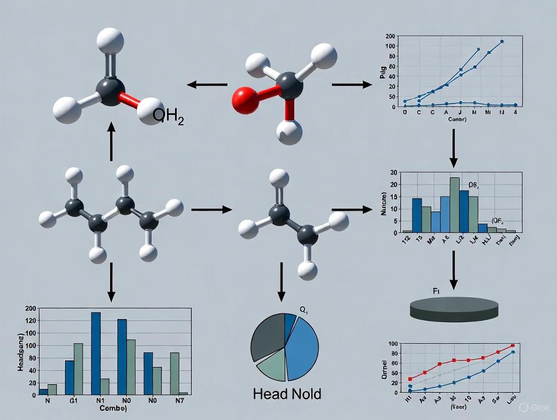
In static headspace gas chromatography (HS-GC), the sealed headspace vial functions as a self-contained, micro-scale ecosystem where a precise thermodynamic equilibrium is established between a sample phase and its vapor phase. This ecosystem is the foundational element enabling the analysis of volatile organic compounds (VOCs) in complex solid and liquid matrices without introducing non-volatile residues into the gas chromatograph [1] [2]. The core principle involves incubating a sample in a sealed vial, allowing volatile analytes to partition between the sample matrix and the headspace gas above it [1]. Once equilibrium is established, a portion of this headspace vapor is introduced into the GC system for separation and detection [3]. This technique is indispensable for applications ranging from residual solvent analysis in pharmaceuticals and blood alcohol content determination to characterizing flavor compounds in foods and volatiles in environmental samples [1]. The reliability of this entire analytical process hinges on a deep understanding of the interactions and equilibrium within the vial ecosystem.
Core Components of the Headspace Vial Ecosystem
The headspace vial ecosystem comprises three fundamental and interacting components: the sample (condensed) phase, the vapor phase (headspace), and the container system itself.
The Sample Phase (Condensed Phase)
The sample phase consists of the original liquid or solid material placed into the vial. This matrix contains the target volatile analytes. The chemical nature of this phase—its polarity, solubility parameters, and viscosity—profoundly influences the release of analytes into the vapor phase [2]. For instance, a polar analyte in a polar solvent will exhibit different partitioning behavior than the same analyte in a non-polar solvent. The volume of the sample phase is a critical experimental parameter, directly affecting the phase ratio and the resulting analyte concentration in the headspace [1].
The Vapor Phase (Headspace)
The vapor phase is the gaseous region above the sample within the sealed vial. It is into this phase that volatile and semi-volatile compounds evaporate [4]. In static headspace analysis, this vapor phase is the portion that is ultimately sampled and injected into the GC. The concentration of an analyte in this headspace ( CG ) is not the original concentration in the sample, but rather an equilibrium concentration that is proportional to the original concentration ( C0 ) [1] [2]. The volume of the headspace is a key variable that analysts can control to optimize sensitivity.
The Container System
The container system includes the vial, septum, and cap, which work together to create a sealed, inert environment for the equilibrium process.
- Vial: Typically made of borosilicate glass (USP Type I) with common volumes of 10 mL, 20 mL, or 22 mL [1] [5]. Vials can be clear or amber (for light-sensitive compounds) and may be silanized to minimize surface adsorption for trace analysis [5].
- Septa and Caps: A proper seal is paramount to prevent the loss of volatile analytes. Septa are typically constructed with PTFE/silicone layers, offering both chemical inertness and effective resealing capability, especially after multiple penetrations by the sampling needle [5]. Caps are either crimp-top, which provide a high-integrity seal for automated systems, or screw-top, which are convenient for manual operation [5].
The following diagram illustrates the relationships and interactions between these core components.
The Thermodynamic Equilibrium Principle
At the heart of static headspace analysis is the principle of thermodynamic equilibrium. After the sealed vial is incubated at a constant temperature, the rates of evaporation and condensation for each volatile component eventually become equal, resulting in steady-state concentrations in both the sample and vapor phases [6] [2]. This state is described by the partition coefficient (K) and the phase ratio (β).
The Partition Coefficient (K)
The partition coefficient, ( K = CS / CG ), is a temperature-dependent constant that defines the distribution of an analyte between the sample phase concentration ( CS ) and the gas phase concentration ( CG ) at equilibrium [2]. A low K value signifies that the analyte has a high volatility or low solubility in the sample matrix, leading to a higher concentration in the headspace. Conversely, a high K value indicates the analyte prefers the condensed phase, resulting in a lower headspace concentration [1] [4].
The Phase Ratio (β)
The phase ratio, ( \beta = VG / VS ), is the ratio of the vapor phase volume ( VG ) to the sample phase volume ( VS ) within the vial [2]. It is a physical, experimentally determined parameter. A smaller β (achieved by using a larger sample volume in a given vial size) generally leads to a higher analyte concentration in the headspace, thereby improving detection sensitivity for many compounds [1].
The Fundamental Quantitative Relationship
The relationship between the original concentration of the analyte in the sample ( C0 ) and the measured concentration in the gas phase ( CG ) is given by the fundamental headspace equation [1] [7]: [ CG = \frac{C0}{K + \beta} ] The detector response (peak area, A) is proportional to ( CG ) [1]: [ A \propto CG = \frac{C_0}{K + \beta} ] This equation reveals that to maximize the detector signal, the sum of ( K + \beta ) must be minimized [1]. This objective drives the optimization of key methodological parameters.
Table 1: Impact of Key Parameters on Detector Response
| Parameter | Effect on Partition Coefficient (K) and Phase Ratio (β) | Resulting Impact on Detector Response |
|---|---|---|
| Increase in Temperature | Decreases K (for most analytes) [1] [7] | Increases [1] [2] |
| Increase in Sample Volume (decreases β) | Decreases β [1] | Increases (especially when K is large) [1] |
| Modification of Sample Matrix | Can increase or decrease K (e.g., salting-out effect) [6] | Can be increased by favoring vapor phase [6] |
Methodologies for Establishing and Sampling Equilibrium
A standardized experimental workflow is essential for obtaining reproducible and quantitative results. The following diagram outlines the key stages of a static headspace analysis.
Sample Preparation and Vial Sealing
The sample, either liquid or solid, is accurately transferred into a headspace vial. For quantitative analysis, consistency in sample volume and matrix composition across vials is critical. The vial is immediately sealed with a septum and a crimp or screw cap to ensure no volatile analytes escape [1] [5]. For solid samples or to aid in the release of analytes, a solvent may be added, or the sample may be suspended in water [1].
Incubation and Equilibration
The sealed vial is placed in a temperature-controlled oven of the headspace autosampler. The temperature and the time are carefully optimized. The temperature must be high enough to facilitate the transfer of analytes into the vapor phase but kept safely below the boiling point of the solvent to avoid excessive pressure [1] [7]. Equilibration time, which is sample-dependent, must be determined experimentally to ensure the system has reached a stable equilibrium before sampling [1]. Modern instruments may incorporate vial shaking to accelerate this process [1].
Sampling the Headspace
Once equilibrium is attained, an automated system samples the headspace. A common method is the valve-and-loop technique, which involves three steps [1]:
- Pressurization: A sampling needle pierces the septum, and the vial is pressurized with carrier gas.
- Venting: The pressurized vapor is allowed to expand into a fixed-volume sample loop.
- Injection: A valve rotates, switching the carrier gas flow to sweep the contents of the sample loop through a heated transfer line into the GC inlet.
An alternative method is pressure-balanced sampling, which uses precise carrier gas pressure regulation to transfer the sample directly from the vial to the column without a sample loop, minimizing dead volume and potential contamination [4].
Quantitative Approaches and Advanced Techniques
Calibration and Quantification
When matrix effects are significant, standard calibration in a pure solvent may be inaccurate. Several matrix-independent quantification techniques are employed:
- Multiple Headspace Extraction (MHE): A series of consecutive extractions are performed from the same vial. The exponential decay of the analyte peak area is used to extrapolate the total analyte content in the sample, effectively canceling out matrix effects [1] [8].
- Standard Addition Method: Known amounts of the analyte standard are added directly to the sample in the vial. This method compensates for matrix effects by ensuring the standard and analyte experience the same matrix environment [8].
- Full Evaporation Technique (FET): A very small sample amount is used in a large vial volume, and the temperature is high enough to completely transfer volatiles to the gas phase. This allows for calibration using standard solutions in any convenient matrix, as the original sample matrix becomes irrelevant [8].
Enhancing Sensitivity
For analytes present at very low concentrations, sensitivity can be enhanced by moving beyond classical static headspace:
- Dynamic Headspace (Purge & Trap): An inert gas continuously purges the sample, sweeping volatiles onto an adsorbent trap. The trapped analytes are then thermally desorbed into the GC, providing a much larger injection volume and lower detection limits compared to static headspace [3] [7].
- Solid-Phase Microextraction (SPME): A fiber coated with a stationary phase is exposed to the headspace. Analytes partition into the coating, concentrating them. The fiber is then thermally desorbed in the GC inlet [6]. Newer devices like SPME Arrow offer higher sorbent capacity and thus greater sensitivity [6].
The Scientist's Toolkit: Essential Materials and Reagents
Table 2: Key Research Reagent Solutions for Headspace Analysis
| Item | Function & Importance |
|---|---|
| Headspace Vials (Borosilicate Glass) | Primary container for the ecosystem. Must be chemically inert and capable of withstanding pressure and temperature. Standard volumes are 10-22 mL [1] [5]. |
| PTFE/Silicone Septa | Provides a gas-tight seal and can be pierced by the sampling needle. The PTFE layer offers chemical inertness, while the silicone provides resealing capability [5]. |
| Crimp or Screw Caps | Secures the septum to the vial. Aluminum crimp caps are for single use and provide a high-integrity seal. Magnetic screw caps are reusable and convenient [5]. |
| Non-Volatile Salts (e.g., NaCl, K₂SO₄) | Used for "salting-out" – increasing the ionic strength of aqueous samples to reduce the solubility of volatile analytes, driving them into the headspace and increasing sensitivity [6]. |
| Internal Standards (e.g., deuterated analogs) | Added in a consistent amount to every sample and standard to correct for instrument variability, minor volume inaccuracies, and sample-to-sample preparation differences, improving quantitative accuracy. |
| High-Purity Gas Standards | Used for instrument calibration in gas-phase standard preparation, particularly useful for the Full Evaporation Technique (FET) or when creating custom calibration mixtures [8]. |
The headspace vial is far more than a simple container; it is a finely tunable micro-ecosystem governed by the predictable laws of thermodynamics. A deep understanding of the interactions between its three core components—the sample, the vapor phase, and the container—allows researchers to manipulate key parameters like temperature, phase ratio, and matrix composition to optimize analysis. By mastering the principles of equilibrium, as defined by the partition coefficient and phase ratio, and by applying robust methodologies and advanced techniques like MHE, scientists can transform static headspace sampling from a simple sample preparation tool into a powerful, quantitative, and indispensable technique for volatile compound analysis across countless industries.
Vapor-liquid equilibrium (VLE) describes the distribution of chemical species between vapor and liquid phases, a fundamental concept in thermodynamics and chemical engineering with critical applications across research and industry [9]. At equilibrium, the concentration of a vapor in contact with its liquid is expressed in terms of vapor pressure, which represents a partial pressure when other gases are present [9]. This equilibrium state is characterized by equivalent temperature, pressure, and partial molar Gibbs free energy for each component across both phases [9]. Understanding VLE is essential for designing separation processes like distillation columns, particularly fractional distillation, which exploits differences in component concentrations between liquid and vapor phases [9].
The composition of phases in mixtures is typically expressed using mole fractions. For a binary mixture with two components, the mole fractions are defined as x₁ = n₁/(n₁ + n₂) and x₂ = n₂/(n₁ + n₂), with the constraint that x₁ + x₂ = 1 [9] [10]. For multi-component mixtures, this relationship extends to x₁ + x₂ + ⋯ + xₙ = 1 [9].
Table 1: Fundamental Variables in Vapor-Liquid Equilibrium
| Variable | Symbol | Description | Application Context |
|---|---|---|---|
| Mole Fraction (Liquid) | xᵢ | Moles of component i divided by total moles in liquid phase | Raoult's Law: pᵢ = xᵢpᵢ* |
| Mole Fraction (Vapor) | yᵢ | Moles of component i divided by total moles in vapor phase | Dalton's Law: pᵢ = yᵢPᵢ |
| Vapor Pressure | pᵢ* | Pressure exerted by pure component i at system temperature | Characterizes volatility |
| Partial Pressure | pᵢ | Pressure contribution from component i in gas mixture | Equilibrium calculations |
| Henry's Constant | kH or H | Proportionality constant for gas solubility | Henry's Law: p = kHx |
Theoretical Foundations of Raoult's Law
Principle and Mathematical Formulation
Raoult's Law, formulated by French chemist François-Marie Raoult in 1887, states that the partial vapor pressure of each component in an ideal mixture is equal to the vapor pressure of the pure component multiplied by its mole fraction in the mixture [11]. Mathematically, for a component i in an ideal solution, this is expressed as:
pᵢ = xᵢpᵢ* [11]
where pᵢ is the partial vapor pressure of component i in the mixture, xᵢ is the mole fraction of component i in the liquid phase, and pᵢ* is the vapor pressure of pure component i at the same temperature [12] [10] [11].
For a mixture containing multiple volatile components, the total vapor pressure P above the solution can be obtained by combining Raoult's law with Dalton's law of partial pressures:
P = p₁ + p₂ + ⋯ = x₁p₁* + x₂p₂* + ⋯ [11]
When a non-volatile solute (with zero vapor pressure) is dissolved in a volatile solvent, the vapor pressure lowering is directly proportional to the solute mole fraction:
Δp = pₐ* - p = pₐ*xᵦ [11]
where pₐ* is the vapor pressure of the pure solvent and xᵦ is the mole fraction of the non-volatile solute [11].
Thermodynamic Basis and Ideal Solutions
From a thermodynamic perspective, compliance with Raoult's Law defines an ideal solution [11]. In such solutions, the chemical potential of each component is given by:
μᵢ = μᵢ* + RTlnxᵢ
where μᵢ* is the chemical potential of the pure component i [11]. At equilibrium, the chemical potential of each component in the liquid phase equals its chemical potential in the vapor phase (μᵢ,liq = μᵢ,vap) [11].
An ideal solution requires that intermolecular forces between unlike molecules are equal to those between like molecules, and that the molar volumes of the components are similar [11]. This is analogous to the ideal gas law, which becomes valid when interactive forces between molecules approach zero [11]. In practice, truly ideal solutions are rare, but the concept provides a valuable reference point for understanding real system behavior.
Limitations and Deviations from Ideal Behavior
In real solutions, deviations from Raoult's Law occur due to variations in intermolecular forces [12] [11]. These deviations provide insight into molecular interactions within the mixture:
Negative deviations occur when the vapor pressure is lower than predicted, indicating stronger attractive forces between unlike molecules than between like molecules [11]. An example is the chloroform-acetone system, where hydrogen bonding enhances intermolecular attraction [11].
Positive deviations occur when the vapor pressure is higher than predicted, indicating weaker attractive forces between unlike molecules [11].
The system of hydrochloric acid and water exhibits such strong negative deviation that it forms a negative azeotrope, where the mixture evaporates without composition change [11]. Most real solutions only approximate Raoult's Law when the liquid phase is nearly pure or when components are chemically similar [11].
Theoretical Foundations of Henry's Law
Principle and Mathematical Formulation
Henry's Law, formulated by William Henry in the early 19th century, states that at constant temperature, the amount of a given gas dissolved in a liquid is directly proportional to its partial pressure above the liquid [13] [10] [14]. Mathematically, this is expressed as:
p = kHx
where p is the partial pressure of the gas, x is the mole fraction of the dissolved gas in the liquid, and kH is Henry's constant, which depends on the solute, solvent, and temperature [13] [10] [14].
Henry's Law can also be expressed using concentration instead of mole fraction:
p = kHcC
where C is the concentration of the dissolved gas and kHc is the Henry's constant in appropriate units [13]. The dimensionless Henry constant is particularly useful and can be expressed as:
Hcc = ca/cg
where ca is the aqueous-phase concentration and cg is the gas-phase concentration [13]. For an ideal gas, the conversion between these forms is given by Hcc = RTHcp, where R is the gas constant and T is temperature [13].
Applications and Practical Significance
Henry's Law has numerous practical applications across scientific and industrial domains:
Carbonated beverages: Under high pressure, CO₂ solubility increases according to Henry's Law. When the container is opened, pressure decreases to atmospheric, solubility decreases, and CO₂ bubbles form [13].
Underwater diving: As divers descend, increased pressure causes more gas to dissolve in body tissues according to Henry's Law. During ascent, if decompression occurs too rapidly, the decreased solubility can cause bubble formation leading to decompression sickness [13].
High-altitude physiology: At high altitudes, reduced oxygen partial pressure leads to lower oxygen concentration in blood, potentially causing hypoxia [13].
Environmental science: Henry's Law governs the exchange of volatile compounds between water bodies and the atmosphere [13].
Henry's Law is most accurate for dilute solutions where the dissolved gas is at low concentration and behaves ideally [10] [14]. It does not apply well at high pressures or when the dissolved gas reacts chemically with the solvent [14].
Henry's Law Constants and Their Variations
Henry's Law constants can be expressed in multiple forms, with atmospheric chemists often defining the Henry solubility as Hscp = ca/p, where ca is the aqueous-phase concentration and p is the partial pressure [13]. The SI unit for Hscp is mol/(m³·Pa) [13].
Table 2: Henry's Law Constant Formulations
| Constant Type | Definition | Common Units | Application Context |
|---|---|---|---|
| Hcp (Solubility) | Hcp = ca/p | mol/(L·atm) | Atmospheric chemistry |
| Hpc (Volatility) | Hpc = p/ca = 1/Hcp | (L·atm)/mol | Environmental engineering |
| Hcc (Dimensionless) | Hcc = ca/cg | Unitless | Partition coefficient studies |
| Hxp (Mixing Ratio) | Hxp = x/p | mol/mol·Pa | Atmospheric modeling |
The Bunsen coefficient (α) and Kuenen coefficient (S) represent additional standardized forms of Henry's Law constants used in specific scientific contexts [13].
The Complementary Relationship Between Raoult's and Henry's Laws
Raoult's Law and Henry's Law represent complementary limiting laws that describe vapor-liquid equilibrium at different concentration ranges [14] [15]. For a binary mixture of components A (solvent) and B (solute):
As the solvent concentration approaches unity (xₐ → 1), its vapor pressure follows Raoult's Law: limₓ→₁(p/x) = p* [14]
As the solute concentration approaches zero (xᵦ → 0), its vapor pressure follows Henry's Law: limₓ→₀(p/x) = K [14]
This complementary relationship is illustrated in vapor pressure diagrams, where Raoult's Law defines the limiting slope as x → 1, and Henry's Law defines the limiting slope as x → 0 [15]. For a binary mixture of pure substances, these laws are complementary: if one law holds for one component, the other law holds for the second component [14].
This relationship has profound implications for understanding solution behavior across concentration ranges. In dilute solutions, the solute molecules are surrounded almost exclusively by solvent molecules, leading to Henry's Law behavior. As concentration increases, solute-solute interactions become significant, and deviations from Henry's Law occur [15].
Figure 1: The Complementary Relationship Between Raoult's and Henry's Laws in Vapor-Liquid Equilibrium
Application in Static Headspace Sampling Research
Fundamental Principles of Headspace Analysis
Static headspace extraction (SHE) is a sample introduction technique for gas chromatography that analyzes the vapor phase above a sample in a sealed vial [7] [16]. This method is particularly valuable for analyzing volatile compounds in complex matrices such as solids, viscous liquids, blood, or medications [16]. In pharmaceutical research, SHE is employed for residual solvents analysis according to USP method 467, ensuring drug product safety [16].
The fundamental equilibrium in headspace analysis can be represented as:
Analyte(sample) ⇌ Analyte(headspace)
with the equilibrium constant expressed as the partition coefficient K = Cs/Cg, where Cs is the concentration in the sample phase and Cg is the concentration in the gas phase [7]. In headspace literature, this partition coefficient is often inverted, with the vapor phase in the numerator [7].
Mathematical Framework for Headspace Quantitation
The relationship between headspace concentration and detector response is described by:
A ∝ Cg = C₀/(K + β) [16]
where A is the chromatographic peak area, Cg is the analyte concentration in the headspace, C₀ is the initial analyte concentration in the sample, K is the partition coefficient, and β is the phase ratio (ratio of vapor phase volume to sample phase volume) [16].
This equation demonstrates that detector response is proportional to the headspace concentration, which depends on the initial concentration divided by the sum of the partition coefficient and phase ratio [16]. To maximize detector response, conditions should minimize the sum K + β, thereby increasing the proportional amount of volatile targets in the headspace [16].
Optimizing Headspace Analysis Parameters
Successful headspace method development requires careful optimization of several key parameters:
Temperature: Increased vial temperature shifts solution-vapor equilibria toward the vapor phase, decreasing the partition coefficient and increasing detector response [7] [16]. However, temperature should remain approximately 20°C below the solvent boiling point to prevent excessive solvent vaporization [16].
Phase ratio (β): Defined as the ratio of headspace volume to sample volume (β = Vg/Vs) [16]. The phase ratio typically ranges between 1-20 in most SHE methods [7]. When the partition coefficient is similar in magnitude to the phase ratio, the phase ratio significantly impacts peak area [7].
Equilibration time: Sufficient time must be allowed for the system to reach equilibrium between the sample and vapor phases [7] [16]. Inadequate equilibration is a leading cause of reproducibility problems in headspace analysis [7].
Matrix effects: Strong solute-solvent interactions can reduce the impact of temperature on the partition coefficient [7]. Interestingly, when non-polar solutes are dissolved in polar solvents at low concentrations, matrix effects can enhance vaporization as the non-polar solute is repelled by the polar solvent [7].
Table 3: Key Parameters in Static Headspace Method Development
| Parameter | Symbol | Definition | Optimization Strategy |
|---|---|---|---|
| Partition Coefficient | K | K = Cs/Cg | Increase temperature; Modify matrix |
| Phase Ratio | β | β = Vg/Vs | Adjust sample volume; Vial size selection |
| Equilibration Time | t_eq | Time to reach equilibrium | Determine experimentally; Minimum 20 min |
| Incubation Temperature | T | Vial heating temperature | Balance sensitivity and solvent boiling point |
| Sample Volume | V_s | Volume of sample in vial | Typically 50% of vial capacity |
Figure 2: Static Headspace Extraction Workflow for Gas Chromatography
Experimental Protocols and Research Toolkit
Standardized Experimental Methodology
For residual solvent analysis in pharmaceutical applications (USP 467), the following protocol provides a representative framework:
Sample Preparation: Precisely weigh solid samples or measure liquid samples and transfer to headspace vials. For solid samples, addition of a minimal solvent volume may improve partitioning behavior [16]. Immediately cap vials with certified septa to prevent volatile loss [16].
Equilibration Conditions: Place vials in a temperature-controlled oven. Typical equilibration temperatures range from 60-120°C, depending on solvent volatility and sample stability [16]. Equilibration times typically range from 15-60 minutes, determined experimentally for each matrix [7].
Instrumental Parameters:
Chromatographic Conditions:
The Scientist's Toolkit: Essential Research Reagents and Materials
Table 4: Essential Materials for Headspace Analysis in Pharmaceutical Research
| Material/Reagent | Specification | Function in Analysis | Quality Considerations |
|---|---|---|---|
| Headspace Vials | 10-20 mL capacity, borosilicate glass | Contain sample during equilibration | Certified for volatile analysis; Consistent dimensions |
| Septa | PTFE/silicone or similar | Seal vials while allowing needle penetration | Low bleed; Minimal analyte adsorption |
| Internal Standards | e.g., d₈-Toluene, Acetonitrile-d₃ | Quantitation reference | Deuterated analogs of analytes; High purity |
| Reference Standards | Certified reference materials | Method calibration and validation | Traceable purity; Appropriate concentration |
| Matrix Modifiers | e.g., salts, water-miscible solvents | Adjust partition coefficient | High purity; Minimal volatile content |
| Calibration Solutions | Prepared in appropriate solvent | Instrument calibration | Prepared gravimetrically; Stored appropriately |
Advanced Techniques: Multiple Headspace Extraction
For challenging matrices where calibration standards cannot be matched to sample composition, Multiple Headspace Extraction (MHE) provides enhanced accuracy [16]. This technique involves performing successive extractions from the same vial:
- The vial is pressurized and an aliquot is taken from the headspace and injected into the GC
- This process is repeated multiple times (typically 3-5 extractions) [16]
- The peak areas decrease exponentially with each extraction
- The total original analyte content is determined by extrapolation [16]
Multiple Headspace Concentration (MHC) is a variant where instead of injecting after each extraction, the aliquots are concentrated in the GC inlet using a cryo trap before analysis [16].
Raoult's Law and Henry's Law provide complementary frameworks for understanding vapor-liquid equilibrium across the concentration spectrum. While Raoult's Law describes solvent behavior in concentrated solutions and ideal mixtures, Henry's Law governs solute behavior in dilute solutions. In static headspace sampling research, these principles form the theoretical foundation for predicting and optimizing analyte partitioning between sample and vapor phases.
The application of these equilibrium principles enables robust analytical methods for volatile compound analysis in pharmaceutical development, particularly for residual solvent testing. Through careful optimization of temperature, phase ratio, and matrix conditions, researchers can leverage these fundamental laws to develop sensitive, reproducible headspace methods that ensure drug product safety and quality.
In the field of static headspace gas chromatography (HS-GC), the partition coefficient, denoted as K, is a fundamental thermodynamic parameter that defines the equilibrium distribution of an analyte between the sample phase (liquid or solid) and the gas phase (headspace) in a sealed vial [17]. This coefficient is the cornerstone of all quantitative static headspace analysis, as it ultimately determines the concentration of the volatile analyte available for injection into the gas chromatograph, thereby dictating the method's sensitivity and reliability [18] [7].
This whitepaper delineates the role of the partition coefficient within the broader equilibrium principles governing static headspace sampling. For researchers in drug development, a precise understanding of 'K' is not merely academic; it is a critical prerequisite for robust method development, ensuring accurate quantification of volatile impurities, residual solvents, and active pharmaceutical ingredients (APIs) [19].
Theoretical Foundation and Mathematical Definition
The partition coefficient (K) is formally defined as the ratio of the analyte's concentration in the sample phase (C_s) to its concentration in the gas phase (C_g) at equilibrium, at a defined temperature [17].
K = Cs / Cg [17]
A lower value of K signifies that the analyte favors the gas phase. For instance, n-Hexane in water at 40°C has a very low K of 0.14, indicating high volatility and a strong tendency to partition into the headspace. Conversely, a high K value indicates the analyte has a greater affinity for the sample matrix. Ethanol in water at 40°C, with a K of 1355, demonstrates this, remaining largely in the liquid phase and resulting in a lower headspace concentration [17]. The relationship between the initial sample concentration (C_0) and the final headspace concentration (C_g) is given by the fundamental headspace equation [18] [17]:
Cg = C0 / (K + β)
Here, β is the phase ratio, defined as the ratio of the gas phase volume (V_g) to the sample phase volume (V_s) in the vial: β = V_g / V_s [18] [17]. This equation synthesizes the two primary factors controlling headspace sensitivity: the chemical nature of the analyte and matrix (captured by K) and the physical geometry of the vial (captured by β). The goal of method development is to minimize the sum K + β to maximize C_g [18].
Distinguishing Partition and Distribution Coefficients
In broader chemical contexts, it is vital to differentiate the partition coefficient from the distribution coefficient. The partition coefficient (P or K_OW) typically refers to the concentration ratio of the un-ionized form of a compound between two immiscible solvents, most commonly octanol and water, and is a measure of its lipophilicity [20]. For ionizable compounds, which constitute approximately 95% of pharmaceuticals, the distribution coefficient (log D) is used, as it represents the ratio of the sum of all forms of the compound (ionized plus un-ionized) in the two phases [20] [21]. The distribution coefficient is pH-dependent, whereas the partition coefficient for a non-ionizable compound is not [20].
Experimental Determination of the Partition Coefficient
Core Methodology and Workflow
The experimental determination of 'K' in a headspace context involves establishing equilibrium in a sealed system and measuring analyte concentrations in both phases. The following workflow outlines the primary stages of this process.
Step-by-Step Protocol:
- Sample Preparation: A known weight or volume of the sample is placed into a headspace vial. For accurate K determination, the initial concentration of the analyte,
C_0, must be known [19]. - Equilibration: The vial is sealed immediately with a septum cap to prevent volatile loss and placed in a thermostatically controlled oven. The vial is heated for a predetermined time to allow the analyte to distribute between the sample and gas phases until equilibrium is achieved [3] [18]. The temperature must be stable, as 'K' is highly temperature-dependent [17].
- Headspace Sampling: Once equilibrium is established, an aliquot of the headspace vapor is extracted. In modern automated systems, this is done by pressurizing the vial with carrier gas and then transferring a portion of the headspace into a sample loop before injection into the GC [3] [18].
- GC Analysis and Quantification: The injected sample is separated and quantified using Gas Chromatography, typically with a Flame Ionization Detector (FID) or Mass Spectrometer (MS) [19]. The peak area (A) is proportional to
C_g[18]. - Calculation of K: With
C_0known andC_gdetermined from the calibration curve, the partition coefficient can be calculated if the phase ratio (β) is known, using the equationC_g = C_0 / (K + β)[17].
Key Research Reagent Solutions
Successful experimental determination of K relies on specific materials and reagents. The following table details essential items and their functions in the workflow.
| Item | Function & Importance |
|---|---|
| Headspace Vials | Sealed containers (e.g., 10-22 mL) that maintain pressure and integrity during heating; a tight seal is critical to prevent volatile loss [18]. |
| Inorganic Salts (e.g., NaCl) | Added to the sample matrix to "salt out" polar organic volatiles, decreasing their solubility in water and lowering the K value, thereby enhancing sensitivity [17]. |
| Gas-Tight Syringe | For manual sampling, it must be heated to prevent condensation of volatiles during transfer from vial to GC inlet [7]. |
| GC Capillary Column | The stationary phase (e.g., 6% cyanopropyl phenyl) is critical for separating volatile compounds; selection impacts resolution of critical pairs [19]. |
| Certified Reference Standards | Pure analytes of known concentration are essential for preparing calibration standards to quantify C_g and validate the method's accuracy and linearity [19]. |
Quantitative Data in Headspace Analysis
The partition coefficient is influenced by the specific analyte-solvent system and temperature. The table below provides K values for common solvents in air-water systems, illustrating the spectrum from high volatility (low K) to low volatility (high K).
Table 1: Partition Coefficients (K) of Common Solvents in Air-Water Systems at 40°C [17]
| Solvent | K Value | Solvent | K Value |
|---|---|---|---|
| Cyclohexane | 0.077 | n-Butylacetate | 31.4 |
| n-Hexane | 0.14 | Ethylacetate | 62.4 |
| Tetrachlorethylene | 1.48 | Methylethylketone | 139.5 |
| 1,1,1-Trichlormethane | 1.65 | n-Butanol | 647 |
| O-Xylene | 2.44 | Isopropanol | 825 |
| Toluene | 2.82 | Ethanol | 1355 |
| Benzene | 2.90 | Dioxane | 1618 |
| Dichloromethane | 5.65 |
Optimizing the Partition Coefficient in Method Development
A primary goal in HS-GC method development is to minimize K to maximize the concentration of analyte in the headspace (C_g) and improve detection sensitivity. The following diagram illustrates the key parameters a researcher can control to achieve this optimization.
Key Optimization Strategies:
- Temperature Control: Increasing the vial temperature is the most effective way to reduce K for most analytes, as it enhances vapor pressure. For example, the K value for ethanol in water drops from 1355 at 40°C to 328 at 80°C [17]. The optimal temperature is typically just below the boiling point of the solvent [18].
- Matrix Modification (Salting-Out Effect): Adding inorganic salts like ammonium sulfate or sodium chloride to an aqueous sample decreases the solubility of polar organic volatiles, promoting their transfer into the headspace and lowering K [17]. The effect is most pronounced for compounds that are already fairly soluble in water.
- Phase Ratio (β) Adjustment: Reducing the phase ratio β by increasing the sample volume in the vial can enhance
C_g. However, this is most effective for analytes with low K values (high volatility). For compounds with high K, changing the sample volume has a minimal effect, and optimizing temperature and matrix is more impactful [18] [17].
The partition coefficient (K) is more than a simple concentration ratio; it is the fundamental thermodynamic constant that governs analyte behavior in static headspace systems. A deep and practical understanding of K, the factors that influence it, and the methods for its determination is indispensable for researchers developing robust and sensitive HS-GC methods. Mastering the manipulation of K through temperature, matrix modification, and vial geometry allows scientists to harness the full power of equilibrium principles, ensuring reliable data in critical applications from drug development to environmental monitoring.
In static headspace gas chromatography (HS-GC), the phase ratio (β) is a fundamental parameter defining the volumetric relationship between the gas (headspace) and condensed (sample) phases within a sealed vial. It is mathematically expressed as β = VG/VL, where VG is the volume of the headspace gas and VL is the volume of the sample liquid [2] [22]. Within the broader context of equilibrium principles governing static headspace sampling, the phase ratio is a critical variable that analysts can directly control to optimize system performance. The equilibrium concentration of an analyte in the gas phase (CG), which is ultimately measured by the GC detector, is governed by the equation: CG = C0 / (K + β) [23] [2]. Here, C0 is the original analyte concentration in the sample, and K is the temperature-dependent partition coefficient.
This equation reveals that to maximize the detector response (A ∝ CG), the sum of K and β must be minimized [23]. For a given analyte-solvent system where K is fixed at a specific temperature, manipulation of the phase ratio β becomes a primary lever for enhancing analytical sensitivity. A thorough understanding of how β interacts with other fundamental parameters is therefore not merely an operational detail but a core aspect of rational, principles-based method development in static headspace research. This guide provides an in-depth examination of phase ratio optimization strategies for researchers and drug development professionals.
Theoretical Foundation: The Equilibrium Principle
The entire process of static headspace analysis is predicated on achieving a thermodynamic equilibrium of volatile analytes between the sample matrix and the inert gas phase above it in a sealed vial [23] [2]. The foundational model for this system is described by the following relationship:
CG = C0 / (K + β)
Where:
- CG: Concentration of the analyte in the gas phase (measured by the GC)
- C0: Original concentration of the analyte in the sample
- K: Partition Coefficient (K = CS / CG), representing the ratio of the analyte's concentration in the sample phase (CS) to its concentration in the gas phase (CG) at equilibrium [23] [24]
- β: Phase Ratio (β = VG / VL) [2] [22]
The following diagram illustrates the core logical relationship between these parameters and the final detector response, which is the ultimate goal of the optimization process.
This fundamental equation demonstrates that the detector signal is directly proportional to the gas-phase concentration CG [23]. Consequently, to maximize the signal for a given C0, the sum (K + β) must be minimized [23]. The phase ratio β is a physical parameter that can be easily and precisely controlled by the analyst through the choice of vial size and sample volume, making it a powerful and straightforward tool for method optimization.
Optimizing Phase Ratio for Different Analyte Types
The partition coefficient (K) indicates an analyte's relative affinity for the sample matrix versus the gas phase. The effectiveness of phase ratio (β) optimization is highly dependent on the value of K, leading to three primary optimization scenarios [24] [22].
Table 1: Guide to Phase Ratio Optimization Based on Analyte Solubility
| Analyte Type | Partition Coefficient (K) | Impact of Increasing Sample Volume (Decreasing β) | Recommended Strategy |
|---|---|---|---|
| High-Solubility Analytes(e.g., Ethanol in water) | K >> β(e.g., ~500) [24] [22] | Negligible ImprovementGas concentration (CG) is dominated by the large K value. Increasing sample volume has minimal effect on CG [24] [22]. | Focus on reducing K instead:• Increase temperature significantly [23] [2].• Use salting-out techniques (e.g., KCl) [24] [22].• Modify solvent to reduce solubility. |
| Intermediate-Solubility Analytes | K ≈ β(e.g., ~10) | Approximately Linear ImprovementIncreasing sample volume (decreasing β) provides a proportional increase in CG [24] [22]. | Balanced approach:• Use a moderate sample volume.• Combine with moderate temperature increase.• A phase ratio of β = 1 (e.g., 10 mL in a 20 mL vial) is often ideal [24]. |
| Low-Solubility Analytes(e.g., n-Hexane in water) | K << β(e.g., ~0.01) [24] [22] | Significant ImprovementGas concentration (CG) is highly sensitive to β. Increasing sample volume (decreasing β) yields a large, proportional increase in CG [24] [22]. | Maximize sample volume:• Use the largest feasible sample volume in a given vial to minimize β [23].• Temperature control is less critical for precision [2]. |
Practical Considerations for Vial and Volume Selection
A general best practice for method development is to begin with a phase ratio of β = 1, which is achieved by using 10 mL of sample in a 20-mL headspace vial [24]. This simplifies initial calculations and provides a balanced starting point for further optimization. When higher sensitivity is required for analytes with low or intermediate K values, using a larger vial (e.g., 20-mL instead of a 10-mL vial) with a greater sample volume is an effective strategy, as it directly reduces the phase ratio β [23]. It is critical to leave at least 50% of the vial's volume as headspace to ensure proper pressurization and sampling by the autosampler [23].
An Integrated Experimental Workflow for Parameter Optimization
Establishing a robust static headspace method requires a systematic approach to optimize all interdependent parameters. The following workflow provides a detailed protocol for method development.
Detailed Experimental Protocol for Phase Ratio and Equilibrium Optimization
This protocol outlines the key experiments needed to characterize and optimize the phase ratio and related parameters for a novel sample type.
Experiment 1: Determining Equilibration Time
- Preparation: Prepare multiple identical samples in headspace vials, ensuring consistent sample volume and matrix [23].
- Time-Course Incubation: Place the vials in the headspace sampler oven set at a constant temperature (e.g., 60°C). Use agitation if available to enhance transfer kinetics [23] [24].
- Sequential Analysis: Remove and analyze vials at increasing time intervals (e.g., 5, 10, 20, 30, 40, 60 minutes).
- Data Analysis: Plot the peak area of the target analytes versus time. The minimum time required for the peak areas to stabilize is the equilibration time. Do not assume this correlates with the partition coefficient K; it must be determined experimentally for each sample type [24] [22].
Experiment 2: Optimizing Phase Ratio (β) and Sample Volume
- Vial Size Comparison:
- Sample Volume Optimization:
- Prepare samples with different volumes (e.g., 2, 5, 10 mL) in the same size vial (e.g., 20-mL) [23].
- Analyze and plot peak area versus sample volume. The optimal volume provides the best sensitivity while maintaining at least 50% headspace and not leading to over-pressurization issues, especially for aqueous samples at high temperatures [23] [22].
Experiment 3: Optimizing Equilibration Temperature
- Temperature Gradient: Prepare identical samples and equilibrate them at a range of temperatures (e.g., 40, 50, 60, 70, 80 °C) for the predetermined equilibration time [23].
- Analysis: Plot the relative peak area against temperature. The response will typically increase with temperature until a plateau is reached. The optimal temperature is at the beginning of this plateau. Note: For aqueous samples, the maximum oven temperature should be kept about 20 °C below the solvent boiling point to prevent excessive pressure [23].
Experiment 4: Evaluating the "Salting-Out" Effect
- Sample Preparation: Prepare a set of samples with increasing concentrations of a non-volatile salt like potassium chloride (KCl) [24] [22].
- Analysis: Analyze the samples and compare peak areas. A significant increase in the response for polar analytes confirms the salting-out effect is beneficial for the method, reducing the effective K value by decreasing the analyte's solubility in the aqueous matrix [24].
The Scientist's Toolkit: Essential Materials and Reagents
A successful headspace analysis requires precise materials and instrumentation. The following table details the key components of a static headspace research system.
Table 2: Essential Research Reagents and Materials for Static Headspace-GC
| Item Name | Specification / Example | Critical Function in the Workflow |
|---|---|---|
| Headspace Vials | 10-mL, 20-mL, 22-mL capacity; chemical inertness [23]. | To contain the sample and headspace gas; volume determines the available range for the phase ratio (β). |
| Gas-Tight Seals | Septa and crimp caps capable of withstanding pressure [23]. | To maintain a sealed, pressurized environment and prevent loss of volatile analytes prior to sampling. |
| Automatic Headspace Sampler | e.g., Agilent 7697A/8697 models with valve-and-loop design [23]. | To automate vial incubation, pressurization, and the transfer of the headspace sample to the GC with high precision. |
| Non-Volatile Salts | Potassium Chloride (KCl), high purity [24] [22]. | "Salting-out" agent to reduce solubility of polar analytes in aqueous matrices, thereby reducing K and increasing CG. |
| Matrix-Matched Standards | Standards prepared in a blank matrix identical to the sample [24]. | For accurate calibration, as the matrix components significantly affect the activity coefficient and partition coefficient (K). |
| GC with Detector | GC system with FID, MS, or other suitable detector. | The core analytical instrument for separating and quantifying the volatile compounds introduced from the headspace sampler. |
Application in Drug Development and Research
The optimization of phase ratio and other headspace parameters is critical in regulated and research environments where reliability and sensitivity are paramount.
- Residual Solvents Analysis (USP <467>): This standardized method for detecting volatile impurities in pharmaceuticals is a quintessential application of static headspace-GC [23] [2]. Precise control of phase ratio, temperature, and equilibration time is mandatory to meet strict regulatory limits and ensure drug safety [23].
- Analysis of Complex Matrices: For solid or viscous drug formulations, headspace sampling avoids introducing non-volatile matrix components into the GC system [23]. In these cases, the phase ratio is optimized alongside other techniques, such as Multiple Headspace Extraction (MHE), which involves repeated sampling from the same vial to achieve accurate quantitation when a blank matrix is unavailable [23].
The phase ratio (β) is a simple concept with a profound impact on the efficacy of static headspace analysis. Its optimization, grounded in the equilibrium principle CG = C0 / (K + β), is not a standalone activity but an integral part of a holistic method development strategy. By systematically investigating and optimizing the phase ratio in concert with temperature, equilibration time, and matrix composition, scientists can develop robust, sensitive, and reliable HS-GC methods. This principles-based approach is essential for advancing research and ensuring quality in critical applications, from pharmaceutical drug development to environmental and food safety monitoring.
In static headspace sampling, the equilibrium distribution of volatile compounds between a sample matrix and the gas phase is governed by fundamental equilibrium principles. A core tenet of this equilibrium is that the vapor pressure of a compound above a solution is directly proportional to its mole fraction in that solution, multiplied by a compound-specific parameter known as the activity coefficient [24]. This coefficient quantifies the degree of intermolecular attraction between the analyte and other species within the sample matrix [24]. A higher activity coefficient indicates stronger repulsive forces between the analyte and matrix, favoring partitioning into the headspace. Conversely, a lower activity coefficient indicates stronger attractive intermolecular forces, effectively "trapping" the analyte in the sample phase and reducing its apparent volatility [25] [24]. Understanding and quantifying these interactions is therefore critical for accurate quantitative analysis in applications ranging from biomarker discovery in biological samples to residual solvent testing in pharmaceuticals [25] [24].
Theoretical Foundation: Activity Coefficients in Headspace Equilibrium
The foundational equation for static headspace analysis is derived from Raoult's Law or Henry's Law (for low analyte concentrations). It describes the concentration of an analyte in the gas phase (CG) relative to its concentration in the original sample (Co) [24]:
CG = Co / K
Here, K is the partition coefficient, a central parameter influenced by the activity coefficient. The value of K is determined by the equilibrium established between the sample (CS) and the headspace gas (CG), such that K = CS / CG [24]. The partition coefficient is itself a function of the phase ratio (β = VG / VL, the ratio of headspace volume to sample volume) and the intermolecular forces captured by the activity coefficient.
The following diagram illustrates the core equilibrium relationship and the key experimental parameters that influence the partition coefficient, K.
Strong intermolecular interactions between a volatile analyte and the sample matrix result in a low activity coefficient. This manifests as a high partition coefficient (K), meaning a significant majority of the analyte remains dissolved in the sample phase [25]. For example, in aqueous solutions, ethanol has a K value of approximately 500 due to hydrogen bonding, meaning it is 500 times more concentrated in the sample than in the headspace. In contrast, a non-polar solvent like n-hexane in water has a very low K value of about 0.01, favoring the headspace by a factor of 100 [24]. These interactions are not limited to simple solutions; in complex biological matrices like blood, proteins such as Human Serum Albumin (HSA) can bind irreversibly to volatile molecules, while lipids can dissolve lipophilic analytes, both mechanisms reducing headspace concentration independent of the true sample concentration [25].
Key Intermolecular Forces and Experimental Evidence
The impact of molecular interactions on volatility is demonstrated by experiments comparing headspace responses of volatile compounds across different matrices. Research shows that lower headspace responses are consistently observed in samples containing proteins or lipids compared to pure water, even when analytes are fortified at equal concentrations [25]. These interactions can arise from various irreversible chemical bonds and forces between the volatile molecules and matrix components [25].
Table 1: Selected Volatile Compounds and Their Properties in Headspace Analysis
| Volatile Compound | Log Kow | Key Functional Group | Primary Interaction Type with Matrix |
|---|---|---|---|
| 1-Hexanol | 1.80 [25] | Hydroxyl (-OH) | Hydrogen Bonding |
| Hexanal | 1.78 [25] | Aldehyde (-CHO) | Dipole-Dipole |
| Benzaldehyde | 1.48 [25] | Aromatic Aldehyde | π-π Stacking, H-binding |
| 2-Octenal | 2.57 [25] | Alkene, Aldehyde | Hydrophobic, Dipole-Dipole |
| Nonanal | 3.27 [25] | Aldehyde | Hydrophobic, Dipole-Dipole |
| 2-Nonanone | 3.16 [25] | Ketone (>C=O) | Dipole-Dipole, H-binding |
The octanol-water partition coefficient (Kow) is a useful predictor for the hydrophilic or lipophilic nature of a compound and its likely behavior in a matrix [25]. Very lipophilic metabolites (high Log Kow) in lipid-rich samples will strongly favor the sample phase over the headspace due to high solubility [25]. Furthermore, the adsorption of small molecules to abundant proteins like HSA represents another significant interaction that influences the first equilibrium in headspace analysis, meaning headspace measurements often reflect only the unbound fraction of these molecules [25].
Experimental Protocols for Investigating Molecular Interactions
Protocol 1: Assessing Matrix Effects on Headspace Concentration
This methodology evaluates how different sample matrices influence the headspace concentration of volatile metabolites [25].
- Test Solutions Preparation: Prepare concentrated stock solutions (10 mg/mL) of target volatile analytes in ethanol. Create an aqueous fortification solution by diluting 10 µL of the stock into 3.0 mL of water. Fortify 1.0 mL of each test medium (e.g., water, bovine serum, intralipid emulsion) with 10 µL of the aqueous fortification solution to yield a final concentration of approximately 0.3 ppm for each analyte [25].
- Headspace Sampling: Transfer 200 µL of the test solution to a 20 mL glass headspace vial. Seal the vial immediately with a magnetic crimp cap. Incubate samples at a set temperature (e.g., 40°C) with agitation at 500 rpm for 10 minutes to reach equilibrium [25].
- SPME Extraction and Analysis: Use a DVB/C-WR/PDMS "arrow" fiber for a 10-minute extraction at the incubation temperature with a stirring speed of 1000 rpm. Following extraction, thermally desorb the analytes in the GC injection port at 230°C for 2 minutes in splitless mode [25].
- GC-MS Analysis: Conduct chromatographic separation using a 30 m × 0.25 mm Stabilwax column. Employ a temperature program: hold at 40°C for 2 minutes, then ramp at 5.5°C per minute to 230°C, with a final hold for 2 minutes. Use helium carrier gas at a constant flow of 1.1 mL/min. Operate the mass spectrometer with an ionization energy of 70 eV and a scan range of m/z 33–400 [25].
- Data Analysis: Compare the raw and internal standard-normalized peak responses for each analyte across the different media. Statistical analysis (e.g., two-way ANOVA) can be used to determine significant differences attributable to the matrix [25].
Protocol 2: Coupling HS-SPME with Inverse Gas Chromatography (IGC) for Quantification
This advanced protocol uses IGC to estimate partition coefficients for more accurate quantification, overcoming the limitation of uneven SPME fiber sensitivity [26].
- IGC Measurement: Pack a GC column with a solid support coated with a stationary phase (e.g., polydimethylsiloxane, PDMS) identical to the SPME fiber coating. Inject pure probe compounds (e.g., n-heptane, toluene) into the IGC system at a series of temperatures (e.g., 100°C to 170°C) at a constant flow rate and column inlet pressure [26].
- Partition Coefficient Calculation: From the measured retention times, calculate the specific retention volumes (Vgo) for each probe. Construct retention diagrams (lnVgo vs. 1/T) to obtain linear relationships. The molar enthalpies of sorption (ΔHmS) are derived from the slopes of these lines, and the relative partition coefficients (Kfh) between the headspace and the SPME fiber are estimated from the intercepts and other thermodynamic data [26].
- HS-SPME of Model Headspace: Prepare a model headspace containing known compositions of vaporized probe compounds. Sample this headspace using the PDMS SPME fiber at controlled temperatures (e.g., 100°C and 130°C) and analyze via GC-MS to obtain experimental composition data [26].
- Data Integration and True Composition Estimation: Use the relative partition coefficients obtained from IGC and the experimental compositions from HS-SPME to estimate the true relative compositions of the compounds in the headspace. The formula for the amount of analyte (n) extracted considers both the fiber/headspace (Kfh) and headspace/sample (Khs) partition coefficients, though the latter can be omitted for simplified model systems [26].
The following workflow integrates these protocols, showing the path from sample preparation to accurate composition estimation.
The Scientist's Toolkit: Essential Reagents and Materials
Successful investigation into activity coefficients and volatility requires specific, high-purity materials and reagents.
Table 2: Key Research Reagent Solutions and Materials
| Item | Typical Specification / Source | Function in Experiment |
|---|---|---|
| Volatile Analytic Standards | Purity 98.0-99.7% (e.g., Sigma-Aldrich) [25] | Target compounds for headspace analysis; represent common metabolites. |
| Deuterated Internal Standard | e.g., Acetophenone-d5 [25] | Aids in quantitative analysis by accounting for instrumental variability. |
| SPME Fiber | 1.10 mm DVB/C-WR/PDMS "Arrow" [25] | Extracts and concentrates volatile analytes from the headspace. |
| Bovine Serum Albumin (BSA) | Fatty acid free [25] | Simulates protein-binding effects in biological samples like blood. |
| Lipid Emulsion | e.g., 20% Intralipid [25] | Simulates lipid-analyte solubility interactions in biological fluids. |
| Protein-Free Serum | Prepared via solvent denaturation & centrifugation [25] | Serves as a control to isolate the effect of proteins from the serum matrix. |
| Chromatographic Support | e.g., Non-acid washed Chromosorb P (60/80 mesh) [26] | Solid support for coating with polymer (e.g., PDMS) in IGC experiments. |
| Polydimethylsiloxane (PDMS) | Molar mass ~95,000 g/mol [26] | Stationary phase for IGC column packing; mimics SPME fiber coating. |
Optimizing Headspace Analysis: Practical Considerations
To overcome the suppressive effects of intermolecular forces and maximize headspace response, several practical strategies can be employed:
- Temperature Control: Increasing the equilibration temperature is an effective way to improve the headspace concentration of analytes with high partition coefficients (K), such as polar compounds in polar matrices. For analytes with a K of 500, a temperature accuracy of ±0.1°C is required to achieve a precision of 5% [24]. Research indicates that headspace responses can be maximized in the temperature range of 60 to 70°C [25].
- Salting Out: The partition coefficient of polar analytes in polar matrices (like water) can be significantly reduced by adding a high concentration of salt, such as potassium chloride. This "salting out" effect decreases the solubility of the analyte in the aqueous phase, driving it into the headspace [24].
- Sample Volume and Phase Ratio: The effect of sample volume is highly dependent on the partition coefficient. For analytes with low K values, increasing the sample volume gives a large proportional increase in headspace concentration. For analytes with high K values, increasing the sample volume has a negligible effect. A common practice is to use 10 mL of sample in a 20 mL vial, creating a phase ratio (β) of 1, which simplifies calculations [24].
- Matrix-Matched Calibration: Because matrix components significantly affect the activity coefficient, it is critical to calibrate instrument response using standards that are matrix-matched to the sample. A significant challenge is obtaining a suitable "blank" matrix, which sometimes requires exhaustive extraction of analytes from a sample to create one [24]. Furthermore, normalization of peak responses to an internal standard does not always account for these strong matrix-analyte interactions [25].
In static headspace gas chromatography (HS-GC), the equilibrium state is a fundamental concept governing the distribution of volatile analytes between the sample matrix and the gas phase. Understanding the kinetic principles that drive analyte migration and stabilization is critical for researchers and drug development professionals seeking to optimize analytical methods for residual solvents, volatile impurities, and active pharmaceutical ingredients [27]. This process is not instantaneous; it involves a dynamic journey of molecules from the sample into the headspace, culminating in a stable state where the rate of analyte evaporation equals the rate of condensation [24]. This technical guide delves into the core mechanisms, mathematical models, and experimental parameters controlling this crucial pre-analytical step, providing a detailed framework within the broader thesis of equilibrium principles in static headspace research.
Theoretical Foundations of Migration and Equilibrium
The Kinetic Process of Analyte Migration
The journey of an analyte from the sample matrix to the headspace is a kinetic process driven by the analyte's inherent vapor pressure and its interaction with the sample matrix. Initially, upon vial sealing and heating, a concentration gradient exists, with the analyte concentration being highest in the sample phase and zero in the headspace. This gradient provides the driving force for mass transfer [27]. The process can be broken down into several stages:
- Desorption from the Matrix: Analyte molecules are released from their binding sites within the sample matrix. The energy required for this step is supplied by the incubation temperature.
- Diffusion to the Interface: Molecules diffuse through the sample to the liquid (or solid)-gas interface.
- Evaporation: Molecules cross the interface and enter the gas phase.
- Diffusion in Headspace: Volatilized analytes diffuse throughout the headspace volume to establish a homogeneous concentration.
Over time, the reverse process—condensation of analyte molecules from the headspace back into the sample—begins to occur at an increasing rate. Equilibrium, a dynamic steady state, is achieved when the rate of transfer from the sample to the headspace is exactly equal to the rate of transfer from the headspace back into the sample [27]. At this point, the concentrations in both phases remain constant, and the system is ready for sampling.
Mathematical Modeling of the Equilibrium State
The relationship at equilibrium is quantitatively described by the partition coefficient (K) and the phase ratio (β), which are incorporated into a key mathematical expression that predicts the detector response [27]:
A ∝ CG = C0 / (K + β)
Where:
- A is the peak area obtained from the GC detector.
- CG is the concentration of the analyte in the gas phase (headspace) at equilibrium.
- C0 is the original concentration of the analyte in the sample.
- K is the partition coefficient, defined as CS/CG, where CS is the analyte concentration in the sample phase at equilibrium.
- β is the phase ratio, defined as VG/VL, the ratio of the headspace volume to the sample volume [24] [27].
This equation shows that to maximize the detector signal (A), the sum of K and β must be minimized. A low K value indicates that the analyte favors the gas phase, while a low β value is achieved by having a larger sample volume relative to the headspace.
Quantitative Data and Experimental Parameters
The following tables summarize the core quantitative relationships and the impact of key experimental parameters on the kinetic journey toward equilibrium and the final analytical signal.
Table 1: Key Quantitative Parameters in Headspace Equilibrium
| Parameter | Symbol & Formula | Definition | Impact on Analysis |
|---|---|---|---|
| Partition Coefficient | K = CS / CG | Measures the distribution of analyte between the sample (S) and gas (G) phases at equilibrium [27]. | A high K means the analyte prefers the sample matrix, leading to a lower headspace concentration. The goal is to minimize K. |
| Phase Ratio | β = VG / VL | The ratio of headspace volume (VG) to sample volume (VL) [27]. | A lower β (more sample, less headspace) increases the analyte concentration in the headspace, improving sensitivity. |
| Detector Response | A ∝ C0 / (K + β) | The chromatographic peak area (A) is proportional to the headspace concentration, which depends on C0, K, and β [27]. | This is the fundamental equation for optimizing headspace sensitivity. Maximizing A is the primary objective. |
Table 2: Impact of Experimental Variables on Kinetics and Equilibrium
| Variable | Effect on Kinetic Process | Effect on Equilibrium Concentration (CG) | Practical Consideration |
|---|---|---|---|
| Temperature | Increases the kinetic energy of molecules, speeding up diffusion and the rate of equilibration [27]. | Generally increases CG by reducing the partition coefficient (K) [24] [27]. | Must be controlled precisely (±0.1°C for high K analytes); kept ~20°C below solvent boiling point [24] [27]. |
| Equilibration Time | Must be sufficient for the system to reach a dynamic steady state. Time is matrix- and analyte-dependent [24]. | No impact if equilibrium is reached; insufficient time gives an unstable, non-equilibrium concentration. | Must be determined experimentally for each new method. Agitation can significantly reduce the time required. |
| Sample Volume (VL) | Affects the path length for diffusion, potentially increasing equilibration time for larger volumes. | Increases CG for analytes with low K by reducing the phase ratio (β). For high K, the effect is minimal [24] [27]. | A best practice is to fill 50% of the vial volume (e.g., 10 mL in a 20 mL vial) to optimize β [27]. |
| Salting-Out | Can alter the kinetics of release from an aqueous matrix. | Significantly reduces K for polar analytes in polar matrices (e.g., water), increasing their concentration in the headspace [24]. | Use of salts like potassium chloride is common for analyzing alcohols or other water-soluble volatiles. |
Experimental Protocols for Kinetic and Equilibrium Studies
Protocol: Determination of Optimal Equilibration Time
Objective: To experimentally determine the minimum time required for a specific analyte-sample matrix combination to reach equilibrium.
- Sample Preparation: Prepare multiple identical samples in headspace vials with the same matrix and analyte concentration (C0).
- Incubation: Place all vials in the headspace sampler oven set at a constant temperature (e.g., 70°C) with agitation enabled.
- Time-Series Sampling: Remove vials from the oven and analyze them sequentially at increasing time intervals (e.g., 5, 10, 20, 30, 40, 60 minutes).
- Data Analysis: Plot the resulting peak area (A) for the target analyte against the equilibration time.
- Interpretation: The equilibration time is determined as the point after which no significant increase in peak area is observed. This indicates that equilibrium has been established, and the system is stable [24].
Protocol: Establishing a Calibration Curve via Multiple Headspace Extraction (MHE)
Objective: To achieve accurate quantification, particularly for complex matrices where a blank matrix is unavailable, by eliminating the matrix effect.
- Principle: MHE involves performing multiple consecutive headspace extractions (cycles) from the same vial. The peak area (A) for each cycle decreases exponentially, allowing for the calculation of the total analyte content [27].
- Procedure:
- A sample is placed in a headspace vial and equilibrated.
- The first analysis is performed as a normal static headspace run (Cycle 1).
- Immediately after, the vial is re-pressurized, and the headspace is sampled and analyzed again (Cycle 2). This process is repeated for several cycles.
- A plot of log(Peak Area) vs. Cycle Number is created, which should be linear.
- The total peak area (representing 100% of the analyte) can be calculated from the regression line of this plot [27].
- Application: This protocol is essential for quantifying analytes in solid samples or complex liquid matrices like blood, polymers, or soil, where the matrix composition cannot be easily replicated for standard preparation [27].
Visualizing Workflows and Relationships
The following diagrams, created using the specified color palette and contrast rules, illustrate the core concepts and experimental workflows.
Diagram 1: Kinetic Migration and Equilibrium State
Diagram 2: Static Headspace Sampling Workflow
The Scientist's Toolkit: Essential Research Reagents and Materials
Table 3: Key Research Reagent Solutions and Materials for Static Headspace Analysis
| Item | Function / Rationale | Application Notes |
|---|---|---|
| Headspace Vials | To contain the sample and maintain a sealed, pressurized environment. | Typically 10-22 mL volume; vials must be chemically inert and used with appropriate caps and septa to prevent volatile loss and withstand pressure [27]. |
| Non-Volatile Salt (e.g., KCl) | "Salting-out" agent used to decrease the solubility of polar analytes in aqueous matrices, effectively reducing the partition coefficient (K) [24]. | Dramatically increases the headspace concentration of analytes like alcohols, improving sensitivity and detection limits. |
| Matrix-Modifying Solvent | A small amount of solvent added to solid samples to assist in the release of analytes, creating more favorable partition coefficients [27]. | Used for solid samples (e.g., polymers, soils) to help analytes migrate into the headspace. Must be non-interfering. |
| Internal Standard Solution | A compound added in a known, constant amount to all samples and calibration standards to correct for analytical variability. | Must be a stable volatile compound not present in the original sample; corrects for minor variations in vial volume, injection volume, and detector response. |
| Calibration Standards | Solutions of the target analyte(s) at known concentrations, used to construct a calibration curve for quantification. | For accurate results, standards should be prepared in a matrix that matches the sample matrix as closely as possible to account for matrix effects on K [24]. |
Implementing Static Headspace Methods in Pharmaceutical and Biomedical Analysis
Static Headspace Gas Chromatography (HS-GC) is a widely adopted sample introduction technique that allows for the analysis of volatile and semi-volatile organic compounds in complex liquid or solid matrices without introducing non-volatile residues into the gas chromatograph system [2]. This technique operates on the fundamental principle of phase equilibrium, where analytes distribute between the sample matrix (liquid or solid phase) and the gas phase (headspace) in a sealed vial under controlled temperature conditions [24] [3]. The theoretical foundation for static headspace analysis is derived from chemical equilibrium principles, particularly the partition coefficient (K), which defines the distribution of an analyte between the sample and gas phases at equilibrium [28].
The governing equation for headspace analysis expresses the relationship between the initial analyte concentration in the sample (C₀) and the measured concentration in the gas phase (CG): CG = C₀ / (K + β) [28] [2]. In this equation, K represents the partition coefficient (CS/CG), where CS is the analyte concentration in the sample phase, and β is the phase ratio (VG/VL), defined as the ratio of headspace volume to sample volume [2]. This relationship highlights that detector response is proportional to the gas phase analyte concentration, which is influenced by both the chemical nature of the analyte-matrix system (K) and the physical dimensions of the vial (β) [28]. A smaller sum of K and β results in higher headspace concentration and improved sensitivity [2].
Equipment and Materials
Research Reagent Solutions and Essential Materials
Table 1: Essential materials and reagents for static headspace analysis.
| Item | Specification/Function |
|---|---|
| Headspace Vials | 10–22 mL capacity; glass vials with gas-tight seals [28] |
| Septa | PTFE/silicone or other appropriate materials; must maintain seal at operating temperatures and pressures [7] |
| Caps | Crimp-top or screw-top; must provide secure seal without vial breakage [7] |
| Internal Standards | Deuterated or structurally similar analogs; correct for analytical variability [24] |
| Matrix Modifiers | Salts (e.g., KCl for "salting out"); increase volatility of polar analytes [24] |
| Calibration Standards | Prepared in matrix-matched solutions; establish quantitative calibration [24] |
Instrumentation
Modern static headspace sampling systems typically consist of several key components [28]. A temperature-controlled oven provides constant temperature incubation for samples before GC analysis. A sampling probe pierces the vial septum and facilitates both gas addition for pressurization and sample transfer. A heated sampling loop of fixed volume ensures repeatable injections, while a heated sampling valve manages flow paths to minimize carryover. Finally, a heated transfer line creates a thermally controlled conduit for transferring the sample from the headspace sampler to the GC inlet.
Step-by-Step Operational Procedure
Vial Preparation
- Sample Introduction: Precisely pipette the appropriate sample volume into a clean headspace vial. For many applications using a 20 mL vial, 10 mL of sample provides a phase ratio (β) of 1, which simplifies calculations [24].
- Internal Standard Addition: When required for quantitative analysis, add a precise volume of internal standard solution directly to the sample in the vial.
- Matrix Modification: If applicable, add matrix modifiers such as salts. For example, high concentrations of potassium chloride can significantly reduce the partition coefficient of polar analytes in polar matrices through the "salting out" effect [24].
- Sealing: Immediately cap the vial with an appropriate septum and seal securely using a crimper or screw cap. Proper sealing is critical to prevent loss of volatile compounds during subsequent heating steps [28].
Equilibration Process
- Vial Loading: Place the prepared vial into the temperature-controlled oven of the headspace autosampler.
- Temperature Equilibration: Set the oven to the optimized temperature and allow sufficient time for the sample to reach thermal equilibrium. The equilibration temperature must be carefully controlled, as variations of ±0.1°C can impact precision by up to 5% for analytes with high K values [24].
- Equilibration Time: Allow the system to reach full equilibrium, where analyte concentrations in the sample and headspace phases remain constant. The required time depends on analyte vapor pressure, concentration, phase ratio, temperature, and agitation [24]. For aqueous samples, ensure the temperature remains approximately 20°C below the solvent boiling point to prevent excessive pressure buildup [28].
Pressurization and Transfer
- Vial Pressurization: The automated system pierces the vial septum with the sampling needle and pressurizes the vial with carrier gas [28] [3].
- Loop Filling: The system vents vial pressure to back-fill the sample loop with the gaseous phase of the sample [28].
- Sample Injection: The sampling valve rotates to inject the contents of the sample loop through the heated transfer line and into the GC inlet for analysis [28] [3].
System Purging
Following injection, the sample pathway is swept with inert gas (typically helium) to remove any residual contaminants and prepare the system for the next analysis [3].
Diagram 1: Static headspace analysis workflow.
Method Development and Optimization
Successful static headspace analysis requires careful optimization of key parameters that influence the partition coefficient (K) and phase ratio (β), ultimately affecting the concentration of analyte in the headspace (CG) [28] [2].
Temperature Optimization
Temperature significantly affects the partition coefficient, with higher temperatures generally increasing volatile transfer to the headspace [2]. However, the effect varies considerably based on analyte properties:
Table 2: Temperature effects on different analyte types.
| Analyte Type | Partition Coefficient (K) | Temperature Effect | Practical Consideration |
|---|---|---|---|
| High Solubility (e.g., Ethanol in water) | High (1350 at 40°C; 330 at 80°C) [28] | Strong positive effect | Requires precise temperature control (±0.1°C for 5% precision) [24] |
| Low Solubility (e.g., n-Hexane in water) | Low (0.01–0.15) [2] | Minimal effect | Temperature increase may even reduce headspace concentration in some cases [24] |
For temperature optimization, conduct a series of experiments across a temperature range (e.g., 40°C–80°C for aqueous samples) with constant equilibration time. Plot detector response versus temperature to identify the optimal setpoint, ensuring the temperature remains approximately 20°C below the solvent boiling point [28].
Phase Ratio Optimization (Sample Volume)
The phase ratio (β = VG/VL) is controlled via sample volume and vial size selection [2]:
- For analytes with high K values (low volatility): Sample volume has minimal impact on headspace concentration. Use approximately 10 mL sample in a 20 mL vial (β = 1) for standardization [24].
- For analytes with intermediate K values (~10): Headspace concentration increases approximately linearly with sample volume [24].
- For analytes with low K values (high volatility): Headspace concentration is highly sensitive to sample volume. Precise volume control is critical for reproducibility [24] [7].
Equilibration Time Determination
Equilibration time is matrix and analyte-specific and must be determined experimentally [24]. Using a representative sample, analyze headspace concentrations at increasing time intervals while holding temperature constant. Plot peak area versus time to identify the point where response plateaus, indicating equilibrium has been reached. Agitation can significantly reduce the required equilibration time for some sample types.
Advanced Applications and Techniques
Multiple Headspace Extraction (MHE)
Multiple Headspace Extraction is used when interfering matrices are present or when calibration standards cannot be made with the same matrix composition [28]. This technique involves performing a series of consecutive headspace extractions from the same vial, with each extraction removing a fraction of the volatile compounds. The data from these multiple extractions are used to calculate the total analyte content in the sample, compensating for matrix effects that might otherwise lead to inaccurate quantitation [28].
Applications Across Industries
Static headspace sampling supports diverse analytical applications across multiple industries:
- Pharmaceuticals: Residual solvent analysis according to USP <467> methods [28]
- Forensic Toxicology: Blood alcohol determination with legally defensible results [28]
- Environmental Monitoring: Analysis of volatile organic compounds in soil and water [28]
- Food and Beverage: Characterization of flavor compounds and quality control [28]
- Polymer Industry: Determination of residual monomers and solvents in polymers [28]
Diagram 2: Factors influencing headspace concentration.
Troubleshooting and Quality Assurance
Common Issues and Solutions
- Poor Precision: Ensure consistent and accurate temperature control throughout equilibration [24]. Verify sample volume precision, particularly for analytes with low K values [7].
- Low Sensitivity: Increase equilibration temperature (for analytes with high K values) [2]. Optimize sample volume to minimize β [24]. Consider matrix modification techniques such as "salting out" for polar analytes [24].
- Carryover Between Samples: Ensure proper system purging between injections [3]. Verify that transfer lines and sampling loops are maintained at adequate temperatures (typically at least 20°C above the oven temperature) to prevent condensation [24].
- Sample Degradation: Reduce equilibration temperature and time. Evaluate chemical stability of analytes under method conditions.
Quality Control Measures
- System Suitability Tests: Perform daily verification of instrument response, retention time stability, and peak shape using reference standards.
- Matrix-Matched Calibration: Prepare calibration standards in a blank matrix that matches the sample composition as closely as possible to account for matrix effects on the partition coefficient [24].
- Blank Analysis: Regularly analyze method blanks to monitor for contamination or carryover. When a true "blank" matrix is unavailable, exhaustively extract a sample using multiple headspace extractions to create a suitable matrix blank [24].
This Standard Operating Procedure provides a comprehensive framework for implementing static headspace analysis from vial preparation through GC injection. By understanding and controlling the equilibrium principles that govern this technique, researchers can develop robust, reproducible methods suitable for a wide range of applications in pharmaceutical, forensic, environmental, and materials science research.
Static Headspace Gas Chromatography (HS-GC) is a premier technique for analyzing volatile organic compounds in complex solid or liquid matrices without introducing non-volatile sample components into the analytical instrument. The core principle involves heating a sealed sample vial to allow volatile analytes to partition between the sample matrix and the gas phase (headspace) above it until equilibrium is established [29] [3]. Once equilibrium is achieved, the composition of the headspace gas provides a reproducible representation of the volatile components in the sample, making it ideal for quantitative analysis [24]. The valve-and-loop autosampler represents a critical technological advancement in this field, enabling fully automated, highly precise, and reproducible injection of the headspace vapor into the gas chromatograph, thereby minimizing human error and maximizing analytical throughput [29] [30].
The fundamental equilibrium governing static headspace analysis is described by the equation: A ∝ CG = C0/(K + β) [29]. In this expression, the detector response (A) is proportional to the analyte concentration in the gas phase (CG). This concentration is determined by the original sample concentration (C0) divided by the sum of the partition coefficient (K)—the ratio of the analyte's concentration in the sample phase to its concentration in the gas phase (K = CS/CG)—and the phase ratio (β)—the ratio of the gas volume to the sample volume in the vial (β = VG/VL) [29] [24]. The valve-and-loop autosampler does not alter these fundamental thermodynamics but provides a mechanically robust system to reliably introduce this equilibrated headspace into the GC for measurement.
The Valve-and-Loop Mechanism: A Detailed Technical Examination
The valve-and-loop autosampler is an integrated system of temperature-controlled and mechanically actuated components designed to transfer the equilibrated headspace from the sample vial to the GC inlet with minimal compositional change or sample loss. Key components of systems like the Agilent 7697A and 8697 models include [29]:
- A Temperature-Controlled Oven: This incubates the sample vials at a constant, user-defined temperature to ensure rapid and reproducible equilibrium is reached before analysis.
- A Heated Sampling Probe: A needle that pierces the vial septum and performs dual functions: introducing pressurization gas and transferring the sample from the vial to the headspace loop.
- A Heated Sampling Loop: A fixed-volume loop (often made of deactivated silica or stainless steel) that stores a precise aliquot of headspace gas for injection, ensuring outstanding injection volume repeatability.
- A Heated Sampling Valve: Typically a multi-port switching valve that controls the flow paths for vial pressurization, loop filling, and injection into the GC. Its heated environment prevents analyte condensation.
- A Heated Transfer Line: A thermally controlled conduit that connects the autosampler to the GC inlet, maintaining the sample in the vapor phase until it enters the chromatograph.
The operational sequence of a valve-and-loop autosampler is a precisely timed, three-step process that ensures repeatable transfers, as illustrated in Figure 1 below.
Figure 1: Valve-and-Loop Autosampler Workflow. This diagram illustrates the three-step process of vial pressurization, loop filling, and injection into the GC.
Step-by-Step Operational Workflow
Vial Pressurization: After the sample vial is heated and equilibrium is reached, the heated sampling probe pierces the septum. The system introduces carrier gas into the vial, raising the internal pressure above the natural vapor pressure of the sample [29] [30]. This overpressure is crucial for driving the subsequent sample transfer.
Loop Filling (Venting): The system then vents the pressurized headspace from the vial. The pressure gradient forces the headspace vapor back through the same probe, through the sampling valve, and into the fixed-volume sample loop. The loop is typically vented to atmosphere during this filling step to ensure it is completely and representatively filled with the headspace gas [29] [30].
Injection: Once the loop is filled, the sampling valve is actuated. This switches the flow path, connecting the sample loop directly to the carrier gas stream flowing into the GC inlet. The carrier gas then sweeps the entire contents of the loop through the heated transfer line and into the GC for chromatographic separation [29]. This "cut-and-transfer" injection is highly repeatable as the loop volume is fixed.
Optimizing Valve-and-Loop Performance: Key Parameters
The performance of a valve-and-loop autosampler is highly dependent on several interlinked parameters. Optimizing these is essential for developing a robust and sensitive analytical method. The most critical parameters directly influence the equilibrium state and the efficiency of the sample transfer.
Table 1: Key Optimization Parameters for Valve-and-Loop Headspace Analysis
| Parameter | Influence on Analysis | Optimization Guidance | Impact on Equilibrium |
|---|---|---|---|
| Equilibration Temperature | Directly affects the partition coefficient (K). Higher temperatures decrease K for most analytes, driving more analyte into the headspace and increasing sensitivity [29] [30]. | Set as high as possible without degrading the sample or compromising the vial seal. Keep at least 20°C below the solvent boiling point [29]. | Critical; temperature must be controlled to ±0.1°C for high-K analytes to achieve 5% precision [24]. |
| Equilibration Time | Time required for analytes to distribute between the sample and gas phase to reach equilibrium [29]. | Determined experimentally; the slowest analyte of interest dictates the minimum time. Agitation can significantly reduce the time required [30]. | Must be sufficient for equilibrium; premature sampling causes poor precision and low results. |
| Sample Volume (Phase Ratio, β) | The phase ratio β = VG/VL affects the concentration in the headspace. A smaller β (larger sample volume in a given vial) increases headspace concentration for analytes with low K values [29]. | For a 20 mL vial, 10 mL of sample is often used (β=1). Fill no more than 50% of the vial to allow adequate headspace [29] [24]. | Directly defined by the phase ratio in the equilibrium equation. |
| Vial Pressurization | Provides the driving force to transfer headspace from vial to loop. Insufficient pressure can cause backflow and double peaks; excessive pressure can damage septa or vials [30]. | Pressure must exceed the natural vapor pressure of the sample at the equilibration temperature. A pressurization delay (~30 s) allows for gas mixing [30]. | Disrupts equilibrium momentarily but is restored quickly if pressure is held stable. |
Advanced Techniques: Multiple Headspace Extraction
For challenging samples where the matrix itself interferes (e.g., by continuously generating volatiles) or where a matrix-matched standard is impossible to prepare, Multiple Headspace Extraction (MHE) is a powerful technique enabled by the automation of valve-and-loop autosamplers [29]. MHE involves performing a series of consecutive extractions (pressurization, loop filling, and injection) from the same sample vial. The exponential decay of the analyte peak areas over these successive extractions can be extrapolated back to time zero to determine the total amount of analyte in the original sample, effectively canceling out matrix effects [29]. A related technique, Multiple Headspace Concentration (MHC), uses a cryo-trap to concentrate the analyte from multiple headspace aliquots before a single injection, thereby boosting sensitivity for trace-level analysis [29].
Experimental Protocol: Determination of Ethanol in Vitreous Humor
The following detailed methodology, adapted from a validated procedure for forensic application, exemplifies the critical role of the valve-and-loop autosampler in generating reliable quantitative data [31].
Research Reagent Solutions and Materials
Table 2: Essential Materials and Reagents for HS-GC Analysis
| Item | Function / Specification | Application Note |
|---|---|---|
| Headspace Vials | 10-20 mL, sealed with PTFE/silicone septa and crimp caps. | Must withstand temperature and pressure; 20 mL vials allow for optimal sample volume and headspace ratio [29] [31]. |
| Internal Standard (IS) Solution | n-Propanol in appropriate solvent. | Compensates for instrument variability and minor preparation errors; chosen for similar vapor pressure behavior to ethanol [31]. |
| Calibration Standards | Ethanol in water or blank matrix at known concentrations (e.g., 0.2, 0.5, 0.75, 1.0, 2.5 mg/mL). | Used to construct the calibration curve for quantification [31]. |
| Control Samples | Blank and spiked quality control (QC) samples. | Verify method accuracy and precision during the analytical run. |
| Salting-Out Agent | High-purity salt (e.g., Potassium Chloride). | Reduces the solubility of polar analytes in the aqueous phase (lowers K), driving them into the headspace and boosting sensitivity [24]. |
Detailed Step-by-Step Procedure
Sample Preparation:
- Pipette 200 µL of the vitreous humor sample, calibrator, or control into a 10 mL headspace vial.
- Add 2000 µL of the internal standard solution (n-propanol) to each vial [31].
- Immediately crimp the vials securely with the septum cap to prevent any loss of volatiles.
Instrumental Configuration:
- GC System: Configure the gas chromatograph with a suitable column (e.g., Zebra BAC1, 30 m × 0.53 mm ID). Use Nitrogen as the carrier gas at a flow rate of 30 mL/min [31].
- Detector: Flame Ionization Detector (FID) with temperatures set appropriately (e.g., 260°C).
- Valve-and-Loop Autosampler: Set the equilibration oven temperature (e.g., 85°C) and time (e.g., 20 minutes). Configure the sample loop volume, pressurization gas pressure, and pressurization time. Ensure the transfer line, valve, and needle temperatures are offset by at least +20°C above the oven temperature to prevent condensation [29] [24].
Automated Analysis Sequence:
- Load the prepared vial tray into the autosampler.
- The autosampler will sequentially move each vial to the heating station, incubate it for the set equilibration time, and then execute the three-step valve-and-loop injection process (pressurize, fill loop, inject).
- The chromatographic run is initiated upon injection.
Data Analysis and Quantification:
- Integrate the peak areas for ethanol and the internal standard (n-propanol) in all chromatograms.
- Calculate the peak area ratio (Ethanol Area / IS Area) for each calibration standard.
- Construct a calibration curve by plotting the area ratio against the known concentration of the standards.
- Use the linear regression equation from the calibration curve to calculate the ethanol concentration in the unknown samples based on their measured area ratios.
Applications in Pharmaceutical and Forensic Science
The valve-and-loop autosampler has become the cornerstone of reliability and reproducibility in numerous regulated and research applications.
Residual Solvents in Pharmaceuticals: The analysis of Class 1, 2, and 3 residual solvents as per United States Pharmacopeia (USP) <467> is a quintessential application. The valve-and-loop system provides the precision and automation needed for compliance, ensuring drug product safety [29] [32]. This principle has been extended to quality control of hand sanitizers, where HS-GC-MS methods determine ethanol/isopropanol content and quantify impurities like acetaldehyde and methanol above safety thresholds [33].
Blood Alcohol and Forensic Toxicology: The determination of ethanol in biological fluids like blood and vitreous humor is a widely used application in forensic laboratories [29] [31]. The high precision and full automation of the valve-and-loop autosampler are critical for generating defensible data in legal proceedings. The technique's ability to handle complex matrices with minimal preparation is a key advantage.
Volatiles in Environmental and Food Products: Environmental labs use this technique to analyze water and soil samples for volatile organic compounds (VOCs) [29]. In the food and beverage industry, it is indispensable for characterizing and quantifying flavor and fragrance compounds, as well as detecting off-odors, to ensure product quality and consistency [29] [32].
The valve-and-loop autosampler is a critical instrument that has profoundly enhanced the practice of static headspace gas chromatography. By providing a fully automated, highly precise, and mechanically robust means of introducing an equilibrated headspace sample into the GC, it has enabled this powerful equilibrium-based technique to meet the demanding requirements of modern analytical laboratories. Its design directly addresses the core challenge of static headspace—the reproducible transfer of a vapor phase—making it an indispensable tool for applications ranging from pharmaceutical quality control and forensic toxicology to environmental monitoring and food safety. The ongoing development and refinement of valve-and-loop technology continue to underpin advances in sensitivity, throughput, and reliability in the analysis of volatile compounds.
Static Headspace Gas Chromatography (SHS-GC) is a premier sample introduction technique that leverages equilibrium principles for the analysis of volatile compounds in complex matrices. This technique involves incubating a sample in a sealed vial, allowing the volatile analytes to partition between the sample matrix and the gaseous headspace above it [3]. After a predetermined equilibration time, an aliquot of this headspace vapor is injected into the Gas Chromatograph for separation and detection [7]. The core of this technique is governed by the equilibrium established between the analyte's concentration in the sample phase and its concentration in the vapor phase, a relationship quantitatively described by the partition coefficient (K), where K = C~S~/C~G~ [24]. C~S~ is the analyte concentration in the sample liquid and C~G~ is the analyte concentration in the headspace gas. The fundamental equation governing the detector response in SHS-GC is A ∝ C~G~ = C~0~/(K + β), where the peak area (A) is proportional to the gas phase concentration (C~G~), which is determined by the original sample concentration (C~0~), the partition coefficient (K), and the phase ratio (β), the latter being the ratio of the vapor volume to the sample volume in the vial [34]. This whitepaper provides an in-depth technical guide on how these principles are applied to three critical applications: residual solvents analysis in pharmaceuticals, blood alcohol determination, and the detection of volatile genotoxic impurities.
Theoretical Foundations of Static Headspace Analysis
The sensitivity and reproducibility of SHS-GC are controlled by optimizing the factors that influence the partition coefficient and the phase ratio.
Key Parameters Influencing Headspace Sensitivity
- Temperature: Increasing the vial temperature shifts the equilibrium towards the vapor phase, decreasing the partition coefficient (K) and increasing the headspace concentration. This effect is most pronounced for analytes with high K values (low volatility) and strong matrix interactions [24] [34]. For aqueous samples, temperature must be controlled with high accuracy (± 0.1°C) to achieve good precision for analytes with high K values [24].
- Phase Ratio (β): The phase ratio, defined as β = V~G~/V~L~, is optimized via sample volume and vial size. For analytes with low K values (high volatility), the phase ratio significantly impacts the peak area, necessitating careful control of sample volume. For analytes with high K values, the impact of the phase ratio is less dramatic [7]. A common practice is to use a 10 mL sample in a 20 mL vial, achieving a phase ratio of 1 [24].
- Matrix Effects (Salting Out): The addition of non-volatile salts like potassium chloride to aqueous samples can significantly reduce the partition coefficient of polar analytes. This "salting out" effect decreases the analyte's solubility in the aqueous phase, forcing a greater proportion into the headspace and increasing sensitivity [24].
- Equilibration Time: The time required for the system to reach equilibrium is matrix- and analyte-dependent and must be determined experimentally. Agitation of the vial during heating can significantly reduce the required equilibration time. Incomplete equilibrium is a leading cause of poor method reproducibility [7] [34].
Table 1: Optimizing Headspace Parameters for Different Analyte Types
| Parameter | Effect on Equilibrium | Analytes with High K (e.g., Ethanol) | Analytes with Low K (e.g., Hexane) |
|---|---|---|---|
| Temperature | ↑ Temperature ↓ K, ↑ C~G~ | Strong positive effect; essential for sensitivity | Lesser effect; can sometimes reduce response |
| Sample Volume | ↑ Volume ↓ β, ↑ C~G~ | Minor improvement in sensitivity | Critical; large impact on sensitivity and precision |
| Salting Out | ↑ Salt ↓ K, ↑ C~G~ | Highly effective for polar analytes in water | Not typically required |
| Equilibration Time | Must be sufficient for equilibrium | Requires longer times due to high solubility | Equilibrium is reached more quickly |
The Static Headspace Workflow
The following diagram illustrates the generalized operational workflow for an automated static headspace analyzer.
Pharmaceutical Application 1: Residual Solvents (USP <467>)
The United States Pharmacopeia (USP) General Chapter <467> is a mandated regulatory method for determining residual solvents in pharmaceutical products, ensuring patient safety by limiting exposure to these potentially toxic compounds [35] [34].
Regulatory Framework and Methodology
USP <467> categorizes solvents into three classes based on toxicity: Class 1 (solvents to be avoided), Class 2 (solvents to be limited), and Class 3 (solvents with low toxic potential) [35]. The chapter provides two primary orthogonal separation procedures (A and B) and a quantitative procedure (C) [35]. Manufacturers have the option to test either the individual components (Active Pharmaceutical Ingredients and excipients) or the final drug product, and may use alternative validated methods as permitted by the USP General Notices [35].
Detailed Experimental Protocol for USP <467>
- Sample Preparation: For a finished product (e.g., a tablet), a representative sample is finely powdered. An exact weight is transferred to a headspace vial, typically 20 mL. An appropriate aqueous solvent, such as Water for Injection (which itself does not require testing if no listed solvents are used in its manufacture [35]), is added to achieve a known concentration, often 100 mg/mL [35]. The vial is immediately sealed with a septum cap.
- Standard Preparation: Standard solutions are prepared containing all target Class 1 and Class 2 solvents at their specified concentration limits (e.g., 0.5 μg/mL for a Class 1 solvent like 1,1,1-Trichloroethane). It is critical that the standard is matrix-matched to the sample as much as possible to account for matrix effects on the partition coefficient [24].
- Headspace Instrument Conditions:
- Equilibration Temperature: 80-85°C [34]
- Equilibration Time: 30-60 minutes (with agitation if available)
- Needle Temperature: 90-100°C
- Transfer Line Temperature: 100-110°C
- Pressurization Gas & Time: Helium or Nitrogen, 0.5-2.0 minutes
- Loop Fill Time: 0.1-0.5 minutes
- Injection Time: 0.5-1.0 minutes
- Gas Chromatography Conditions:
- Column(s): Procedure A: 30 m x 0.32 mm ID, 1.8 μm film G43 (6% cyanopropyl phenyl polysiloxane) or equivalent. Procedure B: 30 m x 0.32 mm ID, 1.0 μm film G16 (polyethylene glycol) or equivalent.
- Carrier Gas: Helium, constant flow ~2.5 mL/min.
- Inlet: Split (10:1 to 20:1), temperature 140-200°C.
- Oven Program: Multiple ramps optimized for the specific procedure to resolve all critical pairs of solvents.
- Detector: Flame Ionization Detector (FID), temperature 250-280°C.
Table 2: USP <467> Residual Solvents: Key Requirements and Options
| Aspect | Requirement / Detail |
|---|---|
| Scope | Applies to all drug substances, excipients, and drug products covered by a USP or NF monograph, irrespective of labeling [35]. |
| Testing Option | Test individual components (Option 1) or the finished product (Option 2) [35]. |
| Class 3 Solvents | Loss on Drying (LOD) may be used if the total Class 3 solvent content is ≤0.5%. If >0.5%, GC must be used [35]. |
| Alternative Methods | Use of appropriately validated alternative methods is permitted under the General Notices [35]. |
| Peak Co-elution | If co-elution occurs in Procedure A, use the orthogonal separation of Procedure B for confirmation and/or quantification [35]. |
Pharmaceutical Application 2: Blood Alcohol Analysis
The determination of blood alcohol concentration (BAC) is a forensic application where SHS-GC is considered the "gold standard" due to its high accuracy, precision, and specificity, producing legally defensible results [36] [34].
Analytical Methodology and Forensic Considerations
SHS-GC provides a direct and robust method for quantifying ethanol in the complex blood matrix. The technique effectively separates ethanol from other volatile compounds that may be present (e.g., acetone, isopropanol), preventing interference [36]. The method involves minimal sample preparation, reducing potential sources of error and contamination.
Detailed Experimental Protocol for Blood Alcohol
- Sample Preparation: A venous blood sample is collected in a vial containing an anticoagulant (e.g., EDTA) and a preservative (e.g., sodium fluoride). An exact volume of whole blood (e.g., 100 μL) is pipetted into a headspace vial. An internal standard solution (e.g., n-propanol or t-butanol) of known concentration is added to the same vial. The vial is immediately sealed. The use of an internal standard corrects for variations in sample volume, injection volume, and minor instrument fluctuations.
- Standard Preparation (Calibrators): Aqueous standard solutions of ethanol are prepared at various concentrations spanning the expected legal and physiological range (e.g., 0.02-0.40 g/dL). Alternatively, certified reference materials of ethanol in whole blood should be used for the highest accuracy. A negative (drug-free) blood control is also prepared.
- Headspace Instrument Conditions:
- Equilibration Temperature: 50-70°C
- Equilibration Time: 15-30 minutes (with agitation)
- Needle/Transfer Line Temperature: 10-20°C above equilibration temperature.
- Gas Chromatography Conditions:
- Column: 30 m x 0.32 mm ID, 1.2 μm film polyethylene glycol (WAX) or equivalent.
- Carrier Gas: Helium or Nitrogen, constant pressure.
- Inlet: Split (10:1), temperature 150-200°C.
- Oven Program: Isothermal or short temperature program (e.g., 40°C for 2-5 min).
- Detector: Flame Ionization Detector (FID), temperature 250°C.
Table 3: Blood Alcohol Concentration (BAC) Correlations and Methods
| BAC % (g/dL) | Effects and Significance | Common Analytical Methods |
|---|---|---|
| 0.01 – 0.05 | Mild relaxation, reduced social inhibition, impaired judgment [37]. | Gas Chromatography (GC), Enzymatic (ADH) [36]. |
| 0.08 – 0.15 | Legal intoxication limit in many jurisdictions; significant impairment of motor skills [37]. | SHS-GC (forensic gold standard) [36] [34]. |
| 0.20 – 0.30 | Nausea, vomiting, confusion [37]. | Enzymatic assay (common in hospitals) [36] [37]. |
| >0.40 | Potentially fatal; risk of coma and respiratory failure [37]. | Breathalyzer (for roadside screening) [37]. |
Pharmaceutical Application 3: Volatile Impurities and Genotoxins
The detection and quantification of volatile genotoxic impurities (GTIs), such as diethyl sulfate (DES), in Active Pharmaceutical Ingredients (APIs) is critical for patient safety, with strict limits often in the parts-per-million (ppm) range or lower [38].
Analytical Challenges and Techniques
GTIs like DES present an analytical challenge due to their low permissible limits and reactivity. While GC-MS and LC-MS offer high sensitivity, they can be complex and costly for routine quality control [38]. SHS-GC, combined with derivatization, provides a robust and sensitive alternative. For example, DES can be derivatized with sodium phenoxide to form ethoxybenzene, which is more volatile and amenable to GC analysis [38].
Detailed Experimental Protocol for Diethyl Sulfate (DES)
- Derivatization and Sample Preparation: A known weight of the API (e.g., Pitolisant hydrochloride) is dissolved in a suitable solvent. A derivatizing agent, sodium phenoxide, is added to the solution. The mixture is heated for a specified time to ensure complete conversion of DES to ethoxybenzene. An aliquot of the derivatized solution is transferred to a headspace vial and sealed [38].
- Standard Preparation: A standard solution of DES is subjected to the same derivatization procedure as the sample to ensure identical recovery. Calibration standards are prepared across the range of interest (e.g., from the Limit of Quantification (LOQ) of 12 ppm to 60 ppm) [38].
- Headspace Instrument Conditions:
- Equilibration Temperature: Optimized based on the volatility of the derivative (e.g., 60-80°C).
- Equilibration Time: Determined experimentally to ensure equilibrium.
- Gas Chromatography Conditions:
- Column: 5% Diphenyl / 95% Dimethyl polysiloxane capillary column (e.g., 30 m x 0.32 mm ID, 1.0 μm film).
- Detection: Mass Spectrometer (MS) or FID. MS provides superior specificity and confirmation for trace-level analysis.
- The method is validated for parameters including specificity, LOD, LOQ, linearity, accuracy, and precision as per ICH guidelines [38].
Table 4: Comparison of Techniques for Volatile Genotoxic Impurity Analysis
| Parameter | GC-HS | GC-MS | LC-MS | HPLC-UV (with derivatization) |
|---|---|---|---|---|
| Sensitivity | High | Very High | Very High | High [38] |
| Selectivity/Specificity | High | Very High | Very High | Moderate |
| Cost | Moderate | High | High | Low [38] |
| Sample Preparation | May require derivatization | Requires derivatization [38] | Minimal | Requires derivatization [38] |
| Operational Simplicity | High | Moderate (skilled analysts) | Moderate (skilled analysts) | High [38] |
| Ideal Use Case | Routine, high-throughput testing of known volatiles | Confirmatory analysis, unknown identification | Non-volatile or polar GTIs | Cost-effective routine testing for specific impurities [38] |
The Scientist's Toolkit: Essential Research Reagents and Materials
Successful implementation of SHS-GC methods relies on the use of specific, high-quality consumables and reagents.
Table 5: Essential Materials for Static Headspace Analysis
| Item | Function and Critical Attributes |
|---|---|
| Headspace Vials | Sealed containers for sample equilibration. Available in 10, 20, 22 mL capacities. Must be chemically inert and withstand pressure. Vial volume choice directly impacts the phase ratio (β) [24] [34]. |
| Septa and Caps | Provide a gas-tight seal to prevent loss of volatile analytes during heating and pressurization. Septa must be thermally stable and non-reactive [34]. |
| Internal Standards | Compounds (e.g., n-propanol, t-butanol) added in known amounts to correct for analytical variability. Must be stable, non-interfering, and behave similarly to the target analytes. |
| Matrix-Matched Standards | Calibration standards prepared in a solvent or blank matrix that mimics the sample composition. Critical for accurate quantification as the matrix affects the partition coefficient (K) [24]. |
| Salting-Out Agents | Non-volatile salts like Potassium Chloride (KCl). Added to aqueous samples to reduce the solubility of polar analytes, thereby decreasing K and increasing headspace concentration [24]. |
| Derivatizing Agents | Reagents like sodium phenoxide. Chemically modify target impurities to enhance their volatility, stability, or detectability for GC analysis [38]. |
Static Headspace Gas Chromatography is a powerful and versatile technique rooted in well-understood equilibrium principles. Its ability to provide clean extracts from complex matrices with minimal preparation makes it indispensable for key pharmaceutical and forensic applications. From ensuring regulatory compliance for residual solvents and worker safety for volatile genotoxins to providing legally defensible results for blood alcohol, SHS-GC delivers the sensitivity, precision, and robustness required in modern analytical laboratories. Continued optimization of the fundamental parameters—temperature, phase ratio, and matrix modification—ensures that SHS-GC will remain a cornerstone technique for the analysis of volatile compounds.
The development of robust analytical methods is a cornerstone of scientific research, particularly in regulated industries such as pharmaceuticals. When framed within the context of equilibrium principles in static headspace sampling research, method development transforms from a purely empirical exercise to a science-driven process. Static headspace sampling, a technique for analyzing volatile compounds in solid or liquid matrices, relies fundamentally on the equilibrium established between the sample matrix and the gas phase (headspace) above it [24]. The underlying theory is based on a form of Raoult's Law or Henry's Law when analyte concentrations are low, which states that the vapor pressure of a compound above a solution is directly proportional to its mole fraction in that solution multiplied by an activity coefficient [24]. This equilibrium-driven approach enables researchers to simultaneously quantify multiple residual solvents in pharmaceutical drug substances with precision and accuracy, as demonstrated in recent Analytical Quality by Design (AQbD) approaches developed under ICH Q14 guidelines [39]. This guide presents a systematic workflow for method development that leverages these equilibrium principles to create robust, reliable analytical methods suitable for quality control and regulatory compliance.
Theoretical Foundation of Static Headspace Analysis
Static headspace analysis operates on well-established thermodynamic principles where a sample is placed in a sealed vial and heated to achieve equilibrium between the sample matrix and the gas phase above it [3]. The fundamental relationship describing this equilibrium is expressed through the partition coefficient (K), defined as K = CS/CG, where CS is the analyte concentration in the sample liquid and CG is the analyte concentration in the headspace gas [24]. The overall relationship for headspace sensitivity is described by the equation: CG = CO/(K + VG/VL), where CO is the original analyte concentration in the sample, VG is the volume of headspace gas, and VL is the sample volume [24].
Several critical factors influence this equilibrium and must be carefully optimized during method development:
Sample Volume: For analytes with high K values (indicating high solubility in the matrix), increasing sample volume does not significantly affect headspace concentration. For analytes with low K values, increasing sample volume provides a proportional increase in headspace concentration [24]. A phase ratio (β = VG/VL) of approximately 1 is often recommended, typically achieved using 10 mL of sample in a 20 mL headspace vial [24].
Temperature Control: Samples with high K values are significantly affected by temperature, and increasing temperature effectively improves headspace concentration. However, precise temperature control is critical—for analytes with K values of 500, a temperature accuracy of ±0.1°C is required to obtain a precision of 5% [24].
Equilibration Time: The time required to reach equilibrium depends on analyte vapor pressure, concentration, phase ratio, and temperature/agitation conditions. Each analyte-sample combination must be individually investigated to determine the required equilibration time [24].
Matrix Effects: The activity coefficient, which relates to the degree of intermolecular attraction between the analyte and other species within the sample, significantly impacts headspace concentration. For polar analytes in polar matrices, the partition coefficient can be significantly reduced by adding high concentrations of salt (e.g., potassium chloride) through a "salting out" effect [24].
Analytical Quality by Design (AQbD) Framework
Modern method development embraces the Analytical Quality by Design (AQbD) approach, which provides a systematic framework for building quality into analytical methods rather than merely testing for it. This approach begins with defining a Quality Target Product Profile (QTPP) that outlines the method's critical performance requirements [39]. Through risk assessment, critical method variables (CMVs) are identified and systematically optimized using experimental designs. The outcome is a Method Operable Design Region (MODR) that defines proven acceptable ranges for method parameters, ensuring robustness throughout the method lifecycle [39]. The AQbD approach aligns with regulatory guidelines such as ICH Q14 and provides a science-based foundation for method development and validation.
AQbD-Based Method Development Workflow
The following diagram illustrates the comprehensive AQbD-based workflow for developing static headspace GC-MS/MS methods, incorporating equilibrium principles at each stage:
Aqbd Method Development Workflow Diagram
Step-by-Step Method Development Protocol
Phase I: Definition of Quality Target Product Profile (QTPP)
The first step in AQbD-based method development involves defining the Quality Target Product Profile (QTPP), which constitutes the foundational goals for the analytical method. For residual solvents analysis in pharmaceuticals, the QTPP typically includes:
- Analytical Scope: Simultaneous analysis of 11 residual solvent impurities (RSIs), including methanol, acetone, dichloromethane (DCM), ethanol, isopropyl alcohol (IPA), and ethyl acetate [39].
- Chromatographic Performance: Resolution ≥2 between all critical pairs, tailing factor ≤2, and theoretical plates >14,000 [39].
- Linearity and Sensitivity: Linear response (R² > 0.98) across the validated range with quantification limits below 10% of the specification limits determined by ICH [39] [40].
- Throughput Considerations: Total analysis time compatible with high-throughput quality control environments, typically under 30 minutes [40].
Phase II: Risk Assessment and Screening Experiments
A systematic risk assessment identifies potential critical method variables (CMVs) that may affect method performance. Taguchi screening and Pareto analysis have successfully identified three key CMVs for headspace GC-MS/MS methods: split ratio, agitator temperature, and ion source temperature [39]. Each factor must be evaluated for its potential impact on equilibrium establishment and method responses:
- Split Ratio: Affects transfer efficiency and sensitivity (range: 1:1 to 1:10 typically evaluated) [40].
- Agitator Temperature: Directly influences equilibrium establishment and partition coefficients (typically evaluated between 70-100°C) [39] [24].
- Ion Source Temperature: Impacts detection sensitivity and fragmentation patterns (typically evaluated between 200-300°C) [39].
Phase III: Multivariate Optimization and MODR Establishment
Multivariate optimization using Central Composite Design (CCD) enables researchers to model the relationship between CMVs and method responses, including number of theoretical plates, resolution, tailing factor, and retention time [39]. Through this approach, the Method Operable Design Region (MODR) can be established with Proven Acceptable Ranges (PARs) for each parameter:
- Split Ratio PAR: 1:20 to 1:25 [39].
- Agitator Temperature PAR: 90 to 97°C [39].
- Ion Source Temperature PAR: 265 to 285°C [39].
These PARs ensure method robustness while maintaining the thermodynamic equilibrium essential for reproducible headspace analysis.
Experimental Protocols and Methodologies
Reagent and Instrument Configuration
| Item | Specification | Function/Purpose |
|---|---|---|
| Dimethylsulfoxide (DMSO) | GC Purity Grade | Sample diluent; higher boiling point (189°C) reduces interference in solvents analysis [40] |
| Helium Carrier Gas | High Purity Grade | Mobile phase for chromatographic separation; constant flow rate of 4.718 mL/min [40] |
| DB-624 Capillary Column | 30 m × 0.53 mm × 3 µm | Stationary phase for separation of volatile compounds [40] |
| Headspace Vials | 20 mL with PTFE/silicone septa | Containment for sample equilibration; maintains closed system [24] |
| Potassium Chloride | Analytical Reagent Grade | "Salting out" agent to reduce partition coefficient of polar analytes [24] |
Detailed Chromatographic Conditions
Based on recent AQbD studies, the following optimized conditions have been established for simultaneous analysis of multiple residual solvents:
- Column Selection: Fused silica column with Advanced Electron Ionisation (AEI) for GC-MS/MS applications [39] or DB-624 capillary column (30 m × 0.53 mm × 3 µm film thickness) for GC-FID applications [40].
- Temperature Program: Initial oven temperature 40°C held for 5 minutes, increased to 160°C at 10°C/min, then to 240°C at 30°C/min with a final hold for 8 minutes [40].
- Carrier Gas: Helium with constant flow rate of 4.718 mL/min (linear velocity of 34.104 cm/s) [40].
- Inlet/Detector Temperatures: Inlet temperature 190°C; FID detector temperature 260°C [40] or ion source temperature 265-285°C for MS detection [39].
- Headspace Conditions: Equilibration time 30 minutes at 90-100°C; syringe temperature 105°C; transfer line temperature 110°C [39] [40].
Sample Preparation Protocol
Standard Solution Preparation: Prepare stock solutions of each target solvent in DMSO GC grade, based on ICH limits [40]. Final concentrations should approximate: 600 µg/mL for methanol, 1000 µg/mL for isopropyl alcohol, 1000 µg/mL for ethyl acetate, 12 µg/mL for chloroform, 1000 µg/mL for triethylamine, and 178 µg/mL for toluene [40].
Sample Solution Preparation: Dissolve 200 mg of drug substance with 5.0 mL DMSO GC grade in a 20 mL headspace vial [40].
Vial Preparation: Transfer 5.0 mL of standard or sample solution to 20 mL HS vial, cap and crimp immediately [40].
Equilibration: Place vials in headspace autosampler and equilibrate for 30 minutes at optimized temperature (90-100°C) with agitation [39] [40].
Method Validation and Performance Data
System Suitability and Chromatographic Performance
| Residual Solvent | Retention Time (min) | Theoretical Plates | Tailing Factor |
|---|---|---|---|
| Methanol | 2.35 ± 0.1 | >14,000 | ≤2 |
| Ethanol | 3.15 ± 0.1 | >14,000 | ≤2 |
| Acetone | 3.68 ± 0.1 | >14,000 | ≤2 |
| Isopropyl Alcohol | 3.91 ± 0.1 | >14,000 | ≤2 |
| Dichloromethane | 4.38 ± 0.1 | >14,000 | ≤2 |
| Ethyl Acetate | 6.39 ± 0.1 | >14,000 | ≤2 |
Validation Parameters and Acceptance Criteria
Comprehensive method validation should demonstrate performance across multiple parameters, with typical acceptance criteria including:
- Specificity: No interference from diluent, sample matrix, or between analytes [40].
- Linearity: Correlation coefficient (r) ≥ 0.999 for all solvents' standard curves across three independent curves with six concentration levels from LQ to 120% of the specification limit [40].
- Precision: Relative standard deviations (RSD) ≤ 10.0% for both repeatability (six individual samples at 100% level) and intermediate precision (second analyst on second day) [40].
- Accuracy: Average recoveries from 95.98% to 109.40% across low, middle, and high spike levels in triplicate [40].
- Limit of Quantification (LQ): Signal-to-noise (S/N) ratio ≥ 10, with LQ values below 10% of the specification limits determined by ICH [40].
- Robustness: Method maintains performance under small, deliberated modifications to chromatographic conditions (oven initial temperature ±5°C, gas linear velocity variations of 29 or 39 cm/s, different column batches) [40].
Troubleshooting and Method Robustness
Even with careful development, analytical methods may encounter challenges that require troubleshooting. The following diagram illustrates a systematic approach to resolving common issues in headspace GC methods, with particular emphasis on equilibrium-related problems:
Headspace Troubleshooting Logic Diagram
Additional common issues and their equilibrium-based solutions include:
Poor Precision: Often related to inadequate temperature control. For analytes with high partition coefficients (K ≈ 500), maintain temperature accuracy of ±0.1°C to achieve 5% precision [24].
Irreproducible Equilibration: Ensure consistent equilibration time by investigating time requirements for each analyte-sample combination. Do not assume correlation between equilibration time and partition coefficient value [24].
Peak Tailing: Adjust split ratio to 10:1 to improve peak shape and make peak area measurement more reproducible [24].
Carryover Effects: Maintain sample, loop, transfer line, and inlet temperatures with at least +20°C offset to avoid sample condensation [24].
Application to Pharmaceutical Analysis
The developed and validated method has been successfully applied to the analysis of residual solvents in losartan potassium raw material, detecting only isopropyl alcohol and triethylamine as residual solvents in the evaluated batch [40]. This application demonstrates that the purification processes applied to this active pharmaceutical ingredient production were capable of removing most solvents from the synthesis step [40]. The method's suitability for regulatory compliance and quality control has been established through its alignment with AQbD principles and validation according to regulatory guidelines [39] [40].
The equilibrium-driven approach ensures accurate quantification of diverse solvent classes, including Class 2 solvents (inherent toxicity—Methanol, Chloroform, Triethylamine, and Toluene) and Class 3 solvents (less toxic—Isopropyl alcohol and Ethyl acetate) as defined by ICH guidelines [40]. This application underscores the method's versatility across different drug substances and its capability to provide reliable data for safety assessment and regulatory submissions.
Static headspace gas chromatography (HS-GC) is a powerful technique for the analysis of volatile organic compounds (VOCs) in complex matrices, underpinned by well-established equilibrium principles. The core tenet of this methodology is that at equilibrium, the concentration of an analyte in the vapor phase (headspace) is directly proportional to its original concentration in the sample matrix. However, the nature of the sample matrix—be it aqueous, solid, or viscous—profoundly influences this equilibrium, thereby affecting the method's sensitivity, accuracy, and reproducibility. This whitepaper provides an in-depth technical guide for researchers and drug development professionals, detailing the theoretical framework and practical strategies to manage matrix effects. It presents optimized experimental protocols, data summaries, and visual workflows to facilitate the development of robust analytical methods grounded in the equilibrium principles of static headspace sampling.
Theoretical Foundations: Equilibrium Principles in Static Headspace
Static headspace analysis operates on the principle of partitioning volatile analytes between the sample matrix (condensed phase) and the vapor phase above it within a sealed vial. After heating to a constant temperature and allowing sufficient time for equilibration, a portion of the headspace gas is injected into a gas chromatograph for analysis [24] [3].
The fundamental relationship governing this equilibrium is described by the following equation [24]:
C_G = C_O / (K + β)
Where:
C_G= Concentration of the analyte in the gas phase (headspace)C_O= Original concentration of the analyte in the sampleK= Partition coefficient (Equation: K = CS / CG, where C_S is the analyte concentration in the sample liquid)β= Phase ratio (β = VG / VL, the ratio of headspace gas volume to sample volume)
The partition coefficient (K) is a critical parameter, representing the affinity of an analyte for the matrix versus the headspace. A high K value indicates the analyte is predominantly in the sample matrix (e.g., ethanol in water due to hydrogen bonding), whereas a low K value signifies a favorable transfer to the headspace (e.g., hexane in water) [24]. The overarching goal of method development is to manipulate experimental variables to maximize C_G for reliable detection, a process entirely dependent on understanding and controlling this equilibrium.
Matrix-Specific Challenges and Optimization Strategies
The sample matrix directly impacts the partition coefficient (K) and the kinetics of how quickly equilibrium is reached. The following sections detail the considerations and optimization strategies for different matrix types.
Aqueous Samples
Aqueous matrices are common but present challenges due to the high solubility of polar volatiles.
- Challenge: High
Kvalues for polar analytes (e.g., alcohols, ketones) result in low headspace concentration, limiting sensitivity [24]. - Salting-Out Effect: Adding high concentrations of salts like potassium chloride disrupts the solvation shell around analyte molecules. This significantly reduces the
Kvalue for polar analytes, driving them into the headspace and enhancing signal response [24]. - Temperature Control: For analytes with high
K, increasing the equilibration temperature is highly effective. However, precision requires rigorous temperature control (±0.1°C for highKanalytes to achieve 5% precision). Note that for aqueous samples, higher temperatures can cause a substantial pressure increase in the vial [24]. - Phase Ratio: Using a standard 20 mL headspace vial with 10 mL of sample provides a phase ratio (β) of 1, which simplifies calculations. For analytes with low
K, a larger sample volume can proportionally increase the headspace concentration [24].
Solid Samples
Solid matrices require strategies to ensure analytes are efficiently released from the sample for partitioning into the headspace.
- Challenge: Volatiles may be trapped within the solid structure, leading to slow diffusion, incomplete release, and heterogeneous distribution, preventing a true equilibrium [8].
- Particle Size Reduction: Grinding or milling the sample increases the surface area-to-volume ratio, facilitating the release of VOCs and reducing equilibration time.
- Full Evaporation Technique (FET): This powerful, matrix-independent approach uses a small sample size in a large vial volume at an elevated temperature. The goal is to completely transfer all volatile analytes into the headspace, effectively eliminating the condensed phase and its matrix effects. This allows for calibration with standard solutions in any convenient matrix [8].
- Use of Headspace Modifiers: Adding a small, measured amount of water or solvent to a solid sample can help by swelling the matrix or dissolving analytes, thereby improving their volatility and the kinetics of reaching equilibrium.
Viscous and Complex Samples
Samples like creams, gels, syrups, and biological fluids present combined challenges of diffusion limitation and strong analyte-matrix interactions.
- Challenge: High viscosity impedes the diffusion of analyte molecules to the sample-headspace interface, prolonging equilibration times and potentially preventing equilibrium within a practical timeframe.
- Agitation: Continuous and vigorous agitation (e.g., using a shaking mechanism in the autosampler) is essential. It disrupts the static matrix layer at the interface, continuously exposes new surfaces, and accelerates the mass transfer of analytes into the headspace.
- Sample Dilution or Dispersal: Diluting the viscous sample with water or a solvent can reduce viscosity and disrupt matrix-analyte interactions. Alternatively, dispersing the sample onto an inert, high-surface-area material like diatomaceous earth or glass wool within the vial can create a more open structure for efficient vaporization.
- Temperature and Time Optimization: Higher temperatures reduce viscosity and increase vapor pressure, but must be balanced against the potential for sample degradation or creating excessive pressure. Equilibration times are generally longer for viscous samples and must be determined experimentally [24].
Table 1: Summary of Matrix Challenges and Primary Mitigation Strategies
| Matrix Type | Primary Challenge | Key Optimization Strategies |
|---|---|---|
| Aqueous | High partition coefficient (K) for polar analytes |
Salting-out, precise temperature control, adjustment of phase ratio |
| Solid | Slow diffusion, analyte trapping, heterogeneity | Particle size reduction, Full Evaporation Technique (FET), use of modifiers |
| Viscous/Complex | Diffusion limitation, strong matrix interactions | Agitation, sample dilution/dispersal, optimized temperature & equilibration time |
Experimental Protocols for Key Experiments
Protocol: Determination of Equilibration Time
Objective: To empirically determine the minimum time required for target analytes to reach equilibrium between the sample matrix and the headspace.
- Sample Preparation: Prepare multiple identical samples in headspace vials.
- Equilibration: Place all vials in the autosampler oven or heating block set at the desired constant temperature.
- Sequential Analysis: Remove vials from the heater and analyze them sequentially after different equilibration times (e.g., 5, 10, 20, 30, 40, 60 minutes). The autosampler can be programmed for this.
- Data Analysis: Plot the peak area (or height) of each target analyte against the equilibration time.
- Determination: The equilibration time is established as the point beyond which no significant increase (>5%) in peak response is observed. Do not assume equilibration time correlates directly with the partition coefficient; it must be determined for each analyte/matrix combination [24].
Protocol: Evaluation of Matrix Effects via Post-Extraction Spike Method
Objective: To quantitatively assess the extent of ionization suppression or enhancement caused by the sample matrix.
- Preparation of Solutions:
- Solution A (Neat Standard): Prepare the analyte at a known concentration in a pure, volatile solvent.
- Solution B (Spiked Matrix): Take a blank matrix (free of the analyte), perform the entire sample preparation procedure, and then spike it with the same concentration of analyte as in Solution A.
- Analysis: Analyze both Solution A and Solution B using the developed HS-GC method.
- Calculation: Calculate the Matrix Effect (ME) as a percentage using the formula:
ME (%) = (Peak Area of Solution B / Peak Area of Solution A) × 100% - Interpretation: An ME of 100% indicates no matrix effect. ME < 100% indicates ion suppression, while ME > 100% indicates ion enhancement [41]. This quantitative assessment is crucial for validating methods, especially in regulated environments like pharmaceutical development [41].
The Scientist's Toolkit: Essential Research Reagent Solutions
Table 2: Key Reagents and Materials for Headspace Method Development
| Item | Function / Application |
|---|---|
| Potassium Chloride (KCl) | A high-concentration salt used in "salting-out" to drive polar analytes from aqueous matrices into the headspace [24]. |
| Inert Vial Stuffing Material | Materials like diatomaceous earth or glass wool are used to disperse solid or viscous samples, creating a larger surface area for volatilization [8]. |
| Matrix-Matched Calibration Standards | Standards prepared in a blank matrix identical to the sample. Essential for accurate quantification as it mimics the partition coefficient and matrix effects of the real sample [24] [41]. |
| Isotope-Labeled Internal Standards | Internal standards (e.g., deuterated analogs of the analyte) are spiked into every sample. They compensate for variability in sample preparation, injection, and matrix effects because they co-elute with the analyte and behave similarly, providing a robust ratio for quantification [41]. |
| Water or Organic Modifiers | Small volumes added to solid samples to swell the matrix or dissolve analytes, improving the release of VOCs and accelerating equilibration. |
Workflow and Pathway Visualizations
The following diagram illustrates the logical decision-making process for selecting the appropriate sample preparation strategy based on the sample matrix, grounded in equilibrium principles.
Matrix-Driven Method Selection Workflow
The experimental workflow for developing and validating a static headspace method, from initial setup to quantitative analysis, is outlined below.
Static Headspace Experimental Workflow
The contemporary laboratory environment is characterized by unrelenting pressure, driven by rapidly growing sample volumes, tightening budgets, rising test complexity, and a shrinking workforce [42]. Traditional manual workflows struggle to match the required scale and speed, leading to significant bottlenecks and inefficiencies across research, clinical diagnostics, and pharmaceutical development [42]. In this challenging landscape, automation has transitioned from a luxury to a critical necessity for laboratories aiming to maintain competitiveness, compliance, and innovation momentum [43]. The global lab automation market, valued at US$6.36 billion in 2025, is projected to advance at a compound annual growth rate (CAGR) of 7.2% through 2030, culminating in a valuation of US$9.01 billion [43]. This growth is primarily fueled by the increasing demand for high-throughput screening, which enables laboratories to efficiently process large volumes of samples in drug discovery and diagnostics while minimizing human intervention for more accurate results [43].
The convergence of automation with specific analytical techniques, particularly static headspace sampling for gas chromatography (GC), represents a particularly powerful synergy. Static headspace analysis is a flexible sample preparation technique used to extract Volatile Organic Compounds (VOCs) from various liquid and solid matrices [3]. When automated, this technique exemplifies how foundational scientific principles—such as equilibrium thermodynamics—can be leveraged at scale to achieve unprecedented levels of efficiency, reproducibility, and throughput. This technical guide explores the integration of automation technologies with high-throughput analysis, framed within the context of equilibrium principles in static headspace sampling research, to provide researchers, scientists, and drug development professionals with a framework for enhancing laboratory performance.
Theoretical Foundations: Equilibrium Principles in Static Headspace Analysis
Static headspace analysis operates on well-established thermodynamic principles governing the distribution of volatile analytes between a sample matrix and the gas phase (headspace) above it in a sealed vial [24] [44]. The entire process is predicated on achieving a state of equilibrium, where the rate of analyte evaporation from the sample equals the rate of its condensation back into the sample phase.
The fundamental relationship describing this equilibrium is expressed mathematically as: A ∝ CG = C0/(K + β) [44]
Where:
- A is the detector response (peak area)
- C0 is the original concentration of the analyte in the sample
- K is the partition coefficient (CS/CG), representing the ratio of the analyte concentration in the sample phase (CS) to its concentration in the gas phase (CG) at equilibrium [44]
- β is the phase ratio (VG/VL), defined as the ratio of the headspace volume (VG) to the sample volume (VL) [44]
To maximize detector response (A), the sum of K and β must be minimized through careful optimization of analytical parameters [44]. The partition coefficient (K) is primarily influenced by temperature and the chemical nature of the sample matrix, while the phase ratio (β) is determined by physical vial dimensions and sample volume [44].
Table 1: Key Parameters Influencing Equilibrium in Static Headspace Analysis
| Parameter | Symbol | Definition | Impact on Equilibrium & Sensitivity |
|---|---|---|---|
| Partition Coefficient | K | Ratio of analyte concentration in sample phase to gas phase (CS/CG) | Lower K values increase headspace concentration. Affected by temperature and matrix composition [44]. |
| Phase Ratio | β | Ratio of headspace volume to sample volume (VG/VL) | Lower β values increase headspace concentration. Optimized by adjusting sample volume and vial size [44]. |
| Equilibrium Temperature | - | Temperature at which the vial is incubated | Higher temperatures generally decrease K for most analytes, increasing volatile transfer to headspace [3] [44]. |
| Equilibrium Time | - | Duration allowed for system to reach equilibrium | Must be sufficient for equilibrium establishment; depends on analyte volatility, temperature, and matrix [3] [24]. |
| Sample Volume | VL | Volume of sample placed in the vial | Increasing volume decreases β, potentially increasing headspace concentration, especially for analytes with intermediate K values [24]. |
The automated headspace sampling process, as implemented in systems like the SCION Versa and HT3 samplers or Agilent 7697A and 8697 models, consists of three fundamental stages that leverage these equilibrium principles: (1) Sample Equilibration, where the sealed vial is heated to a precise temperature to facilitate the migration of volatile compounds into the headspace until equilibrium is established; (2) Sampling, where the system pressurizes the vial with carrier gas and transfers an aliquot of the headspace to a sample loop; and (3) Injection, where the contents of the sample loop are introduced into the GC inlet for subsequent separation and detection [3] [44].
The Automation Ecosystem: Technologies Driving High-Throughput Analysis
The modern automated laboratory represents an integrated digital ecosystem rather than a collection of isolated instruments [42]. This ecosystem comprises several interconnected technological components that work in concert to streamline workflows from sample preparation to data analysis.
Robotic Automation Systems
Robotic systems serve as the physical workhorses of laboratory automation, performing high-precision tasks at speeds and volumes impossible for human operators [42]. These include:
- Robotic arms and automated liquid handlers that manage sample preparation, dilution, and transfer
- Plate handlers and sample sorters that manage sample flow through the system
- Integrated workcells that combine multiple functions into seamless workflows
These systems provide the essential foundation for high-throughput screening (HTS) and routine sample preparation, particularly in pharmaceutical applications where thousands of compounds may need screening [42]. In the context of headspace analysis, automated samplers enable continuous, unattended operation of dozens or even hundreds of samples with precise control over critical equilibrium parameters such as temperature and timing [3].
Data Management and Intelligence Systems
As throughput increases, so does the volume and complexity of generated data, necessitating sophisticated data management solutions:
- Laboratory Information Management Systems (LIMS) act as the digital backbone, centralizing data management, tracking sample journeys, and automating routine documentation [42]
- Electronic Laboratory Notebooks (ELNs) facilitate experimental documentation and knowledge management
- AI and Machine Learning (ML) tools accelerate data analysis, process complex datasets, identify patterns, and enable predictive decision-making [42]
The integration of these systems creates a connected environment that ensures higher transparency, seamless workflows, and faster data-driven decision-making [43]. For headspace analysis, this integration enables automated data processing, trend analysis, and real-time monitoring of system performance and quality control metrics.
Modular and Flexible Automation Platforms
Contemporary automation strategies increasingly emphasize modularity and flexibility over fixed, rigid systems [43]. This approach allows laboratories to:
- Implement automation incrementally, minimizing initial capital investment
- Customize systems to match specific workflow requirements
- Scale capacity as needs evolve
- Adapt to changing research priorities
This modular paradigm is particularly valuable for analytical laboratories implementing headspace techniques, as it allows for appropriate matching of automation levels to specific application needs—from standalone autosamplers to fully integrated analytical workstations.
Quantitative Benefits: Measuring the Impact of Automation
The implementation of automation technologies yields measurable improvements across multiple performance dimensions. Laboratories adopting automation report significant gains in operational efficiency, data quality, and economic performance [42].
Table 2: Quantifiable Benefits of Laboratory Automation
| Performance Dimension | Measurable Impact | Supporting Evidence |
|---|---|---|
| Operational Efficiency | Streamlined tasks from sample to result; support for continuous 24/7 operations; reduced turnaround times [42]. | Automated systems remove manual handoffs and bottlenecks, enabling uninterrupted processing [42]. |
| Cost Reduction | Reduced labor costs; minimized error-related expenses; decreased reagent and consumable waste through tighter process control [42]. | Automation helps manage increasing sample volumes with fewer new hires and less overtime [42]. |
| Data Quality & Reproducibility | Greater consistency and improved confidence in findings; enhanced data integrity and traceability [42]. | Automation ensures every sample is processed identically, improving reproducibility [42]. |
| Staff Utilization | Improved job satisfaction and retention; redirection of skilled personnel from repetitive tasks to higher-value work [42]. | Teams freed from repetitive, low-value tasks can focus on work that utilizes their expertise [42]. |
These benefits collectively contribute to a compelling return on investment (ROI) case for laboratory automation. One documented case study reported savings of approximately $240,000 over two years when compared to manual Selenium-based testing approaches [45]. Another organization reported reducing a process from two weeks of work to just two hours through implementation of autonomous testing systems [45].
Experimental Protocols: Automated Static Headspace Analysis for Residual Solvents
The application of automated headspace analysis is particularly well-established in the pharmaceutical industry for determining residual solvents in active pharmaceutical ingredients (APIs), as mandated by regulatory standards such as USP method 467 [44]. The following detailed protocol, adapted from a validated method for analyzing residual solvents in losartan potassium, demonstrates the integration of automation principles with equilibrium-based static headspace analysis [40].
Research Reagent Solutions
Table 3: Essential Materials and Reagents for Headspace Analysis of Residual Solvents
| Item | Function/Application | Specifications/Considerations |
|---|---|---|
| Headspace Vials | Containment of sample during equilibration and sampling | 20 mL capacity; must maintain seal integrity to prevent volatile loss [40] [44] |
| Dimethylsulfoxide (DMSO) | Sample diluent | High purity GC grade; high boiling point (189°C) minimizes interference [40] |
| DB-624 Capillary Column | Chromatographic separation of volatile compounds | 30 m × 0.53 mm × 3 µm film thickness; appropriate for volatile compound separation [40] |
| Helium Carrier Gas | Mobile phase for GC separation | High purity; constant flow rate of 4.718 mL/min [40] |
| Certified Reference Standards | Method calibration and quantification | Individual solvents at known concentrations for preparing calibration curves [40] |
Instrumentation and Analytical Conditions
- Gas Chromatograph: Agilent 7890A equipped with Flame Ionization Detector (FID)
- Headspace Sampler: Agilent 7697A with automated sampling capability
- Analytical Column: DB-624 capillary column (30 m × 0.53 mm × 3 µm film thickness)
- Carrier Gas: Helium at constant flow rate of 4.718 mL/min (linear velocity of 34.104 cm/s)
- Oven Temperature Program:
- Initial temperature: 40°C held for 5 min
- Ramp 1: 10°C/min to 160°C
- Ramp 2: 30°C/min to 240°C held for 8 min
- Total Run Time: 28 min [40]
Sample Preparation Protocol
Standard Solution Preparation: Prepare stock solutions of each target solvent (methanol, isopropyl alcohol, ethyl acetate, chloroform, triethylamine, toluene) in DMSO at concentrations based on ICH limits [40]. Final concentrations should be:
- Methanol: 600 µg/mL
- Isopropyl alcohol: 1000 µg/mL
- Ethyl acetate: 1000 µg/mL
- Chloroform: 12 µg/mL
- Triethylamine: 1000 µg/mL
- Toluene: 178 µg/mL
Sample Solution Preparation: Accurately weigh 200 mg of losartan potassium API into a 20 mL headspace vial. Add 5.0 mL of DMSO using a precision pipette [40].
Vial Sealing: Immediately cap and crimp vials to prevent loss of volatile compounds [40].
Mixing: Vortex vials for 1 minute to ensure complete dissolution and homogenization [40].
Automated Headspace Analysis Parameters
- Equilibration Temperature: 100°C
- Equilibration Time: 30 min
- Syringe Temperature: 105°C
- Transfer Line Temperature: 110°C
- Inlet Temperature: 190°C
- Detector Temperature: 260°C
- Split Ratio: 1:5
- Pressurization Time: 1 min [40]
Method Validation Parameters
The method should be validated according to appropriate regulatory guidelines (e.g., RDC 166/2017 for ANVISA Brazil or ICH guidelines) to demonstrate:
- Selectivity: No interference from sample matrix at retention times of target analytes
- Linearity: Correlation coefficient (r) ≥ 0.999 over concentration ranges from LQ to 120% of specification limits
- Precision: Relative standard deviations (RSD) ≤ 10.0% for repeatability and intermediate precision
- Accuracy: Average recoveries between 90-110% across low, middle, and high concentration levels
- Robustness: Consistent performance under small, deliberate variations in chromatographic conditions [40]
Workflow Visualization: Automated Headspace Analysis
Diagram 1: Automated static headspace analysis workflow highlighting the critical equilibrium establishment step.
Market Context and Implementation Considerations
Headspace Samplers Market Dynamics
The growing adoption of automated headspace analysis is reflected in market trends. The global headspace samplers market was valued at approximately USD 1.2 billion in 2023 and is projected to reach around USD 2.3 billion by 2032, growing at a CAGR of 7.6% [46]. This growth is fueled by:
- Stringent regulatory requirements in pharmaceutical, environmental, and food and beverage sectors
- Increasing demand for precise and efficient sample preparation methods
- Rising awareness and implementation of environmental regulations governing VOC monitoring [46]
The market is segmented into static and dynamic headspace samplers, with static samplers currently holding significant market share due to their simplicity, cost-effectiveness, and reliability for volatile component analysis [46]. However, dynamic headspace samplers are anticipated to grow at a faster rate as industries place greater emphasis on detecting ultra-trace levels of volatile compounds [46].
Implementation Strategy for Laboratory Automation
Successful implementation of laboratory automation requires a strategic, phased approach:
- Readiness Evaluation: Identify high-frequency, predictable workflows that would benefit most from automation, such as sample preparation or data entry [42]
- Pilot Deployment: Start with a focused pilot project to validate effectiveness and integration before wider rollout [42]
- Cross-Functional Team Assembly: Ensure buy-in from all stakeholders, including operators, data managers, and compliance personnel [42]
- Success Metric Definition: Establish clear metrics from day one, such as reducing turnaround times or error rates [42]
The integration of automation technologies with high-throughput analytical techniques such as static headspace analysis represents a paradigm shift in laboratory science. By leveraging equilibrium principles within automated workflows, laboratories can achieve unprecedented levels of efficiency, reproducibility, and throughput while maintaining the scientific rigor required for research and regulatory compliance. The implementation framework presented in this guide provides a pathway for laboratories to harness these technologies effectively, with appropriate consideration of both technical fundamentals and practical implementation strategies.
As automation technologies continue to evolve—increasingly incorporating artificial intelligence, machine learning, and predictive analytics—the potential for further enhancing laboratory efficiency will only expand. Organizations that strategically embrace these technological advances will be positioned to accelerate discovery cycles, improve operational performance, and maintain competitive advantage in an increasingly demanding scientific landscape.
Advanced Optimization and Troubleshooting for Enhanced Sensitivity and Reproducibility
In static headspace-gas chromatography (HS-GC), temperature is the most critical parameter controlling the equilibrium between the sample and vapor phases. It directly dictates the analytical sensitivity and the safety of the process. The fundamental relationship is governed by the partition coefficient (K), defined as K = CS/CG, where CS is the analyte concentration in the sample phase and CG is the analyte concentration in the headspace gas [47]. An optimal temperature method maximizes the transfer of volatiles into the headspace for detection without causing sample degradation or unsafe pressure levels, ensuring robust and reproducible results for researchers in fields from pharmaceuticals to environmental science.
Theoretical Foundation: Temperature and Equilibrium
The core relationship in static headspace analysis, derived from the equilibrium principles in a sealed vial, is expressed in the following equation [48] [7]:
A ∝ CG = C0 / (K + β)
In this equation:
- A is the peak area obtained from the GC detector.
- CG is the concentration of the analyte in the gas phase (headspace).
- C0 is the original concentration of the analyte in the sample.
- K is the partition coefficient (K = CS/CG).
- β is the phase ratio (β = VG/VL), the ratio of the headspace volume (VG) to the sample volume (VL) [47].
The primary effect of increasing the vial temperature is a reduction in the partition coefficient (K) for most analytes, thereby increasing CG and the detector response (A) [7]. However, the magnitude of this effect is highly dependent on the analyte's solubility in the sample matrix, creating a critical distinction between soluble and insoluble compounds.
Diagram 1: Temperature's primary effect is reducing the partition coefficient (K), which increases headspace concentration. The strength of this effect depends on analyte solubility.
Practical Optimization Strategy
The Interplay of Temperature, Solubility, and Safety
A method development strategy must balance the powerful influence of temperature with its practical limits. The relationship between temperature and analyte solubility reveals two distinct scenarios, as illustrated in the experimental data below.
Table 1: Quantitative Impact of Temperature on Analyte Response
| Analyte | Matrix | Partition Coefficient (K) at 40°C | Partition Coefficient (K) at 80°C | Fold Increase in Peak Area (40°C to 80°C) | Key Implication |
|---|---|---|---|---|---|
| Ethanol | Water | ~1350 [48] | ~330 [48] | 6.3-fold [47] | High sensitivity to temperature; requires precise control (±0.1°C for 5% precision [24]) |
| n-Hexane | Water | ~0.15 [47] | ~0.01 [24] | ~1.1-fold (Minor increase) [47] | Low sensitivity to temperature; sample volume and phase ratio are more critical |
For analytes with high solubility and high K values (like ethanol in water, where K >> β), temperature is the dominant factor. Even a small temperature increase causes a large decrease in K, significantly boosting the headspace concentration [24] [47]. Consequently, achieving high precision requires extremely accurate temperature control, with one source noting a requirement of ±0.1°C to obtain a precision of 5% for analytes with K values around 500 [24]. In contrast, for analytes with low solubility and low K values (like n-hexane in water, where K << β), the partition coefficient is already small. Increasing the temperature has a minimal effect on the headspace concentration, as the system is already heavily favored toward the gas phase [24] [47]. For such analytes, adjusting the sample volume to change the phase ratio (β) is a more effective strategy [24].
While pushing temperatures higher can improve sensitivity, two critical safety-related constraints must be respected. First, the vial temperature must be kept at least 20°C below the boiling point of the sample solvent to prevent rapid vaporization and a dangerous pressure increase within the vial, which can cause analyte loss upon needle insertion [24] [7]. Second, excessively high temperatures can lead to sample degradation for thermally labile analytes or generate unwanted artifacts, compromising the analysis [7].
Diagram 2: A strategic workflow for temperature optimization must account for analyte solubility and critical safety limits.
Supporting Parameters for Robust Methods
Other parameters must be optimized in conjunction with temperature to achieve a robust and sensitive method.
Equilibration Time: The time required to reach a stable equilibrium is sample-dependent and must be determined experimentally for each analyte-matrix combination; it cannot be assumed from the K value [24]. Agitation of the vial during heating can significantly reduce the time needed to reach equilibrium.
Salting-Out Effect: For polar analytes in aqueous matrices, adding a high concentration of a salt like potassium chloride reduces the partition coefficient (K) by decreasing the solubility of the analytes in the water, thus driving more analyte into the headspace [24].
Instrument Settings: The sample loop, transfer line, and GC inlet must be maintained at a temperature at least 20°C higher than the vial oven to prevent condensation of the vapor sample, which would lead to peak broadening and poor reproducibility [24].
Experimental Protocol for Temperature Optimization
The following detailed methodology can be employed to systematically determine the optimal equilibration temperature for a static headspace method.
Aim: To establish the equilibration temperature that provides maximum detector response without exceeding the solvent boiling point safety margin or causing analyte degradation.
Materials and Reagents: Table 2: The Scientist's Toolkit for HS-GC Temperature Optimization
| Item | Function & Specification |
|---|---|
| Static Headspace Sampler | An automated system (e.g., Agilent 7697A/8697) with a temperature-controlled oven, sampling needle, valve, and loop for reproducible vapor transfer [48]. |
| Gas Chromatograph | Fitted with a suitable detector (FID, MS) for separating and detecting the target volatiles [49]. |
| Headspace Vials | Sealed vials (typically 10-22 mL) capable of withstanding internal pressure; 20 mL vials with a 10 mL sample are often ideal for a phase ratio (β) of 1 [24] [48]. |
| Gas-Tight Syringe | For manual headspace sampling in non-automated setups [7]. |
| Inert Sealing Septa & Caps | Critical for maintaining a gas-tight seal and preventing loss of volatiles [48]. |
| Salt (e.g., KCl) | Used for "salting-out" polar analytes in aqueous solutions to improve volatility [24]. |
| Matrix-Matched Standards | Calibration standards prepared in a blank matrix identical to the sample to accurately account for matrix effects on the partition coefficient [24]. |
Procedure:
- Sample Preparation: Prepare a standard of the target analyte at a known concentration in the appropriate sample matrix. For a 20 mL headspace vial, a sample volume of 10 mL is often used to simplify the phase ratio (β = VG/VL = 1) [24]. If applicable, add a known amount of salt (e.g., potassium chloride).
- Instrument Setup: Configure the headspace sampler and GC with a constant sample loop volume, carrier gas pressure, and transfer line temperature (offset by at least +20°C from the maximum vial temperature). Set a constant equilibration time that is suspected to be sufficient for equilibrium.
- Temperature Gradient Experiment: Load replicate vials of the prepared standard into the autosampler. Program the method to run these vials at a series of increasing temperatures (e.g., 40°C, 50°C, 60°C, 70°C, 80°C). Ensure the maximum temperature is at least 20°C below the solvent's boiling point [7].
- Data Collection and Analysis: For each temperature, record the peak area (or height) of the target analyte. Plot the detector response against the equilibration temperature.
- Identify the Optimal Temperature: The optimal temperature is identified as the point where the response curve begins to plateau. A continued, sharp increase indicates that equilibrium may not have been fully reached at lower temperatures. A sudden decrease or the appearance of new peaks could indicate thermal degradation, invalidating that and higher temperatures.
Temperature optimization in static headspace analysis is a fundamental process that directly manipulates underlying equilibrium principles to enhance analytical performance. The strategy is not simply "the hotter, the better," but a careful balance. For soluble analytes, temperature is a powerful yet delicate tool requiring extreme precision, while for insoluble analytes, its utility is limited. The theoretical goal of minimizing the partition coefficient (K) is always constrained by the practical imperatives of safety—staying well below solvent boiling points—and preserving analyte integrity. A systematic, experimentally-driven optimization protocol that respects this balance is therefore essential for developing sensitive, safe, and robust static headspace methods in rigorous scientific and industrial settings.
In static headspace-gas chromatography (HS-GC), equilibration time is the critical period required for volatile analytes to establish a stable concentration between the sample matrix and the gas phase (headspace) within a sealed vial. Determining this precise endpoint is fundamental to analytical accuracy, as injections taken before or after true equilibrium can lead to significant quantitative errors [24] [50]. This technical guide, framed within broader equilibrium principles in static headspace research, details the theoretical and practical methodologies for accurately determining this point, ensuring reliable and reproducible results for scientists in pharmaceutical, food, and environmental fields.
Static headspace analysis operates on the principle of partitioning, where volatile compounds distribute themselves between the sample (liquid or solid) and the inert gas phase above it in a sealed vial. The system is incubated at a controlled temperature until the net transfer of analytes between the two phases ceases, at which point thermodynamic equilibrium is achieved [50] [3]. The central goal is to inject an aliquot of this headspace gas only once the analyte concentration within it has stabilized.
The core mathematical expression governing this partitioning is:
A ∝ CG = C0 / (K + β) [50]
Where:
- A is the detector response (peak area).
- CG is the concentration of the analyte in the gas phase (headspace).
- C0 is the initial concentration of the analyte in the original sample.
- K is the partition coefficient (CS/CG), representing the ratio of the analyte's concentration in the sample phase to its concentration in the gas phase at equilibrium [24].
- β is the phase ratio (VG/VL), the ratio of the headspace gas volume to the sample liquid volume [24].
The equilibration time is the period required for K to become constant for a given set of experimental conditions. Failure to reach this point means CG is not stable, making accurate quantitation impossible.
Key Factors Influencing Equilibration Dynamics
Several experimental parameters critically influence the rate at which equilibrium is achieved and the final concentration of analyte in the headspace. The following table summarizes these key factors and their effects.
Table 1: Key Factors Affecting Headspace Equilibration and Analyte Response
| Factor | Mechanism of Action | Impact on Equilibration & Sensitivity |
|---|---|---|
| Temperature [24] [50] | Increases vapour pressure of analytes, driving them into the gas phase. | Generally shortens equilibration time and increases CG for analytes with high K (soluble in matrix). For low K analytes, excessive heat may reduce CG. Requires precise control (±0.1°C) for good precision. |
| Sample Volume (Phase Ratio β) [24] [50] | Changing the sample volume (VL) alters the phase ratio (β = VG/VL). | For analytes with low K (high volatility), a larger sample volume (smaller β) significantly increases CG. For high K analytes, volume change has minimal effect. A common practice is 10 mL in a 20 mL vial (β=1). |
| Agitation [24] | Mechanical shaking or stirring of the vial. | Significantly reduces the time required to reach equilibrium by enhancing mass transfer from the sample interior to its surface. |
| Salting-Out Effect [24] | Addition of high concentrations of salts (e.g., KCl) to aqueous samples. | For polar analytes in polar matrices, salting-out reduces the partition coefficient (K) by decreasing the analytes' solubility in the aqueous phase, thereby increasing CG and improving sensitivity. |
| Matrix Properties [24] [50] | The physical and chemical composition of the sample (viscosity, polymer content, etc.). | A complex and profound effect. Matrix components influence the activity coefficient of the analyte. Matrix-matched calibration is essential for accurate quantitation. |
Methodologies for Determining Equilibration Time
There is no universal equilibration time; it must be determined empirically for each analyte-matrix combination. The following experimental protocol provides a robust workflow for this determination.
Core Experimental Protocol: Time-Profilings
This method is the most direct way to identify the point of equilibrium.
- Sample Preparation: Prepare multiple identical samples of the target matrix, spiked with the analyte(s) of interest at a representative concentration. Ensure all vials are filled with the same volume and sealed immediately [50].
- Instrument Setup: Place the samples in the headspace autosampler. Set the oven to a constant, appropriate temperature. Keep all other instrument parameters (loop size, transfer line temperature, carrier gas flow) constant throughout the experiment [24] [50].
- Sequential Analysis: Program the autosampler to inject vials after a series of progressively longer equilibration times. For example, analyze vials after 5, 10, 20, 30, 45, 60, 90, and 120 minutes of equilibration.
- Data Analysis: Plot the peak area (or height) of the target analyte against the equilibration time.
Data Interpretation and Endpoint Identification
The plot generated from the time-profiling experiment will show one of three trends, as visualized in the following logic workflow.
Interpreting the Time-Profile Curve:
- The Plateau: The equilibration time is identified as the point where the curve flattens, indicating that the headspace concentration (CG) has stabilized. The minimum time required to reach this plateau is the optimal equilibration time for the method [24] [50].
- The Decrease: A subsequent decrease in peak area after an initial rise suggests analyte degradation or matrix decomposition at the elevated incubation temperature. This necessitates a lower equilibration temperature or a shorter, non-equilibrium method [24].
- The Asymptote: A continuous, slow increase that fails to form a clear plateau within a practical timeframe suggests equilibrium is very slow. Parameters like agitation or temperature should be optimized to accelerate the process.
Advanced Applications and Considerations
The Scientist's Toolkit: Essential Research Reagents and Materials
Table 2: Essential Materials for Static Headspace Research
| Item | Function & Importance |
|---|---|
| Sealed Headspace Vials [50] | Provide a closed system to prevent loss of volatiles. Common sizes are 10 mL and 20 mL. A secure seal is critical for maintaining system integrity and pressure. |
| Inert Septa & Caps [50] | Prevent contamination of the sample and adsorption of analytes. Must be thermally stable to withstand incubation temperatures without off-gassing. |
| Non-Volatile Salts (e.g., KCl) [24] | Used for "salting-out" to decrease the solubility of polar analytes in aqueous matrices, thereby increasing their headspace concentration and improving sensitivity. |
| Matrix-Matched Calibration Standards [24] | Solutions used for instrument calibration that mimic the sample's matrix composition. Essential for compensating matrix effects on the partition coefficient (K) to ensure accurate quantitation. |
| Chemical Standards | High-purity analyte compounds used for preparing calibration standards and for method development and validation. |
| Thermostatically-Controlled Oven [50] | Provides precise and uniform heating of headspace vials. Temperature accuracy of ±0.1°C is often required for high precision with soluble analytes [24]. |
Addressing Complex Matrices: Multiple Headspace Extraction
For complex solid matrices or samples where obtaining a blank matrix for calibration is impossible (e.g., polymers, soils), Multiple Headspace Extraction (MHE) is a powerful technique [24] [50]. MHE is a stepwise, exhaustive extraction from a single vial. The vial is pressurized and sampled multiple times in succession, with each extraction reducing the amount of analyte remaining. By extrapolating the peak areas from these multiple extractions to zero, the total original amount of analyte can be calculated without needing a matrix-matched standard [50].
Determining the true equilibration point is not a matter of arbitrary timing but a systematic process of empirical investigation. The stability of the headspace concentration, verified through rigorous time-profiling, is the only reliable indicator that thermodynamic equilibrium has been achieved. By understanding the factors that influence partitioning and adhering to a structured experimental protocol, researchers can establish robust, accurate, and precise static headspace methods. This rigorous approach ensures data integrity across diverse applications, from quantifying residual solvents in pharmaceuticals to characterizing flavors in food, solidifying the role of static headspace-GC as a cornerstone technique in modern analytical chemistry.
Static headspace gas chromatography (HS-GC) is a powerful technique for analyzing volatile organic compounds (VOCs) in complex matrices, from pharmaceuticals and food products to environmental samples. The fundamental principle governing this technique is equilibrium partitioning of analytes between the sample matrix and the gas phase (headspace) in a sealed vial. According to the underlying theory, which is based on a form of Raoult's Law or Henry's Law at low concentrations, the vapor pressure of a compound above a solution is proportional to its mole fraction in that solution [24]. The core relationship describing this equilibrium is expressed mathematically as:
A ∝ CG = C0 / (K + β) [51]
Where:
- A is the detector response (peak area)
- CG is the analyte concentration in the gas phase
- C0 is the original analyte concentration in the sample
- K is the partition coefficient (CS/CG), describing how the analyte distributes itself between the sample and gas phases
- β is the phase ratio (VG/VL), the ratio of headspace volume to sample volume
The primary goal in optimizing static headspace sensitivity is to maximize CG, which is achieved by minimizing the sum of K and β. The salting-out effect provides a powerful thermodynamic means to manipulate the partition coefficient (K) in favor of the gas phase, thereby enhancing volatile recovery and analytical sensitivity [24] [51].
Theoretical Foundation of Salting-Out Effects
Molecular Mechanisms of Salting-Out
The addition of electrolytes to aqueous samples alters the physicochemical environment in ways that significantly impact volatile analyte solubility. When salts dissolve, they interact strongly with water molecules through ion-dipole interactions, effectively structuring the surrounding water molecules into hydration shells. This process reduces the availability of free water molecules to solvate organic analytes, thereby decreasing their solubility—a phenomenon known as "cosmotropic" effect [52] [53].
The resulting increase in the activity coefficient of volatile compounds makes them more "uncomfortable" in the aqueous phase, driving their partitioning into the headspace. For polar analytes in polar matrices, the partition coefficient (K) can be significantly reduced by adding very high concentrations of salt [24]. The effectiveness of this process depends on the specific ion's properties, following the Hofmeister series, which ranks ions based on their ability to salt out proteins and other organics from solution [52].
Quantitative Relationship Between Salt Addition and Volatile Recovery
The efficacy of salting-out follows a predictable pattern based on salt concentration and ionic strength. At low salt concentrations, electrostatic shielding (screening) effects dominate, leading to a gradual decrease in analyte solubility. As salt concentration increases further, the solution becomes progressively structured, with a corresponding non-linear increase in volatile partitioning into the headspace [52].
Different salts exhibit varying effectiveness based on their position in the Hofmeister series. The lyotropic number of ions correlates with their salting-out efficiency: smaller ions with higher charge densities (such as SO₄²⁻ and Mg²⁺) exert stronger salting-out effects due to their greater hydration energy and ability to structure water molecules [53].
Table 1: Effectiveness of Common Salts in Salting-Out Applications
| Salt | Relative Effectiveness | Key Applications | Mechanism |
|---|---|---|---|
| Potassium Carbonate | High | General headspace analysis | High ionic potential, strong water structuring |
| Ammonium Sulfate | High | Protein precipitation, LLPS studies | High solubility, multi-valent ions |
| Sodium Citrate | High | Biological samples, food analysis | Chelating properties, high ionic strength |
| Magnesium Sulfate | Medium-High | QuEChERS pesticide extraction | Rapid dissolution, exothermic hydration |
| Sodium Chloride | Medium | Environmental analysis, USP methods | Readily available, consistent performance |
| Calcium Chloride | Medium | Metal chelate extraction | High hydration energy |
Experimental Implementation and Methodologies
Salt Selection and Preparation
Choosing the appropriate salt is critical for method optimization. The most effective salts are typically those with high solubility and ions that produce multiple charged species upon dissociation, such as sodium sulfate (Na₂SO₄), which yields triple-molar amounts of ions [53]. Salt purity should be consistently high to prevent contamination or introduction of volatile compounds that could interfere with analysis.
Salt solutions are typically prepared at saturation or near-saturation concentrations to maximize the salting-out effect. For direct addition to samples, salts should be anhydrous to avoid dilution effects. The optimal salt concentration is determined experimentally for each analyte-matrix combination, though saturation (recognized by the presence of undissolved salt crystals) often provides maximum enhancement [53].
General Workflow for Salting-Out Assisted Headspace Analysis
The following diagram illustrates the standardized workflow for implementing salting-out in static headspace analysis:
Step-by-Step Protocol:
Sample Preparation: Weigh or pipette a representative sample aliquot into a headspace vial. For liquid samples, typical volumes range from 2-10 mL in a 20-mL vial to maintain an optimal phase ratio [24] [51].
Salt Addition: Add the predetermined optimal amount of salt directly to the sample. For many applications, saturation is achieved with 2-4 g of salt per 5-10 mL of aqueous sample [53].
Vial Sealing: Immediately cap the vial with a septum and crimp cap to prevent loss of volatiles. Proper sealing is critical for maintaining system integrity throughout the equilibration process [51].
Equilibration: Place vials in the headspace autosampler and equilibrate at constant temperature with agitation if available. Typical equilibration times range from 10-60 minutes, depending on the sample matrix and analyte properties [24] [3].
Headspace Sampling and Analysis: Using an automated headspace sampler, pressurize the vial, transfer an aliquot of the headspace to a sample loop, and inject into the GC system [51].
Quantitative Method Optimization Experiments
To systematically optimize salting-out conditions, researchers should conduct controlled experiments varying salt type and concentration while monitoring detector response. The following protocol outlines this optimization process:
Experimental Design:
- Prepare identical sample aliquots spiked with target analytes at concentrations near the expected detection limit.
- Add different salts (e.g., NaCl, (NH₄)₂SO₄, K₂CO₃, MgSO₄) at varying concentrations (0%, 10%, 25%, saturation).
- Include a control with no salt addition for baseline comparison.
- Maintain all other parameters constant (temperature, equilibration time, vial size, sample volume).
- Analyze replicates (n=3-5) to assess precision and significant differences.
Data Analysis:
- Calculate mean peak areas or heights for each analyte under each condition.
- Compute enhancement factors relative to the no-salt control.
- Perform statistical analysis to identify significant improvements.
- Select optimal salt and concentration based on maximum response and precision.
Table 2: Sample Optimization Data Structure for Salting-Out Experiments
| Salt Type | Concentration (% saturation) | Mean Peak Area (n=3) | Relative Standard Deviation (%) | Enhancement Factor vs. Control |
|---|---|---|---|---|
| Control (No salt) | 0% | 12,450 | 4.2 | 1.00 |
| Sodium Chloride | 25% | 18,880 | 3.8 | 1.52 |
| Sodium Chloride | 50% | 22,150 | 3.5 | 1.78 |
| Sodium Chloride | 100% | 25,990 | 3.1 | 2.09 |
| Ammonium Sulfate | 25% | 21,220 | 4.1 | 1.70 |
| Ammonium Sulfate | 50% | 28,740 | 3.3 | 2.31 |
| Ammonium Sulfate | 100% | 35,880 | 2.9 | 2.88 |
| Magnesium Sulfate | 25% | 19,560 | 3.9 | 1.57 |
| Magnesium Sulfate | 50% | 26,320 | 3.4 | 2.11 |
| Magnesium Sulfate | 100% | 32,150 | 3.0 | 2.58 |
Advanced Applications and Case Studies
Pharmaceutical Analysis
In pharmaceutical quality control, salting-out has been successfully implemented for residual solvent analysis according to USP method <467>. The technique improves detection of Class 1 and Class 2 solvents, ensuring product safety and regulatory compliance. For analysis of ethanol in blood—a common forensic application—salt addition significantly enhances sensitivity and precision, which is critical for defensible legal results [51].
Environmental Analysis
The U.S. Environmental Protection Agency Method 8330A for nitroaromatic and nitramine explosives in water employs salting-out extraction with acetonitrile and sodium chloride. This approach demonstrated substantial improvements in recovery efficiency compared to conventional solvent extraction, particularly for challenging compounds like HMX and RDX [53].
Food and Flavor Analysis
In wine analysis, saturation with sodium sulfate (2.1 g per 6.0 mL sample) was found to optimally enhance the recovery of aroma compounds in solid-phase microextraction (SPME) headspace experiments. The high solubility and triple-ion yield upon dissociation made sodium sulfate particularly effective for this application [53].
The Scientist's Toolkit: Essential Research Reagents
Table 3: Key Reagents for Salting-Out Assisted Headspace Analysis
| Reagent/Material | Function/Application | Technical Considerations |
|---|---|---|
| Ammonium Sulfate | High-efficiency salting-out for polar volatiles | Multi-valent ions, high solubility; follows direct Hofmeister series |
| Sodium Chloride | General-purpose salting-out agent | Readily available, cost-effective; minimal background interference |
| Potassium Carbonate | Enhanced recovery of alcohols and ketones | High ionic potential; effective at lower concentrations |
| Magnesium Sulfate | QuEChERS methods, pesticide analysis | Rapid dissolution; often used in combination with other salts |
| Sodium Citrate | Buffered salting-out applications | Provides pH control along with salting-out effect |
| Headspace Vials (20 mL) | Sample containment and equilibration | Sufficient headspace volume for accurate sampling |
| Septa and Crimp Caps | Vial sealing | Critical for maintaining integrity during equilibration |
Salting-out effects represent a powerful, yet often underutilized, strategy for enhancing volatile compound recovery in static headspace analysis. By systematically manipulating the partition coefficient through electrolyte addition, researchers can significantly improve analytical sensitivity while maintaining the simplicity and cleanliness of static headspace sampling. When implemented following the optimized protocols outlined in this guide, salting-out provides a robust means to push detection limits, improve precision, and expand the application range of static headspace gas chromatography across pharmaceutical, environmental, food, and biomedical fields. As equilibrium-based techniques continue to evolve, the strategic application of fundamental physicochemical principles like salting-out will remain essential for advancing analytical science.
Agitation and Its Role in Accelerating Equilibrium
In static headspace gas chromatography (HS-GC), achieving equilibrium between the sample matrix and the gaseous headspace is a prerequisite for precise and accurate quantitative analysis. The period required to reach this equilibrium can be a significant limiting factor in analytical throughput. Within this framework, agitation emerges as a critical mechanical intervention to accelerate the attainment of equilibrium. By enhancing mass transfer rates of volatile analytes from the bulk sample to the headspace, agitation directly addresses the kinetic barriers imposed by slow diffusion coefficients in liquid matrices. This technical guide examines the fundamental principles and practical methodologies of agitation, detailing its role in optimizing static headspace sampling for researchers and drug development professionals focused on streamlining analytical protocols for volatile compound analysis [54] [55].
Theoretical Principles of Equilibrium and Mass Transfer
The Headspace Equilibrium Equation
The foundational principle of static headspace analysis is described by the equilibrium equation [24] [22]:
C_G = (C_O) / (K + β)
Where:
C_G= Analyte concentration in the gas phase (headspace)C_O= Analyte concentration in the original sampleK= Partition coefficient (Equation 2)β= Phase ratio (V_G / V_L)
The partition coefficient K is defined as [24] [22]:
K = C_S / C_G
Where:
C_S= Analyte concentration in the sample liquidC_G= Analyte concentration in the headspace gas
The Role of Agitation in Mass Transfer
In direct immersion SPME and liquid sample analysis, the diffusion of analytes through the liquid matrix is often the rate-limiting step due to the relatively low diffusion coefficients in liquids compared to gases [55]. Without agitation, a stagnant layer or "boundary layer" develops around the fiber or at the liquid-gas interface, creating a significant resistance to mass transfer [55].
Agitation serves to disrupt this boundary layer and reduce the effective thickness of this stagnant region. By creating laminar or turbulent flows within the sample matrix, agitation actively transports analyte molecules from the bulk solution to the vicinity of the fiber or the headspace interface, thereby dramatically accelerating the equilibration process [54] [55].
Table 1: Impact of Agitation on Analytical Parameters in Static Headspace Analysis
| Parameter | Static Condition (No Agitation) | With Agitation | Mechanism of Improvement |
|---|---|---|---|
| Time to Equilibrium | Significantly longer (hours in some cases) | Reduced by up to 50-90% [55] | Enhanced convective mass transfer |
| Analytical Precision | Lower due to incomplete equilibrium | Higher reproducibility | Consistent mixing reduces vial-to-vial variability |
| Sensitivity | Lower for slow-diffusing analytes | Improved signal for high molecular weight compounds | Increased analyte flux to the headspace |
| Boundary Layer | Thick, creating high mass transfer resistance | Thin and disrupted | Mechanical shearing at the interface |
Experimental Protocols for Agitation Optimization
Protocol for Establishing Optimal Agitation Parameters
This protocol outlines a systematic approach for determining the most effective agitation conditions for a given sample matrix.
1. Equipment and Reagents:
- HS-GC system with automated agitator capability
- Standard 20 mL headspace vials
- Matrix-matched standards containing target analytes
- Internal standard solution
2. Experimental Procedure:
- Step 1: Prepare a series of identical samples in headspace vials.
- Step 2: Set a constant equilibration temperature based on analyte stability and solvent boiling point [56].
- Step 3: Program the autosampler to agitate samples at varying intensities (e.g., 250, 500, 750 rpm) and patterns (continuous vs. pulsed).
- Step 4: For each agitation condition, analyze samples at different time intervals (e.g., 5, 10, 20, 30, 45, 60 min).
- Step 5: Plot peak area (or area ratio relative to internal standard) versus time for each agitation condition.
3. Data Analysis:
- The equilibration time is determined as the point beyond which no statistically significant increase in peak area is observed (plateau region).
- The optimal agitation condition is the one that achieves the shortest equilibration time without causing sample foaming or septum damage.
Protocol for Investigating Agitation with Complex Matrices
For complex matrices such as blood, urine, or protein-rich formulations, a modified approach is necessary.
1. Special Considerations:
- Viscous Samples: Agitation intensity may need to be increased to overcome viscosity.
- Foaming Samples: Reduced agitation speed or anti-foaming agents may be required.
- Heterogeneous Solids: A combination of temperature and vigorous agitation is needed for uniform heating and extraction [54].
2. Procedure:
- Follow the main protocol above, but include a wider range of agitation speeds.
- Monitor for matrix-specific issues like foam formation, emulsion, or protein denaturation.
- Compare results against a non-agitated control to quantify the improvement.
The diagram below illustrates the decision pathway for selecting and optimizing agitation parameters based on sample matrix properties and analytical goals, integrating key parameters from experimental designs [54] [57] [56].
Interaction of Agitation with Other Method Parameters
Agitation does not function in isolation; its effectiveness is modulated by several other critical headspace parameters.
Agitation and Temperature
Temperature exerts a profound influence on the partition coefficient K. For analytes with high K values (indicating high solubility in the matrix), increasing temperature significantly reduces K, driving more analyte into the headspace [24] [22]. Agitation works synergistically with temperature by ensuring that this thermally liberated analyte is rapidly transported to the headspace, preventing re-equilibration at the interface and shortening the time needed to achieve a uniform headspace concentration [55].
Agitation and the Salting-Out Effect
The addition of salts like potassium chloride or sodium sulfate to aqueous samples increases ionic strength, reducing the solubility of hydrophobic volatile compounds and driving them into the headspace—a phenomenon known as the "salting-out effect" [54] [56]. Agitation enhances this effect by ensuring rapid and uniform dissolution of the salt and facilitating the mass transfer of the displaced volatile compounds from the entire sample volume to the headspace [54].
Agitation in Headspace vs. Direct Immersion Modes
The benefits of agitation are more pronounced in direct immersion SPME compared to headspace SPME. In direct immersion, the analytes must diffuse through the entire liquid matrix to reach the fiber. In headspace mode, the fiber is exposed to the gaseous phase, and agitation primarily accelerates the transfer of analytes from the liquid to the headspace [55]. For volatile analytes, the concentration in the headspace may be high, and diffusion in the gas phase is fast, so the relative impact of agitation might be less critical than for semi-volatile compounds [55].
Table 2: Optimized Experimental Conditions for Accelerated Equilibrium [54] [57] [56]
| Parameter | Recommended Range | Synergistic Effect with Agitation |
|---|---|---|
| Equilibration Temperature | 45–150 °C (sample dependent) | Higher temperature reduces K, agitation accelerates response [54] [56] |
| Equilibration Time | Application-dependent (e.g., 10-60 min with agitation) | Agitation reduces the time required to reach equilibrium [54] [55] |
| Sample Volume | ~10 mL in a 20 mL vial (β = VG/VL = 1) [24] [22] | Agitation ensures efficient extraction regardless of phase ratio |
| Salting-Out | Saturation with KCl or NaCl | Agitation ensures salt dissolves and effect is uniform [54] [56] |
| Agitation Speed | 250–750 rpm (instrument dependent) | Directly reduces boundary layer thickness [55] |
The Scientist's Toolkit: Essential Research Reagent Solutions
Table 3: Key Materials and Reagents for Headspace Method Development with Agitation
| Item | Function/Benefit | Application Notes |
|---|---|---|
| Headspace Vials (20 mL) | Standardized container for ensuring consistent headspace-to-sample volume ratio (β) | Using 10 mL sample in a 20 mL vial (β=1) simplifies calculations [24] [22] |
| Magnetic Stir Bars | Provides mechanical agitation for liquid samples when using stirrer hotplates | Critical for HSSE (Headspace Sorptive Extraction) techniques [54] |
| Polydimethylsiloxane (PDMS) Coated Stir Bars | Thick sorbent phase for HSSE; high capacity for non-polar volatiles | PDMS volume is 50-250x greater than SPME fibers, greatly increasing sensitivity [54] |
| Salt (e.g., KCl, NaCl) | Induces "salting-out" effect to drive polar volatiles from aqueous phase to headspace | Significantly reduces partition coefficient (K) for polar analytes [54] [56] |
| Internal Standards (e.g., deuterated analogs) | Corrects for vial-to-vial variation in recovery and instrument fluctuation | Essential for achieving high precision, especially in complex matrices [57] |
| Temperature-Calibrated Agitator/Incubator | Provides precise and reproducible control of temperature and agitation | Temperature accuracy of ±0.1°C is required for high K analytes for 5% precision [24] [22] |
Agitation is not merely a supportive technique but a fundamental parameter that directly governs the kinetics of equilibrium in static headspace analysis. Its role in disrupting the stagnant boundary layer and enhancing convective mass transfer is critical for reducing analysis time, improving detection sensitivity, and achieving robust analytical precision. For researchers in pharmaceutical development and other fields requiring rapid and reliable volatile compound analysis, a systematic approach to agitation optimization—conducted in concert with temperature and salting-out effects—provides a powerful strategy for enhancing throughput and data quality in headspace-based analytical methods.
Static headspace gas chromatography (HS-GC) is a powerful technique for analyzing volatile compounds in complex matrices, prized for its minimal sample preparation and clean introduction of analytes into the gas chromatograph. However, its effectiveness hinges on the precise control of equilibrium conditions governed by fundamental physicochemical principles. This technical guide examines the core challenges of poor sensitivity, carryover, and irreproducible results through the lens of equilibrium theory, providing researchers in drug development with targeted, actionable solutions. By exploring the intimate relationship between the partition coefficient (K), phase ratio (β), and practical method parameters, this work delivers a structured framework for optimizing HS-GC methods to achieve robust, reliable, and sensitive analyses in pharmaceutical applications.
Static headspace sampling operates on the principle of analyzing the gas layer (the headspace) above a solid or liquid sample sealed within a vial [58]. The technique is exceptionally suited for volatile analytes and is widely adopted for residual solvents analysis in pharmaceuticals, blood alcohol testing, and flavor profiling in foods and beverages [58] [59]. Its primary advantage lies in its ability to introduce only volatile components into the GC system, thereby minimizing contamination from non-volatile matrix components and significantly reducing instrument downtime and maintenance [58] [47].
The entire process is governed by the establishment of equilibrium between the sample phase and the gas phase [58]. After a sample is sealed in a vial and heated, volatile compounds migrate from the sample into the headspace until their concentrations in the two phases stabilize. Once this equilibrium is established, a portion of the headspace gas is withdrawn and injected into the GC for analysis [3]. The critical understanding is that the concentration of an analyte measured by the GC detector is not its original concentration in the sample, but its concentration in the gas phase at equilibrium. This gas-phase concentration is a function of the original sample concentration and the specific equilibrium conditions within the vial [47].
Core Equilibrium Principles in Headspace Analysis
A clear grasp of the chemical equilibrium inside the headspace vial is paramount for diagnosing and resolving analytical issues. Two key parameters control this system.
The Headspace Equilibrium Equation
The relationship between the detector response and the original sample concentration is described by the following fundamental equation [58] [47]: A ∝ CG = C0 / (K + β)
Where:
- A is the peak area obtained from the GC detector.
- CG is the concentration of the analyte in the gas phase (headspace).
- C0 is the original concentration of the analyte in the sample.
- K is the partition coefficient (distribution coefficient).
- β is the phase ratio.
To maximize detector response (A), the sum of K and β must be minimized, thereby increasing CG [58]. The following sections define K and β and explain how they can be manipulated.
The Partition Coefficient (K)
The partition coefficient is defined as K = CS / CG, where CS is the analyte's concentration in the sample phase at equilibrium [58] [47]. It is a temperature-dependent expression of the analyte's relative solubility in the sample matrix versus its tendency to volatilize into the gas phase.
- A high K value indicates the analyte favors the sample matrix (e.g., ethanol in water), resulting in a lower concentration in the headspace.
- A low K value indicates the analyte favors the gas phase (e.g., n-hexane in water), leading to a higher headspace concentration [47].
The Phase Ratio (β)
The phase ratio is defined as β = VG / VL, representing the ratio of the headspace gas volume (VG) to the sample liquid volume (VL) [58] [47]. It is a physical parameter determined by the analyst's choice of vial size and sample volume. A best practice is to leave at least 50% of the vial volume as headspace to optimize the equilibration process [58] [56].
The following diagram illustrates the logical relationship between the core equation, its parameters, and the common problems addressed in this guide.
Troubleshooting Common Problems: A Principles-Based Approach
Poor Sensitivity
Root Cause: Low concentration of the target analyte in the headspace (low CG), resulting from a high value of (K + β) in the equilibrium equation [58] [47].
Solutions and Experimental Protocols:
Optimize Incubation Temperature:
- Principle: Increasing temperature typically decreases the partition coefficient (K) for soluble analytes, driving more analyte into the headspace [58] [47]. For example, the K value for ethanol in water decreases from ~1350 at 40 °C to ~330 at 80 °C, significantly increasing the detector response [58].
- Protocol: Perform a temperature gradient experiment. Incubate identical standard samples at various temperatures (e.g., 50, 60, 70, 80 °C) for a fixed time. Plot peak area versus temperature to identify the point of diminishing returns. Critical Note: The maximum oven temperature should be kept around 20 °C below the solvent boiling point to avoid excessive pressure and potential leakage [58] [56].
Employ the Salting-Out Effect:
- Principle: Adding high concentrations of salt (e.g., NaCl, KCl) to aqueous samples reduces the solubility of polar analytes, thereby lowering the partition coefficient (K) and increasing their headspace concentration [24] [56].
- Protocol: Saturate an aqueous standard sample with an anhydrous salt like NaCl. Compare the peak area to an identical standard without salt. The effect is most pronounced for polar analytes with high K values [24]. Note that salts like sodium hydroxide or sodium sulfate may be better suited for specific sample matrices [56].
Adjust the Phase Ratio (β):
- Principle: Reducing the phase ratio (β = VG/VL) by increasing the sample volume or using a smaller vial decreases the denominator in the equilibrium equation, increasing CG [58].
- Protocol: Prepare a series of standard samples with increasing volumes in the same vial size (e.g., 1, 2, 3 mL in a 10-mL vial) and compare responses. Alternatively, compare the same sample volume in different vial sizes (e.g., 2 mL in a 10-mL vial vs. 20-mL vial) [58]. Ensure at least 50% headspace is maintained for effective equilibration [56].
Table 1: Summary of Strategies to Improve Sensitivity
| Strategy | Parameter Targeted | Mechanism of Action | Typical Experimental Range |
|---|---|---|---|
| Increase Temperature | Partition Coefficient (K) | Decreases K, favoring analyte transfer to gas phase [58] [47] | 40°C to 20°C below solvent BP [58] |
| Salting-Out | Partition Coefficient (K) | Reduces analyte solubility in aqueous matrix, lowering K [24] [56] | Saturation with NaCl or KCl [24] |
| Increase Sample Volume | Phase Ratio (β) | Decreases β, increasing the proportional amount of analyte in headspace [58] [47] | 50-70% vial fill volume [58] [56] |
Irreproducible Results
Root Cause: Inconsistent conditions affecting the equilibrium, leading to variable CG between runs. This is often due to poor control of temperature, timing, or vial integrity [60] [47].
Solutions and Experimental Protocols:
Ensure Complete Equilibrium:
- Principle: An insufficient equilibration time means the system has not reached a stable state, making the gas-phase concentration sensitive to minor timing inconsistencies [60].
- Protocol: Conduct a time-profile experiment. Incubate standard samples for varying times (e.g., 5, 10, 15, 20, 30 minutes) at a fixed temperature. Plot peak area versus time; the point where the area plateaus indicates the minimum required equilibration time, which is typically 15-30 minutes [60]. Use automated systems for uniform heating and injection timing [60].
Control Temperature with High Precision:
- Principle: For analytes with high K values (soluble analytes), even minor temperature fluctuations can cause significant changes in headspace concentration. For a compound with a K of 500, a temperature accuracy of ±0.1°C is required for a precision of 5% [24] [47].
- Protocol: Regularly calibrate the headspace sampler's thermostat. Use a certified thermometer to verify the internal temperature of the sample oven. Avoid overloading the oven with vials, which can lead to temperature gradients.
Guarantee Vial Seal Integrity:
- Principle: A leaky vial septum or improperly crimped cap allows analytes to escape, preventing equilibrium establishment and causing low or variable results [60].
- Protocol: Visually inspect septa for defects and ensure caps are tight without deformation [56]. Systematically replace septa after a certain number of uses or as part of routine preventive maintenance. Consistently adjust the crimper to the appropriate settings for the vial/cap combination used [60] [56].
Carryover and High Background
Root Cause: Contamination of the sampling system (needle, transfer line, valve, inlet) with analyte residues from a previous sample, which are then injected in a subsequent run [60].
Solutions and Experimental Protocols:
Optimize System Flushing and Baking:
- Principle: Automated valve-and-loop systems, like the Agilent 7697A, use a carrier gas to flush the sample pathway after each injection, preventing residual sample from remaining in the loop or transfer line [58] [3].
- Protocol: If carryover is suspected, increase the flush time or pressure in the method. For dynamic headspace systems that use a trap, implement a high-temperature "bake" cycle after desorption to remove any lingering contaminants from the trap [3].
Maintain Proper System Temperatures:
- Principle: If the sampling needle, transfer line, or injection valve is not hot enough, less volatile analytes can condense, causing carryover and memory effects [60].
- Protocol: Set the temperatures of the transfer line, valve, and sample loop to be at least 20°C higher than the oven temperature to prevent condensation [24] [56].
Implement a Rigorous Blank Program:
- Principle: Running blank vials (e.g., containing only solvent or matrix) helps identify the source of contamination, whether it's from the system, vials, or septa [60].
- Protocol: Run a blank sample immediately after a high-concentration standard. If the blank shows the analyte peak, it confirms carryover. Systematically clean the injection system, replace the inlet liner, or use pre-cleaned, disposable vials to resolve the issue [60].
Table 2: Troubleshooting Guide for Common Headspace Issues
| Symptom | Primary Root Cause | Immediate Action | Long-Term Solution |
|---|---|---|---|
| Poor Sensitivity | High (K + β) [58] [47] | Increase incubation temperature; Add salt | Systematically optimize temperature, volume, and matrix |
| Poor Repeatability | Inconsistent equilibrium [60] | Check vial seals; Standardize prep | Extend equilibration time; Control temperature precisely; Automate |
| Carryover/Ghost Peaks | System contamination [60] | Run blank samples; Check temps | Increase flush time/bake cycle; Maintain all components >20°C above oven temp [24] [60] |
| Retention Time Drift | Unstable flow/temperature [60] | Check for leaks; Verify carrier gas pressure | Calibrate temperature controllers; Use EPC for gas pressure [60] |
Advanced Techniques and the Scientist's Toolkit
For particularly challenging matrices or trace-level analysis, advanced techniques beyond standard static headspace are required.
- Multiple Headspace Extraction (MHE): This technique involves performing a series of consecutive headspace extractions from the same vial. It is primarily used for quantitative analysis in complex solid matrices or when a blank matrix is unavailable for calibration, as it can mathematically determine the total analyte content in the sample [58].
- Solid-Phase Microextraction (SPME): A solvent-free alternative where a coated fiber is exposed to the headspace to adsorb analytes, which are then thermally desorbed in the GC inlet. SPME can offer higher sensitivity than conventional static headspace and is well-suited for profiling applications, such as ignitable liquid analysis in fire debris [61].
The workflow below integrates standard and advanced headspace techniques.
The Scientist's Toolkit: Key Research Reagent Solutions
Table 3: Essential Materials for Headspace GC Method Development
| Item | Function & Selection Criteria |
|---|---|
| Headspace Vials (10-mL, 20-mL) | Containers for sample incubation. Larger vials allow for a larger sample volume, reducing the phase ratio (β) for more sensitive analysis of compounds with low K [58]. |
| Septum & Cap | Ensures a gas-tight seal. Must be compatible with the incubation temperature to prevent degradation and leakage, which is a common cause of poor precision [60] [56]. |
| Non-Volatile Salts (e.g., NaCl, KCl) | Induces the "salting-out" effect, reducing the partition coefficient (K) of polar analytes in aqueous samples and improving sensitivity [24] [56]. |
| Anhydrous Salts (e.g., CaCl₂) | Used in advanced techniques like Water Removal by Hydrate Formation (WRHF) to dramatically improve sensitivity for low-volatility solutes in aqueous samples by removing water vapor [62]. |
| SPME Fibers (e.g., PDMS) | An alternative sampling tool for trace analysis. The fiber coating adsorbs analytes from the headspace, which are then desorbed in the GC inlet, offering high concentration efficiency [61]. |
| Narrow Bore Inlet Liner | Improves transfer efficiency and peak shape by reducing band broadening, leading to sharper peaks and better resolution [56]. |
Mastering static headspace analysis requires moving beyond a "black box" application of the technique and developing a deep understanding of the underlying equilibrium principles. The relationship defined by A ∝ C0 / (K + β) provides a powerful diagnostic and optimization framework. Problems of poor sensitivity, irreproducibility, and carryover are not isolated failures but are directly traceable to poor control of the parameters in this equation. By methodically optimizing temperature to manage the partition coefficient, adjusting volumes to control the phase ratio, and rigorously maintaining system integrity, researchers and drug development professionals can transform a problematic headspace method into a robust, reliable, and highly sensitive analytical tool.
Static headspace gas chromatography (HS-GC) is a powerful technique for analyzing volatile compounds in complex matrices. However, its fundamental principle—relying on the equilibrium partitioning of analytes between the sample matrix and the gas phase—inherently introduces a significant limitation: matrix effects. These effects cause the chemical and physical properties of the sample matrix to directly influence an analyte's partition coefficient (K), thereby affecting the analytical signal intensity and making accurate quantification challenging [63].
In pharmaceutical analysis, where precise quantification of trace-level volatiles like residual solvents or genotoxic impurities is critical, these matrix effects are particularly problematic. Studies have demonstrated a significant influence of protein content on the signal intensity of ethanol in aqueous solutions, where the distribution coefficient of the analyte between the condensed phase and the headspace varies substantially with changes in matrix composition [63]. Similar challenges arise with solid samples, polar analytes in polar matrices, and complex formulations where consistent equilibria are difficult to achieve [64].
The Full Evaporation Technique (FET) was developed as a revolutionary approach to overcome this fundamental limitation. By fundamentally altering the phase equilibrium dynamics, FET effectively eliminates the influence of the sample matrix on quantitative results, enabling robust, sensitive, and universally applicable methods across diverse analytical scenarios [63] [65].
Theoretical Foundation: The Equilibrium Principle Behind FET
The Core Principle of FET
Traditional static headspace analysis operates within a closed vial containing a condensed sample (liquid or solid) and a headspace gas phase. Analytes distribute themselves between these two phases according to their matrix-dependent partition coefficients (K). The concentration in the gas phase, which is injected into the GC, is therefore a function of the matrix [63].
FET颠覆了这一范式。In FET, a very small sample aliquot (typically < 100 µL for liquids or < 100 mg for solids) is introduced into a standard headspace vial (10-20 mL). The vial is then heated to a sufficiently high temperature, causing the complete transfer of volatile analytes from the condensed phase into the vapor phase, while the non-volatile sample matrix components remain in the vial [65] [66]. This process satisfies the "criterion of full evaporation," where the partition coefficient (K) becomes irrelevant because there is effectively no condensed phase for the analytes to partition back into [63].
The relationship can be summarized as:
- Traditional HS-GC:
C_gas = C_total / (K + β), where β is the phase ratio, and K is matrix-dependent. - FET-HS-GC:
C_gas ≈ C_total, as K is effectively bypassed, eliminating the matrix effect.
A Comparative Workflow: FET vs. Traditional Static Headspace
The following diagram illustrates the critical procedural differences between traditional static headspace and the Full Evaporation Technique, highlighting how FET prevents matrix interaction.
Quantitative Performance and Applications
Key Advantages and Performance Metrics of FET
The implementation of FET provides significant and measurable advantages over traditional methods, particularly in sensitivity and applicability. The following table summarizes core performance data and applications documented in recent research.
| Application / Analyte | Matrix | Key Performance Metric | Comparison to Traditional Methods | Reference |
|---|---|---|---|---|
| NDMA (Nitrosamines) | Pharmaceutical tablets (Metformin) | Quantitation Limit: 0.25 ppb | Significant improvement over traditional LC-MS; enables testing at <10% of Acceptable Intake (AI). | [65] |
| Ethanol | Aqueous protein solutions (Albumin) | Elimination of protein matrix effect | Internal standard calibration failed to fully compensate; FET showed excellent robustness. | [63] |
| Water | Solid pharmaceutical products | Simple, fast, reliable determination | Avoids dissolution hurdles and Karl Fischer titration reagents; uses <20 mg sample. | [66] |
| Wide Boiling Point Analytes | Complex matrices (e.g., tea) | More comprehensive and sensitive profile | Better recovery of higher boiling/polar compounds with high distribution constants. | [64] |
Universal Application Across Drug Products
A major strength of FET is its potential as a universal method. Research on nitrosamine analysis demonstrates that the same FET-SHSGC-NPD method was successfully applied to over ten different pharmaceutical products, including valsartan, metformin, and ranitidine, with minimal to no modifications required [65]. This "plug-and-play" capability is a direct result of eliminating the matrix-specific partition coefficient, drastically reducing method development time for new products.
Experimental Protocol: Implementing FET for Ultratrace Analysis
This section provides a detailed, step-by-step methodology for implementing FET, based on an ultrasensitive analysis of nitrosamines in pharmaceutical products [65].
Research Reagent Solutions
The following table lists the essential materials and reagents required to perform the FET analysis for nitrosamines.
| Reagent/Material | Specification / Preparation | Primary Function |
|---|---|---|
| Pyrogallol Solution | 20 mg/mL in isopropanol | Serves as an effective inhibitor of in situ nitrosation during analysis. |
| Phosphoric Acid | 0.1% v/v added to the pyrogallol diluent | Acidifies the medium, further stabilizing analytes and preventing decomposition. |
| Isopropanol | Analytical grade | Serves as the primary diluent due to its volatility and compatibility. |
| NDMA Standard | Prepared at 50 µg/mL in isopropanol, then serially diluted with diluent. | Used for instrument calibration and determining the method's detection limit. |
| Headspace Vials | 10 mL volume, sealed with crimp caps. | The reaction vessel where full evaporation and sample introduction occur. |
Step-by-Step Workflow
The experimental workflow for FET is methodical, requiring attention to sample preparation and instrument parameters to ensure complete evaporation and accurate analysis.
Critical Method Parameters and Optimization
- Sample Size: The sample mass must be small enough to allow for complete evaporation of the analytes of interest at the chosen temperature. The sensitivity in ppb is inversely proportional to the sample size [65].
- Temperature Optimization: The headspace oven temperature must be high enough to achieve full evaporation quickly but below the decomposition temperature of the sample matrix. For the nitrosamine method, 115°C was effective despite NDMA's boiling point of 151°C [65].
- Inhibition of Artifacts: For sensitive analytes like nitrosamines, the addition of an inhibitor solution (e.g., pyrogallol and phosphoric acid) is critical to prevent in situ formation of artifacts during heating, which would lead to false positives [65].
The Full Evaporation Technique represents a significant conceptual and practical advancement in static headspace analysis. By operating under the "criterion of full evaporation," FET successfully decouples the analyte response from the sample matrix, thereby overcoming the most significant limitation of traditional HS-GC [63] [65]. This principle holds true across a wide range of applications, from the analysis of water in solids [66] and ethanol in protein solutions [63] to the ultrasensitive determination of potent nitrosamine impurities in pharmaceuticals [65].
The technique offers a compelling combination of simplicity, sensitivity, and universality. The instrumentation required is standard in most analytical laboratories, and the sample preparation is often simpler than that of liquid-based methods, as it avoids complex dissolution steps and the search for a perfect diluent [66]. Furthermore, the ability to use a single, validated method across multiple product types—a "universal method"—dramatically increases testing efficiency and reduces method development time, which is crucial in a fast-paced regulatory environment [65].
In the context of equilibrium principles, FET redefines the system by removing the two-phase partitioning equilibrium that governs traditional headspace. Instead, it establishes a condition where the quantitative transfer of the analyte is the defining factor. For researchers and drug development professionals dealing with complex and variable matrices, the Full Evaporation Technique provides a robust, reliable, and powerful tool to ensure accurate quantification, ultimately supporting drug safety and quality.
Method Validation, Comparative Analysis, and Future Directions in Headspace Sampling
In the regulated environments of pharmaceutical development and food safety, analytical method validation provides documented evidence that a test procedure is suitable for its intended purpose [67]. It is a critical component of quality assurance, ensuring that the data generated for releasing drug batches or ensuring the safety of food additives is reliable, accurate, and reproducible [68]. This process establishes the performance characteristics of a method through defined laboratory studies, confirming that it meets the requirements for its specific analytical application [67].
The International Council for Harmonisation (ICH), the U.S. Food and Drug Administration (FDA), and the United States Pharmacopeia (USP) all mandate method validation for compliance [67] [69] [68]. This technical guide delves into three core validation parameters—Sensitivity, Linearity, and Precision—framed within the context of static headspace gas chromatography (HS-GC), a technique whose fundamental principle is the equilibrium partitioning of analytes. Understanding this equilibrium is paramount for developing and validating robust methods.
Theoretical Foundation: Equilibrium Principles in Static Headspace Analysis
Static headspace sampling operates on the principle of partitioning volatile analytes between the sample matrix (liquid or solid) and the gas phase (headspace) in a sealed vial until equilibrium is reached [70]. The concentration of an analyte in the headspace ((CG)) is directly related to its original concentration in the sample ((C0)) and is governed by the partition coefficient (K) and the phase ratio (β), as described by the fundamental equation:
(CG = C0 / (K + β)) [70]
Where:
- (C_G) = Analyte concentration in the gas phase (headspace)
- (C_0) = Original analyte concentration in the sample
- K = Partition coefficient ( (CS / CG) ), a temperature-dependent measure of the analyte's distribution between the sample and gas phases [70]
- β = Phase ratio ( (VG / VL) ), the ratio of headspace volume to sample volume [70]
The following diagram illustrates the relationship between these core equilibrium factors and the resulting analytical performance parameters discussed in this guide.
This relationship means that the optimization of a headspace method—through adjustments in temperature, sample volume, and matrix modification—is essentially an exercise in minimizing K and β to maximize (C_G), which directly enhances sensitivity, linearity, and precision [70].
Sensitivity: Limits of Detection and Quantitation
Definitions and Regulatory Importance
Sensitivity defines the lowest levels of an analyte that a method can reliably detect or quantify. It is characterized by two key parameters:
- Limit of Detection (LOD): The lowest concentration of an analyte that can be detected, but not necessarily quantified, under the stated conditions of the method. It is a limit test [67].
- Limit of Quantitation (LOQ): The lowest concentration of an analyte that can be quantitatively determined with acceptable precision and accuracy [67].
Establishing these limits is critical for methods designed to detect trace-level impurities, such as residual solvents in pharmaceuticals [70] or food additives [71].
Methodologies for Determination
Two primary methodologies are accepted for determining LOD and LOQ:
- Signal-to-Noise Ratio (S/N): This is a common practical approach, especially in chromatographic methods. The LOD is generally determined at an S/N of 3:1, while the LOQ is determined at an S/N of 10:1 [67].
- Standard Deviation and Slope Method: This method is based on the calibration curve and is gaining popularity. It involves the formula: LOD = 3.3σ / S and LOQ = 10σ / S where 'σ' is the standard deviation of the response (y-intercept) and 'S' is the slope of the calibration curve [67] [72].
It is critical to note that the calculation of these limits is only the first step. The method's performance at the LOD and LOQ must be validated through the analysis of a sufficient number of samples spiked at those levels [67].
Setting Acceptance Criteria
While traditional approaches may report LOD/LOQ as absolute values, a more robust, risk-based approach evaluates them relative to the product's specification tolerance. This ensures the method is fit-for-purpose in a regulated environment [69].
Table 1: Recommended Acceptance Criteria for LOD and LOQ Relative to Specification Tolerance
| Parameter | Excellent | Acceptable | Context |
|---|---|---|---|
| LOD / Tolerance × 100 | ≤ 5% | ≤ 10% | For two-sided specifications [69] |
| LOQ / Tolerance × 100 | ≤ 15% | ≤ 20% | For two-sided specifications [69] |
Linearity and Range
Definitions and Relationship
Linearity is the ability of an analytical method to elicit test results that are directly proportional to the concentration of the analyte within a given range [67] [72]. Range is the interval between the upper and lower concentrations of analyte that have been demonstrated to be determined with acceptable precision, accuracy, and linearity [67].
Experimental Protocol and Data Evaluation
To establish linearity, a minimum of five concentration levels across a specified range are prepared and analyzed [67] [72]. A calibration curve is generated by plotting the instrumental response against the theoretical concentration.
The data is typically evaluated by performing a linear regression analysis, which provides the coefficient of determination (r²). However, a high r² value alone is not a sufficient indicator of linearity. A more rigorous assessment involves an examination of the residuals (the difference between the observed and predicted values) [69]. A plot of the residuals should show no systematic pattern, and a statistical test should confirm the absence of a significant quadratic effect, ensuring the response is truly linear [69].
Application in Headspace Analysis and Minimum Ranges
In headspace analysis, the linear range is intrinsically linked to the partitioning equilibrium. If the concentration of analyte becomes too high, it can saturate the headspace or alter the partition coefficient, leading to non-linearity. The range must be established to ensure the detector response remains linear and the equilibrium model holds true.
Regulatory guidelines specify minimum ranges for different types of analytical procedures.
Table 2: Typical Minimum Ranges for Analytical Procedures as per Guidelines
| Type of Procedure | Minimum Specified Range | Example |
|---|---|---|
| Assay (Drug Substance/Product) | 80% - 120% of test concentration | For a 100 mg/mL assay, range would be 80-120 mg/mL [67] |
| Impurity Testing | Reporting level - 120% of specification | For an impurity spec of 0.5%, range may be 0.1% - 0.6% [68] |
| Content Uniformity | 70% - 130% of test concentration | - [67] |
| Dissolution Testing | ±20% over the specified range | - [67] |
Precision
The Dimensions of Precision
Precision, the closeness of agreement between a series of measurements obtained from multiple sampling of the same homogeneous sample, is typically subdivided into three tiers [67] [68]:
- Repeatability (Intra-assay Precision): Expresses the precision under the same operating conditions over a short interval of time (e.g., same analyst, same instrument, same day). It is assessed with a minimum of nine determinations covering the specified range or six determinations at 100% of the test concentration [67].
- Intermediate Precision: Demonstrates the reliability of results within a single laboratory under normal variations, such as different days, different analysts, or different equipment. An experimental design is used to monitor the effects of these individual variables [67].
- Reproducibility (Ruggedness): Represents the precision between different laboratories, as assessed in collaborative studies [67]. The term "ruggedness" is falling out of favor and is addressed under intermediate precision in ICH guidelines [67].
A Risk-Based Approach to Acceptance Criteria
Precision is commonly reported as the Relative Standard Deviation (RSD) or Coefficient of Variation (%CV). However, a more advanced and product-aware approach evaluates precision as a percentage of the product specification tolerance or margin. This directly links method performance to the risk of out-of-specification (OOS) results [69].
Table 3: Recommended Acceptance Criteria for Precision and Accuracy (Bias)
| Parameter | Calculation | Recommended Acceptance Criteria (Analytical Method) | |
|---|---|---|---|
| Repeatability | (Repeatability Std Dev * 5.15) / (USL - LSL) | ≤ 25% of Tolerance [69] | |
| Accuracy (Bias) | Bias / (USL - LSL) | ≤ 10% of Tolerance [69] |
USL: Upper Specification Limit; LSL: Lower Specification Limit. The constant 5.15 represents the span covering 99% of a normal distribution. For one-sided specifications, the margin (USL - Mean or Mean - LSL) is used with a constant of 2.575 [69].
Case Study: Validation of a Static Headspace-GC-MS Method
A recent study on detecting residual solvents in β-cyclodextrin provides an excellent example of applying these principles within an equilibrium-driven framework [71].
- Objective: To develop and validate a static headspace GC-MS (SH-GC-MS) method for detecting trichloroethylene (TCE) and toluene (TOL) in β-cyclodextrin, ensuring compliance with the 1 ppm regulatory limit for food additives [71].
- Equilibrium Optimization: The method was optimized by carefully controlling factors affecting the partition coefficient (K) and phase ratio (β):
- Temperature: The equilibration temperature was optimized to 60°C, increasing the headspace concentration of the analytes without causing excessive pressure.
- Time: A 45-minute equilibration time was established to ensure thermodynamic equilibrium was reached.
- Salting Out: Contrary to common practice, the study found that adding salt (CaCl₂) adversely affected recovery, highlighting the need for matrix-specific optimization [71].
- Validation Results:
- Linearity: Excellent correlation (R² > 0.99) over a concentration range of 0.05–10 mg/L for both TCE and TOL [71].
- Accuracy: Demonstrated by recovery rates between 91.7–106.0%, well within acceptable limits [71].
- Precision: The method showed high precision, with Relative Standard Deviations (RSDs) between 1.0–8.9% [71].
The following workflow diagram summarizes the key experimental steps in this validation, from sample preparation through to the final calculation of validation parameters.
The Scientist's Toolkit: Essential Reagents and Materials
The following table details key materials required for method validation, particularly in the context of headspace analysis.
Table 4: Key Research Reagent Solutions for Headspace Method Validation
| Item | Function / Purpose | Application Example |
|---|---|---|
| Certified Reference Standards | To establish accuracy, linearity, and prepare calibration curves. Provides the known "true value" for comparison. | Using a certified Toluene standard to create a calibration curve for residual solvent analysis [71]. |
| Placebo or Blank Matrix | To demonstrate specificity and accuracy by proving the absence of interference from the sample matrix. | Using β-cyclodextrin free of residual solvents to prepare spiked samples for recovery studies [71]. |
| Internal Standard (IS) | To correct for sample-to-sample variability in sample preparation and instrument response, improving precision and accuracy. | Using toluene-d8 as an internal standard in the analysis of residual solvents [71]. |
| High-Purity Salts (e.g., KCl) | To modify the partition coefficient (K) via "salting-out," increasing the concentration of polar analytes in the headspace. | Adding potassium chloride to an aqueous sample to improve the sensitivity of ethanol detection [24]. |
| Headspace Vials, Caps, Septa | To provide a hermetically sealed, inert environment for the sample to reach equilibrium without loss of volatiles. | Using 20 mL headspace vials to analyze residual solvents in a pharmaceutical drug product [70]. |
The validation of sensitivity, linearity, and precision is a non-negotiable requirement for generating reliable data in regulated environments. Moving beyond the simple calculation of %RSD and r² to a risk-based approach—where these parameters are evaluated against product specification tolerances—ensures that analytical methods are not just scientifically sound but are truly fit-for-purpose. For techniques like static headspace analysis, this validation is deeply intertwined with the fundamental principles of equilibrium and partitioning. A thorough understanding of both the regulatory guidelines and the underlying physical chemistry is essential for developing robust, defensible, and high-quality analytical methods.
Headspace gas chromatography (HS-GC) is a premier technique for analyzing volatile organic compounds (VOCs) in complex matrices, prized for its ability to introduce clean samples into the chromatographic system, thereby minimizing non-volatile residue accumulation [2]. At its core, headspace analysis examines the vapor phase, or "headspace," above a solid or liquid sample sealed within a vial [3]. This technique is broadly divided into two methodologies: static headspace, which relies on the establishment of a thermodynamic equilibrium, and dynamic headspace, which operates on a principle of continuous extraction [2] [73]. The selection between these methods is critical, as it directly influences method sensitivity, detection limits, and applicability to different sample types.
This article frames the comparison within the foundational context of equilibrium principles in static headspace sampling research. The establishment of equilibrium is not merely a procedural step but the theoretical bedrock that governs analyte partitioning between the sample matrix and the gas phase, ultimately determining the concentration available for measurement and the reliability of quantitative results [2] [24]. Understanding this chemical system is paramount for analysts to optimize methods, control variables, and interpret data accurately, especially when dealing with complex matrices common in pharmaceutical and environmental research.
Theoretical Foundations of Static Headspace Sampling
The Equilibrium Principle in a Sealed System
In static headspace analysis, the sample is placed in a sealed vial and heated at a controlled temperature to facilitate the transfer of volatile analytes from the sample matrix into the gas phase [2] [24]. The system is allowed to reach a state of thermodynamic equilibrium, where the rate at which analyte molecules escape from the condensed phase into the headspace equals the rate at which they return [2]. At this point, the concentrations in both phases become constant, and a portion of the headspace gas is extracted and introduced into the GC system for analysis [3].
The chemical equilibrium in the vial is quantitatively described by a few key parameters and equations. Figure 1 illustrates the core theoretical model of a static headspace vial.
Figure 1. Theoretical model of a static headspace vial at equilibrium. The system is defined by the volumes of the sample (V~S~) and gas (V~G~) phases, the analyte concentrations in each phase (C~S~ and C~G~), and the partition coefficient (K). The resulting equilibrium relationship governs the gas-phase concentration measured by the GC [2] [24].
The central parameter is the partition coefficient (K), defined as the ratio of an analyte's concentration in the sample phase to its concentration in the gas phase at equilibrium (K = C~S~/C~G~) [2]. This coefficient is a measure of the analyte's volatility or solubility within a specific sample matrix and is highly dependent on temperature and chemical composition [2] [24]. A high K value indicates strong affinity for the sample matrix (e.g., ethanol in water), while a low K value indicates a preference for the gas phase (e.g., n-hexane in water) [2].
The relationship between the original analyte concentration in the sample (C~0~) and the measured gas-phase concentration (C~G~) is given by: C~G~ = C~0~ / (K + β) [2] [24] where β is the phase ratio (β = V~G~/V~S~), the ratio of headspace volume to sample volume [2]. This equation is fundamental, demonstrating that the detected signal (C~G~) is inversely proportional to the sum of K and β. Therefore, to maximize sensitivity, the goal is to minimize this sum [2].
Controlling the Equilibrium System
Theoretical principles provide a direct roadmap for method optimization in static headspace. Analysts can manipulate K and β to enhance gas-phase analyte concentrations [2].
- Temperature Control: Increasing the vial temperature is the most effective way to reduce K for analytes with high solubility in the matrix, thereby significantly increasing C~G~ [2] [24]. For example, the peak area for ethanol in water can increase over 6-fold from 40°C to 80°C [2]. However, temperature must be controlled with high precision (±0.1°C) for soluble compounds to achieve good reproducibility [24].
- Sample Volume Adjustment: Modifying the phase ratio (β) by changing the sample volume can also affect sensitivity. This strategy is most effective for analytes with intermediate K values. For analytes with very high K, changing sample volume has a negligible effect, while for those with very low K, increasing sample volume can substantially boost the headspace concentration [24].
- Matrix Modification (Salting Out): Adding a high concentration of salt (e.g., potassium chloride) to an aqueous sample reduces the solubility of polar analytes, effectively lowering the partition coefficient K and pushing more analyte into the headspace [24] [74].
Dynamic Headspace: Principles of Continuous Extraction
Fundamental Mechanism: Purge and Trap
Dynamic headspace sampling, commonly known as purge and trap, operates on a non-equilibrium, continuous extraction principle [2] [75]. Instead of allowing a closed system to reach equilibrium, an inert gas (the purge gas) is continuously bubbled through the sample or passed over its headspace [3] [74]. This continuous gas flow actively and continuously strips volatile compounds from the sample matrix [74].
The extracted analytes are not injected directly into the GC. Instead, they are swept by the gas stream and focused onto an adsorbent trap [2] [3]. This trap, often a multi-bed sorbent tube, is designed to retain a broad spectrum of volatile and semi-volatile compounds [74]. After a predetermined purge time, the trap is rapidly heated to desorb the concentrated analytes, which are then transferred via a carrier gas into the GC column for separation and analysis [3] [75]. Following desorption, the trap is baked at a high temperature and flushed with inert gas to remove any residual contaminants, preparing it for the next analysis [3]. Figure 2 illustrates this multi-step workflow.
Figure 2. Dynamic headspace (purge and trap) workflow. The process involves continuously purging the sample, concentrating analytes on a trap, thermally desorbing them into the GC, and finally baking the trap clean [3] [74] [75].
Key Technical Considerations
The dynamic approach introduces several critical technical components:
- Adsorbent Trap Selection: The choice of sorbent material is crucial. Multi-bed tubes containing different adsorbents (e.g., Tenax, silica gel, activated charcoal) are often used to capture a wide range of analytes varying in polarity and volatility [74] [75].
- Dry Purging: For aqueous samples, a dry purge step after trapping is essential. A stream of inert gas is passed through the trap to remove residual water vapor, which would otherwise interfere with the chromatographic analysis and detector performance [74].
- Cold Trapping: Upon desorption, using a cryogenic trap at the head of the GC column focuses the analyte band, preventing peak broadening and significantly enhancing sensitivity and chromatographic resolution [74].
Direct Technical Comparison: Static vs. Dynamic Headspace
A direct comparison of the two techniques reveals a clear trade-off between operational simplicity and analytical performance, particularly regarding sensitivity.
Table 1. Quantitative Performance Comparison: Static vs. Dynamic Headspace
| Performance Parameter | Static Headspace | Dynamic Headspace (Purge & Trap) |
|---|---|---|
| Typical Detection Limits | ~10 ppb [76] | ~0.5 ppb or lower [76] |
| Relative Sensitivity | Baseline | 20 to 125 times greater for specific VOCs at the same concentration [76] |
| Mechanism | Equilibrium-based partitioning [2] | Continuous extraction (non-equilibrium) [74] |
| Analyte Transfer | Partial (limited by partition coefficient) [24] | Near-total (analytes are purged to completion) [75] |
| Key Advantage | Simplicity, robustness, high throughput [3] [77] | Superior trace-level detection [3] [77] |
Table 2. Characteristics and Application Fit
| Characteristic | Static Headspace | Dynamic Headspace (Purge & Trap) |
|---|---|---|
| Optimal Use Cases | Samples with high volatile content (e.g., blood alcohol, residual solvents) [24] [75] | Trace-level analysis in complex matrices (e.g., environmental pollutants, flavors) [73] [75] |
| Matrix Complexity | Can struggle with strong matrix-analyte interactions (e.g., polar analytes in solids) [74] | Handles complex matrices more effectively via continuous purging [74] |
| Throughput & Maintenance | High throughput, lower maintenance [3] [75] | More maintenance-intensive (e.g., trap aging, foaming) [74] [75] |
| Automation | Easily automated [3] | Requires more sophisticated, fully automated systems [74] |
| Regulatory Prevalence | USP <467> for residual solvents [2] | EPA Method 524.2 for drinking water [2] |
The dramatic difference in sensitivity stems from the fundamental mechanisms. Static headspace only transfers a small, equilibrium-driven fraction of the total analytes into the GC [24]. In contrast, dynamic headspace actively removes and concentrates nearly all available volatiles from the sample, resulting in a much larger amount of analyte reaching the detector [75]. For instance, at a 10 ppb standard concentration, dynamic headspace produced peak areas that were 20 times greater for methyl tert-butyl ether and 60 times greater for 1,3-dichloropropene compared to static headspace [76].
Essential Research Reagents and Instrumentation
Successful implementation of headspace techniques requires specific reagents and instrumental components. The following toolkit outlines the essential materials for method development.
Table 3. The Scientist's Toolkit: Key Research Reagents and Materials
| Item | Function | Application Notes |
|---|---|---|
| Headspace Vials | Sealed container for sample equilibration/purging. | Standard 20-22 mL vials with PTFE/silicone septa are common [2] [24]. |
| Chemical Standards | Calibration and quantification. | Must be matrix-matched to account for partition coefficient (K) effects [24]. |
| Salting-Out Agents | Reduces solubility of polar analytes in aqueous matrices. | Potassium chloride (KCl) is typical; use high concentrations for maximum effect [24] [74]. |
| Multi-Bed Sorbent Tubes | Traps and concentrates volatiles during dynamic sampling. | Essential for DHS; contains layered adsorbents (e.g., Tenax, charcoal) for a broad analyte range [74]. |
| Inert Purge Gas | Carrier for volatile extraction. | High-purity Helium or Nitrogen for static; used for purging in dynamic [74] [75]. |
| Internal Standards | Controls for analytical variability. | Deuterated or structurally similar analogs not found in the native sample. |
Detailed Experimental Protocols
Protocol for Static Headspace Analysis of VOCs in Water
This method is adapted from EPA-referenced methodologies for the determination of volatile organic compounds [76].
- Sample Preparation: Pipette 5 mL of the aqueous standard or sample into a 22 mL headspace vial. For calibration standards, prepare concentrations ranging from 0.5 to 200 ppb. Add a fixed concentration of internal standard (e.g., 25 ppb). Seal the vial immediately with a crimp cap containing a PTFE/silicone septum [76].
- Equilibration: Place the vial in the static headspace autosampler and heat with agitation. Typical equilibration temperatures range from 60°C to 80°C, and times can vary from 10 to 30 minutes, depending on the analytes [76] [24].
- Instrument Parameters (Exemplar):
- Injection: The pressurized vial is sampled with a heated gas-tight syringe or via a loop system. The headspace aliquot is injected into the GC inlet in split or splitless mode, with the inlet temperature offset at least 20°C above the loop temperature to prevent condensation [24].
Protocol for Dynamic Headspace (Purge and Trap) Analysis of VOCs in Water
This protocol is designed for achieving maximum sensitivity and is based on EPA Method 8260 [76].
- Sample Preparation: Pipette 5 mL of the aqueous standard or sample into a 22 mL headspace vial. Prepare a calibration curve with concentrations as low as 0.5 ppb. Add the internal standard and seal the vial [76].
- Dynamic Headspace Parameters (Exemplar):
- Thermal Desorption: The trap is rapidly heated (~250°C) to desorb the trapped analytes. The volatiles are transferred to the GC column in a reverse-flow of carrier gas. A cryo-trap (cold trapping) at the column head may be used to focus the analyte band [76] [74].
- Trap Baking: After desorption, the trap is heated to an elevated temperature and flushed with inert gas to ensure it is clean and free of carryover for the next run [3].
Advanced Applications and Future Trends
Addressing Complex Matrices with Advanced Dynamic Techniques
When traditional static or dynamic methods are insufficient for complex samples, advanced variants have been developed.
- Full Evaporative Technique (FET): This adaptation is used for volatile compounds in difficult-to-analyze matrices like viscous liquids or semi-solids. The sample and matrix are completely evaporated inside the vial, fully liberating volatiles regardless of their matrix affinity. The entire headspace is then collected onto an adsorbent trap for analysis, effectively eliminating the matrix effect [74].
- Multi-Volatile Method (MVM): This comprehensive profiling strategy uses sequential extraction at different temperatures or flow rates to sequentially release a wide range of volatiles, from light to heavy. This is ideal for applications requiring a complete volatile profile, such as flavor fingerprinting in food or forensic analysis [74].
The Rise of Non-Separative and Fast Analysis
A notable trend in headspace research is the coupling of headspace sampling directly to mass spectrometry (HS-MS) without a chromatographic separation step. This non-separative approach is gaining traction for fast, unbiased sample classification and fingerprinting, such as in food authentication and quality control, where speed is prioritized over individual compound identification [73].
The choice between static and dynamic headspace is a fundamental decision in analytical method development. Static headspace offers a robust, simple, and high-throughput solution for samples where the analytes are relatively abundant and the matrix is not overly retentive. Its operation is firmly grounded in the well-understood principles of thermodynamic equilibrium. In contrast, dynamic headspace (purge and trap) provides significantly higher sensitivity and is the method of choice for trace-level analysis and challenging matrices, albeit with greater instrumental complexity and maintenance requirements.
For the researcher, the decision matrix is clear: when the application is governed by regulatory methods like USP <467> or involves high-concentration volatiles, static headspace is the appropriate, efficient choice. When the analytical question demands the ultimate sensitivity for trace-level contaminants, complex fragrance profiling, or analysis of strongly adsorbing solid matrices, dynamic headspace is the superior technical solution. As instrumentation advances, the gap between these techniques may narrow, but their core principles—equilibrium versus continuous extraction—will continue to define their distinct and complementary capabilities in the scientist's analytical arsenal.
The analysis of volatile organic compounds (VOCs) is crucial across numerous scientific and industrial fields, including pharmaceutical development, environmental monitoring, and food and fragrance sciences. Among the various techniques available for VOC analysis, static headspace (HS) and headspace solid-phase microextraction (HS-SPME) have emerged as two prominent solvent-free sample preparation methods. Both techniques leverage the fundamental principle of extracting volatile analytes from the headspace above a sample, yet they operate on distinct mechanical and thermodynamic principles that directly influence their sensitivity, efficiency, and application suitability.
This technical guide provides an in-depth comparison of these two techniques, with a specific focus on their sensitivity and extraction efficiency, framed within the core context of equilibrium principles governing static headspace sampling research. Understanding these principles is paramount for researchers, scientists, and drug development professionals to select the optimal method for their specific analytical challenges, develop robust methods, and accurately interpret results.
Fundamental Principles and Instrumentation
Static Headspace (HS) Extraction
Static headspace is a well-established technique where a sample is placed in a sealed vial and heated until the volatile compounds reach an equilibrium between the sample matrix and the gas phase (headspace) above it [7] [78]. Once equilibrium is established, a representative portion of this headspace is injected directly into the gas chromatograph (GC) or gas chromatography-mass spectrometry (GC/MS) system [79].
The instrumental setup typically involves a dedicated headspace autosampler. The process follows three key steps, as illustrated in Figure 1:
- Sample Equilibration: The sealed vial is heated in a temperature-controlled oven, allowing volatiles to partition into the headspace until dynamic equilibrium is achieved [3].
- Sampling: The vial is pressurized with carrier gas, and then a sample loop is filled with the pressurized headspace vapor [7] [79].
- Injection: The content of the sample loop is transferred via a heated transfer line into the GC inlet for analysis [3] [79].
Figure 1: Static Headspace Workflow. The process involves equilibration, pressurized sampling, and GC injection.
A defining characteristic of static headspace is that it is an equilibrium technique [7]. The concentration of an analyte in the headspace (C~G~) at equilibrium is governed by its partition coefficient (K) and the phase ratio (β), which is the ratio of the vapor phase volume to the sample phase volume in the vial [79]. The fundamental relationship is described by the equation:
A ∝ C~G~ = C~0~ / (K + β) [79]
Where:
- A is the detector response (peak area).
- C~0~ is the original concentration of the analyte in the sample.
- K is the partition coefficient (concentration in sample phase / concentration in gas phase).
- β is the phase ratio (volume of gas phase / volume of sample phase).
This equation is central to method development in static headspace. To maximize sensitivity (A), conditions must be adjusted to minimize the sum (K + β). This is typically achieved by increasing the temperature (which generally decreases K) or by adjusting the sample volume to optimize the phase ratio [7] [79].
Headspace Solid-Phase Microextraction (HS-SPME)
HS-SPME is a non-exhaustive extraction technique that combines sampling, extraction, and concentration into a single step [80] [81]. Instead of directly injecting the headspace, a fused-silica fiber coated with a thin layer of polymeric stationary phase is exposed to the headspace above the sample. Volatile analytes adsorb onto or absorb into the fiber coating until an equilibrium is reached among the sample matrix, the headspace, and the fiber coating [80]. The fiber is then retracted and thermally desorbed in the hot injection port of a GC, releasing the concentrated analytes onto the chromatographic column [81].
The workflow, depicted in Figure 2, involves:
- Vial Equilibration: The sample vial is heated and agitated to accelerate the partitioning of volatiles into the headspace.
- Fiber Exposure: The SPME fiber, housed within a needle assembly, pierces the vial septum and is exposed to the headspace, where analytes are extracted by the fiber coating.
- Analyte Desorption: The fiber is retracted and the needle is introduced into the hot GC inlet, where the collected analytes are thermally desorbed for analysis.
Figure 2: HS-SPME Workflow. The process involves vial equilibration, fiber extraction from the headspace, and thermal desorption in the GC.
HS-SPME is also an equilibrium-based technique, but its sensitivity is determined by the equilibrium concentration of the analyte on the fiber, not just in the headspace [80]. The amount of analyte extracted by the fiber at equilibrium (n) is given by:
n = (K~fs~ V~f~ C~0~ V~s~) / (K~fs~ V~f~ + K~hs~ V~h~ + V~s~) [80]
Where K~fs~ and K~hs~ are the fiber/sample and headspace/sample distribution constants, and V~f~, V~h~, and V~s~ are the volumes of the fiber coating, headspace, and sample, respectively. This multi-phase equilibrium allows HS-SPME to achieve high sensitivity through pre-concentration on the fiber, making it particularly suited for trace analysis.
Comparative Analysis: Sensitivity and Extraction Efficiency
Direct comparative studies and application-specific data consistently demonstrate distinct performance differences between static headspace and HS-SPME, particularly regarding sensitivity and efficiency.
Direct Comparative Studies
A foundational study comparing techniques for analyzing French olive oils found that classical static headspace was "not suited to the characterization of olive oil volatile compounds because of low sensitivity" compared to HS-SPME and other techniques [82]. HS-SPME was highlighted for its ability to characterize key volatile compounds contributing to flavor and was noted as a more appropriate technique for routine quality control [82].
A more recent study (2023) on honey volatiles provided quantitative data, verifying "the superiority of the HS-SPME to static headspace technique... exhibiting four- to nine-fold higher sensitivity" [83]. This significant enhancement in sensitivity is a direct result of the pre-concentration step inherent to the SPME process.
For the analysis of BTEX (Benzene, Toluene, Ethylbenzene, and Xylenes) in aqueous samples, both techniques achieved detection limits in the nanogram-per-milliliter (ng ml⁻¹) range, demonstrating their applicability for volatile analytes [78]. However, the study concluded that HS-SPME was "the most sensitive, selective and least time consuming technique," making it particularly appropriate for routine analysis [78].
Table 1: Quantitative Comparison of Static Headspace and HS-SPME Performance
| Performance Metric | Static Headspace (HS) | HS-SPME | References |
|---|---|---|---|
| Relative Sensitivity | Lower (base technique) | 4 to 9 times higher than HS | [83] |
| Typical Detection Limits | ≤ ng mL⁻¹ range (e.g., for BTEX) | ng mL⁻¹ to pg mL⁻¹ range (e.g., for BTEX) | [78] |
| Concentration Mechanism | Equilibrium-based gas phase sampling | Pre-concentration via sorption onto a coated fiber | [80] [81] |
| Analysis Time | Requires full equilibrium; can be slower for some applications | Can be faster as full equilibrium not always mandatory | [78] |
| Reproducibility | Good, but sensitive to matrix effects and equilibrium conditions | Good repeatability noted | [82] [83] |
Factors Governing Extraction Efficiency
The efficiency of both techniques is governed by several key parameters, which are rooted in their respective equilibrium principles.
Table 2: Key Parameters Affecting Extraction Efficiency
| Parameter | Impact on Static Headspace | Impact on HS-SPME |
|---|---|---|
| Temperature | Increases volatility, shifting equilibrium to gas phase (decreases K), thereby increasing signal. Must be balanced to avoid solvent vaporization. | Increases transfer to headspace but can decrease fiber coating/analyte distribution constant (K~fs~) for some compounds; requires optimization. |
| Phase Ratio (β) | Critical. Smaller β (more sample, less headspace) increases sensitivity, especially for analytes with high K. | Less critical than in HS, as sensitivity is primarily governed by fiber coating affinity and volume. |
| Equilibration Time | Essential. Must be sufficient for the system to reach a stable equilibrium between the sample and its headspace. | Can be used in non-equilibrium conditions if timing is strictly controlled, but quantitative work benefits from equilibrium. |
| Salting-Out Effect | Can significantly decrease solubility of analytes in aqueous samples (decrease K), driving them into the headspace. | Similarly effective in driving volatile analytes into the headspace, from where they are extracted by the fiber. |
| Agitation | Not typically used in automated systems. | Magnetic agitation is commonly used to enhance mass transfer from the sample to the headspace, reducing equilibration time. |
For static headspace, the phase ratio (β) is a critical parameter, particularly when the partition coefficient (K) is of a similar order of magnitude to β [7] [79]. For analytes with low volatility or strong matrix effects (K >> β), the phase ratio has little effect, whereas for highly volatile analytes (K << β), the sample volume must be carefully controlled to ensure reproducibility [7].
In HS-SPME, the choice of fiber coating is paramount. Coatings such as polydimethylsiloxane (PDMS), divinylbenzene (DVB), and Carboxen (CAR) are common, with mixed-phase coatings (e.g., DVB/CAR/PDMS) offering a broader affinity for analytes of varying volatilities and polarities [82] [81]. The higher concentration capacity of a stir bar sorptive extraction (SBSE) device with a PDMS coating compared to a DVB/CAR/PDMS SPME fiber was attributed to the larger volume of the polymeric coating, underscoring the relationship between coating volume and sensitivity [82].
Experimental Protocols and Applications
Detailed Methodologies from Cited Studies
Protocol 1: HS-SPME for Honey Volatiles [83]
- Sample Preparation: Dilute honey with ultrapure water at a ratio of 2:1 (honey:water). Use multiple internal standards (e.g., chlorobenzene, benzophenone) for normalization.
- HS-SPME Conditions: Incubate sample at 60°C with agitation. Expose a DVB/CAR/PDMS fiber to the headspace for a specified extraction time.
- GC-MS Analysis: Desorb the fiber in the GC inlet. Use a medium-polarity column (e.g., Varian CP-Select 624) for separation. Determine experimental Retention Indexes (RI) for suspect screening.
- Key Findings: This optimized method identified and quantified 53 volatile compounds in Greek honey, with concentrations ranging from 3.1 μg kg⁻¹ to 20 mg kg⁻¹.
Protocol 2: Static Headspace for Citrus Leaf VOCs [84]
- Sample Preparation: Place intact or crushed citrus leaves directly into a headspace vial without any solvent or salt addition.
- Static HS Conditions: Equilibrate the vial at 100°C for 15 minutes.
- GC-MS Analysis: Transfer an aliquot of the headspace to the GC-MS for separation and identification.
- Key Findings: This simple, solvent-free method allowed for the rapid profiling of 83 volatile metabolites from 42 citrus cultivars, successfully discriminating between mandarin and orange groups based on their volatile profiles.
The Scientist's Toolkit: Essential Research Reagents and Materials
Table 3: Key Materials and Their Functions in Headspace Analysis
| Item | Primary Function | Technical Considerations |
|---|---|---|
| Headspace Vials | Contain the sample and maintain a sealed environment for equilibrium. | Typically 10-22 mL capacity; must use vials and seals capable of withstanding pressure and temperature. |
| SPME Fiber Assembly | Device for extracting and concentrating analytes from the headspace. | Consists of a fiber holder and replaceable fibers with various coatings (e.g., PDMS, DVB/CAR/PDMS). |
| Internal Standards | Correct for variability in sample preparation, injection, and matrix effects. | Should be stable, non-interfering, and mimic the behavior of target analytes (e.g., deuterated analogs). |
| Non-Volatile Salts | Modify the ionic strength of aqueous samples to reduce analyte solubility. | Salts like sodium chloride (NaCl) or sodium sulfate are used to "salt-out" volatiles, enhancing headspace concentration. |
The choice between static headspace and HS-SPME is not a matter of one technique being universally superior, but rather hinges on the specific analytical requirements and the underlying equilibrium principles.
Static headspace is a robust, straightforward technique ideal for analyzing highly volatile compounds present at relatively high concentrations (e.g., residual solvents in pharmaceuticals [79], ethanol in blood [79]). Its operational simplicity, good reproducibility, and compatibility with virtually any matrix make it excellent for routine quality control [82] [79]. However, its main limitation is lower sensitivity, as it relies on analyzing the equilibrium vapor concentration without a pre-concentration step.
HS-SPME, in contrast, excels in trace analysis and the characterization of complex volatile profiles due to its pre-concentration capability, which often grants it a significant sensitivity advantage—by an order of magnitude or more in some cases [83]. It is the preferred method for applications requiring the identification and quantification of a wide range of volatiles and semi-volatiles at low levels, such as in food flavor profiling [83], environmental analysis [78], and bioanalysis [80]. This enhanced sensitivity comes with a slightly more complex setup involving fragile fibers and the need to optimize more parameters, including fiber coating selection.
Ultimately, the decision should be guided by the nature of the analytes, their expected concentration, and the required detection limits. For high-concentration volatiles where simplicity and robustness are key, static headspace is a powerful tool. For the challenging demands of trace-level analysis and detailed volatile characterization, HS-SPME's superior sensitivity and efficiency make it the more appropriate choice.
In static headspace-gas chromatography (HS-GC), the fundamental principle governing analyte partitioning between the sample and vapor phase is the equilibrium principle, mathematically defined by the equation A ∝ CG = C0/(K + β) [85]. In this equation, the detector response (A) is proportional to the analyte concentration in the gas phase (CG), which is determined by the initial sample concentration (C0), the partition coefficient (K) representing the analyte's distribution between the sample and gas phases, and the phase ratio (β) defined as the ratio of headspace volume to sample volume [85]. The core objective of method optimization in static headspace is to minimize the sum (K + β), thereby maximizing CG and the resulting detector signal [85]. This whitepaper provides a comprehensive technical guide for assessing critical data quality parameters—Method Detection Limits (MDLs), Relative Standard Deviations (RSDs), and Extraction Yields—within the context of this equilibrium framework, ensuring reliable and reproducible analytical results for researchers and drug development professionals.
Theoretical Foundations: Equilibrium Principles in Headspace Sampling
The efficiency of static headspace extraction is governed by the thermodynamic equilibrium established between the analyte in the sample matrix and the vapor phase above it. This equilibrium state is influenced by several critical, interdependent parameters [85]:
- Partition Coefficient (K): This temperature-dependent parameter is the ratio of the analyte's concentration in the sample phase (CS) to its concentration in the gas phase (CG) [85]. A lower K value indicates a greater propensity for the analyte to reside in the headspace, thus enhancing sensitivity. Temperature is the most critical factor affecting K; increasing incubation temperature typically decreases K for volatile compounds, driving more analyte into the headspace until a point of diminishing returns is reached [85].
- Phase Ratio (β): Defined as the ratio of the headspace volume (VG) to the sample volume (VS) in the vial, the phase ratio directly impacts the concentration of analyte available for injection [85]. For a given vial size, increasing the sample volume decreases β, which in turn increases the detector response, as a greater proportion of the analyte is transferred to the headspace [86] [85].
- Equilibration Time: The time required for the system to reach equilibrium is matrix- and analyte-dependent. Insufficient time leads to low recovery, while excessive time offers no analytical benefit and reduces throughput [86] [40].
The following diagram illustrates the workflow for optimizing a headspace method based on these equilibrium principles, highlighting the critical parameters and their interactions.
Experimental Design for Headspace Method Optimization
A robust assessment of extraction yield and precision requires a structured approach to experimental design. The traditional one-variable-at-a-time (OVAT) approach is inefficient and fails to capture interaction effects between parameters. A multivariate approach using Design of Experiments (DoE) is highly recommended for efficient and statistically sound optimization [86].
Central Composite Design (CCD) Approach
A study optimizing headspace conditions for volatile petroleum hydrocarbons (VPHs) in water employed a central composite face-centered (CCF) design to simultaneously evaluate sample volume, temperature, and equilibration time [86]. The response variable was the chromatographic peak area per microgram of analyte (Area per μg), directly measuring extraction efficiency [86]. Analysis of variance (ANOVA) of the model showed global significance (R² = 88.86%, p < 0.0001), confirming the model's reliability for predicting optimal conditions and identifying significant main, quadratic, and interaction effects [86].
Detailed Protocol: DoE for Headspace Optimization
The following protocol outlines a general procedure for implementing a CCD to optimize a headspace method, adaptable for various applications.
- Step 1: Define Factor Ranges: Identify critical parameters (e.g., incubation temperature, equilibration time, sample volume, salt concentration) and establish practical minimum and maximum levels for each based on preliminary experiments or literature.
- Step 2: Generate Experimental Matrix: Use statistical software to create a design matrix (e.g., CCF). This matrix specifies the exact parameter combinations for each experimental run, including center points to estimate pure error and model curvature [86].
- Step 3: Execute Experiments: Prepare headspace vials according to the design matrix. For aqueous samples, this involves transferring defined volumes of spiked matrix into vials, often with the addition of a non-volatile salt like sodium chloride (NaCl) to improve partitioning (salting-out effect) [86]. Seal vials immediately with PTFE/silicone septa and aluminum crimp caps to prevent analyte loss [86].
- Step 4: Data Analysis and Model Fitting: Analyze samples using the GC method and record the response (e.g., peak area). Fit the data to a quadratic model and perform ANOVA to identify significant factors and interaction effects. The fitted model allows for predicting the optimal combination of parameters that maximizes the response (extraction yield) [86].
Core Metrics for Data Quality Assessment
Method Detection Limits (MDLs) and Limits of Quantification (LOQ)
The MDL is the minimum concentration of an analyte that can be detected with a specified degree of confidence. The LOQ is the lowest concentration that can be quantitatively measured with acceptable precision and accuracy. In a study of sulfonamides and tetracyclines, MDLs were estimated to be in the low ng/mL range (0.48-2.64 ng/mL) [87]. For a residual solvents method, the Limit of Quantitation (LQ) was determined by preparing decreasing concentrations of analyte and establishing the level where the signal-to-noise (S/N) ratio is ≥ 10:1 [40]. Another approach involves establishing precision and accuracy at the low end of the calibration curve, as demonstrated by a method validated per ICH Q2(R1) guidelines, which assessed linearity from 0.1–20 μg mL⁻¹ [86].
Relative Standard Deviation (RSD) for Precision
Precision, expressed as RSD (or coefficient of variation), measures the degree of repeatability of an analytical method.
- Repeatability (Intra-day Precision): Assessed by analyzing six individual samples at 100% concentration level within the same day [40]. RSD values should typically be ≤ 10.0% for the method to be considered precise [40].
- Intermediate Precision (Inter-day Precision): Evaluates the influence of random events on different days, often involving a second analyst performing the analysis on a second day [40]. A study on pharmaceutical residuals demonstrated RSDs ≤ 10.0% for both repeatability and intermediate precision [40]. Another multi-residue method reported RSDs better than 11.08% [87].
Extraction Yield and Accuracy
Extraction yield reflects the efficiency of the headspace process in transferring the analyte from the sample to the instrument and is typically assessed through accuracy/recovery experiments [40].
- Protocol for Recovery Assessment: Spike the target analyte into the blank matrix at a minimum of three concentration levels (low, middle, high) in triplicate [40]. Process these samples using the optimized headspace method and quantify the results against a calibration curve. The recovery is calculated as (Measured Concentration / Spiked Concentration) × 100%. A validated method for residual solvents demonstrated excellent accuracy with average recoveries ranging from 95.98% to 109.40% [40]. Similarly, a multi-residue method reported recoveries from 72.01 to 97.39% [87].
Table 1: Summary of Data Quality Metrics from Cited Studies
| Analytical Method | Analyte / Matrix | Metric | Result / Value | Reference |
|---|---|---|---|---|
| HS-GC-FID | VPHs / Water | Linearity Range | 0.1 - 20 μg mL⁻¹ | [86] |
| LC-MS | Sulfonamides, etc. / Milk | Method Detection Limits (MDLs) | 0.48 - 2.64 ng/mL | [87] |
| HS-GC-FID | Residual Solvents / API | Limit of Quantitation (LQ) | Below 10% of ICH specification | [40] |
| HS-GC-FID | Residual Solvents / API | Precision (RSD) | ≤ 10.0% | [40] |
| LC-MS | Sulfonamides, etc. / Milk | Precision (RSD) | < 11.08% | [87] |
| HS-GC-FID | Residual Solvents / API | Accuracy (Recovery) | 95.98% - 109.40% | [40] |
| LC-MS | Sulfonamides, etc. / Milk | Accuracy (Recovery) | 72.01% - 97.39% | [87] |
The Scientist's Toolkit: Essential Research Reagents and Materials
The following table details key materials and reagents essential for conducting rigorous headspace experiments and data quality assessments.
Table 2: Essential Research Reagents and Materials for Headspace-GC Analysis
| Item | Function / Purpose | Example from Literature |
|---|---|---|
| DB-1 / DB-624 GC Column | Non-polar / mid-polar stationary phase for separation of volatile compounds. | DB-1 column for VPHs [86]; DB-624 for residual solvents [40]. |
| Headspace Vials (10-22 mL) | Containment vessel for sample; must be sealed to maintain volatile integrity. | 20 mL headspace vials used for sample preparation [86] [40]. |
| PTFE/Silicone Septa & Crimp Caps | Provides a hermetic seal to the vial, preventing loss of volatile analytes. | Vials sealed immediately after preparation to prevent analyte loss [86]. |
| High-Purity Analytical Standards | Used for calibration, preparation of spiked samples, and method validation. | Analytical-grade standards dissolved in methanol for calibration [86]. |
| Internal Standards (IS) | Corrects for sample-to-sample variation in injection volume and sample matrix effects. | Note: For HS-SPME, a single IS for "quantitation" is incorrect; full calibration is required [88]. |
| Sodium Chloride (NaCl) | "Salting-out" agent; reduces solubility of analytes in aqueous phase, improving headspace yield. | Supplementing samples with 1.8 g of NaCl to improve partitioning [86]. |
| Ultrapure Water / Diluent | Sample matrix or diluent; must be free of target analytes to avoid background interference. | Ultrapure water (18.2 MΩ·cm) used for preparations and blanks [86]. Dimethylsulfoxide (DMSO) used as diluent for API [40]. |
Method Validation and Regulatory Compliance
A comprehensively optimized and assessed method must be formally validated to be fit for regulatory purpose. Validation should be performed in accordance with international guidelines such as ICH Q2(R1) or specific regional guidelines like the Brazilian RDC 166/2017 [40]. Key validation parameters include [40] [89]:
- Specificity/Selectivity: Ability to unequivocally assess the analyte in the presence of matrix components.
- Linearity: The method's ability to produce results directly proportional to analyte concentration. Demonstrated by a correlation coefficient (r) ≥ 0.999 [40].
- Robustness: A measure of the method's capacity to remain unaffected by small, deliberate variations in method parameters (e.g., oven temperature ±5°C, gas velocity changes) [40] [89].
The final validated protocol, developed through systematic optimization and rigorous quality assessment, provides a reliable tool for sensitive and precise quantification of volatile compounds, supporting critical decisions in drug development and environmental monitoring [86] [40].
Volatile Organic Compound (VOC) analysis is a critical component in environmental monitoring, pharmaceutical quality control, and food and flavor science. Among the various techniques available, headspace analysis has emerged as a predominant method for extracting and pre-concentrating VOCs from complex liquid and solid matrices. This technique prevents non-volatile residue accumulation in the instrument, thereby simplifying sample preparation and extending equipment longevity [2]. Headspace sampling operates primarily in two modalities: static and dynamic, each with distinct mechanistic approaches and applications. The foundational principle underlying static headspace, in particular, is the establishment of equilibrium between the sample and its vapor phase, a process governed by well-defined thermodynamic parameters [90] [2].
This case study provides a systematic comparison of headspace techniques, with a specific focus on the equilibrium principles governing static headspace sampling. It further explores advanced hyphenated techniques such as Multiple Headspace Extraction (MHE) and their application in addressing complex analytical challenges, particularly in the pharmaceutical industry. By examining experimental protocols, key parameters, and real-world case studies, this work aims to serve as a technical guide for researchers and drug development professionals in selecting and optimizing headspace methodologies for VOC analysis.
Theoretical Foundations of Static Headspace Analysis
Static headspace analysis is an equilibrium-based technique where a sample is sealed in a gas-tight vial and heated to a controlled temperature, allowing volatile analytes to partition between the sample matrix and the headspace gas above it [3]. The fundamental relationship dictating the concentration of an analyte in the gas phase is described by the equation: A ∝ CG = C0/(K + β) [90].
In this equation:
- A is the detector response (peak area).
- CG is the concentration of the analyte in the gas phase at equilibrium.
- C0 is the original concentration of the analyte in the sample.
- K is the partition coefficient (or distribution coefficient), defined as K = CS/CG, where CS is the analyte's concentration in the sample phase at equilibrium [2].
- β is the phase ratio, defined as β = VG/VS, representing the ratio of the gaseous phase volume (VG) to the sample phase volume (VS) within the vial [90] [2].
To maximize detector response, the sum of K and β must be minimized. This is achieved by optimizing two key factors:
- Temperature: The partition coefficient (K) is highly temperature-dependent. Increasing the temperature typically decreases K, driving more analyte into the headspace and increasing CG [90] [2]. The effect is most pronounced for analytes with high solubility in the sample matrix (where K >> β). For example, the K value for ethanol in water decreases from ~1350 at 40 °C to ~330 at 80 °C, significantly boosting detector response [90].
- Sample Volume and Vial Size: The phase ratio (β) is directly manipulated by changing the sample volume or vial size. Using a larger vial or increasing the sample volume decreases β, which in turn increases the gas-phase concentration (CG) and detector response [90]. A best practice is to leave at least 50% of the vial volume as headspace.
The following diagram illustrates the core process and equilibrium principle of static headspace analysis.
Figure 1: The Static Headspace Process and Equilibrium Principle.
Systematic Comparison of Headspace Techniques
While both static and dynamic headspace sampling aim to transfer volatile analytes to a gas chromatograph for separation and detection, their operational principles and performance characteristics differ significantly. The table below provides a systematic comparison of these two primary headspace techniques.
Table 1: Comparison of Static and Dynamic Headspace Techniques
| Feature | Static Headspace | Dynamic Headspace (Purge and Trap) |
|---|---|---|
| Fundamental Principle | Equilibrium partitioning between sample and a static gas phase [2] [3] | Continuous extraction by sweeping the sample with inert gas and trapping analytes [2] [3] |
| Process Overview | Sample is heated in a sealed vial; a portion of the equilibrated headspace is injected into the GC [90] | Volatiles are purged from the sample and concentrated on an adsorbent trap, which is then heated to desorb analytes into the GC [2] |
| Key Strength | Simple, robust, and excellent for volatile targets; minimal sample preparation [90] | Higher sensitivity for trace-level and semi-volatile compounds due to pre-concentration [3] |
| Typical Workflow | Equilibrate -> Pressurize Vial -> Sample Loop Fill -> Inject to GC [90] | Purge Sample -> Trap Volatiles -> Desorb Trap -> Inject to GC [2] |
| Relative Sensitivity | High for low-boiling, highly volatile compounds [3] | Superior, enables trace-level detection [3] |
| Matrix Effects | Can be significant, as K is matrix-dependent [2] | Reduced by exhaustive extraction, but matrix can still influence purge efficiency |
| Automation | Highly automated with commercial autosamplers [90] | Automated, but can be more complex due to trap management |
The following workflow diagram visually contrasts the steps involved in these two techniques.
Figure 2: Static vs. Dynamic Headspace Workflow Comparison.
Advanced Equilibrium Techniques: Multiple Headspace Extraction (MHE)
For complex matrices where preparing matrix-matched calibration standards is difficult or impossible (e.g., polymers, gels, solid pharmaceuticals), Multiple Headspace Extraction (MHE) is a powerful quantitative technique derived from static headspace principles [91]. MHE involves performing a series of sequential static headspace extractions (purge and regeneration cycles) from the same sample vial [91]. The peak areas from these successive injections form a decaying exponential curve. By extrapolating this curve to zero, the total peak area corresponding to the exhaustive extraction of the analyte can be calculated, allowing for quantification without matrix-matched standards [91].
This technique is particularly valuable in pharmaceutical analysis for quantifying volatile impurities in drug products and packaging materials, such as styrene in polystyrene polymers, formaldehyde in gelucire excipients, and N-nitrosodimethylamine (NDMA) in ranitidine drug products [91]. While traditionally considered costly and time-consuming with conventional GC, the advent of Selected Ion Flow Tube Mass Spectrometry (SIFT-MS) has transformed MHE into a cost-effective approach. SIFT-MS enables rapid, chromatography-free analysis, reducing a 30-minute GC run to under two minutes and significantly increasing throughput [91].
Experimental Protocols and Case Studies
Case Study 1: Screening of VOCs in Electronic Cigarette Liquids
- Objective: To screen a convenience sample of 146 e-liquids for 20 target VOCs using a static headspace technique [92].
- Protocol:
- Sample Preparation: Approximately 1 gram of each e-liquid was accurately weighed into a pre-weighed 40 cm³ amber volatile organic analysis vial, which was immediately sealed [92].
- Equilibration: The samples were equilibrated for 24 hours at room temperature (22°C) to allow the VOCs to partition into the headspace [92].
- Headspace Transfer: After equilibration, the headspace from each vial was transferred to an evacuated canister [92].
- Analysis: The collected headspace was quantitatively analyzed for 20 target VOCs using a preconcentrator/gas chromatography/mass spectrometer (GC/MS) system. Tentatively identified compounds were also screened [92].
- Key Findings: The study identified concerning levels of certain compounds, including 2,3-butanedione (diacetyl) at the highest concentrations in "brown" flavor types, and benzene (a known carcinogen) at concentrations up to 1.6 ppm in a fruit flavor type [92]. This protocol demonstrated the effectiveness of static headspace as a rapid screening tool with minimal sample preparation.
Case Study 2: Quantitative Analysis of Volatile Impurities using MHE-SIFT-MS
- Objective: To quantify volatile impurities in challenging drug product matrices without matrix-matched standards [91].
- Protocol (e.g., for NDMA in Ranitidine):
- Sample Preparation: Powdered ranitidine tablets were placed directly into 20-mL headspace vials without dissolution [91].
- Automated MHE Analysis: An automated autosampler performed multiple headspace extractions. The system used a purge tool to repeatedly pressurize, sample, and purge the vial headspace [91].
- SIFT-MS Analysis: For each extraction, a 2.5-mL headspace sample was injected via syringe into a SIFT-MS instrument at a steady rate (e.g., 50 µL/s). The SIFT-MS instrument utilized soft chemical ionization (H₃O⁺, NO⁺, O₂⁺•) for real-time, chromatography-free analysis of NDMA and other volatiles like residual solvents (isopropyl alcohol, ethanol) [91].
- Quantification: The peak areas from the multiple extractions were used to perform an MHE calculation, extrapolating to the total analyte content. The method achieved limits of quantification (LOQs) in the very low nanogram per gram range [91].
- Key Findings: The MHE-SIFT-MS workflow allowed for direct analysis of powdered tablets without dissolution or derivatization. The analysis was highly repeatable, and the MHE calibration remained stable for at least four weeks, enabling subsequent quantitative analysis from a single headspace injection. Throughput reached up to 12 samples per hour [91].
Table 2: Key Parameters and Performance in MHE-SIFT-MS Case Studies [91]
| Analyte | Matrix | Key Challenge | MHE-SIFT-MS Solution | Reported Performance |
|---|---|---|---|---|
| Formaldehyde | Gelucire 44/14 (excipient) | Mutagenic impurity; difficult chromatography | Direct analysis from headspace without derivatization | Calibration stable >4 weeks; throughput: 12 samples/h |
| N-Nitrosodimethylamine (NDMA) | Powdered Ranitidine Tablets | Potent carcinogen; complex matrix | Direct analysis of powder; no dissolution | LOQs in low ng/g; analysis in presence of Class 3 solvents |
| Styrene | Polystyrene Polymer | Impossible matrix-matched standards | Full MHE quantification | High repeatability (<2.5% RSD at optimal temp) |
The Scientist's Toolkit: Essential Reagents and Materials
Successful headspace analysis requires careful selection of consumables and reagents to ensure reproducibility, accuracy, and sensitivity.
Table 3: Essential Research Reagent Solutions for Headspace Analysis
| Item | Function and Importance |
|---|---|
| Headspace Vials | Sealed vials (common: 10-mL, 20-mL, 22-mL) to contain the sample and maintain pressure integrity. Larger vials allow for a lower phase ratio (β), enhancing sensitivity [90]. |
| Gas-Tight Seals & Caps | Critical to prevent loss of volatile analytes during incubation and to withstand vial pressurization. Typically include a septum and a crimp or screw cap [90]. |
| Internal Standards | Isotopically labeled or chemically similar analogs of target analytes. Added to the sample to correct for variability in sample preparation, injection, and matrix effects, improving quantitative accuracy. |
| Non-Volatile Salts | (e.g., NaCl, Na₂SO₄). Added to aqueous samples to decrease the solubility of analytes (salting-out effect), which reduces the partition coefficient (K) and increases their concentration in the headspace [90]. |
| Adsorbent Traps | (For Dynamic HS). Containing materials like Tenax, carbon molecular sieves, or graphitized carbon. Used to trap and concentrate volatiles during the purge cycle [2]. |
| Matrix Modifiers | Solvents or chemicals added to solid samples to assist in creating more favorable partition coefficients (K) or to facilitate the release of analytes from the matrix [90]. |
Static Headspace Analysis (SHS) is a sophisticated sample introduction technique for gas chromatography (GC) that leverages the fundamental principle of phase equilibrium to analyze volatile compounds. The technique involves sampling the gas layer (headspace) above a solid or liquid sample contained within a sealed vial, after the system has reached thermodynamic equilibrium [93]. This equilibrium state is governed by the partition coefficient (K), defined as K = C~S~/C~G~, where C~S~ is the analyte concentration in the sample phase and C~G~ is the analyte concentration in the gas phase [7]. The core relationship describing detector response in SHS is expressed mathematically as A ∝ C~G~ = C~0~/(K + β), where A is the chromatographic peak area, C~0~ is the initial analyte concentration in the sample, and β is the phase ratio (V~G~/V~S~), representing the volume of gas divided by the volume of the sample in the vial [93]. This fundamental equation demonstrates that to maximize detector response, analysts must minimize the sum of K and β through careful optimization of temperature, sample volume, and matrix conditions.
Contemporary research has pushed SHS techniques toward higher temperature regimes and innovative coupled methodologies to expand application boundaries. These emerging trends enable the analysis of less volatile compounds, improve sensitivity for trace-level analysis, and extend headspace techniques to non-volatile compounds through derivatization and gas-evolving reactions. This whitepaper examines these advanced applications within the framework of equilibrium principles, providing researchers and drug development professionals with cutting-edge methodologies and protocols.
Theoretical Foundations: Equilibrium Principles in Method Development
The effectiveness of any SHS method hinges on manipulating equilibrium conditions to maximize the concentration of target analytes in the vapor phase. Three critical parameters govern this process: temperature, phase ratio, and matrix effects.
Temperature Optimization: Temperature profoundly influences the partition coefficient. Increasing vial temperature shifts the phase equilibrium toward the vapor phase, decreasing the K value and thereby increasing the concentration of analyte in the headspace and the resulting detector response [7] [93]. As demonstrated in Figure 8, higher equilibration temperatures yield significantly higher chromatographic responses. However, practical limitations exist—the maximum oven temperature should remain approximately 20°C below the solvent boiling point to prevent excessive pressure buildup and potential sample degradation [93].
Phase Ratio (β) Considerations: The phase ratio, defined as β = V~G~/V~S~, represents the relative volumes of the gas and liquid phases within the vial [93]. When the partition coefficient (K) is similar in magnitude to β, the phase ratio significantly impacts detector response. In such cases, the phase ratio should be minimized by using larger sample volumes or smaller vials. Conversely, when K >> β (for low volatility analytes) or K << β (for highly volatile analytes), the phase ratio has less influence on results [7].
Matrix Effects and Modifications: Sample matrix composition significantly affects the partition coefficient through solute-solvent interactions. Strong intermolecular interactions between analyte and matrix can reduce the impact of temperature on vaporization [7]. Matrix modification techniques, such as adding salt to aqueous solutions or using appropriate solvents for solid samples, can favorably alter K values to enhance volatile release into the headspace.
Emerging Applications and Coupled Techniques
High-Temperature SHS for Pharmaceutical Residual Solvents
The analysis of residual solvents in active pharmaceutical ingredients (APIs) represents a critical application of high-temperature SHS. A recent development for losartan potassium API analysis exemplifies this trend, where a method was validated for six residual solvents—methanol, ethyl acetate, isopropyl alcohol, triethylamine, chloroform, and toluene [40]. This method employed aggressive temperature conditions with an incubation temperature of 100°C for 30 minutes, using dimethylsulfoxide (DMSO) as the sample diluent [40]. The high boiling point of DMSO (189°C) enabled these elevated temperatures without significant solvent interference.
Table 1: Optimized HS-GC Conditions for Losartan Potassium Residual Solvents
| Parameter | Specification | Rationale |
|---|---|---|
| Column | DB-624 capillary (30 m × 0.53 mm × 3 µm) | Optimal for volatile separation |
| Carrier Gas | Helium at 4.718 mL/min | Constant flow for retention time stability |
| Oven Program | 40°C (5 min) → 160°C @ 10°C/min → 240°C @ 30°C/min (8 min) | Effective separation of diverse solvents |
| Headspace | 30 min equilibration at 100°C | Enhanced sensitivity for high-boiling solvents |
| Split Ratio | 1:5 | Balanced sensitivity and resolution |
| Injection | Pressurized vial with valve-and-loop system | Reproducible quantitative transfer |
The method demonstrated excellent performance characteristics, with quantification limits below 10% of ICH specification limits, relative standard deviations ≤ 10.0%, correlation coefficients (r) ≥ 0.999 for all calibration curves, and accuracy ranging from 95.98% to 109.40% recovery [40]. When applied to an actual API batch, the method detected only isopropyl alcohol and triethylamine, demonstrating the purification process's effectiveness in removing most synthesis solvents.
Gas-Evolving Headspace Techniques for Non-Volatile Compounds
A groundbreaking advancement in headspace analysis is the development of gas-evolving techniques that extend SHS to non-volatile compounds through chemical reactions that generate volatile derivatives. This approach was elegantly demonstrated in the quantification of vanadium pentoxide (V~2~O~5~), where the target compound undergoes a redox reaction with oxalic acid under acidic conditions to produce carbon dioxide [94]:
C~2~O~4~^2−^ + V~2~O~5~ + 6H^+^ → 2VO^2+^ + 2CO~2~(g) + 3H~2~O
In this methodology, the headspace vial serves dual purposes as both a microreactor and sampling chamber, with the quantitatively evolved CO~2~ measured by GC [94]. This technique represents a paradigm shift in headspace analysis, enabling the indirect quantification of non-volatile inorganic compounds through monitoring of gaseous reaction products.
Table 2: Validation Parameters for Gas-Evolving Headspace Method for V~2~O~5~
| Parameter | Result | Significance |
|---|---|---|
| Reproducibility | Excellent | Reliable for quality control |
| Spike Recovery | Satisfactory | Accurate for complex matrices |
| Linear Range | Not specified | Suitable for industrial applications |
| Advantages | No sample pretreatment required | Cost-effective with high throughput |
Similar principles were applied to formaldehyde determination in pharmaceutical excipients, where formaldehyde was derivatized to diethoxymethane using acidified ethanol in the headspace vial prior to analysis [95]. This approach overcame the analytical challenges posed by formaldehyde's high reactivity, low molecular weight, and poor detector sensitivity.
Enhanced Sensitivity Through Dual-Needle Sampling
Innovative instrumentation approaches have further expanded SHS capabilities. Dual-needle sampling technology, which allows total or partial sampling of the vapor phase from a pre-equilibrated headspace vial, provides enhanced sensitivity compared to conventional single-needle systems [8]. This technique generates a flow through the headspace vial via a dual-needle system that can be directed to the front of a GC column, through a sample loop, or through a trapping system for thermal desorption.
The theoretical foundation of this approach reveals that the maximum mass of an analyte in the gaseous phase (M~g~) is achieved when the gas/condensed phase ratio (V~g~/V~c~) equals the square root of the partition coefficient (K) [8]: M~g~ = M~max~ at V~g~/V~c~ = K^0.5^
This configuration enables larger sample injections, extends the applicability of matrix-independent full evaporation technique (FET) methodology, and improves sensitivity for trace-level analysis, achieving detection in the low parts-per-billion range for compounds such as butadiene and isoprene in polymers [8].
Experimental Protocols for Advanced SHS Applications
Protocol: Residual Solvent Analysis in APIs
Materials: Losartan potassium API; DMSO GC grade; residual solvent standards (methanol, isopropyl alcohol, ethyl acetate, chloroform, triethylamine, toluene) [40].
Instrumentation: Agilent 7890A GC with 7697A headspace sampler, FID detector, DB-624 column (30 m × 0.53 mm × 3 µm) [40].
Procedure:
- Prepare standard solution containing all six solvents in DMSO at concentrations based on ICH limits: methanol (600 µg/mL), isopropyl alcohol (1000 µg/mL), ethyl acetate (1000 µg/mL), chloroform (12 µg/mL), triethylamine (1000 µg/mL), toluene (178 µg/mL) [40].
- Transfer 5.0 mL of standard solution to 20 mL headspace vial, cap immediately.
- Prepare sample solution by dissolving 200 mg losartan potassium in 5.0 mL DMSO in 20 mL headspace vial.
- Equilibrate vials in headspace oven for 30 minutes at 100°C.
- Set GC conditions: oven temperature program: 40°C (5 min hold), ramp to 160°C at 10°C/min, then to 240°C at 30°C/min (8 min hold); carrier gas: helium at 4.718 mL/min; split ratio: 1:5; inlet temperature: 190°C; FID temperature: 260°C [40].
- Inject headspace sample using pressurized vial with valve-and-loop system.
Validation Parameters:
- Selectivity: Analyze diluent, individual standards, mixture, API, and spiked API.
- Linearity: Three independent curves with six concentration levels from LQ to 120% of specification limit.
- Precision: Six replicates at 100% level for repeatability; second analyst on second day for intermediate precision.
- Accuracy: Spike recovery at low, middle, and high levels in triplicate.
- Robustness: Evaluate small modifications to initial temperature, gas velocity, and column batch [40].
Protocol: Gas-Evolving Headspace for Vanadium Pentoxide
Materials: V~2~O~5~ sample; oxalic acid (99.5%); sulfuric acid (98.3%); distilled water [94].
Instrumentation: TriPlus 300 headspace sampler coupled with Agilent 7890A GC, TCD detector, HayeSep Q packed column (2 m × 1/8 inch) [94].
Procedure:
- Weigh appropriate amount of V~2~O~5~ sample into headspace vial.
- Add optimized concentrations of oxalic acid and sulfuric acid solutions.
- Immediately seal vial to prevent gas escape.
- Optimize reaction conditions: temperature 60°C, reaction time 10 minutes [94].
- Equilibrate vial in headspace sampler with sufficient time for complete reaction and equilibrium.
- Set GC conditions: isothermal oven temperature 120°C; injector temperature 150°C; TCD temperature 150°C; carrier gas: nitrogen [94].
- Monitor CO~2~ production as quantitative measure of V~2~O~5~ content.
Validation Approach:
- Demonstrate complete conversion of V~2~O~5~ to CO~2~ under optimized conditions.
- Establish measurement precision through replicate analyses.
- Evaluate analytical reliability through spike recovery experiments [94].
Visualization of Advanced SHS Workflows
Diagram 1: High-temperature SHS workflow for pharmaceutical residual solvent analysis, showing sample preparation, equilibration at elevated temperatures (100°C), automated sampling, and chromatographic analysis with specific conditions optimized for sensitive detection [40].
Diagram 2: Gas-evolving headspace technique for vanadium pentoxide quantification, demonstrating how non-volatile compounds can be analyzed indirectly through monitoring of gaseous reaction products (CO~2~) generated by redox chemistry in a sealed headspace vial [94].
The Scientist's Toolkit: Essential Research Reagents and Materials
Table 3: Essential Research Reagents and Materials for Advanced SHS Applications
| Item | Specification | Function | Application Example |
|---|---|---|---|
| Headspace Vials | 20-22 mL, amber available | Containment vessel for sample equilibration, serves as microreactor | All applications [40] [95] |
| Aprotic Solvents | DMSO, GC grade | High-boiling diluent for high-temperature SHS | Residual solvent analysis [40] |
| Derivatization Reagents | Oxalic acid, p-toluenesulfonic acid | Convert non-volatile targets to volatile species | Gas-evolving techniques [94] [95] |
| Acid Catalysts | Sulfuric acid (98.3%) | Provide acidic medium for redox reactions | V~2~O~5~ determination [94] |
| Reference Standards | Target analytes in GC purity | Method development, calibration, quantification | All quantitative applications [40] |
| DB-624 Columns | 30 m × 0.53 mm × 3 µm | Optimal separation of volatile compounds | Residual solvent analysis [40] |
| Polar Wax Columns | ZB-WAX, 30 m × 0.25 mm × 0.25 µm | Separation of polar volatile derivatives | Formaldehyde as diethoxymethane [95] |
| Hayesep Q Packed Columns | 2 m × 1/8 inch | Separation of permanent gases | CO~2~ analysis in gas-evolving techniques [94] |
The emerging trends in high-temperature static headspace and coupled techniques represent significant advancements in analytical science, expanding the applicability of SHS to challenging analytical problems. Through manipulation of equilibrium principles—optimizing temperature, phase ratio, and matrix conditions—researchers can achieve enhanced sensitivity for traditional volatile compound analysis. Furthermore, innovative approaches such as gas-evolving reactions and dual-needle sampling technologies have fundamentally expanded the scope of headspace analysis to include non-volatile compounds and trace-level determinations. These advanced methodologies, framed within the core principles of phase equilibrium, provide drug development professionals and researchers with powerful tools for quality control, method development, and analytical problem-solving across diverse industries.
Conclusion
The effective application of static headspace sampling hinges on a deep understanding of its underlying equilibrium principles. Mastery of the partition coefficient, phase ratio, and the factors influencing them—temperature, matrix composition, and sample preparation—is paramount for developing robust, sensitive, and reproducible methods. While static headspace offers distinct advantages in simplicity and automation for volatile analytes, its limitations in sensitivity for trace analysis or challenging matrices are well-documented. The comparative analysis with dynamic and SPME techniques provides a clear decision framework for method selection based on analytical requirements. For pharmaceutical researchers, future directions will likely involve further automation, integration with advanced detection systems, and the development of more sophisticated calibration approaches like Multiple Headspace Extraction to handle complex matrices. Continued advancement in these areas will solidify static headspace's critical role in drug development, quality control, and clinical analysis, ensuring the safety and efficacy of pharmaceutical products through reliable volatile compound analysis.
References
- 1. Headspace Sampling Fundamentals [https://www.agilent.com/en/product/gas-chromatography/gc-sample-preparation-introduction/what-is-headspace?srsltid=AfmBOor6Hn2DhVZcrIhq5fbrUoayxZVvxLpeoyZm0oh73RRVtbTFXcIL]
- 2. Headspace Sampling [https://www.chromatographyonline.com/view/headspace-sampling]
- 3. Principles of Headspace Analysis [https://scioninstruments.com/blog/principles-of-headspace-analysis/]
- 4. Exploring Headspace Sampling in Volatile Sample Analysis [https://www.azom.com/article.aspx?ArticleID=22979]
- 5. Get a quick overview of headspace sampling vials:bottle, ... [https://www.membrane-solutions.com/get-a-quick-overview-of-headspace-sampling-vials]
- 6. Is Headspace Sampling Quantitative? [https://www.chromatographyonline.com/view/headspace-sampling-quantitative-0]
- 7. From Gas to Gas: Fundamentals of Static Headspace ... [https://www.chromatographyonline.com/view/from-gas-to-gas-fundamentals-of-static-headspace-extraction-gas-chromatography]
- 8. Methodologies of quantitative headspace analysis using ... [https://www.sciencedirect.com/science/article/abs/pii/S000326700001374X]
- 9. Vapor–liquid equilibrium [https://en.wikipedia.org/wiki/Vapor%E2%80%93liquid_equilibrium]
- 10. Raoult's law and Henry's law - Thermodynamics Of Fluids [https://fiveable.me/thermodynamics-fluids/unit-10/raoults-law-henrys-law/study-guide/ET8L0vuLKvc0HHKM]
- 11. Raoult's law [https://en.wikipedia.org/wiki/Raoult%27s_law]
- 12. Raoult's Law [https://chem.libretexts.org/Bookshelves/Physical_and_Theoretical_Chemistry_Textbook_Maps/Supplemental_Modules_(Physical_and_Theoretical_Chemistry)/Physical_Properties_of_Matter/Solutions_and_Mixtures/Ideal_Solutions/Changes_In_Vapor_Pressure_Raoult's_Law]
- 13. Henry's law [https://en.wikipedia.org/wiki/Henry%27s_law]
- 14. Henry's Law - Chemistry LibreTexts [https://chem.libretexts.org/Bookshelves/Physical_and_Theoretical_Chemistry_Textbook_Maps/Supplemental_Modules_(Physical_and_Theoretical_Chemistry)/Physical_Properties_of_Matter/Solutions_and_Mixtures/Ideal_Solutions/Dissolving_Gases_In_Liquids_Henry's_Law]
- 15. Raoult Law - an overview [https://www.sciencedirect.com/topics/chemistry/raoult-law]
- 16. Headspace Sampling Fundamentals [https://www.agilent.com/en/product/gas-chromatography/gc-sample-preparation-introduction/what-is-headspace?srsltid=AfmBOopg1kWtBohgouLP-th3CUEJw76OfW6iPPn1CDFNY02wpXPL35bo]
- 17. What is the partition coefficient? [https://www.analytics-shop.com/us/partition-coefficient]
- 18. Headspace Sampling Fundamentals [https://www.agilent.com/en/product/gas-chromatography/gc-sample-preparation-introduction/what-is-headspace?srsltid=AfmBOopta_VhrAfmvXn8inhpSakVa7PeYgmweAHm71zxknvJf2j_dzvk]
- 19. Development and validation of a headspace gas ... [https://pmc.ncbi.nlm.nih.gov/articles/PMC3146089/]
- 20. Partition coefficient [https://en.wikipedia.org/wiki/Partition_coefficient]
- 21. The Hidden Crux of Correctly Determining Octanol–Water ... [https://pmc.ncbi.nlm.nih.gov/articles/PMC12326361/]
- 22. Optimizing Sample Introduction for Headspace GC [https://www.chromatographyonline.com/view/optimizing-sample-introduction-headspace-gc]
- 23. Headspace Sampling Fundamentals [https://www.agilent.com/en/product/gas-chromatography/gc-sample-preparation-introduction/what-is-headspace?srsltid=AfmBOoprMMpRoREFHEpyRJlAjcNH1b26nocWNSrJyCtq6G-tsc4JzBc1]
- 24. Static Headspace Sampling [https://www.chromatographyonline.com/view/static-headspace-sampling]
- 25. Examination of Intermolecular Forces Influencing ... [https://pmc.ncbi.nlm.nih.gov/articles/PMC11943694/]
- 26. Quantitative chemical analysis of volatile compounds via ... [https://www.sciencedirect.com/science/article/abs/pii/S2468170918300183]
- 27. Headspace Sampling Fundamentals [https://www.agilent.com/en/product/gas-chromatography/gc-sample-preparation-introduction/what-is-headspace?srsltid=AfmBOors14iC7-jQVcByuUlr4ZCScRLgxY0xyWwxjbLFyhAemtFeWs-z]
- 28. Headspace Sampling Fundamentals [https://www.agilent.com/en/product/gas-chromatography/gc-sample-preparation-introduction/what-is-headspace?srsltid=AfmBOoqJcRyAA4AvMOGO3IgaWp0lJsvxbnBm9Xant37HxOvKeZX1Ue6e]
- 29. Headspace Sampling Fundamentals [https://www.agilent.com/en/product/gas-chromatography/gc-sample-preparation-introduction/what-is-headspace?srsltid=AfmBOooH1zzUM22Lztg8i4JtIZelo6bfyW7eo18nEfxNiLKs9Fy6yeTe]
- 30. Headspace Sampling, Part II: Instrumentation [https://www.chromatographyonline.com/view/headspace-sampling-part-ii-instrumentation]
- 31. Development and validation of headspace gas ... [https://pmc.ncbi.nlm.nih.gov/articles/PMC11737362/]
- 32. Headspace Gas Chromatography: Types and Uses [https://www.phenomenex.com/knowledge-center/gc-knowledge-center/headspace-gc-principles-applications?srsltid=AfmBOopwreY0En8P1iG4wsYdLW9KmcVdnvMgvbO_06Cqb9H1KyANp7Gi]
- 33. Development and validation of a headspace GC-MS ... [https://pmc.ncbi.nlm.nih.gov/articles/PMC8762429/]
- 34. Headspace Sampling Fundamentals [https://www.agilent.com/en/product/gas-chromatography/gc-sample-preparation-introduction/what-is-headspace?srsltid=AfmBOooId9Z7thEaE7mW2l5JgCt2tZfd-XHWC3d3YiN8DXh7MQzfpCSa]
- 35. FAQs: Residual Solvents | USP [https://www.usp.org/frequently-asked-questions/residual-solvents]
- 36. Alcohol determination in the clinical laboratory [https://pubmed.ncbi.nlm.nih.gov/7446484/]
- 37. Blood alcohol content [https://en.wikipedia.org/wiki/Blood_alcohol_content]
- 38. Development and validation of a method for quantitative ... [https://pmc.ncbi.nlm.nih.gov/articles/PMC12076629/]
- 39. AQbD-based headspace GC-MS/MS method development ... [https://pubs.rsc.org/en/content/articlehtml/2025/an/d5an00653h?page=search]
- 40. Analytical Method by Headspace‐Gas Chromatography for ... [https://pmc.ncbi.nlm.nih.gov/articles/PMC12614197/]
- 41. Compensate for or Minimize Matrix Effects? Strategies ... [https://pmc.ncbi.nlm.nih.gov/articles/PMC7412464/]
- 42. The return on investment of lab automation [https://diagnostics.roche.com/global/en/article-listing/lab-leaders/article/roi-of-automation-lab.html]
- 43. Lab Automation Market Trends You Can't Afford to Miss in 2025 [https://www.marketsandmarketsblog.com/lab-automation-market-trends-you-cant-afford-to-miss-in-2025.html]
- 44. Headspace Sampling Fundamentals [https://www.agilent.com/en/product/gas-chromatography/gc-sample-preparation-introduction/what-is-headspace?srsltid=AfmBOorwY9JPyapPX2PRTOdRR3nDGkGQ8_ovogNJV9-OXvPE920a6cW0]
- 45. 11 Best AI Test Automation Tools for 2025: The Third Wave [https://testguild.com/7-innovative-ai-test-automation-tools-future-third-wave/]
- 46. Headspace Samplers Market Report | Global Forecast ... [https://dataintelo.com/report/global-headspace-samplers-market]
- 47. Headspace Sampling | LCGC International [https://www.chromatographyonline.com/view/headspace-sampling-0]
- 48. Headspace Sampling Fundamentals [https://www.agilent.com/en/product/gas-chromatography/gc-sample-preparation-introduction/what-is-headspace?srsltid=AfmBOorWBRz4tDu0RIeffQ5Juy6MDPfT1fnqUdvvvVSR96rLqBLtSVB9]
- 49. Static headspace-gas chromatography with mass ... [https://www.sciencedirect.com/science/article/pii/S0003267024012133]
- 50. Headspace Sampling Fundamentals [https://www.agilent.com/en/product/gas-chromatography/gc-sample-preparation-introduction/what-is-headspace?srsltid=AfmBOoqCKzHtUSfMdaAGt3g7yJ6zW87Yzon52_fg4i_jAEK5UbmMzHkg]
- 51. Headspace Sampling Fundamentals [https://www.agilent.com/en/product/gas-chromatography/gc-sample-preparation-introduction/what-is-headspace?srsltid=AfmBOootcWpkUhlxw1Nkp5rbjJ-8rgiSoMLZ0yaiFWTU_uwazWHkYr1V]
- 52. A Unified Description of Salt Effects on the Liquid–Liquid ... [https://pmc.ncbi.nlm.nih.gov/articles/PMC10906038/]
- 53. Salting-out Liquid-Liquid Extraction (SALLE) [https://www.chromatographyonline.com/view/salting-out-liquid-liquid-extraction-salle]
- 54. Headspace-based approaches for volatile analysis: A review [https://www.koreascience.kr/article/JAKO202513550406322.page]
- 55. Headspace Extraction - an overview [https://www.sciencedirect.com/topics/immunology-and-microbiology/headspace-extraction]
- 56. Headspace Sampling Optimization [https://scioninstruments.com/us/blog/headspace-sampling-optimization/]
- 57. Optimization of headspace extraction conditions for volatile ... [https://pubs.rsc.org/en/content/articlelanding/2025/ay/d5ay01147g]
- 58. Headspace Sampling Fundamentals [https://www.agilent.com/en/product/gas-chromatography/gc-sample-preparation-introduction/what-is-headspace?srsltid=AfmBOoo081U7YIliYKT8_lAhrYAkgzTqzkALfgJhwOvLM6nOgbENsxus]
- 59. Headspace Gas Chromatography: Types and Uses [https://www.phenomenex.com/knowledge-center/gc-knowledge-center/headspace-gc-principles-applications?srsltid=AfmBOoop3iQUYPSlFif6kUjQjghnM2cXcpUONQnTdJTUNtvKj79oq-vn]
- 60. Common Issues And Solutions in Headspace Sampling ... [https://www.alwsci.com/news/common-issues-and-solutions-in-headspace-sampl-85029965.html]
- 61. Implementation of SPME and Rapid GC-MS as a Screening ... [https://pmc.ncbi.nlm.nih.gov/articles/PMC10938352/]
- 62. Increasing the sensitivity of headspace analysis of low ... [https://www.sciencedirect.com/science/article/abs/pii/S0021967314005019]
- 63. Elimination of matrix effects for static headspace analysis ... [https://www.sciencedirect.com/science/article/abs/pii/S0021967399006111]
- 64. The LCGC Blog: Thinking Out of the Box on Headspace ... [https://www.chromatographyonline.com/view/lcgc-blog-thinking-out-box-headspace-sampling-gas-chromatography]
- 65. A Full Evaporation Static Headspace Gas Chromatography ... [https://link.springer.com/article/10.1208/s12248-021-00669-8]
- 66. Full evaporation headspace gas chromatography with ... [https://www.sciencedirect.com/science/article/abs/pii/S0731708521005513]
- 67. Analytical Method Validation: Back to Basics, Part II | LCGC International [https://www.chromatographyonline.com/view/analytical-method-validation-back-basics-part-ii]
- 68. Validation of Stability-Indicating HPLC Methods for Pharmaceuticals: Overview, Methodologies, and Case Studies | LCGC International [https://www.chromatographyonline.com/view/validation-of-stability-indicating-hplc-methods-for-pharmaceuticals-overview-methodologies-and-case-studies]
- 69. Establishing Acceptance Criteria for Analytical Methods | BioPharm International [https://www.biopharminternational.com/view/establishing-acceptance-criteria-analytical-methods]
- 70. Headspace Sampling Fundamentals [https://www.agilent.com/en/product/gas-chromatography/gc-sample-preparation-introduction/what-is-headspace?srsltid=AfmBOooJLusgMSGw3VpQTPZ1DQFZms5jU6sVPXAutTguJlPGOQhXwq-u]
- 71. Application of static headspace GC–MS for detection ... [https://www.sciencedirect.com/science/article/abs/pii/S0963996924013620]
- 72. Validation of Analytical Methods | IntechOpen [https://www.intechopen.com/chapters/57909]
- 73. Volatile sampling by headspace techniques [https://www.sciencedirect.com/science/article/abs/pii/S0165993615001612]
- 74. Troubleshooting Common Issues in Headspace Sampling ... [https://www.pgeneral.com/news/troubleshooting-common-issues-in-headspace-sampling-for-gas-chromatography-2/]
- 75. Gas Chromatography: Types and Uses | Phenomenex Headspace [https://www.phenomenex.com/knowledge-center/gc-knowledge-center/headspace-gc-principles-applications]
- 76. Detection Limits of EPA Method 8260 with Static and ... [https://www.chromatographyonline.com/view/detection-limits-epa-method-8260-static-and-dynamic-headspace-gc-ms-analysis]
- 77. Static or Dynamic Headspace Analysis ? [https://scioninstruments.com/blog/static-or-dynamic-headspace-analysis/]
- 78. Static headspace, solid-phase microextraction and ... [https://www.sciencedirect.com/science/article/abs/pii/S000326700000862X]
- 79. Headspace Sampling Fundamentals [https://www.agilent.com/en/product/gas-chromatography/gc-sample-preparation-introduction/what-is-headspace?srsltid=AfmBOop4T25JwEudjgUJJOvUW5ZRaKRy6rSNX799rXUHdt3nIiaTnVez]
- 80. Headspace solid-phase microextraction: Fundamentals ... [https://www.sciencedirect.com/science/article/pii/S2772582022000328]
- 81. Solid Phase Microextraction (SPME) [https://www.sigmaaldrich.com/US/en/applications/analytical-chemistry/sample-preparation/solid-phase-microextraction?srsltid=AfmBOooo4iyR1l09yGsT0tyDUGyVSGXhA8J7_kYPNgkia1rleO5w3L3Z]
- 82. Comparison of static headspace, headspace solid phase ... [https://pubmed.ncbi.nlm.nih.gov/14664533/]
- 83. Development and validation of a high-throughput ... [https://www.sciencedirect.com/science/article/pii/S2405844023085195]
- 84. Optimization of a static headspace GC-MS method and its ... [https://pmc.ncbi.nlm.nih.gov/articles/PMC9772436/]
- 85. Headspace Sampling Fundamentals [https://www.agilent.com/en/product/gas-chromatography/gc-sample-preparation-introduction/what-is-headspace?srsltid=AfmBOooxvzsp-uSN99EMDjw8DIumk70prAXevX1XztQnPFs55Dy-Sp7U]
- 86. Optimization of headspace extraction conditions for volatile ... [https://pubs.rsc.org/en/content/articlehtml/2025/ay/d5ay01147g]
- 87. Validation of a liquid chromatography-mass spectrometry ... [https://pubmed.ncbi.nlm.nih.gov/17187807/]
- 88. Commentary: “Quantitative” vs quantitative Headspace ... [https://www.sciencedirect.com/science/article/abs/pii/S0889157522005737]
- 89. Method Validation - an overview [https://www.sciencedirect.com/topics/engineering/method-validation]
- 90. Headspace Sampling Fundamentals [https://www.agilent.com/en/product/gas-chromatography/gc-sample-preparation-introduction/what-is-headspace?srsltid=AfmBOorEHhZUF0-DXyAvS6feo2R0kTPVgvYXNSp7lg0wPe_3vz810lQY]
- 91. A Fast, Simplified Approach to Quantitative Headspace ... [https://www.chromatographyonline.com/view/fast-simplified-quantitative-headspace-analysis-volatile-impurities-drug-packaging-products]
- 92. Headspace analysis for screening of volatile organic ... [https://pmc.ncbi.nlm.nih.gov/articles/PMC6129974/]
- 93. Headspace Sampling Fundamentals [https://www.agilent.com/en/product/gas-chromatography/gc-sample-preparation-introduction/what-is-headspace?srsltid=AfmBOoog18vVqcziUA5QYVRJa_ES43-vYKemJPZJPwSWZ6Da6lkR5Qzg]
- 94. Gas-Evolving Headspace Technique toward Highly ... [https://www.sciencedirect.com/science/article/abs/pii/S0021967325007496]
- 95. A Robust Static Headspace GC-FID Method to Detect and ... [https://pmc.ncbi.nlm.nih.gov/articles/PMC5857322/]