Integrating Sample Preparation into Smartphone-Compatible Lab-on-a-Chip Devices: From Concept to Clinical Application
The integration of complete sample preparation workflows into smartphone-compatible lab-on-a-chip (LoC) devices represents a transformative frontier in point-of-care diagnostics, environmental monitoring, and food safety testing.
Integrating Sample Preparation into Smartphone-Compatible Lab-on-a-Chip Devices: From Concept to Clinical Application
Abstract
The integration of complete sample preparation workflows into smartphone-compatible lab-on-a-chip (LoC) devices represents a transformative frontier in point-of-care diagnostics, environmental monitoring, and food safety testing. This article provides a comprehensive analysis for researchers and professionals on the motivations, technological enablers, and practical methodologies for developing truly integrated 'sample-to-answer' systems. We explore the foundational principles driving this convergence, detail advanced fabrication and integration techniques, address critical troubleshooting and optimization challenges, and present rigorous validation frameworks. By synthesizing recent advances in microfluidics, materials science, and smartphone technology, this review aims to accelerate the development of portable, accessible, and powerful analytical platforms that democratize molecular analysis beyond traditional laboratory settings.
The Drive Toward Integrated Systems: Why Smartphone-Compatible LoC Devices are Revolutionizing Point-of-Need Analysis
Technical Support Center
Troubleshooting Guides
Q1: The smartphone camera fails to detect or quantify a colorimetric signal from the microfluidic chip. What steps should I take?
A: This issue often stems from suboptimal imaging conditions. Follow this protocol to resolve it:
- Check Ambient Lighting: Conduct the assay in a controlled lighting environment or use an inexpensive, portable dark box to shield the chip from variable ambient light. The smartphone camera sensor can be affected by glare and shadows [1].
- Utilize Onboard Flash: Use the smartphone's LED flash as a consistent, uniform light source for illumination. This enhances the reproducibility of colorimetric readings [1].
- Leverage Smartphone Capabilities: Use a stand to maintain a fixed distance and angle between the phone and the chip. Employ the smartphone's touch-to-focus and exposure lock features to ensure a sharp, consistently lit image. High Dynamic Range (HDR) mode can also help capture a wider range of color and light details [1].
- Calibrate with Standards: Always image a set of standard solutions with known analyte concentrations alongside your sample. Use these to generate a calibration curve, which corrects for device-to-device variations in camera performance [2].
Q2: Electrolytic bubble pumps in my microfluidic device are not generating sufficient flow pressure. How can I troubleshoot this?
A: Inadequate bubble generation points to issues with the electrodes or the applied power.
- Verify Electrode Fabrication: Ensure the carbon black-polydimethylsiloxane (C-PDMS) composite electrodes are fully cured and properly integrated into the microfluidic structure. Check for cracks or poor adhesion that would increase electrical resistance [2].
- Confirm Electrical Connectivity: Use a multimeter to check for continuity between the smartphone-powered controller (e.g., Arduino), the printed circuit board (PCB), and the C-PDMS electrodes. Ensure all connections are secure [2].
- Inspect Electrolyte Solution: The pump relies on the electrolysis of water. Verify that the buffer solution in the pumping chamber is not depleted and has sufficient ionic conductivity to support the required current [2].
- Optimize Voltage and Timing: The controller script must apply the correct voltage for a specific duration to generate bubbles of the right volume. Review and adjust the voltage pulse sequence in the control software to achieve the desired liquid displacement [2].
Q3: My on-chip ELISA is showing high background noise or poor sensitivity compared to a standard plate reader. What could be the cause?
A: Discrepancies in assay performance often relate to reagent handling and incubation.
- Optimize Flow Control: Inconsistent flow rates from the micropumps can lead to incomplete washing, leaving unbound reagents that cause high background. Calibrate your bubble pumps to ensure precise and repeatable fluidic actuation for each wash step [2].
- Review Assay Kinetics: Scaling down an ELISA to a microchip requires optimization of incubation times. The reduced diffusion distances in microchannels can speed up binding, but flow rates must be slow enough to allow adequate antigen-antibody interaction. Systematically vary incubation periods to maximize signal-to-noise [2].
- Validate Reagent Stability: Ensure that all reagents, particularly enzyme conjugates (e.g., Horseradish Peroxidase-labeled antibodies), are stable and have been stored correctly. Degraded reagents will lead to a weak or absent signal [2].
Q4: The entire system lacks portability due to multiple external peripherals. How can I make it more self-contained?
A: The goal is to leverage the smartphone's integrated capabilities to the fullest.
- Consolidate Power: Use the smartphone's USB On-The-Go (OTG) feature to power peripheral components like the Arduino microcontroller or LED light sources directly, eliminating the need for separate batteries or power supplies [2].
- Simplify Electronics: Design custom PCBs that are smaller and dedicated solely to the functions required for your assay, replacing general-purpose development boards like Arduino for a more compact form factor [1].
- Embrace 3D Printing: Use 3D printing to create a custom cradle that holds the smartphone, microfluidic chip, and minimal electronics in a single, robust, and portable unit [1].
Frequently Asked Questions (FAQs)
Q1: What smartphone specifications are most critical for analytical detection?
A: The most important components are the camera, processor, and connectivity [1].
- Camera: Prioritize larger sensor size (a smaller value for
1/x"), larger pixel size, and optical image stabilization. High megapixel counts are less critical than large pixels, which capture more light. - Processor: A capable processor is necessary for on-device data processing and running analysis apps.
- Connectivity: 4G/5G and Wi-Fi enable data transmission to cloud-based analysis servers, which is useful for complex processing like machine learning models [1].
Q2: Can I use a basic smartphone model for this research?
A: Yes. Research demonstrates that even mid- and low-range smartphone models can be effective for quantitative colorimetric and fluorescent detection when the assay and imaging conditions are properly optimized. The key is system-level calibration and controlled imaging, not necessarily the highest-end hardware [1].
Q3: How can I perform complex data analysis without a powerful desktop computer?
A: Several strategies exist:
- On-Device Apps: Develop lightweight mobile applications that perform essential calculations and generate results directly on the smartphone.
- Cloud Computing: Transmit captured data (e.g., an image) to a cloud server for more intensive processing using machine learning or AI algorithms. The results are then sent back to the phone, leveraging the smartphone's connectivity [1].
Q4: What are the primary motivations for using smartphones in lab-on-a-chip development?
A: The motivations are multifaceted, focusing on accessibility, cost, and integration [1]:
- Ubiquity and Connectivity: Smartphones are a globally pervasive technology, with networks covering most of the world's population.
- Integrated Package: They combine a powerful computer, high-resolution camera, sensors, and user interface in a single, handheld device.
- Cost-Effectiveness: Leveraging mass-produced consumer electronics avoids the high cost of developing custom analytical instruments from scratch.
- Democratization: This approach has the potential to make sophisticated diagnostic tools accessible in rural, remote, and low-resource settings that lack traditional laboratory infrastructure [1] [3].
Experimental Protocols & Visualization
Key Experimental Protocol: Smartphone-Interfaced Microfluidic ELISA for BDE-47 Detection
This protocol summarizes a method for detecting an environmental contaminant using a smartphone-powered system [2].
1. Device Fabrication:
- Microfluidic Chip: Fabricate polydimethylsiloxane (PDMS) layers via soft lithography or laser etching to create channels, detection chambers, and waste chambers.
- Integrated Electrodes: Create micropumps by filling recessed, interdigitated electrode patterns with a Carbon Black-PDMS (C-PDMS) composite (e.g., 5-25% carbon by weight). Cure at 100°C. These electrodes act as electrolytic pumps.
2. System Assembly:
- Assemble the microfluidic chip by bonding the PDMS layers.
- Connect the on-chip C-PDMS electrodes to a smartphone-powered controller (e.g., Arduino) via a custom PCB.
- Mount the chip and smartphone in a stable, portable cradle, ensuring the camera is aligned with the detection chamber.
3. Assay Execution (Competitive ELISA):
- Immobilize Antigen: Pre-coat the detection chamber with BDE-C2-BSA conjugate.
- Load Reagents: Introduce the sample (containing BDE-47 analyte) and a Horseradish Peroxidase (HRP)-labeled VHH (Nanobody) into the chip.
- Automated Fluid Handling: Use the smartphone to send commands via USB to the controller. The controller applies a voltage sequence to the C-PDMS electrodes, generating electrolytic bubbles that pump the fluid mixture through the channel for the competitive binding reaction.
- Washing: Activate specific pumps to flow wash buffer through the detection chamber to remove unbound reagents.
- Signal Development: Pump a colorimetric HRP substrate (e.g., TMB) into the chamber. The enzymatic reaction produces a color change.
4. Detection & Analysis:
- Image Capture: Use the smartphone camera to capture an image of the detection chamber under consistent lighting (e.g., using the LED flash).
- Data Processing: Analyze the image color intensity (e.g., in the blue channel for TMB) using a custom app or by transmitting the data to a cloud server. Compare the signal to a calibration curve from standards to determine analyte concentration.
Experimental Workflow Diagram
System Integration Logic Diagram
The Scientist's Toolkit: Research Reagent Solutions
The following table details key materials used in the development and operation of smartphone-interfaced lab-on-a-chip devices, based on the cited research.
| Item | Function & Application |
|---|---|
| Carbon Black-PDMS (C-PDMS) Composite | Used to fabricate low-cost, disposable, and electrochemically stable electrodes integrated directly into microfluidic chips. These electrodes function as electrolytic pumps by generating gas bubbles upon applied voltage [2]. |
| Polydimethylsiloxane (PDMS) | An elastomeric polymer that is the primary material for soft lithography-based microfluidic device fabrication. It is gas-permeable, optically transparent, and biocompatible [2]. |
| Variable Domain of Heavy Chain Antibodies (VHH/Nanobodies) | Used as sensitive and stable recognition elements in immunoassays like ELISA. Their small size can improve assay kinetics in microfluidic environments [2]. |
| Horseradish Peroxidase (HRP) Conjugates | An enzyme commonly used as a label for antibodies in ELISA. It catalyzes a reaction with a colorimetric substrate (e.g., TMB), producing a measurable signal detectable by a smartphone camera [2]. |
| Portable Dark Box | A simple, low-cost enclosure that shields the microfluidic chip from variable ambient light during imaging, ensuring consistent and reproducible camera readings [1]. |
FAQs and Troubleshooting for Smartphone-LoC Integration
This section addresses common technical challenges researchers face when developing and using smartphone-based Lab-on-a-Chip (LoC) systems.
1. Question: How can I improve the consistency and reproducibility of colorimetric measurements taken with a smartphone camera?
Inconsistent lighting and camera settings are primary sources of error in quantitative colorimetric analysis. Variations can arise from ambient light intensity/color, capture distance/angle, and phone-specific image processing algorithms [4].
- Solution: Implement a multi-faceted approach to standardize imaging conditions.
- Use a Customizable Platform: Leverage free software platforms like
appuente, which provide a framework for chip identification, guided imaging procedures, and integrated image processing to minimize user error and variability [4]. - Incorporate On-Chip Calibration: Design your microfluidic device to include color references, chart references, or controls within the same field of view as your sample. This allows for post-processing correction of light and color aberrations [4].
- Control the Light Source: Utilize the smartphone's built-in LED flash as a consistent, uniform primary light source. For critical applications, a simple, 3D-printed dark box can eliminate ambient light interference [4].
- Explore Advanced Color Spaces: During image processing, convert from the standard RGB color space to others like HSV or CIE Lab, which can better separate chromatic information from lighting intensity [4].
- Use a Customizable Platform: Leverage free software platforms like
2. Question: My LoC device requires precise fluid control. How can I manage this without bulky external hardware?
A core challenge in deploying LoC technology is miniaturizing and integrating all necessary fluid handling components [5].
- Solution: Focus on innovative chip design and leverage smartphone capabilities.
- Optimize Chip Architecture: Design microfluidic cartridges that use capillary action, passive pumping, or pre-stored reagents to move fluids. Some successful disposable cartridges integrate magnetic stir bars and turning valves actuated by a small, external base unit, keeping the form factor relatively small [6].
- Utilize Smartphone Connectivity: While the smartphone itself may not pump fluids, its communication ports (e.g., USB-C) can be used to power and control compact, low-power external fluidic modules, creating a more portable system than traditional benchtop equipment [7].
3. Question: What are the key considerations for ensuring my smartphone-based diagnostic meets regulatory standards?
Navigating the path to regulatory approval is a significant hurdle for any new diagnostic tool.
- Solution: Integrate regulatory thinking early in the development process.
- Adhere to Established Criteria: Design your system with the WHO's ASSURED (Affordable, Sensitive, Specific, User-friendly, Rapid and robust, Equipment-free, Deliverable) or REASSURED (which includes Real-time connectivity) criteria as a benchmark [4].
- Implement Data Security: For systems that connect to the cloud, the challenge of securely and trustfully linking to central medical databases must be addressed through a multi-disciplinary approach involving technologists and social scientists to ensure patient data protection [5].
- Validate Rigorously: Conduct extensive testing to demonstrate that your smartphone-based system is at least as sensitive and specific as the gold-standard laboratory method (e.g., ELISA for proteins or PCR for nucleic acids) for your target analyte [6].
4. Question: How can I effectively power heating or detection modules for my LoC device in field settings?
Many molecular assays, like those for pathogen detection, require a heating step for amplification [5].
- Solution: Create integrated but modular systems.
- Leverage Smartphone Power: The smartphone battery can be used to power compact, low-energy peripheral devices, such as a miniaturized potentiostat for electrochemical detection or a small heating element for isothermal amplification [7] [4].
- Prioritize Assay Selection: Choose biochemical assays that are compatible with field-use. Isothermal amplification methods (e.g., LAMP, RPA) that operate at a single temperature are often more suitable than traditional PCR, which requires thermal cycling and more power [5].
5. Question: My research involves complex data from LoC devices. How can smartphones assist with data analysis and clinical decision-making?
The computational power of smartphones enables more than just displaying a result; it can provide sophisticated analysis and support [5] [7].
- Solution: Integrate advanced algorithms and connectivity.
- Incorporate Machine Learning: Use the smartphone's GPU to run deep learning algorithms for tasks like image analysis (e.g., counting cells, interpreting complex signal patterns) or to combine multiple sensor inputs (chemical and physical) for improved diagnostic sensitivity and specificity [5] [7].
- Enable Real-Time Connectivity: Utilize the smartphone's connection to the internet to transmit results to cloud-based databases for physician review, population health monitoring, or integration with electronic health records, fulfilling the "real-time connectivity" aspect of the REASSURED criteria [4].
Experimental Protocol: Smartphone-Based Colorimetric Immunoassay
This protocol outlines a method for detecting protein biomarkers from a saliva sample using a microfluidic chip and a smartphone for imaging and analysis, adapted from research on chronic respiratory disease diagnostics [6] and the appuente platform [4].
1. Goal: To quantitatively measure the concentration of a specific protein biomarker (e.g., Interleukin-8 (IL-8)) in a 10 µL human saliva sample.
2. Principle: The assay is a sandwich fluorescence immunoassay. The target protein is captured by antibodies immobilized in a microfluidic well array and detected with a fluorescently-labeled secondary antibody. The smartphone camera, with a specific filter, captures the fluorescence intensity, which is correlated to analyte concentration [6].
Workflow: Smartphone-Based Colorimetric Immunoassay
3. Materials and Reagents:
- Customizable Smartphone App Platform:
appuentemobile and web apps for test guidance, imaging, and data management [4]. - Microfluidic Chip: A disposable chip with a well array pre-loaded with capture antibody-coated fluorescently coded microbeads [6].
- Smartphone with Accessories: A smartphone running the custom app, and a simple 3D-printed attachment to hold an emission filter and ensure a fixed distance from the chip.
- Reagents: Wash buffer, fluorescently-labeled detection antibody, and a series of standard solutions with known biomarker concentrations for calibration.
4. Procedure: 1. Chip Preparation & Sample Loading: Use the smartphone app to scan the chip's ID for tracking. Follow the app's on-screen instructions to load 10 µL of the saliva sample (or standard) into the designated inlet on the microfluidic chip [6] [4]. 2. Incubation and Washing: The chip is designed to autonomously guide the sample through the wells. The app will start a timer for the incubation period (typically 30-40 minutes). Follow subsequent app prompts to perform wash steps by adding wash buffer to the inlet [6]. 3. Detection: Add the fluorescent detection antibody to the chip and allow a second incubation, as timed by the app. 4. Imaging: After a final wash, place the smartphone into the imaging attachment. The app will automatically activate the LED flash, set the camera's focus and exposure, and capture an image of the chip's well array through the emission filter [4]. 5. Analysis and Reporting: The app processes the image, identifying the wells and measuring the fluorescence intensity. It compares the intensity against the on-chip calibration curve (from the standards) to calculate the biomarker concentration in the sample. The result is displayed on the screen and can be securely transmitted to a cloud database [4].
The Scientist's Toolkit: Essential Research Reagents & Materials
The following table details key components required for developing smartphone-integrated LoC diagnostic systems.
| Item | Function & Application in LoC Research |
|---|---|
Customizable Software Platform (e.g., appuente) |
Provides a free, customizable framework for developing smartphone apps that guide users, control imaging, process data, and enable cloud connectivity, dramatically accelerating prototyping [4]. |
| Disposable Microfluidic Cartridge | The consumable chip that performs the assay; often made of plastic (e.g., PDMS, PMMA) or biodegradable materials. It integrates microchannels, valves, and reaction chambers for sample preparation, separation, and detection [5] [6]. |
| Fluorescently-Coded Microbeads | Enable multiplexed detection of several analytes simultaneously in a single sample. Different biomarkers can be bound to beads with unique fluorescent signatures, which are then detected in a microfluidic well array [6]. |
| Isothermal Amplification Reagents | Used for nucleic acid amplification (e.g., for pathogen detection) at a constant temperature, making them more suitable for portable, smartphone-powered devices than traditional PCR, which requires thermal cycling [5]. |
| Lateral Flow Immunoassay (LFIA) Strips | A well-established technology for rapid, simple testing. Smartphones can be used to read these strips quantitatively, not just qualitatively, by analyzing the test and control line intensities with the camera [4]. |
| Smartphone with Imaging Accessories | The core analytical instrument. Its camera is used for optical detection (colorimetry, fluorescence), its CPU for analysis, and its connectivity for data transmission. Simple accessories like filters or dark boxes improve reproducibility [7] [4]. |
Logical Workflow: From Sample to Answer with Integrated Data Analysis
This diagram illustrates the integrated data and analytical workflow from sample introduction to final result, highlighting the role of the smartphone at each stage.
Smartphone LoC Data Analysis Workflow
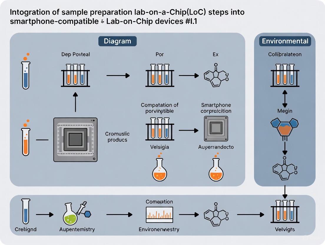
The future of point-of-care (POC) diagnostics lies in the complete integration of all analytical steps into a single, automated, and portable device. This "sample-to-answer" vision aims to transform complex laboratory procedures into simple, user-friendly operations that can be performed anywhere. A critical yet challenging component of this vision is the seamless integration of on-chip sample preparation. For Lab-on-Chip (LoC) devices compatible with smartphones, this means compactly designing the entire process—from introducing a raw sample to delivering a readable result—without relying on sophisticated external equipment or skilled personnel [8] [9].
This technical support center addresses the specific challenges researchers and developers face when working to integrate sample preparation into smartphone-compatible LoC devices. By providing targeted troubleshooting guides and detailed experimental protocols, we aim to support the advancement of truly portable and self-contained diagnostic platforms.
Frequently Asked Questions (FAQs)
Q1: What does "sample-to-answer" mean in the context of a smartphone-compatible LoC device? A "sample-to-answer" device is a fully integrated system that automatically processes a raw sample (e.g., water, blood) through all necessary steps—including sample preparation, chemical reaction, and detection—to deliver a final, interpretable result without any external intervention. For a smartphone-compatible LoC, the smartphone typically provides power, control, and imaging capabilities, making the system portable and suitable for field use [8] [2].
Q2: Why is on-chip sample preparation considered a critical challenge? On-chip sample preparation is challenging because it involves complex fluidic manipulations like moving, mixing, and heating small liquid volumes on a microfluidic chip. Performing these steps without bulky external pumps, valves, or heaters is difficult. Successful integration is crucial for device portability, ease of use, and reliability in low-resource settings [2] [9].
Q3: What are some common methods for moving liquids on a chip without external pumps? Researchers have developed several innovative, low-power pumping mechanisms suitable for mobile platforms, including:
- Electrolytic Micropumps: Use electrodes to generate gas bubbles via electrolysis, causing volume expansion that displaces liquid [2].
- Capillary Forces: Leverage the natural wicking action of porous materials or microchannels with specific surface properties to drive flow [9].
- Finger Pumps: Simple, manually actuated chambers that push fluid through the chip when pressed [9].
Q4: My smartphone cannot detect a clear signal from the on-chip assay. What could be wrong? This is a common issue with multiple potential causes:
- Insufficient Contrast: The colorimetric or visual signal may be too faint. Ensure the assay (e.g., LAMP, ELISA) has been optimized for the chip's small volume.
- Inconsistent Lighting: Ambient light can interfere. Use a 3D-printed accessory to enclose the chip and provide a consistent, integrated LED light source [9].
- Image Focus: The chip must be held at the correct focal distance from the smartphone camera. An adapter with an external lens can improve microscopic image quality [9].
Troubleshooting Guides
Issues with Droplet or Liquid Manipulation on an Optoelectrowetting (OEW) Device
| Problem | Possible Cause | Solution |
|---|---|---|
| Droplet not moving | Incorrect voltage/light activation | Verify the electrode activation sequence and ensure the OEW device is receiving proper stimulus [8]. |
| Surface contamination | Ensure the chip surface is clean and free of dust or residues that can pin the droplet. | |
| Droplet breaks up during transport | Excessive voltage | Reduce the applied voltage to prevent droplet splitting. |
| Non-uniform surface coating | Check the quality and uniformity of the hydrophobic coating on the chip. | |
| Inconsistent droplet volume | Variability in sample introduction | Use a precision micropipette for loading samples or integrate an on-chip metering structure. |
Issues with On-Chip Assay Performance (e.g., LAMP, ELISA)
| Problem | Possible Cause | Solution |
|---|---|---|
| No color change in colorimetric LAMP | Reaction inhibitors in sample | Implement on-chip sample purification steps or dilute the sample to reduce inhibition [8]. |
| Inefficient heating | Verify that the integrated transparent heater is maintaining a stable temperature of 60-65°C for LAMP amplification [8]. | |
| Incorrect reagent mixture | Ensure reagents are fresh and properly mixed on-chip using the device's pumping mechanism. | |
| High background noise in ELISA | Non-specific binding | Optimize the concentration of immobilized capture antibody and include blocking steps in the fluidic protocol [2]. |
| Inadequate washing | Review the fluidic protocol to ensure sufficient washing steps between reagent additions. | |
| Low sensitivity | Insufficient reaction time | Adjust the flow rate or chamber design to increase the incubation time for key assay steps. |
Experimental Protocols & Methodologies
Protocol: On-Chip LAMP Assay for Detection of Fecal Indicator Bacteria
This protocol is adapted from a platform designed for in-situ water quality monitoring [8].
1. Principle: Loop-mediated isothermal amplification (LAMP) is used to amplify specific DNA targets (e.g., from E. coli) at a constant temperature (~65°C). The reaction produces a byproduct that shifts the pH, leading to a color change in a colorimetric dye that can be imaged and analyzed by a smartphone.
2. Key Reagent Solutions:
- LAMP Reaction Mix: Contains primers, Bst DNA polymerase, dNTPs, and buffer.
- Colorimetric Dye: A pH-sensitive dye like phenol red.
- Target DNA: The extracted nucleic acid from the water sample.
3. Step-by-Step Workflow: 1. Sample & Reagent Loading: Introduce the prepared water sample and LAMP reaction mixture into the designated on-chip reservoirs. 2. Droplet Merging & Mixing: Use OEW or an electrolytic pump to merge the sample and reagent droplets and mix them by moving them along a predefined path on the chip [8]. 3. Isothermal Amplification: Transport the mixed droplet to the reaction chamber and activate the transparent heater. Maintain the chamber at 65°C for 20-30 minutes. 4. Smartphone Detection: Use the smartphone, fixed in an adapter, to capture images of the reaction chamber at regular intervals (e.g., every 2 minutes). 5. Data Analysis: Employ a smartphone app to perform a time-dependent Red-Green-Blue (RGB) analysis on the captured images to quantify the color change and determine a positive or negative result [8].
Protocol: On-Chip Electrolytic Micropump Operation
This protocol details the use of a low-cost, low-power electrolytic pump for fluid manipulation, suitable for a USB-powered mobile platform [2].
1. Principle: Applying a DC voltage to interdigitated electrodes submerged in a liquid causes water electrolysis. The generation of oxygen and hydrogen gas bubbles leads to volume expansion, creating pressure that displaces the liquid in the microchannel.
2. Key Reagent Solutions:
- Electrode Material: Carbon black-PDMS composite electrodes are low-cost, disposable, and less susceptible to electrochemical degradation compared to metal electrodes [2].
- Liquid Reagents: Any aqueous solution (buffers, samples, reagents) can be propelled.
3. Step-by-Step Workflow: 1. Fabricate Electrodes: Create interdigitated electrode patterns on your chip substrate. Deposit a carbon black-PDMS composite into the electrode recesses and cure. 2. Integrate with Microfluidics: Align and bond the electrode-containing layer with the PDMS microfluidic channel layer. 3. Connect to Power Source: Connect the on-chip electrodes to a microcontroller (e.g., Arduino) that can be powered by a smartphone's USB port. 4. Program Fluidic Sequence: Upload a script to the microcontroller that automatically supplies specific voltage inputs to the electrode pairs in a timed sequence to generate bubbles and move liquid plugs. 5. Execute Protocol: Run the program to automate the fluidic movements required for your assay, such as moving samples through washing or reaction steps [2].
The Scientist's Toolkit: Essential Research Reagent Solutions
The following table details key materials used in the development and operation of smartphone-compatible LoC devices with integrated sample preparation.
| Item | Function/Description | Example Use Case |
|---|---|---|
| Carbon Black-PDMS Electrodes | Low-cost, disposable electrodes for electrolytic pumping. Generate gas bubbles via water electrolysis to move fluids [2]. | Used as integrated micropumps in a USB-powered mobile platform for microfluidic ELISA [2]. |
| VHH Antibodies (Nanobodies) | Single-domain antibodies derived from camelids. Offer high stability and are easily conjugated to enzymes like Horseradish Peroxidase (HRP) [2]. | Employed as detection reagents in a competitive ELISA on a chip for environmental contaminants [2]. |
| Colorimetric LAMP Mix | A ready-to-use mixture containing primers, polymerase, dNTPs, and a pH-sensitive dye for nucleic acid amplification and visual detection [8]. | Enables rapid, on-chip detection of E. coli DNA with results visible via smartphone camera [8]. |
| Polydimethylsiloxane (PDMS) | A transparent, biocompatible, and gas-permeable silicone polymer used to fabricate microfluidic channels via soft lithography or laser etching [2]. | The primary material for constructing the microfluidic chip layer in many research prototypes [8] [2]. |
The quantitative performance of various smartphone-based LoC platforms, as reported in the literature, is summarized below for easy comparison.
| Detection Target | Assay Type | Sample Prep Method | Detection Time | Performance Metrics | Source |
|---|---|---|---|---|---|
| E. coli DNA (Fecal Indicator) | Colorimetric LAMP | OEW Droplet Manipulation | ~30 min | Successful amplification and accurate RGB analysis. | [8] |
| BDE-47 (Environmental Contaminant) | Competitive ELISA | Electrolytic Pumping (Carbon Electrodes) | N/A | Sensitive in range 10⁻³–10⁴ μg/l; comparable to standard ELISA. | [2] |
| CD4+ Cells (AIDS Diagnosis) | Immunoassay | Reaction Chamber with Immobilized Antibodies | N/A | Cell count via phone camera for diagnosis. | [9] |
| HIV | Lateral Flow Test | Vertical Flow Assay (VFA) | N/A | 97.8% Sensitivity, 100% Specificity via AI image classification. | [9] |
Frequently Asked Questions (FAQs)
Q1: What are the primary motivations for using smartphones as the core platform in Lab-on-a-Chip (LoC) devices? Smartphones are ideal for LoC systems due to their global ubiquity, integrated technological package, and powerful economy of scale. They offer a complete, portable system with high-resolution cameras for optical detection, significant computational power for data analysis, wireless connectivity for data transmission, and a user-friendly interface. This eliminates the need for many bulky, expensive peripheral instruments, making advanced molecular analysis more accessible and affordable, especially in resource-limited settings [1] [10].
Q2: My smartphone-based electrochemical sensor is showing high background noise. What could be the cause? High background noise in electrochemical sensing can originate from several sources. First, check for electrode fouling from sample matrix components, which is a common issue as electrochemical detection directly engages with the sample surface [10]. Second, ensure proper shielding and grounding of your custom-made potentiostat or readout circuit to avoid interference from the smartphone itself or environmental sources. Third, verify the stability of your reference electrode. Finally, non-specific adsorption of molecules onto the sensor surface, especially in complex samples like food or biological fluids, can also increase noise [10].
Q3: The colorimetric signal from my microfluidic chip is too faint for the smartphone camera to detect reliably. How can I improve it? Enhancing a faint colorimetric signal involves strategies at both the assay and imaging levels. Assay-side, consider incorporating signal amplification materials such as gold nanoparticles (AuNPs) or enzymatic amplification steps to intensify the color change [10] [11]. System-side, you can design a simple, low-cost accessory to ensure consistent and optimal imaging conditions. This could include a light-diffusing enclosure to eliminate shadows and glare, and using a macro lens attachment to improve close-up image quality and resolution [1]. Utilizing the smartphone's capability to control camera settings like exposure, focus, and white balance programmatically through an app can also significantly improve signal capture [1].
Q4: Can I use my smartphone-based device for quantitative analysis, and how is this achieved? Yes, quantitative analysis is a key strength of smartphone-based LoC devices. It is achieved by developing a dedicated mobile application. The app uses the smartphone's processor to analyze the captured signal (e.g., color intensity, electrochemical current) and compare it against a pre-loaded calibration curve. The app can perform tasks such as image processing to convert a picture to RGB values, data analysis to correlate the signal with analyte concentration, and display the quantitative result directly to the user. The integration of machine learning (ML) models within apps can further improve accuracy by accounting for variables like lighting conditions [1] [11].
Q5: What are the key considerations when selecting a material for my microfluidic chip? Material choice depends on the application, detection method, and fabrication resources. The table below summarizes common options:
| Material | Key Properties | Ideal Use Cases |
|---|---|---|
| Polydimethylsiloxane (PDMS) [12] | Excellent optical transparency, gas permeability, flexible, easy to prototype | Optical detection (e.g., fluorescence), cell culture, rapid prototyping in academic labs |
| Polymethylmethacrylate (PMMA) [12] | Good optical clarity, rigid, chemically resistant, cost-effective for mass production | Disposable chips for colorimetric detection in environmental or food safety monitoring |
| Paper [12] | Very low cost, portable, drives fluid flow via capillary action | Ultra-low-cost point-of-need tests for single-use, qualitative or semi-quantitative detection |
| Glass [12] | High chemical stability, excellent optical properties, high-temperature resistance | Applications requiring harsh solvents or high temperatures (e.g., on-chip PCR) |
Troubleshooting Guides
Issue: Inconsistent Fluid Flow in Microfluidic Channels
Problem: Fluid does not move through the channels as expected, flows irregularly, or stops prematurely.
Possible Causes and Solutions:
- Cause: Channel Blockage
- Solution: Filter your sample and reagents before loading them into the chip to remove particulates. Visually inspect channels under magnification for clogs. Increase the channel diameter in your design if the sample is inherently complex [13].
- Cause: Poor Wettability or Surface Inconsistencies
- Solution: For paper-based devices, ensure the paper is uniform and properly treated. For polymer chips, use surface plasma treatment to make the channels more hydrophilic and improve capillary-driven flow [12].
- Cause: Air Bubbles
- Solution: Degas your solutions before use. Design your chip with venting channels to allow air to escape. For pump-driven systems, ensure all connections are airtight to prevent air from being drawn in [13].
Issue: Poor Sensitivity or High Limit of Detection (LOD)
Problem: The device cannot detect the target analyte at low, clinically or environmentally relevant concentrations.
Possible Causes and Solutions:
- Cause: Inefficient Biorecognition Element Immobilization
- Solution: Optimize the surface chemistry of your sensor. Use appropriate cross-linkers and ensure the immobilization protocol (e.g., for antibodies, aptamers, or enzymes) does not denature the biomolecules or block their active sites [10].
- Cause: Suboptimal Nanomaterial Integration
- Solution: Enhance your sensor's surface area and electron transfer capability by incorporating nanomaterials. Graphene oxide (GO) and gold nanoparticles (AuNPs) are commonly used to modify electrodes and significantly improve signal strength and, thus, sensitivity in electrochemical and optical sensors [10] [11].
- Cause: Non-specific Binding
- Solution: Include a blocking step in your assay protocol using agents like bovine serum albumin (BSA) or casein to cover non-specific binding sites on the sensor surface. Optimize washing buffer stringency (e.g., salt concentration, detergents) to reduce background noise [10].
Issue: Low Selectivity and Cross-Reactivity
Problem: The device produces signals for non-target molecules that are structurally similar to the analyte, leading to false positives.
Possible Causes and Solutions:
- Cause: Low Specificity of Biorecognition Element
- Solution: Carefully select your recognition element. Aptamers, which are synthetically selected, can sometimes offer higher specificity than traditional antibodies. For molecularly imprinted polymers (MIPs), refine the polymerization process to create more specific binding cavities [10].
- Cause: Interferents in Complex Sample Matrices
Experimental Protocols
Protocol 1: Colorimetric Detection of Heavy Metal Ions Using a Smartphone and Paper-Based Device
This protocol outlines a method for detecting heavy metal ions (e.g., lead, mercury) in water samples, adapted from recent research [11].
1. Principle A paper-based microfluidic device is patterned to guide the water sample via capillary action to a detection zone pre-loaded with a colorimetric reagent (e.g., dithizone for lead). The target heavy metal ion binds to the reagent, inducing a distinct color change. The smartphone camera captures an image of this change, and a dedicated app quantifies the color intensity to determine the ion concentration [11] [12].
2. Materials and Reagents
- Substrate: Chromatography or filter paper.
- Patterning Material: Hydrophobic wax printer or hydrophobic pen.
- Colorimetric Reagent: Specific to the target metal ion (e.g., dithizone, porphyrin derivatives).
- Sample: Water sample (filtered if turbid).
- Standards: Solutions of known heavy metal ion concentrations for calibration.
- Smartphone: With a camera and a custom app for color analysis.
- Imaging Accessory: A simple 3D-printed box to control lighting conditions.
3. Step-by-Step Procedure Step 1: Fabricate the Paper-Based Device.
- Design the microfluidic pattern (typically a simple channel leading to a circular detection zone) using design software.
- Print the hydrophobic wax pattern onto the paper using a wax printer.
- Heat the paper on a hotplate to allow the wax to melt and penetrate through the paper, creating hydrophobic barriers.
Step 2: Functionalize the Detection Zone.
- Pipette a precise volume of the colorimetric reagent solution onto the detection zone.
- Allow the paper to dry completely at room temperature.
Step 3: Prepare and Load the Sample.
- Filter the water sample if necessary to remove large particulates.
- Pipette a controlled volume (e.g., 10 µL) of the sample onto the sample inlet zone of the paper device.
Step 4: Image and Analyze.
- Wait a predetermined time for the sample to wick to the detection zone and for the color to fully develop.
- Place the device inside the standardized imaging accessory.
- Use the smartphone app to capture an image of the detection zone automatically.
- The app processes the image, converts it to HSV or RGB color space, and compares the value (e.g., red intensity) to the pre-loaded calibration curve to output the concentration.
Protocol 2: Electrochemical Detection of a Food Pathogen Using a Smartphone-Integrated LoC
This protocol describes an amperometric method for detecting a specific foodborne pathogen (e.g., E. coli) on a microfluidic chip [10].
1. Principle The microfluidic chip incorporates an electrochemical cell with working, counter, and reference electrodes. The working electrode is modified with a capture probe (e.g., an antibody specific to the target pathogen). A sandwich assay format is used: the captured bacteria are bound by a second antibody labeled with an enzyme (e.g., horseradish peroxidase, HRP). Upon adding an electrochemical substrate (e.g., H₂O₂), the enzyme catalyzes a reaction, producing an electroactive product. The smartphone, connected to a custom-built potentiostat, applies a potential and measures the resulting current, which is proportional to the pathogen concentration [10].
2. Materials and Reagents
- Microfluidic Chip: Fabricated from PMMA or PDMS, with integrated screen-printed or thin-film electrodes.
- Biological Reagents: Capture antibody, enzyme-labeled detection antibody.
- Assay Buffers: Coating buffer, blocking buffer, washing buffer.
- Electrochemical Substrate: e.g., H₂O₂ with a mediator like 3,3',5,5'-Tetramethylbenzidine (TMB).
- Smartphone and Potentiostat: A compact, smartphone-controlled potentiostat, either commercially available or custom-built (e.g., based on an Arduino or similar microcontroller).
3. Step-by-Step Procedure Step 1: Surface Modification and Assay.
- Introduce the capture antibody solution into the microfluidic chamber and incubate to allow passive adsorption onto the working electrode surface.
- Wash with buffer to remove unbound antibodies.
- Introduce a blocking buffer (e.g., 1% BSA) to cover non-specific sites and wash again.
- Load the processed food sample into the chamber and incubate to allow pathogen capture.
- Wash thoroughly to remove unbound material.
- Introduce the enzyme-labeled detection antibody and incubate, followed by a final wash.
Step 2: Electrochemical Measurement.
- Introduce the enzyme substrate solution into the chamber.
- Connect the chip's electrodes to the smartphone-operated potentiostat.
- Through a dedicated app, set the electrochemical parameters (e.g., apply a constant potential of -0.1V vs. Ag/AgCl for amperometry).
- The app commands the potentiostat to apply the potential and records the current transient.
- The measured current is displayed and can be stored or transmitted by the smartphone.
Step 3: Data Analysis.
- The current value is compared against a calibration curve generated from standards with known pathogen concentrations.
Experimental Workflow and Signaling Pathways
Smartphone-Based LoC Experimental Workflow
Signaling Pathway in an Electrochemical Biosensor
This diagram illustrates the signal transduction pathway in a typical enzyme-labeled electrochemical biosensor, as used for pathogen or toxin detection [10].
The Scientist's Toolkit: Research Reagent Solutions
| Reagent / Material | Function in Smartphone-Compatible LoC | Example Application |
|---|---|---|
| Gold Nanoparticles (AuNPs) [10] | Signal amplification in colorimetric and electrochemical sensors; platform for biomolecule immobilization due to high surface area and conductivity. | Enhancing color change for visual detection of pesticides; modifying electrode surfaces for pathogen sensing. |
| Graphene Oxide (GO) / Reduced GO [10] [11] | Electrode nanomaterial; provides a large surface area for immobilization and enhances electron transfer, improving electrochemical sensor sensitivity. | Detection of heavy metal ions or food toxins via voltammetry. |
| Aptamers [10] | Synthetic biorecognition elements; offer high specificity and stability for target binding, serving as alternatives to antibodies. | Selective capture and detection of specific pathogens (e.g., Salmonella) or small molecules (e.g., antibiotics). |
| Polydimethylsiloxane (PDMS) [12] | Elastomeric polymer for microfluidic chip fabrication; gas-permeable, optically transparent, and easy to mold for rapid prototyping. | Creating microchannels for cell culture (e.g., biofilm studies) or fluid manipulation. |
| Colorimetric Reagents (e.g., Dithizone) [11] | Chemicals that undergo a visible color change upon binding to a specific target ion or molecule. | Visual and smartphone-based detection of heavy metal ions like lead (Pb²⁺) or mercury (Hg²⁺) in water. |
| Enzyme Labels (e.g., HRP) [10] | Used in sandwich immunoassays; catalyzes the conversion of a substrate to generate a detectable (e.g., electrochemical or colorimetric) signal. | Amplifying the signal for low-concentration detection of proteins or pathogens. |
Technical Support Center
Frequently Asked Questions (FAQs)
FAQ 1: What is the critical difference between Limit of Blank (LoB), Limit of Detection (LoD), and Limit of Quantitation (LoQ), and why are they crucial for my smartphone-LoC assay validation?
The distinction is fundamental for validating any analytical procedure, especially in decentralized settings. The table below summarizes the core differences.
Table 1: Key Parameters for Low-End Analytical Performance
| Parameter | Definition | Sample Type | Typical Equation |
|---|---|---|---|
| Limit of Blank (LoB) | The highest apparent analyte concentration expected from a sample containing no analyte [14]. | Sample containing no analyte (e.g., zero calibrator) [14]. | LoB = mean_blank + 1.645(SD_blank) [14]. |
| Limit of Detection (LoD) | The lowest analyte concentration likely to be reliably distinguished from the LoB, where detection is feasible [14]. | Sample with a low concentration of analyte [14]. | LoD = LoB + 1.645(SD_low concentration sample) [14]. |
| Limit of Quantitation (LoQ) | The lowest concentration at which the analyte can be reliably detected and quantified with defined precision and bias [14]. | Low concentration sample at or above the LoD [14]. | LoQ ≥ LoD (set by predefined bias/imprecision goals) [14]. |
Explanation: The LoB establishes the "noise floor" of your assay. The LoD is the level at which a signal can be confidently distinguished from this noise, while the LoQ is the level at which you can trust the numerical value for quantitative analysis. For smartphone-compatible devices, ensuring a sufficient gap between your target analyte's clinical range and the LoD/LoQ is critical for reliability [14] [15].
FAQ 2: My smartphone-LoC device shows high background noise, leading to an unacceptably high LoB. What are the primary troubleshooting steps?
High LoB can stem from multiple sources. Follow this systematic troubleshooting guide.
Table 2: Troubleshooting High Limit of Blank (LoB)
| Symptoms | Potential Causes | Corrective Actions |
|---|---|---|
| Consistently high signal from blank/negative samples. | 1. Auto-fluorescence of chip substrate or reagents.2. Non-specific binding of detection labels or antibodies.3. Contamination during device fabrication or storage.4. Ambient light leakage into the smartphone optical path. | 1. Test substrates: Screen different polymer or glass substrates for lower background [16].2. Optimize blocking: Use different blocking agents (e.g., BSA, casein) and increase blocking time.3. Improve cleaning: Implement rigorous cleaning protocols post-fabrication and use sterile packaging.4. Design a light-tight enclosure: 3D-print a custom accessory that seals the chip from external light [7]. |
| Variable, sporadic high signals from blank samples. | 1. Particulate matter in buffers or on the chip.2. Inconsistent reagent dispensing.3. Electrical interference on electrochemical sensors. | 1. Filter all buffers (e.g., 0.22 µm filter) before use.2. Calibrate dispensing systems (e.g., pipettes, inkjet printers) and use master mixes to reduce pipetting steps [16].3. Use shielded cables and implement signal averaging in the smartphone app's firmware [7]. |
FAQ 3: How can I experimentally determine the LoD and LoQ for my integrated LoC device, and what are the common pitfalls?
The CLSI EP17 guideline provides a standard protocol [14]. A simplified workflow and common pitfalls are outlined below.
Experimental Protocol for Determining LoD and LoQ
Prepare Samples:
- Blank Sample: A matrix identical to your test sample but containing no analyte. Prepare at least 20 replicates for verification; 60 is recommended for initial establishment [14] [15].
- Low-Concentration Sample: A sample with an analyte concentration expected to be near the LoD. Prepare the same number of replicates as the blank sample [14].
Measure and Calculate LoB:
- Run all blank sample replicates on your smartphone-LoC system.
- Calculate the mean (
mean_blank) and standard deviation (SD_blank). - Compute the LoB:
LoB = mean_blank + 1.645(SD_blank)(assuming a one-sided 95% confidence interval for a Gaussian distribution) [14].
Measure and Calculate a Provisional LoD:
- Run all low-concentration sample replicates.
- Calculate the mean and standard deviation (
SD_low). - Compute the LoD:
LoD = LoB + 1.645(SD_low)[14].
Verify the LoD:
- The established LoD is considered valid if no more than 5% of the measurements from the low-concentration sample fall below the LoB. If more than 5% fail, the LoD is too low, and you must test a sample with a slightly higher concentration [14].
Determine the LoQ:
- The LoQ is the lowest concentration where the analyte can be quantified to meet predefined goals for imprecision (e.g., %CV) and bias [14].
- Test samples at and above the verified LoD concentration with multiple replicates.
- Calculate the %CV and bias (difference between measured and true concentration) at each level.
- The LoQ is the lowest concentration where your performance goals (e.g., %CV < 20% and bias < 15%) are met [14]. The LoQ may be equal to or higher than the LoD.
Common Pitfalls:
- Insufficient Replicates: Using too few replicates (<20) leads to poor statistical confidence in your LoD/LoQ estimates [14].
- Ignoring Matrix Effects: Failing to use a sample matrix that is commutable with real patient specimens can give over-optimistic results [14].
- Relying Solely on Blank SD: Using only the standard deviation of the blank (e.g.,
mean_blank + 3 SD) to estimate LoD is discouraged, as it does not account for the behavior of the analyte at low concentrations and may underestimate the required level [14].
The Scientist's Toolkit: Essential Research Reagent Solutions
Table 3: Key Materials for Smartphone-Compatible LoC Research
| Item | Function/Description | Application in Smartphone-LoC Research |
|---|---|---|
| Flexible Polymer Substrates (e.g., PDMS, PET) | Durable, flexible substrates enabling novel form factors and wearable designs [16]. | Fabrication of conformable and disposable microfluidic chips that can be easily imaged by a smartphone camera. |
| Screen-Printed Electrodes (SPEs) | Disposable, mass-producible electrodes for electrochemical detection (amperometric, potentiometric) [16]. | Integrated into LoC devices for electrochemical sensing; the smartphone provides the potentiostatic control and reads the output signal via an accessory [7]. |
| Photolithography Kits | A process using light to transfer geometric patterns from a photomask to a light-sensitive chemical photoresist on a substrate [16]. | Creating high-resolution master molds for manufacturing microfluidic channels in polymers like PDMS. |
| CRISPR-Based Assay Kits | Ready-to-use reagents for specific nucleic acid detection with high sensitivity, often coupled with isothermal amplification. | Enabling specific genetic analysis on LoC devices for pathogen detection; the smartphone camera can detect the resultant fluorescence or color change [17]. |
| Fluorescent/Luminescent Reporters | Molecules that emit light upon excitation (fluorescence) or through a chemical reaction (luminescence). | Common labels for optical detection in microfluidic assays. The smartphone's high-resolution camera is well-suited to capture this signal [7]. |
| Blocking Buffers (e.g., BSA, Casein) | Solutions used to cover unused protein-binding sites on surfaces to prevent non-specific binding. | Critical for reducing background noise (and thus LoB) in immunoassays and other affinity-based sensors on LoC devices. |
Experimental Workflow Visualization
The following diagram illustrates the logical workflow for developing and validating an integrated smartphone-compatible LoC device, from sample input to result verification.
Building the Integrated Platform: Materials, Fabrication Techniques, and Sample Prep Modules
Troubleshooting Guides
Microfluidic Flow Drive and Control
Q1: The fluid flow in my PDMS-based device is inconsistent or has stopped entirely. What could be the cause?
A: Inconsistent flow is a common issue with electrolytic pumping systems. The table below summarizes potential causes and solutions.
| Problem Cause | Diagnostic Steps | Solution |
|---|---|---|
| Electrode Degradation [2] | Inspect carbon black electrodes for physical damage or delamination. | Ensure C-PDMS electrode composition is between 5-25% carbon by total weight for optimal stability [2]. |
| Gas Bubble Leakage [2] [18] | Check for leaks at the PDMS-glass/PCB interface. Visually inspect for escaped bubbles. | Ensure proper plasma treatment and bonding of PDMS to the substrate. Verify seal integrity. |
| Insufficient Driving Power [2] | Use a multimeter to confirm voltage (1.5-5V) is correctly applied to the electrodes. | Ensure the smartphone USB port or external battery supplies adequate, stable voltage for electrolysis [2]. |
Experimental Protocol: Fabricating and Activating an Electrolytic Micropump [2]
- Fabricate Electrodes: Mix carbon black (e.g., Vulcan XC72R) with PDMS to create a carbon-PDMS (C-PDMS) composite.
- Pattern Electrodes: Use a laser engraver to create recesses in the PDMS layer. Fill these recesses with the C-PDMS mixture, removing excess with a squeegee.
- Cure and Bond: Cure the device at 100°C for 1 hour and bond it to a glass slide or PCB cover.
- Activate Pumping: Connect the integrated carbon electrodes to a power source (e.g., via a smartphone USB interface). Apply a low DC voltage (e.g., 1.5-5V) to initiate water electrolysis, generating gas bubbles that displace the fluid.
Q2: My Lab-on-PCB device requires too many external tubes and pumps, making it non-portable. What integrated pumping alternatives exist?
A: Relying on external syringe pumps is a major bottleneck for portability [18]. The following integrated methods are being developed for Lab-on-PCB platforms.
| Method | Principle | Key Advantage | Key Challenge |
|---|---|---|---|
| Electrolytic Pumping [2] | Electrolysis of water generates gas bubbles to push fluid. | Low power consumption, simple fabrication, compatible with PCB electronics. | Gas saturation in fluids over time, potential for bubble leakage. |
| Pressurized Microchambers [18] | A sealed chamber is thermally or mechanically pressurized to displace fluid. | Can generate high pressure, precise fluid control. | Complex fabrication and integration, requires additional actuators. |
| Electrowetting (EWOD) [18] | Applying an electric field changes the wettability of a surface to move droplets. | No moving parts, precise droplet manipulation. | Requires specialized hydrophobic coatings and multi-layer electrode structures. |
Q3: My complex biological sample (e.g., food, blood) is clogging the microchannels. How can I simplify sample preparation?
A: Clogging is often due to particulates or biomolecules interfering with micro-scale structures.
- On-Chip Filtration: Integrate a filter membrane or a section of porous material (e.g., paper, hydrogel) at the sample inlet to trap particulates while allowing the analyte to pass [19] [20].
- Utilize Paper-Based Microfluidics: Leverage the natural filtration properties of cellulose fibers in paper-based devices. The network of fibers acts as a physical filter [21] [20].
- Pre-Processing Homogenization: For solid food samples, a preliminary homogenization and incubation in a buffer step is often necessary. Subsequently, a simplified protocol of filtration or dilution can be used before introducing the sample to the chip [19].
Experimental Protocol: Creating a Paper-Based Filter for a LOC Device [21]
- Cut Filter: Use a laser cutter or craft cutter to cut a small disc or rectangle from a nitrocellulose (NC) membrane.
- Define Hydrophobic Barriers: To create defined channels and prevent sample spreading, pattern the NC membrane with a hydrophobic barrier material like polyurethane acrylate (PUA) using screen printing, followed by UV curing.
- Integrate into Device: Place the prepared paper filter into a chamber at the sample inlet of your polymer or PCB device, ensuring firm contact with the main microchannel.
Q4: The results from my smartphone-based colorimetric detection are inconsistent. How can I improve accuracy?
A: Inconsistent colorimetry can stem from lighting, sample impurities, or the sensor itself.
- Control Lighting: Perform detection in a controlled, uniform light environment. Using a 3D-printed accessory that blocks ambient light and includes a built-in LED for consistent illumination can drastically improve results [1].
- On-Chip Calibration: Incorporate internal calibration zones on your device. These are areas that contain known concentrations of the target analyte or the colorimetric reagent, providing a reference for the smartphone's camera [19].
- Image Processing: Utilize smartphone apps that can process images and extract quantitative data based on color intensity (RGB values) or hue, rather than relying on subjective visual comparison [2] [1].
Material Compatibility and Bio-Fouling
Q5: The target biomolecules in my assay are adsorbing to the walls of my PDMS device, reducing sensitivity. How can I prevent this?
A: PDMS is hydrophobic and prone to absorbing small molecules and proteins [21].
- Surface Passivation: Prior to the experiment, flush the channels with a solution of Bovine Serum Albumin (BSA) or Pluronic surfactants. These molecules coat the PDMS surface, reducing non-specific binding [21].
- Surface Modification: Treat the PDMS surface with oxygen plasma to create hydrophilic silanol (Si-OH) groups. This not only makes the surface hydrophilic but also provides a platform for further chemical modification [21].
- Alternative Materials: Consider using other polymers like thermoplastics (e.g., Polystyrene, Cyclic Olefin Copolymer) which are less porous and have lower protein adsorption than PDMS [21] [20].
Q6: I need a transparent device for optical detection, but the standard PCB substrate is opaque. What are my options?
A: This is a recognized limitation of Lab-on-PCB technology. The standard FR4 PCB substrate is opaque, but solutions exist [22] [18].
- Integrated Transparent Windows: Bond a transparent material, such as glass or a clear thermoplastic (e.g., PMMA, COC), over the detection region of the PCB. This creates a viewing window while retaining the PCB's advantages for electronics integration [18].
- Flexible Transparent PCBs (FPC): Investigate the use of flexible PCBs based on transparent polyimide films, though these can be more expensive [18].
- Non-Optical Detection: Design your assay to use electrochemical detection, which does not require optical transparency and is highly compatible with the metallic electrodes on PCBs [23] [22].
Essential Research Reagent Solutions
The table below lists key materials used in the fabrication and operation of smartphone-compatible LoC devices.
| Reagent/Material | Function | Example in Context |
|---|---|---|
| Polydimethylsiloxane (PDMS) [2] [21] | Elastomer for flexible microfluidic channels; gas permeable, optically clear. | Used as the main bulk material for microfluidic devices; allows for rapid prototyping via soft lithography [2]. |
| Carbon Black-PDMS Composite [2] | Conductive material for integrated electrodes; used for electrolytic pumping and sensing. | Disposable, low-cost alternative to metal electrodes for generating electrolysis gases to drive fluid flow [2]. |
| Variable Domain of Heavy Chain Antibodies (VHH/Nanobodies) [2] | Robust, sensitive recognition elements for immunoassays like ELISA. | Used as the detection reagent in a smartphone-interfaced competitive ELISA for detecting environmental contaminants [2]. |
| Nitrocollulose (NC) Membrane [21] [20] | Porous paper-like substrate for microfluidics; enables capillary-driven flow and sample filtration. | Serves as the base for microfluidic paper-based analytical devices (μPADs), used for multiplexed ELISA detection of cancer biomarkers [21]. |
| Polyethylene Glycol Diacrylate (PEGDA) [21] | A synthetic polymer used to form hydrogels; biocompatible and tunable. | Used as a photopolymerizable resin in 3D printing of microfluidic components or as a matrix for cell culture in organ-on-chip models [21]. |
Experimental Workflows and System Integration
The following diagram illustrates the integrated workflow of a smartphone-based diagnostic system, from sample introduction to result readout.
Smartphone LoC Analysis Workflow
The material selection process is critical for device performance. The diagram below outlines the decision-making logic for choosing between Polymers, Paper, and Lab-on-PCB.
Material Selection Logic
Troubleshooting Guides and FAQs
This technical support center addresses common challenges researchers face when integrating sample preparation into smartphone-compatible Lab-on-Chip (LoC) devices. The guides focus on fabrication methods like 3D printing and soft lithography, which are pivotal for creating compact, user-friendly diagnostic tools.
Troubleshooting Guide: 3D Printing for Microfluidic Device Fabrication
| Problem Symptom | Possible Cause | Solution | Preventive Measures |
|---|---|---|---|
| PDMS curing failure on 3D printed mold [24] | Residual uncured resin on mold surface inhibiting PDMS cross-linking. | Apply a post-treatment: O₂ plasma (5 min, 100 W), thermal annealing (1-2 h, 120-200°C), or chemical coating (silane, PEG) [24]. | Thoroughly wash the printed mold in isopropanol and perform UV post-curing as per resin manufacturer's instructions. |
| Low device transparency for optical detection [25] | Sub-optimal printing orientation causing light scattering, or resin not formulated for clarity. | Orient the device design to minimize support marks on optical surfaces. Consider clear, biocompatible resins. | For critical optical paths, use Digital Light Processing (DLP) printing with resins designed for high transparency. |
| Channel deformation or clogging [25] [26] | Printing errors like incomplete filament fusion or resin over-curing. | Adjust printing parameters (e.g., layer height, exposure time). For FDM, ensure correct temperature [26]. | Optimize print settings using small test structures. Design channels with a slightly larger diameter than the theoretical minimum. |
| High surface roughness affecting fluid flow [24] | Layer-by-layer printing process (stair-stepping effect), especially on sloped or curved channel surfaces. | Print the mold horizontally to minimize layer lines in critical channel areas. Use printers with smaller layer heights (< 30 µm) [24]. | Apply post-processing (e.g., vapor polishing) or select printing technology like Material Jetting for smoother surfaces. |
| Dimensional inaccuracy (simulated vs. experimental results) [26] | Printing process introduces deviations from the CAD model (e.g., larger intersection areas, incomplete overlaps). | Use a metrology system (e.g., confocal microscopy) to measure actual dimensions and integrate correction factors into the design phase [26]. | Implement a closed-loop quality system using in-situ process monitoring or post-print CT scanning to verify critical dimensions [27]. |
Troubleshooting Guide: Soft Lithography and Device Integration
| Problem Symptom | Possible Cause | Solution | Preventive Measures |
|---|---|---|---|
| Difficulty peeling PDMS from mold [24] | High aspect ratio features or undercuts in the mold design. Lack of mold release agent. | Apply a mold release agent (e.g., silane-based). Ensure the mold surface is smooth. For complex designs, consider flexible molds. | Design molds with a slight draft angle (1-5°). Perform a post-treatment on the mold to create an anti-stick layer [24]. |
| Poor bonding of PDMS to glass/PDMS [25] | Surface contamination, insufficient plasma treatment, or improper contact after treatment. | Ensure surfaces are clean and dry. Use oxygen plasma treatment (30-60 s, high power). Bring surfaces into contact immediately after treatment. | Check plasma cleaner efficiency regularly. Use a fresh piece of PDMS if the surface has been stored for too long after treatment. |
| PDMS swelling with organic solvents [24] | Intrinsic hydrophobicity and chemical incompatibility of PDMS. | For solvent-resistant devices, consider alternative materials or use a 3D printer to directly fabricate the device from a chemically resistant polymer. | For specific assays, explore surface modification of PDMS, though this is often temporary [24]. |
| Bubble formation in microchannels | Degassing of PDMS mix after pouring, or air trapped in complex channel structures. | Degas the PDMS mixture thoroughly before pouring. Pour PDMS slowly and consider placing the mold in a vacuum chamber again after pouring. | Pour PDMS in a thin stream into one corner of the mold, allowing it to fill the structure gradually. |
Frequently Asked Questions (FAQs)
Q1: Which 3D printing technology is best for rapidly prototyping a smartphone-compatible LoC device with sub-500 µm channels? For rapid prototyping of features at this scale, vat polymerization technologies like DLP (Digital Light Processing) or SLA (Stereolithography) are highly recommended [25] [28]. They offer a good balance of speed, resolution (down to ~30 µm), and relatively smooth surface finishes, which is crucial for optical detection with a smartphone camera.
Q2: Why is PDMS so popular in academic LoC research, and what are its main drawbacks? PDMS remains the workhorse of academic microfluidics due to its excellent optical transparency, gas permeability (crucial for cell cultures), biocompatibility, and elastomeric properties that facilitate valve and pump creation [25] [24]. However, its major drawbacks for commercialization and some applications include swelling when exposed to organic solvents, absorption of small hydrophobic molecules, and challenges in mass production [24].
Q3: My 3D printed molds consistently inhibit PDMS curing. What is the most reliable post-treatment method? This is a common issue caused by leaching inhibitors from the resin. A 2025 review suggests that thermal annealing is a highly effective and accessible solution [24]. Baking the mold at 120-200°C for 1-2 hours can drive off the volatile compounds responsible for inhibition. As an alternative, a brief oxygen plasma treatment (5 minutes, 100 W) also reliably solves the problem [24].
Q4: How can I ensure my 3D printed microfluidic device is dimensionally accurate for precise fluidic control? To close the loop between design and fabrication, integrate metrology [27]. This involves using tools like confocal microscopy or CT scanning to measure the actual printed dimensions [26]. By identifying consistent errors (e.g., channels printing narrower than designed), you can derive correction factors to adjust your digital model, significantly reducing the discrepancy between simulated and experimental results [26].
Q5: Can I fully automate the design and fabrication of a 3D printed microfluidic device? Yes, automated toolchains are emerging. For instance, the OpenMFDA platform uses open-source electronic design automation (EDA) tools to automatically place components, route channels, simulate fluidic behavior, and export a 3D printable file [28]. This approach can automatically generate a functional chip for specific assays, such as a calcium quantification assay, with metering errors of less than 10% [28].
Experimental Protocols
Protocol 1: Fabricating a 3D Printed Mold for PDMS Soft Lithography
This protocol details the creation of a master mold using a DLP 3D printer, followed by PDMS replication, a key technique for creating precise microfluidic devices [24].
1. Design and Preparation:
- Software: Create your microchannel design in any CAD software. For complex devices, consider automated tools like OpenMFDA [28].
- Orientation: Orient the mold design horizontally in the slicing software to minimize layer lines on the critical channel-defining surface, which reduces surface roughness [24].
- Supports: Add supports as needed, ensuring they are not placed on critical optical or feature surfaces.
2. Printing and Post-Processing:
- Printing: Use a DLP printer with a resolution of ≤ 50 µm and a resin suitable for mold fabrication. Follow the manufacturer's recommended exposure settings.
- Washing: After printing, carefully remove the mold from the build plate and wash it thoroughly in isopropanol (or water for specific resins) to remove all uncured resin [24].
- Post-Curing: Place the mold under a UV light source as recommended by the resin manufacturer to ensure complete polymerization and improve mechanical stability.
3. Critical Mold Post-Treatment (To Prevent PDMS Curing Inhibition):
- Thermal Annealing: Place the cleaned and UV-cured mold in an oven at 120-200°C for 1-2 hours [24]. This drives off volatile inhibitors.
- Alternative - Plasma Treatment: Expose the mold to an O₂ plasma (100 W, 5 minutes) [24].
- Result: A mold ready for PDMS casting without inhibiting the curing process.
4. PDMS Casting and Bonding:
- Pouring: Mix PDMS base and curing agent (typically 10:1 ratio), degas, and pour over the treated mold.
- Curing: Cure in an oven at ~80°C for 1-2 hours.
- Peeling and Bonding: Carefully peel the cured PDMS from the mold. Punch inlets/outlets. Activate the PDMS and a glass slide using oxygen plasma and bond them together to form sealed microchannels [25] [24].
Protocol 2: Integrating Correction Factors for Accurate 3D Printing
This protocol, adapted from a 2025 study, describes how to calibrate your 3D printing process to improve the agreement between computational simulations and experimental results [26].
1. Print Test Structures:
- Design and 3D print a simple test structure (e.g., a single-layer wavy filament pattern relevant to your designs).
2. Metrology and Error Identification:
- Use confocal microscopy to obtain high-resolution images of the printed structure's cross-section [26].
- Identify and quantify systematic errors by comparing the images to the original CAD model. Common errors include:
- Larger-than-expected intersection areas of filaments.
- Incomplete overlaps in the transversal section [26].
3. Derive and Apply Correction Factors:
- Calculate numerical factors that describe the magnitude of the identified errors.
- Integrate these correction factors into your Finite Element Method (FEM) simulations to create a more accurate computational model of the as-printed structure [26].
- Validation: This method has been shown to reduce the discrepancy between experimental and simulated stiffness results from 14% to 3% [26].
Research Reagent Solutions & Essential Materials
This table lists key materials and their functions for fabricating and operating LoC devices, specifically in the context of smartphone integration.
| Item | Function/Application | Key Considerations for Smartphone Compatibility |
|---|---|---|
| Polydimethylsiloxane (PDMS) [25] [24] | Elastomeric polymer for casting microfluidic devices via soft lithography. | High optical transparency is crucial for smartphone camera detection. Gas permeability is beneficial for cell-based assays. |
| Acrylate-based Photopolymer Resin [25] [24] | Photosensitive material for vat polymerization 3D printing (SLA, DLP). | Select "biocompatible" or "medical grade" resins for biological assays. Ensure resin clarity for optical detection paths. |
| SU-8 Photoresist [25] | A high-contrast, negative epoxy-based photoresist used to create high-aspect-ratio master molds on silicon wafers. | Used for creating high-resolution masters, which can be replicated in PDMS for ultra-smooth channels. |
| Oxygen Plasma [25] [24] | Surface treatment for activating PDMS and glass surfaces to enable irreversible bonding. | Essential for creating sealed, leak-free devices. Also used as a post-treatment for 3D printed molds. |
| Silanizing Reagents [24] | Used as a mold release agent on 3D printed molds to facilitate PDMS demolding. | Prevents damage to delicate PDMS features when peeling from the mold, preserving channel integrity. |
Workflow and Process Diagrams
Diagram 1: 3D Printing Correction Factor Integration
This diagram visualizes the protocol for integrating 3D printing correction factors into the simulation workflow, enhancing the precision of device fabrication [26].
Diagram 2: Mold Post-Treatment for PDMS Curing
This diagram illustrates the decision-making process for selecting an appropriate post-treatment method for a 3D printed mold to prevent PDMS curing inhibition [24].
This technical support center provides troubleshooting and methodological guidance for researchers developing and using integrated sample preparation modules in smartphone-compatible Lab-on-a-Chip (LoC) devices.
Troubleshooting Guides
Common Issues with On-Chip Cell Lysis
| Problem Description | Possible Root Cause | Solution & Verification Method |
|---|---|---|
| Low Lysis Efficiency leading to insufficient nucleic acid yield [29] | Inefficient mixing of lysis buffer with sample [29]; Incorrect pH for alkaline lysis method [29] | - For chemical lysis: Implement a droplet-based mixing system to improve contact [29].- For Gram-negative bacteria (e.g., E. coli), ensure pH ≥10; for Gram-positive, consider alternative methods like electrochemical lysis [29]. |
| Channel Fouling by cell debris post-lysis [29] | Adsorption of cellular material to channel walls [29] | - Incubate microchannel surface with 5% Pluronic F-127 to prevent attachment [29].- Implement acoustic streaming devices to induce shear stress without physical contact [29]. |
| Incomplete Lysis of Robust Cells (e.g., Gram-positive bacteria) [29] | Homogeneous alkaline solution is ineffective within a short time frame [29] | - Switch to or combine with mechanical methods (e.g., bead beating, acoustofluidic devices with sharp edges) [29].- Use non-ionic surfactants combined with lysozymes [29]. |
Common Issues with Nucleic Acid Purification and Extraction
| Problem Description | Possible Root Cause | Solution & Verification Method |
|---|---|---|
| Low Purity/Co-elution of Inhibitors affecting downstream amplification [29] [30] | Inefficient washing steps; Non-specific binding to solid-phase matrix [30] | - Optimize wash buffer composition and volume in the extraction domain [30].- Use magnetic beads and ensure proper separation under magnetic field [29]. |
| Low Extraction Yield from complex raw samples [30] | Sample volume is too low for the sample type [30]; Nucleic acids lost during capture [30] | - Ensure minimum sample volume: Whole Blood (~90 nL), Saliva (~10 µL), Urine (~50 µL) [30].- Validate the nucleic acid capture method (e.g., silica membrane, bead type) for your sample matrix [30]. |
| Clogging in Microfluidic Channels when using raw samples [30] | Presence of particulates in crude samples (e.g., stool, whole blood) [30] | - Incorporate a pre-filtration or cell separation module upstream of the lysis chamber [30].- Design channels with appropriate geometry to reduce clogging risk [30]. |
Common Issues with Smartphone-LoC Integration
| Problem Description | Possible Root Cause | Solution & Verification Method |
|---|---|---|
| Weak or No Detection Signal on smartphone [10] [1] | Poor optical alignment; Low sensitivity of camera sensor; Incompatible fluorescent dye/assay chemistry [1] | - Use 3D-printed cradles for precise phone-chip alignment [1].- For fluorescence, use a external LED and a high-quality emission filter [1].- Test assay with a positive control to confirm smartphone camera sensitivity [12]. |
| High Background Noise in optical detection [10] [12] | Ambient light interference; Autofluorescence of chip material [12] | - Use an opaque enclosure to shield the chip during detection [1].- Select chip materials with low autofluorescence (e.g., Cyclic Olefin Copolymer instead of PDMS) [12]. |
| Inconsistent Fluidic Flow affecting sample/reagent timing [30] | Inefficient passive capillary flow; Clogging; Bubble formation [30] | - Treat surfaces to modify hydrophilicity/hydrophobicity [30].- Design channels with appropriate geometry (e.g., serpentine for mixing) and de-gas reagents before loading [30] [12]. |
Frequently Asked Questions (FAQs)
Q1: What are the key advantages of integrating sample preparation into a single chip?
Integrating sample preparation (sample-in-answer-out systems) offers numerous benefits [29] [30]:
- Cost & Time Efficiency: Reduces reagent volumes and automates labor-intensive steps, speeding up the entire process.
- Portability: Enables true point-of-care (POC) testing outside central labs.
- Robustness & Safety: Minimizes cross-contamination and user handling of toxic reagents.
Q2: Should I choose PCR or isothermal amplification for my integrated smartphone-LoC device?
Isothermal amplification methods (like LAMP) are often better suited for POC devices [29] [12]. They do not require rapid thermal cycling, simplifying the hardware needed for temperature control. This reduces power consumption and device complexity, making integration with a smartphone platform more feasible [29].
Q3: Which chip material is best for my application?
Material choice involves trade-offs [12]:
- PDMS: Excellent for prototyping, gas-permeable (good for cell culture), but can absorb small molecules.
- PMMA/Polystyrene: Inexpensive, good for mass production via injection molding, with reasonable optical clarity.
- Cyclic Olefin Copolymer (COC): Features low autofluorescence and high chemical resistance, ideal for optical detection.
- Paper: Extremely low-cost and portable for simple tests, but limited in fluidic complexity.
Q4: How can I improve the sensitivity of smartphone-based optical detection?
- Nanomaterials: Use gold nanoparticles (AuNPs) or graphene oxide (GO) to enhance signals due to their excellent optical and electrical properties [10].
- Lens Attachment: Simple external lenses can magnify the signal onto the camera sensor [1].
- AI-Powered Analysis: Develop a smartphone app that uses machine learning algorithms to process images and distinguish faint signals from noise [1].
Experimental Protocols
Protocol 1: On-Chip Alkaline Lysis for Bacterial Cells
This protocol describes a common chemical lysis method suitable for Gram-negative bacteria [29].
- Chip Priming: Load the lysis chamber and microchannels with the alkaline lysis buffer (e.g., pH 12-13).
- Sample Introduction: Introduce the bacterial sample (e.g., E. coli in culture medium) into the inlet port.
- Mixing and Incubation: Use integrated micropumps or droplet-based flow to mix the sample and buffer thoroughly. Incubate for approximately 2 minutes [29].
- Neutralization (Optional): For downstream compatibility, a neutralization buffer may be introduced in a subsequent chamber.
- Verification: Assess lysis efficiency off-chip by comparing total cell count before and after lysis using a hemocytometer.
Protocol 2: Nucleic Acid Purification Using Silica-Coated Magnetic Beads
This is a standard method for extracting and purifying nucleic acids on-chip [29] [30].
- Lysate Transfer: The lysate from Protocol 1 is moved to a mixing chamber containing silica-coated magnetic beads in a binding solution (e.g., high-salt buffer).
- Binding: Mix for 5-10 minutes to allow nucleic acids to adsorb onto the beads.
- Capture: Activate an external magnet to immobilize the bead-nucleic acid complex against the chamber wall.
- Washing: Remove the supernatant. Introduce and remove 2-3 wash buffers (e.g., ethanol-based) while the magnet is active to remove impurities.
- Elution: Add a low-ionic-strength elution buffer (e.g., Tris-EDTA, TE) and mix. Deactivate the magnet and collect the purified nucleic acids.
Reagents and Materials Toolkit
| Item | Function & Application Notes |
|---|---|
| Silica-Coated Magnetic Beads | Solid-phase matrix for binding, washing, and eluting nucleic acids; core to many integrated systems [29] [30]. |
| Pluronic F-127 | A surfactant used to coat microchannels and prevent adsorption of proteins and cell debris, reducing fouling [29]. |
| Gold Nanoparticles (AuNPs) | Nanomaterial used as a signal label or to enhance electrochemical and optical detection signals on sensors [10]. |
| Lysis Buffer (Alkaline) | Chemical lysis reagent; typically NaOH and SDS; effective for many cell types, but efficiency depends on pH and incubation time [29]. |
| Lysozyme | Enzyme used in combination with surfactants to break down the peptidoglycan cell walls of Gram-positive bacteria [29]. |
| Polydimethylsiloxane (PDMS) | A common, flexible, and transparent elastomer used for rapid prototyping of microfluidic chips [12]. |
Workflow and Troubleshooting Diagrams
Integrated Sample Preparation Workflow
Systematic Troubleshooting Methodology
Capillary-driven flow microfluidics has emerged as an attractive technology for developing robust, cost-effective, and simple-to-operate diagnostic devices. This technology enables fluid movement in microchannels through capillary action without requiring external pumping systems or power sources. By exploiting the inherent surface tension and wettability properties of fluids in microscale channels, passive microfluidics provides autonomous liquid handling that is particularly valuable for point-of-care (POC) diagnostics and lab-on-chip (LOC) devices intended for use in resource-limited settings [31] [32].
The significance of capillary-driven microfluidics lies in its ability to simplify device operation while maintaining analytical capabilities. These systems allow for pre-coating of reagents into microchannels, creating ready-to-use devices that require only the introduction of a patient sample to initiate testing. When combined with smartphone-based detection systems, capillary-driven LoC devices represent a powerful platform for real-time diagnostic results at or near the patient site, aligning with the World Health Organization's "ASSURED" criteria for POC devices (Affordable, Sensitive, Specific, User-friendly, Rapid and robust, Equipment-free, and Deliverable to end-users) [31] [32].
Fundamental Principles of Capillary Action
Physics of Capillary Flow
Capillary-driven flow operates on the principle that liquids spontaneously move through narrow channels or porous media due to surface tension effects at the liquid-air interface and the adhesive forces between the liquid and channel walls. This phenomenon occurs when the combination of surface tension and wettability overcomes the effects of gravity and liquid viscosity [31].
The flow is continuous once initiated and stops when the capillary is filled, creating a fully autonomous liquid handling system. The key parameters governing capillary flow include surface tension (γ), contact angle (θ), channel geometry, and fluid properties such as density (ρ) and viscosity [31].
Mathematical Foundation
The fundamental mathematics describing capillary flow is derived from the Young-Laplace equation and the Lucas-Washburn equation, which relate capillary pressure to fluid viscosity and pressure drop at the meniscus. Jurin's law provides a simplified model for capillary rise:
Where h is the height of liquid rise, γ is the surface tension between liquid and air, θ is the contact angle at the interface, r is the radius of the capillary, g is gravitational acceleration, and ρ is the density of the rising liquid [31].
For rectangular microchannels, the equation modifies to:
Where a and b represent the width and depth of the microchannels, respectively [31].
Figure 1: Fundamental factors governing capillary action in microchannels
Troubleshooting Guide: Common Experimental Challenges
Flow Rate and Timing Issues
| Problem | Possible Causes | Solutions |
|---|---|---|
| Inconsistent flow rates | Variability in channel dimensions, surface wettability inconsistencies | Implement flow resistance mechanisms, use consistent fabrication protocols, incorporate porous materials at outlets [31] |
| Asynchronous fluid movement | Pressure differences at inlets, channel dimension variations | Integrate flow resistance mechanisms, vary microchannel dimensions strategically, use synchronization channels [31] [33] |
| Slow capillary flow | High fluid viscosity, suboptimal surface wettability, narrow channels | Increase channel width (>160μm for rapid flow), use surface treatments to enhance wettability, optimize fluid composition [33] |
| Premature flow stoppage | Insufficient capillary pressure, air bubble formation, channel blockages | Ensure adequate channel hydrophilicity, implement bubble traps, use degassed solutions [32] |
Clogging and Contamination Problems
| Problem | Possible Causes | Solutions |
|---|---|---|
| Channel clogging | Particle aggregation in suspensions, polymer precipitation, cell clustering | Reverse flow direction to dislodge blockages, use solvent treatments (ethanol, acetone), apply microwave heating method (5 min at 500-700W) [34] |
| Backflow and cross-contamination | Pressure differences at inlets, improper channel design | Incorporate small flow bridges, use vacuum storage chambers, design appropriate flow resistance [31] |
| Bubble formation and trapping | Air introduction during loading, outgassing from materials, rapid pressure changes | Implement bubble traps, use degassed solutions, apply hydrophobic coatings to specific regions, utilize semi-open channel designs [32] [35] |
| Clogging in filtration elements | Stagnation zones in circular pillars, excessive particle loading | Use triangular posts instead of circular pillars in DLD devices, implement crossflow filtration instead of dead-end filtration [36] |
Device Fabrication and Performance Challenges
| Problem | Possible Causes | Solutions |
|---|---|---|
| Evaporation in open channels | Large surface-to-volume ratio, extended incubation times | Apply impermeable layers (glass slides), use oil enclosure methods, reduce incubation temperatures [35] |
| Reagent degradation or instability | Improper storage, inadequate stabilization in dry form | Optimize reagent cross-linking techniques, use appropriate stabilizers for dry reagents, implement protective coatings [31] |
| Inconsistent device-to-device performance | Fabrication variability, material property fluctuations | Standardize surface treatment protocols, implement quality control measures, use automated fabrication where possible [33] |
| Poor droplet uniformity | Inconsistent channel dimensions, variable surface properties | Optimize capillary channel design (typically 90μm width for timing control), ensure precise fabrication [35] [33] |
Frequently Asked Questions (FAQs)
Q1: What are the main advantages of passive capillary-driven microfluidics compared to active microfluidics for POC diagnostics?
Capillary-driven systems offer several key advantages: they eliminate the need for external power sources and pumping systems, significantly reducing device complexity and cost. They provide inherent simplicity of operation—often requiring only the dipping of the device into a sample—and enable pre-storage of reagents within microchannels for true "ready-to-use" functionality. These characteristics make them particularly suitable for resource-limited settings and align well with the development of smartphone-compatible LoC devices [31] [37] [32].
Q2: How can I control flow timing and sequence in a completely passive capillary system?
Advanced flow control can be achieved through channel design without active components. Using narrow "timing channels" (approximately 90μm width) can delay meniscus motion significantly (to <0.7 mm/min), while wider channels (>160μm) enable rapid filling. By strategically combining different channel widths and implementing synchronization channels, you can create precise sequential and parallel flow processing for multiple solutions in an integrated system [33].
Q3: What materials are most suitable for capillary-driven microfluidic devices?
Common materials include PDMS (polydimethylsiloxane), PMMA (polymethyl methacrylate), glass, and silicon with hydrophilic surface treatments. PDMS is widely used due to its ease of fabrication and oxygen permeability, while glass and treated silicon provide excellent impermeability to prevent evaporation. The choice depends on your specific application requirements regarding surface properties, fabrication constraints, and compatibility with detection methods [31] [35] [33].
Q4: How can I prevent evaporation in capillary-driven devices, especially during incubation steps?
Effective evaporation prevention strategies include: (1) using impermeable layers such as glass slides bonded to PDMS channels, (2) implementing oil enclosure methods where oil surrounds aqueous droplets in dedicated chambers, and (3) designing semi-enclosed channel architectures that minimize air exchange while maintaining capillary function. These approaches are particularly important for nucleic acid amplification applications where volume stability is critical [35].
Q5: What methods exist for controlling flow direction in passive capillary systems?
Flow direction can be controlled by strategically designing the meniscus pressures at inlet wells and regulating channel fluidic conductance. By creating pressure differentials between inlets through well size and meniscus curvature manipulation, you can preprogram flow directions without active components. This approach has been demonstrated to control up to 10 different solutions in an integrated system [33].
Figure 2: Systematic troubleshooting workflow for capillary microfluidics
Essential Research Reagent Solutions and Materials
| Component | Function | Application Notes |
|---|---|---|
| PDMS (Polydimethylsiloxane) | Primary device fabrication material | Offers high gas permeability, transparency, and ease of fabrication; requires surface treatment for hydrophilic capillary flow [37] [35] |
| PMMA (Polymethyl methacrylate) | Alternative polymer substrate | Provides rigidity and better impermeability than PDMS; suitable for mass production approaches [31] |
| Hydrophilic surface treatments | Modify surface wettability | HMDS, oxygen plasma, silane coatings create hydrophilic surfaces essential for spontaneous capillary action [33] |
| BSA solutions | Flow medium and blocking agent | 1% (w/v) BSA commonly used to standardize flow characteristics and prevent non-specific binding [33] |
| Parylene C coatings | Vapor barrier | Reduces evaporation through PDMS; requires complex deposition process [35] |
| Glass substrates | Impermeable layer | Effectively prevents evaporation when bonded to PDMS channels [35] |
| Oil enclosure media | Evaporation prevention | Mineral oil or fluorinated oils create protective barrier around aqueous droplets in reaction chambers [35] |
Experimental Protocols for Critical Procedures
Device Priming and Sample Loading Protocol
- Surface Preparation: Ensure all microchannels have been properly treated for hydrophilicity using oxygen plasma or appropriate chemical treatments.
- Quality Control Check: Visually inspect channels under magnification for defects or contamination before use.
- Priming Solution Application: Introduce priming solution (typically 1% BSA in buffer) to the sample inlet using a positive displacement pipette to minimize bubble formation.
- Flow Initiation: Allow capillary action to draw the priming solution completely through the channel network.
- Sample Introduction: Once primed, introduce the actual sample using the same technique, ensuring consistent pipette angle and pressure.
- Flow Monitoring: Observe initial flow to identify any flow anomalies or bubble formation.
Note: Using a positive displacement pipette (e.g., Microman M10) is recommended to prevent air bubble introduction during the dispensing process [33].
Clog Removal Protocol
- Initial Assessment: Determine the location and severity of clogging through microscopic examination.
- Reverse Flow Attempt: Disconnect output and attempt to flush backward through the system using a hand-held syringe with appropriate solvent.
- Solvent Treatment: For persistent clogs, flush with ethanol, isopropanol, acetone, or their mixtures with water (particularly effective for hydrophobic polymer precipitates).
- Microwave Treatment:
- Remove any metal components from the device
- Place in standard kitchen microwave for 5 minutes at 500-700 watts
- Reinstall ports and flush with solvent immediately after treatment
- Repeat if Necessary: For severe clogs, repeat the entire procedure until patency is restored [34].
Flow Timing Calibration Protocol
- Channel Characterization: Measure actual flow rates in different channel widths using standard solutions.
- Timing Channel Implementation: Incorporate 90μm width channels for desired delay elements based on calibration data.
- Synchronization Validation: Test simultaneous arrival of multiple fluid fronts at junctions using colored dyes or tracer particles.
- Device-Specific Calibration: Establish flow timing parameters for each new device design, as variations can occur between fabrication batches.
- Documentation: Record flow characteristics for future reference and quality control purposes [33].
Integration with Smartphone-Compatible LoC Platforms
Capillary-driven microfluidics is particularly well-suited for integration with smartphone-based detection systems in diagnostic applications. The passive nature of fluid handling complements the portability of smartphone detection, creating truly equipment-free diagnostic platforms except for the smartphone itself [31].
Key integration considerations include:
- Optical compatibility: Device materials must allow clear optical transmission for smartphone camera detection
- Timing synchronization: Capillary flow timing must align with detection timepoints
- Storage stability: Pre-loaded reagents must maintain stability during device shelf life
- Manufacturing scalability: Designs must enable cost-effective mass production for widespread distribution
Recent advances have demonstrated capillary-driven devices with chemiluminescence-based sandwich ELISA detection that is compatible with smartphone imaging, highlighting the practical viability of this integration for POC diagnostics [31] [32].
The future optimization of these integrated systems will focus on increasing analytical sensitivity while maintaining the simplicity and cost-effectiveness that makes capillary-driven microfluidics so promising for healthcare applications in both developed and resource-limited settings.
This case study examines the development and implementation of the iPOC3D system, an integrated diagnostic strategy that combines 3D-printed microfluidic auto-mixing chips with smartphone-based analysis for rapid hemoglobin quantification. This technology addresses critical needs in point-of-care (POC) diagnosis, particularly for conditions like anemia in resource-limited settings where access to traditional laboratory facilities is constrained [38] [39].
The system represents a significant advancement in mobile healthcare by integrating sample preparation, biochemical reaction, and optical detection into a single, stand-alone platform. By leveraging the widespread availability of smartphones and the design flexibility of 3D printing, the iPOC3D system enables rapid anemia screening with minimal user intervention, requiring only a finger-prick blood sample (∼5 μl) and providing results in approximately 60 seconds [38] [40].
Experimental Protocols & Methodologies
Device Fabrication and Materials
The successful replication of the iPOC3D system requires careful attention to fabrication protocols and material selection.
3D Printing Process:
- Design Software: Solid modeling was performed using the AutoCAD 360 app on a smartphone or computer [38].
- Printer Specifications: Fabrication utilized a D3 ProJet 1200 micro-stereolithography (SLA) 3D printer with a 30-μm resolution and 585-dpi printing capability [38] [39].
- Printing Material: VisiJet FTX Clear resin was used, comprising triethylene glycol diacrylate, sobornyl methacrylate, and 2%–3% photoinitiator phenylbis (2,4,6-trimethylbenzoyl)-phosphine oxide [38].
- Post-Printing Processing: Printed devices were cleaned with isopropyl alcohol and blown dry. Uncured resin within microchannels was flushed out using an air compressor [38].
Surface Treatment for Hydrophilicity:
- Solution Preparation: 1.82 M potassium hydroxide (KOH) was mixed into pure ethylene glycol solution [38].
- Treatment Protocol: 3D printed devices were soaked in the solution at 55°C for 2 hours to create a hydrophilic surface [38].
Bonding Protocol (Optional):
- For applications requiring bonding to glass, 3% of photoinitiator 2-(2-bromoisobutyryloxy)ethyl methacrylate (BrMA) was introduced into the clear resin before printing [38].
- The 3D printed layer was annealed to Silane-Prep glass slides pretreated with aminoalkylsilane [38].
- The assembled layers were exposed to 365-UV light for 8 minutes to create an irreversible covalent bond [38].
Analytical Protocol for Hemoglobin Detection
Reagent Preparation:
- The colorimetric assay is based on the 3,3′,5,5′-tetramethylbenzidine (TMB) and hydrogen peroxide (H2O2) oxidation-reduction reaction with hemoglobin as a catalyst [38].
- The amount of hemoglobin determines the charge transfer status of TMB, displayed in various colors [38].
Sample Testing Procedure:
- Collect approximately 5 μl of finger-prick blood [38].
- Dilute blood sample 10 times with appropriate buffer [38].
- Introduce the diluted blood sample into the 3D printed sampling well [38].
- The auto-mixing chip automatically combines blood with reagents in a 1:1 volume ratio via capillary action [38].
- Observe the color change in the view-window within 1 second of mixing [38].
- Capture the color result using the smartphone camera through the 3D printed housing with a 5× gel lens [38].
Smartphone Analysis:
- A custom color-scale analytical app written in-house extracts RGB (red, green, and blue) peak values in the region of interest (ROI) in the view-window [38].
- RGB values are converted to CIE L*a*b* color space values corresponding to hemoglobin concentrations via calibration using a MATLAB mathematic model [38].
Technical Specifications and Performance Data
System Specifications
Table 1: Technical specifications of the iPOC3D system
| Parameter | Specification | Notes |
|---|---|---|
| Sample Volume | ∼5 μl | Finger-prick blood [38] |
| Assay Time | 1 second (mixing) <60 seconds (total test) | Capillary-driven auto-mixing [38] [39] |
| Detection Method | Colorimetric | TMB/H₂O₂ reaction catalyzed by hemoglobin [38] |
| Smartphone Integration | AutoCAD 360 app (design) + Custom color-scale analytical app (readout) | Android platform [38] |
| Cost per Test | ∼$0.50 | Excluding initial device investment [40] |
| Blood Sample Preparation | 10x dilution | Prior to loading on chip [38] |
Analytical Performance
Table 2: Performance metrics of the iPOC3D system for hemoglobin detection
| Performance Metric | Result | Comparative Standard |
|---|---|---|
| Diagnostic Accuracy (a.u.c.) | 0.97 | Training set: nanemia = 16, nhealthy = 6 [38] |
| Mixing Efficiency | Complete in 1 second | Capillary force without external pumps [38] |
| Correlation with Clinical Analyzers | High consistency | Clinical Complete Blood Count (CBC) tests [38] |
| Diagnostic Sensitivity | Comparable to clinical standards | Clinical hematology analyzer [38] |
| Diagnostic Specificity | Comparable to clinical standards | Clinical hematology analyzer [38] |
The Scientist's Toolkit: Essential Research Reagent Solutions
Table 3: Key reagents and materials for the iPOC3D hemoglobin analysis system
| Reagent/Material | Function/Application | Specifications/Notes |
|---|---|---|
| VisiJet FTX Clear resin | 3D printing material for microfluidic chips | Triethylene glycol diacrylate base; provides optical transparency for imaging [38] [39] |
| 3,3′,5,5′-Tetramethylbenzidine (TMB) | Chromogenic substrate | Oxidized by H₂O₂ with hemoglobin catalyst; produces color change [38] |
| Hydrogen Peroxide (H₂O₂) | Oxidizing agent | Reacts with hemoglobin in presence of TMB [38] |
| Ethylene Glycol | Surface hydrophilization | Mixed with KOH for surface treatment to enable capillary flow [38] |
| Potassium Hydroxide (KOH) | Base catalyst for surface treatment | 1.82 M concentration in ethylene glycol for hydrophilic surface [38] |
| Isopropyl Alcohol | Post-printing cleaning | Removes uncured resin from printed devices [38] |
Technical Support Center
Troubleshooting Guides
Issue: Incomplete or Slow Capillary Flow in Microchannels
- Potential Cause: Insufficient surface hydrophilicity
- Solution: Verify ethylene glycol/KOH treatment was performed at 55°C for exactly 2 hours. Ensure complete flushing of uncured resin with compressed air before treatment [38].
- Prevention: Always characterize contact angle after surface treatment to confirm hydrophilicity.
Issue: Poor Color Development in View Window
- Potential Cause: Suboptimal reagent mixing or degradation
- Solution: Freshly prepare TMB and H₂O₂ solutions. Verify blood dilution ratio is exactly 10:1. Check that microchannel design enables complete mixing within 1 second [38].
- Prevention: Aliquot and freeze reagents; use within 2 weeks of preparation.
Issue: Inconsistent Smartphone Readout
- Potential Cause: Variable lighting conditions or camera misalignment
- Solution: Use the 3D printed housing with integrated 5× gel lens to standardize distance and lighting. Perform daily calibration with reference standards [38].
- Prevention: Conduct all readings in the housing with consistent ambient lighting.
Issue: Channel Blockage or Printing Defects
- Potential Cause: Incomplete resin removal or printer resolution issues
- Solution: Increase compressed air pressure during cleaning. Verify 30-μm printer resolution is maintained. Replace resin cartridges if polymerization is inconsistent [38] [39].
- Prevention: Regular printer maintenance and use of fresh printing resin.
Issue: Weak Bonding Between 3D Printed Layer and Glass Substrate
- Potential Cause: Incorrect BrMA concentration or insufficient UV exposure
- Solution: Ensure exactly 3% BrMA photoinitiator in resin. Verify UV exposure for full 8 minutes at 365 nm [38].
- Prevention: Quality control check of BrMA concentration before printing.
Frequently Asked Questions (FAQs)
Q1: What smartphone specifications are required for the iPOC3D system? A: The system was demonstrated on Android platforms with the AutoCAD 360 app for design and a custom color-scale analytical app for readout. The camera should have a minimum of 8MP resolution, and the phone must support the development and installation of custom applications [38].
Q2: Can this system be adapted for other blood biomarkers? A: Yes, the authors note that with modifications to chip design and reagents, similar setups could measure protein content, cholesterol, glycated hemoglobin, and other biomarkers [38] [41].
Q3: What is the shelf life of the 3D printed chips? A: While the study didn't explicitly report shelf life, properly cleaned and stored chips (protected from dust and UV light) maintained functionality for multiple tests. The limiting factor is typically reagent stability rather than chip integrity [38].
Q4: How does the diagnostic accuracy compare to standard laboratory equipment? A: In a training set of 22 clinical samples, the system showed consistent measurements of blood hemoglobin levels (a.u.c. = 0.97) with comparable diagnostic sensitivity and specificity to standard clinical hematology analyzers [38].
Q5: Can the system be used without smartphone connectivity? A: The color reaction can be visually compared to reference standards, but quantitative results require the smartphone app for RGB analysis and conversion to hemoglobin concentration [38].
Q6: What are the advantages of 3D printing over traditional microfabrication for this application? A: 3D printing enables rapid prototyping (1 hour vs. 1-2 days for PDMS), eliminates need for cleanroom facilities, allows complex 3D geometries without multilayer bonding, and reduces cost from hundreds of dollars to minimal material cost [38] [39].
System Workflow and Signaling Pathway
Diagram 1: Integrated workflow for 3D printed auto-mixing chip hemoglobin analysis showing the complete process from device fabrication to result interpretation.
The 3D printed auto-mixing chip for hemoglobin analysis represents a significant advancement in point-of-care diagnostics, successfully integrating sample preparation, microfluidic mixing, and smartphone-based detection into a single, cost-effective platform. The system's ability to perform rapid (under 60 seconds), low-cost (∼50 cents per test) hemoglobin measurements with clinical-grade accuracy positions it as a viable solution for resource-limited settings [38] [40].
For researchers pursuing similar integration of sample preparation steps into smartphone-compatible Lab-on-Chip devices, this case study highlights several critical success factors: the importance of capillary-driven design for pump-free operation, the value of 3D printing flexibility in prototyping complex microstructures, and the utility of machine learning algorithms for accurate result interpretation despite biological variability [38] [42]. Future work in this field should focus on expanding the panel of detectable biomarkers, further minimizing sample volume requirements, and enhancing the robustness of these systems for real-world deployment by non-expert users.
Frequently Asked Questions (FAQs)
Q1: What are the key advantages of integrating biosensing elements with smartphones? Integrating biosensing elements with smartphones creates a powerful, portable laboratory. Smartphones provide the computational power, wireless connectivity, and high-resolution cameras necessary for data processing, real-time analysis, and remote transmission of results [10] [1]. This synergy significantly simplifies complicated laboratory protocols and automates advanced data handling, making sophisticated biochemical analysis accessible for point-of-care (POC) and point-of-need testing in resource-limited environments [43] [44].
Q2: How do I choose between electrochemical, optical, and impedance-based detection methods? The choice depends on your target analyte, required sensitivity, and the operational context.
- Electrochemical methods (voltammetry, amperometry) are prized for their high sensitivity, low cost, and low power needs, making them highly suitable for miniaturized portable devices [10].
- Optical methods (absorbance, fluorescence) leverage the smartphone's high-quality camera for detection. They are versatile but can be affected by sample turbidity and may require additional components like LEDs [10] [1].
- Impedance-based detection is a label-free method excellent for monitoring binding events (e.g., antigen-antibody) or cellular changes in real-time [22]. Consider a hybrid approach where one method is used for detection and another for validation to enhance reliability.
Q3: Why is my biosensor signal weak or inconsistent? A weak signal can stem from several issues related to the biosensing interface and the sample:
- Probe Immobilization: Inconsistent probe (antibody, DNA, aptamer) attachment to the transducer surface is a common cause. Ensure your immobilization chemistry (e.g., using Au-Thiol bonds on gold electrodes) is robust and reproducible [10] [45].
- Biofouling: Non-specific adsorption of molecules from complex samples (like blood or food matrices) can block the active sensing surface. Using biocompatible materials like hydrogels or implementing surface passivation with BSA or PEG can mitigate this [46].
- Sensor Degradation: The sensing elements, especially biological ones, can degrade if not stored properly. Ensure storage in appropriate buffers and conditions. Using synthetic probes like Peptide Nucleic Acids (PNA), which offer higher stability than DNA, can also improve longevity [45].
Q4: How can I manage and secure the data generated by my smartphone-based biosensor? Data handling is a critical and often overlooked component. Best practices include:
- On-Device Processing: Utilize the smartphone's processor to run apps for initial data analysis, such as converting a pixel intensity from a camera image into a concentration value [43] [1].
- Secure Transfer: Leverage wireless connectivity to transmit encrypted results to cloud servers or healthcare providers for further analysis and storage [43].
- Privacy Compliance: Adhere to data protection regulations (e.g., GDPR) by implementing anonymization techniques and obtaining explicit user consent for data collection and usage [43].
Q5: What materials are best for fabricating a smartphone-compatible microfluidic chip? The choice of material is a trade-off between application needs, fabrication capabilities, and cost.
- PDMS: Excellent for rapid prototyping in research labs due to its optical transparency and gas permeability. However, it absorbs small hydrophobic molecules and is not ideal for mass production [47] [48].
- Thermoplastics (PMMA, PS): Offer good optical properties and are more suitable for mass production via injection molding. They are more chemically inert than PDMS [48].
- Paper: An ultra-low-cost material that drives fluid flow via capillary action, eliminating the need for pumps. Ideal for single-use, disposable tests in low-resource settings [47] [48].
- Printed Circuit Boards (PCB): Gaining traction for Lab-on-PCB as they allow seamless integration of microfluidics with electronic sensors and electrodes, simplifying device architecture and enabling scalable manufacturing [22].
Troubleshooting Guides
Signal Instability and High Background Noise
This issue manifests as signal drift or an excessively high background, which obscures the true detection signal.
- Problem: Non-specific binding of non-target molecules to the sensor surface.
- Solution: Implement a rigorous surface blocking step after probe immobilization. Common blocking agents include Bovine Serum Albumin (BSA), casein, or commercial blocking buffers. The table below summarizes key reagents for surface preparation [46].
- Problem: Unstable electrical connections or fluctuating power supply in electrochemical systems.
- Solution: Check all physical connections for loose wires or corrosion. Use a stable, regulated power source or ensure the smartphone battery is sufficiently charged. Shielding cables can reduce electromagnetic interference.
- Problem: Environmental interference, such as temperature fluctuations or ambient light (for optical sensors).
- Solution: Perform calibrations immediately before measurements. For optical detection, use a dedicated darkbox or enclosure to isolate the sensor from ambient light [1].
Poor Fluidic Control in Microfluidic Chips
This includes issues like bubble formation, channel clogging, and inconsistent flow rates.
- Problem: Bubble formation during sample loading or operation.
- Solution: Degas your samples and reagents before loading them into the chip. If using external pumps, ensure all connections are airtight. Designing microchannels with appropriate capillary forces and incorporating bubble traps can also help [47].
- Problem: Clogging from particulate matter in biological samples (e.g., whole blood, soil extracts).
- Problem: Inconsistent flow rates in capillary-driven (paper/pump-free) systems.
- Solution: Ensure the storage conditions of the paper or capillary strips are controlled, as humidity can affect flow. Use materials with consistent pore sizes and pre-treat channels with surfactants to standardize wettability [48].
Smartphone Integration and Data Acquisition Errors
These problems arise when interfacing the biosensor with the smartphone for readout and analysis.
- Problem: Low image quality or inconsistent focus for optical detection.
- Solution: Use a fixed-focus jig or holder to maintain a consistent distance and angle between the smartphone camera and the sensor. Utilize smartphone apps that allow manual control over focus, exposure, and white balance instead of relying on fully automatic settings [1].
- Problem: Mobile app crashes or fails to process data correctly.
- Solution: This is often a software bug. Ensure you are using the latest version of the app. Clear the app's cache and data, or reinstall it. For custom-built apps, implement comprehensive error handling and data validation routines [43].
- Problem: Inconsistent results between different smartphone models.
- Solution: This is a common challenge due to variations in cameras, sensors, and processors. Develop a calibration protocol specific to each device model. Alternatively, use an external, standardized reference material for on-device calibration during each use [1].
Experimental Protocols for Key Detection Methodologies
Protocol: Fabrication of a Paper-Based Microfluidic Chip for Colorimetric Detection
This protocol outlines the creation of a low-cost, pump-free device ideal for smartphone-based colorimetric assays [47] [48].
Workflow Overview:
Title: Workflow for paper-based chip colorimetric assay.
Step-by-Step Procedure:
- Design: Create a channel and detection zone pattern using computer-aided design (CAD) software.
- Print: Use a wax printer to deposit a hydrophobic barrier onto chromatographic paper. The wax defines the hydrophilic microfluidic channels.
- Heat: Bake the printed paper on a hotplate (~120-150°C) for 1-2 minutes. This causes the wax to melt and penetrate through the paper, creating a complete hydrophobic barrier.
- Functionalize: Pipette specific reagents (e.g., enzymes, antibodies, chemical indicators) onto the defined detection zones and allow them to dry.
- Sample Application: Apply the liquid sample to the sample inlet pad. Capillary action will drive the sample through the channels to the detection zones.
- Image Acquisition: Place the chip in a standardized imaging box with uniform LED lighting. Use a smartphone holder to fix the position and capture an image of the detection zones after the reaction is complete.
- Analysis: Use a smartphone app (e.g., a color picker tool or custom image analysis algorithm) to quantify the color intensity in the detection zones.
Protocol: Electrochemical Detection using Smartphone-Integrated Potentiostat
This protocol describes the setup for conducting sensitive electrochemical measurements like amperometry using a smartphone [10] [44].
Workflow Overview:
Title: Workflow for smartphone electrochemical detection.
Step-by-Step Procedure:
- Electrode Modification: Clean the working electrode (e.g., gold, carbon). Immobilize the biorecognition element (e.g., aptamer, antibody) onto the electrode surface. This often involves:
- For gold electrodes: incubating with a thiolated probe to form a self-assembled monolayer.
- For carbon electrodes: drop-casting a solution of nanomaterial (e.g., graphene oxide, AuNPs) to enhance surface area and conductivity, followed by probe attachment [10].
- Assembly: Integrate the modified electrode into a microfluidic chip or a small-volume electrochemical cell containing all three electrodes (working, reference, counter).
- Connection: Connect the electrochemical cell to a miniaturized potentiostat that interfaces with the smartphone via the audio jack, USB-C port, or wirelessly (Bluetooth).
- Measurement: Open the dedicated smartphone app. Select the electrochemical technique (e.g., Amperometric i-t curve) and set parameters (applied potential, duration). Introduce the sample and initiate the measurement from the app.
- Data Handling: The app records the current response, which is proportional to the analyte concentration. Results can be displayed, stored on the phone, or transmitted to a cloud for further analysis [43].
Research Reagent Solutions and Essential Materials
Table 1: Key Materials for Biosensor Integration in Smartphone-Compatible LoC Devices
| Category | Item | Function in the Experiment | Key Considerations |
|---|---|---|---|
| Probe Molecules | Antibodies | High-specificity molecular recognition for proteins and pathogens [10]. | Susceptible to denaturation; requires cold chain. |
| Aptamers | Synthetic DNA/RNA strands with high affinity for targets; more stable than antibodies [10]. | Can be chemically synthesized and modified. | |
| Peptide Nucleic Acids (PNA) | Synthetic DNA mimic with a neutral backbone; enables stronger, more stable hybridization with DNA/RNA targets [45]. | Resistant to enzymatic degradation; high thermal stability. | |
| Signal Enhancement | Gold Nanoparticles (AuNPs) | Enhance electrical conductivity and provide a large surface area for probe immobilization [10]. | Can be used in both electrochemical and colorimetric assays. |
| Graphene Oxide (GO) | High surface area scaffold with oxygen functional groups for stable probe attachment [10]. | Excellent for pre-concentrating analytes near the electrode. | |
| Substrate Materials | PDMS | Elastomer for rapid prototyping of microfluidic chips; gas-permeable for cell culture [47] [48]. | Can absorb small hydrophobic molecules; not ideal for mass production. |
| Paper | Ultra-low-cost substrate for capillary-driven, pump-free fluidics [47] [48]. | Ideal for single-use, disposable diagnostic tests. | |
| Thermoplastics (PMMA, PS) | Transparent polymers suitable for mass production via injection molding [48]. | More chemically inert than PDMS. | |
| PCB (Printed Circuit Board) | Substrate for Lab-on-PCB, enabling seamless integration of electronics and microfluidics [22]. | Excellent for scalability and creating fully integrated systems. | |
| Surface Chemistry | Polyethylene Glycol (PEG) | Used to passivate surfaces and minimize non-specific binding [46]. | Improves signal-to-noise ratio in complex samples. |
| Biocompatible Hydrogels | 3D polymer networks that can encapsulate biorecognition elements; enhance biocompatibility [46]. | Useful for wearable sensors and maintaining biomolecule activity. |
Performance Data and Comparison
Table 2: Comparison of Biosensing Detection Methods for Smartphone Integration
| Detection Method | Measured Signal | Typical Limit of Detection (LOD) | Key Advantages | Key Challenges in Integration |
|---|---|---|---|---|
| Electrochemical (Amperometry) | Current (Amperes) | Pico- to femtomolar levels [10] | High sensitivity; low power; easily miniaturized [10]. | Susceptible to surface fouling; requires stable reference electrode. |
| Optical (Colorimetry) | Absorbance / Color Intensity | Varies (e.g., ~0.5 mM for Nitrite) [44] | Simple setup; directly uses smartphone camera [1]. | Affected by ambient light; lower sensitivity than electrochemical. |
| Optical (Fluorescence) | Fluorescence Intensity | e.g., 5–10 CFU/mL for E. coli [44] | Very high sensitivity and specificity [1]. | Requires external LEDs/light source and optical filters. |
| Impedance-Based | Impedance / Capacitance | Label-free, real-time monitoring of binding events [22]. | No need for labels; suitable for cell analysis [22]. | Can be affected by non-specific binding; data analysis can be complex. |
Overcoming Integration Hurdles: Technical Challenges and Optimization Strategies for Robust Performance
FAQs: Understanding and Addressing Non-Specific Binding
What is non-specific binding and how does it affect my assay? Non-specific binding (NSB) occurs when analytes interact with surfaces other than the intended target, such as the sensor matrix, sample tubing, or container walls [49]. This adsorption, driven by non-covalent bonding forces like electrostatic or hydrophobic interactions, can significantly affect assay accuracy by reducing the available analyte concentration, leading to inconsistent recovery rates, higher signal at high concentrations, lower signal at low concentrations, and poor chromatographic peak shapes [50]. In the context of smartphone-compatible LoC devices, NSB can foul microfluidic channels and optical surfaces, compromising the device's sensitivity and quantitative reliability.
How can I identify if non-specific binding is occurring in my experiment? Signs of NSB include sample values falling below the blank value, inconsistent sample extraction recovery calculations for standard curves, and issues with chromatographic peak shapes and system carryover [51] [50]. You can investigate NSB using methods like linear dilution (where the interference of non-specific binding decreases with dilution, causing test data to change significantly) or recovery experiments (where the recovery rate falls outside the acceptable 80-120% range) [51] [50]. For optical LoC systems, an unexplained baseline drift or a reduction in signal-to-noise ratio can also indicate surface fouling.
What are the main factors that contribute to non-specific binding? The occurrence and extent of NSB are primarily determined by three factors [50]:
- The Solid Surface: The material of the consumables and fluidic paths (e.g., glass, polypropylene, polystyrene, metal in LC lines) each have different adsorption principles, such as ion-exchange or hydrophobic effects [50].
- The Solution Composition: Complex biological matrices like plasma, with their proteins and lipids, can sometimes attenuate adsorption compared to simpler matrices like urine, bile, or cerebrospinal fluid [50].
- The Analyte Properties: Large molecule drugs (e.g., peptides, proteins, nucleic acids) and compounds like cationic lipids are more prone to NSB due to their amphoteric nature and strong electrostatic or hydrophobic properties [50].
Which types of analytes are most susceptible to non-specific binding? Large molecule and charged analytes are particularly susceptible. This includes peptides, proteins, peptide-drug conjugates (PDCs), and nucleic acid drugs due to their amphoteric nature and large structure [50]. Cationic lipids also show pronounced adsorption because their structure includes a positively charged head group (electrostatic effect) and a long chain tail (hydrophobic effect) [50].
Troubleshooting Guides: Symptoms and Solutions
Guide for Managing General Non-Specific Binding
| Symptom | Possible Cause | Recommended Solution |
|---|---|---|
| Low analyte recovery, inconsistent results [51] [50] | Adsorption to container/fluidic path walls [50] | Use low-adsorption consumables; add surfactants (e.g., Tween) or competitors like BSA to the solution [49] [50]. |
| Signal suppression, poor peak shape in analysis [52] [50] | Analyte interacting with metal LC lines/columns [50] | Use a low-adsorption (passivated) liquid phase system and columns; add chelating agents (e.g., EDTA) to the mobile phase [50]. |
| High background signal on biosensor | Fouling from complex sample matrix (e.g., plasma) [49] | Change buffer conditions (add salt, detergent); use alternative surface chemistries (PEG, zwitterionic polymers) [49]. |
| Poor peak shape (tailing) in chromatography [52] | Interactions with active silanol groups on silica column | Add buffer to both aqueous and organic mobile phases to block active sites [52]. |
| Low signal response for large molecules (peptides, proteins) [50] | Poor solubility and strong adsorption | Optimize solvent type, pH, and composition to improve solubility; screen desorption agents [50]. |
Quantitative Data for Method Selection
Table: Strategies for Mitigating Matrix Effects in Analytical Assays
| Mitigation Strategy | Typical Implementation | Key Considerations | Applicable Sample Matrix |
|---|---|---|---|
| Sample Dilution [51] | Linear dilution series | Can reduce interference; may impact sensitivity and detection limit. | Various, particularly for initial screening. |
| Recovery Experiments [51] | Spiking with low/medium/high analyte concentrations | Recovery rate of 80-120% is typically acceptable; validates assay accuracy. | All matrices to validate method. |
| Surface Passivation [49] | Use of PEG-amine, ethylenediamine, or zwitterionic polymer brushes | Critical for microfluidic channels and optical sensors in LoC devices. | All matrices, especially complex biofluids. |
| Buffer Additives [49] | 0.005-0.05% P20 detergent; 0.5 M NaCl; 3 mM EDTA; 0.1-10 mg/ml carboxyl methyl dextran | Can minimize NSB; requires compatibility with downstream detection. | Serum, plasma, urine. |
| Reference Surface [49] | Use of deactivated sensor surface in SPR | Allows for compensation of bulk refractive index effects and NSB. | Optical biosensing applications. |
Experimental Protocols
Protocol 1: Investigating NSB Using Linear Dilution and Recovery
Principle: A linear dilution test checks if measured concentration changes disproportionately with dilution, indicating NSB. A recovery experiment checks accuracy by spiking a known amount of analyte into the matrix [51] [50].
Materials:
- Test analyte
- Biological matrix (e.g., plasma, urine)
- Low-adsorption microcentrifuge tubes
- Phosphate Buffered Saline (PBS)
- Bovine Serum Albumin (BSA)
- Surfactant (e.g., Tween-20)
Procedure:
- Sample Preparation: Prepare a series of sample dilutions in the intended matrix (e.g., 1:2, 1:5, 1:10) using low-adsorption tubes.
- Analysis: Analyze all dilutions using your established method (e.g., on your LoC device or LC system).
- Recovery Spike: Take a sample aliquot and spike it with a known concentration of the analyte.
- Calculation: Calculate the recovery rate using the formula:
Recovery (%) = (Measured Concentration after spike - Measured Concentration before spike) / Added Concentration * 100%[51]. - Interpretation: A recovery rate of 80-120% is generally acceptable. A consistent change in measured concentration with dilution or a recovery rate outside the acceptable range indicates significant NSB.
Protocol 2: Surface Passivation for a Microfluidic Flow Cell
Principle: This protocol describes passivating a 3D-printed microfluidic flow cell to minimize fouling, which is critical for maintaining optical clarity and assay performance in smartphone-integrated devices [53].
Materials:
- SLA 3D-printed microfluidic flow cell
- Ethylenediamine or PEG-amine solution (1-10 mg/mL) [49]
- Phosphate Buffered Saline (PBS), pH 7.4
- Peristaltic pump and tubing
Procedure:
- Initial Cleaning: Flush the flow cell thoroughly with ethanol followed by deionized water to remove any manufacturing residues.
- Surface Activation: Flush the flow cell with a basic solution (e.g., 0.1 M NaOH) for 10-15 minutes, then rinse with copious amounts of deionized water.
- Passivation: Recirculate the ethylenediamine or PEG-amine solution through the flow cell for 2-4 hours at a slow flow rate (e.g., 10-20 µL/min).
- Rinsing: Flush the flow cell with PBS to remove any unbound passivation agent.
- Validation: Test passivation effectiveness by running a complex matrix (e.g., 10% plasma in PBS) through the cell and monitoring for baseline drift or unexpected adsorption on your detection system.
Workflow and Strategy Diagrams
Diagram Title: Non-Specific Binding Troubleshooting Workflow
Diagram Title: Integrated Strategy to Mitigate Matrix Effects
The Scientist's Toolkit: Essential Reagents & Materials
Table: Key Research Reagent Solutions for Mitigating NSB
| Item | Function/Benefit | Example Use Cases |
|---|---|---|
| Low-Adsorption Tubes/Plates [50] | Surface-passivated plastic consumables to minimize analyte loss. | Sample collection and storage for proteins, peptides, nucleic acids. |
| Zwitterionic Polymers (e.g., pCBMA) [49] | Highly effective antifouling surface coating for sensors and chips. | Coating microfluidic channels in LoC devices used with biofluids. |
| Surfactants (e.g., Tween-20, CHAPS) [50] | Disperse analytes uniformly, weakening hydrophobic NSB. | Added to sample buffers or running buffers for complex analytes. |
| Bovine Serum Albumin (BSA) [49] [50] | Acts as a competing protein to saturate non-specific binding sites. | Used as an additive in sample diluents or for blocking sensor surfaces. |
| Carboxyl Methyl Dextran [49] | Additive that reduces NSB by interacting with the sample matrix. | Added to samples (0.1-10 mg/ml) prior to analysis in biosensor systems. |
| EDTA (Chelating Agent) [50] | Chelates metal ions, reducing NSB of certain analytes (e.g., nucleic acids) to metal surfaces. | Added to mobile phases in LC systems or sample collection buffers. |
| Passivated LC Columns/Systems [50] | Chromatographic systems with treated metal surfaces to minimize adsorption. | Analysis of challenging molecules like phosphorylated compounds and nucleic acid drugs. |
This technical support center provides troubleshooting guides and FAQs to help researchers address specific issues encountered when developing and operating the fluidic control systems within smartphone-compatible Lab-on-a-Chip (LoC) devices.
Troubleshooting Guide: Common Fluidic Failure Modes and Solutions
The tables below summarize common failure modes, their root causes, and specific corrective actions to improve the reliability of your microfluidic connections [54].
Mechanical and Connection Failures
| Failure Mode | Root Cause | Corrective Action |
|---|---|---|
| Leaks at connections | Overtightened or worn ferrule; Thread mismatch; Tubing size mismatch [55]. | Inspect and replace worn ferrules; Ensure fitting and port threads match (e.g., 10-32, ¼"-28) [55]. |
| Channel Blockages | Particle accumulation; Bubble entrapment [54]. | Pre-filter samples; Incorporate bubble traps or degassing modules into chip design. |
| Flow Disruptions | Kinked or twisted tubing; Poor channel alignment [54]. | Use "No-Twist" fittings; Verify component alignment during assembly [55]. |
Electrical, Chemical, and Thermal Failures
| Failure Mode | Root Cause | Corrective Action |
|---|---|---|
| Power Supply Issues | Voltage fluctuations; Battery fatigue for integrated pumps/sensors [54]. | Use stable power supplies; Implement routine battery monitoring and charging protocols. |
| Sample Contamination | Residual chemicals in system; Incompatible materials [54]. | Implement stringent cleaning protocols; Assess material chemical compatibility before selection. |
| Overheating | Inadequate heat dissipation; Improper sensor calibration [54]. | Integrate active cooling methods; Ensure proper calibration of temperature sensors. |
Frequently Asked Questions (FAQs)
1. How can I make my liquid scanning probe more robust to obstructions and gap distance variations?
Integrating a microfluidic bypass channel can passively address these issues [56].
- DC Mode: When filled with liquid, the bypass acts as a resistor. If the main flow path is obstructed, liquid is diverted through the bypass, preventing leakage and high-pressure buildup [56].
- AC Mode: When filled with gas, the bypass has capacitive properties. Monitoring the phase shift between gas-liquid interfaces allows for real-time monitoring of the probe-to-sample gap distance [56].
2. What is the significance of the thread designation (e.g., 10-32) on microfluidic fittings?
This English mechanical designation describes the nut's threads [55].
- The first number (e.g., 10 or 1/4") indicates the outside diameter of the threaded part.
- The second number (e.g., 32 or 28) indicates the number of threads per inch (TPI). A higher TPI value generally means the fitting can withstand a higher pressure, making threads like 10-32 common for high-pressure microfluidic applications compared to ¼"-28 [55].
3. Why is the integration of smartphones particularly advantageous for LoC devices in resource-limited settings?
Smartphones are a powerful, globally available platform that directly addresses key LoC challenges [44] [1].
- Ubiquity and Connectivity: Mobile networks are available to 95% of the world's population, enabling data transmission and remote diagnosis [1].
- Integrated Sensors and Processing: Smartphone cameras and processors can function as sophisticated detection and analysis tools, replacing bulky, expensive external instruments [44] [1].
- Economy of Scale: The massive smartphone market drives down the cost of advanced components, making the overall analytical platform more affordable than bespoke instruments [1].
Experimental Workflow for Connection Reliability Testing
The diagram below illustrates a robust methodology for testing and validating the reliability of fluidic interfaces in your LoC device.
The Scientist's Toolkit: Essential Research Reagent Solutions
The table below lists key materials and their functions for developing and testing fluidic interfaces in smartphone-compatible LoC devices.
| Item | Function in Fluidic Control & Interfacing |
|---|---|
| PDMS (Polydimethylsiloxane) | An elastomeric polymer used to fabricate flexible, transparent microfluidic chips via soft lithography; ideal for prototyping [44]. |
| Paper Microfluidics | Low-cost substrates that transport fluids via capillary action, eliminating the need for pumps; used for colorimetric assays [44]. |
| Microfluidic Bypass Channel | A passive design element that improves operational robustness by preventing leaks during flow obstructions and enabling gap distance monitoring [56]. |
| Knurled/Winged Head Fittings | "Finger-tight" fittings that enable fast, tool-free, and leak-free connections, enhancing ease-of-use and experimental setup speed [55]. |
| Fluidic Plugs | Used to securely close off unused ports in valves and multi-port connectors to prevent leaks [55]. |
The integration of sample preparation into smartphone-compatible Lab-on-a-Chip (LoC) devices presents unique challenges for preserving biomolecule integrity. Two of the most critical factors are shear forces, generated by fluid flow through microchannels, and surface interactions with device materials. Understanding and managing these factors is essential for developing robust, reliable point-of-care diagnostic tools for researchers and drug development professionals.
This technical support center provides foundational knowledge, troubleshooting guides, and detailed protocols to help you diagnose and resolve common issues related to biomolecule stability in microfluidic environments.
Fundamentals: Shear Forces and Surface Interactions
Understanding Shear Forces in Microfluidic Devices
Shear forces in miniaturized systems arise when a fluid experiences a velocity gradient. There are two primary modes of shear to consider [57]:
- Laminar Shear: Occurs when parallel flow strata move at different velocities. This is the dominant mode in monolithic media and is generally less destructive [57].
- Turbulent Shear: Results from anti-parallel or counter-current forces, often described as a "grinding effect." This is the dominant mode in porous particle columns and imposes greater stress on biomolecules, making it a greater concern for stability [57].
Several factors independently affect a solute's susceptibility to shear:
- Solute Shear Strength: Lipid-enveloped viruses can have their envelopes stripped, brittle capsids can break, and large DNA can be nicked [57].
- Solute Size: Larger solutes are more likely to simultaneously occupy flow strata moving in different directions and/or at different rates [57].
- Viscosity: Higher viscosity is directly proportional to increased shear stress due to greater friction [57].
Critical Surface Interaction Mechanisms
The high surface-to-volume ratio of microfluidic devices makes biomolecule integrity highly susceptible to surface interactions. The microchannel interface is the site where immobilized biomolecules promote cell capture, sense analytes, or provide enzymatic readouts [58]. Common issues include:
- Nonspecific Binding: Unwanted adsorption of proteins or other biomolecules to channel walls, which can deplete samples and reduce assay sensitivity.
- Analyte Adsorption: Loss of target molecules to the device surface, severely impacting quantitative performance. This varies between materials and is affected by sample matrix changes [59].
- Surface-Induced Denaturation: Some materials can destabilize the native structure of proteins upon contact.
Troubleshooting Guide: Common Issues and Solutions
Frequently Asked Questions (FAQs)
Q1: Can the high shear rates in my microfluidic pump or narrow channels denature my proteins? For many small, globular proteins, the risk is lower than often assumed. A controlled study on horse cytochrome c (104 amino acids) found that even shear rates as high as ~2 × 10^5 s⁻¹ did not significantly destabilize the folded protein. The research suggested that extraordinary shear rates on the order of ~10^7 s⁻¹ would be required to denature typical small, globular proteins in water [60]. However, larger or more complex biomolecules like large DNA plasmids, viruses, or extracellular vesicles are more susceptible to shear damage [57].
Q2: I'm observing a loss of analyte in my LoC device. Could this be due to surface adsorption? Yes, analyte adsorption (or binding) is a common problem that can severely impact quantitative performance [59]. The degree of adsorption varies between surface materials and is affected by the sample matrix. It is crucial to investigate filter and surface binding during method development by comparing instrument response for filtered vs. unfiltered samples, or by analyzing system suitability standards to establish baseline recovery.
Q3: What is the best way to handle samples heavy in particulates for my microfluidic device? For samples very heavy in particulates, using a prefilter can prevent blockage of the main filter membrane. Be aware that most prefilters are glass fibre, which is incompatible with protein filtration. For protein samples, identify a filter with a PVDF or PES prefilter material [59].
Q4: My biomolecule lost function/activity after flowing through the device. Is it definitely shear denaturation? Not necessarily. Early studies on shear denaturation were often complicated by experimental design. Shear was frequently applied for prolonged periods, and observed effects could reflect gradual surface denaturation at air-liquid or solid-liquid interfaces, or aggregation, rather than the direct consequence of shear forces [60]. It is critical to design experiments that can distinguish between these phenomena.
Symptom-Based Troubleshooting Table
The following table outlines common symptoms, their potential causes, and recommended solutions.
Table 1: Troubleshooting Guide for Biomolecule Integrity Issues
| Symptom | Potential Cause | Solution |
|---|---|---|
| Loss of protein activity or enzyme function [60] | Surface-induced denaturation at air-liquid or solid-liquid interfaces. | Use biocompatible surface coatings; minimize air-fluid interfaces; use surfactants. |
| Drop in analyte recovery / signal [59] | Analyte adsorption to device walls or filters. | Pre-treat surfaces with blocking agents (e.g., BSA); use low-binding polymers (e.g., PVDF, PES) for filters [59]; rinse filters with solvent to pre-clean. |
| Fragmentation of large DNA or viruses [57] | Turbulent shear stress, especially at high flow rates. | Reduce flow rate; use flow paths that promote laminar over turbulent flow (e.g., monoliths) [57]; avoid sudden constrictions in channels. |
| Clogging of microchannels [59] | Particulates in sample; protein aggregation. | Implement a prefilter compatible with your sample (e.g., PVDF/PES for proteins) [59]; optimize sample preparation to reduce particulates. |
| Unstable assay performance | Nonspecific binding of reagents or sample components. | Implement interfacial engineering to create a bio-inert surface [58]; functionalize surfaces with hydrophilic polymers (e.g., PEG). |
Experimental Protocol: Investigating Filter Binding
A critical step in method development is to evaluate whether your sample preparation steps, such as filtration, are causing loss of analyte [59].
Objective: To quantify the loss of analyte due to adsorption onto a filter membrane.
Materials:
- Your sample matrix
- Standard solution of the target analyte
- Syringe filters of different materials (e.g., Nylon, PVDF, PES, PTFE)
- HPLC or other suitable analytical instrument for quantification
Method:
- Prepare a standard solution of your analyte at a known concentration in the relevant solvent/buffer.
- Split the solution into two aliquots.
- Test Aliquot: Pass the solution through the syringe filter, discarding the first few drops if necessary, and collect the filtrate for analysis.
- Control Aliquot: Do not filter this aliquot.
- Analyze both the test and control aliquots using your standard analytical method (e.g., HPLC, MS).
- Compare the instrument response (e.g., peak area) for the analyte in the filtered vs. unfiltered sample.
Interpretation:
- High Recovery (>95%): The filter material is suitable for your analyte and matrix.
- Low Recovery (<95%): Significant adsorption is occurring. You should test other filter materials (e.g., switch from Nylon to PVDF) or pre-clean the filter with a solvent rinse [59].
Table 2: Filter Material Compatibility Guide
| Filter Material | Recommended Application / Compatibility | Notes on Nonspecific Binding |
|---|---|---|
| PVDF (Polyvinylidene fluoride) | Organic solvents, aqueous and aggressive solutions, proteins and peptides. | Hydrophilic PVDF gives very low nonspecific binding for low molecular weight analytes [59]. |
| PES (Polyethersulphone) | Aqueous solutions, tissue culture media, proteins and peptides. | More suitable for proteins and peptides than Nylon or glass fibre [59]. |
| Nylon | Aqueous and organic solutions (general purpose). | Avoid for proteins/peptides; shows very high binding [59]. |
| PTFE (Polytetrafluoroethylene) | Aggressive solvents, acids, and bases. | Hydrophilic PTFE gives low nonspecific binding [59]. |
| Glass Fibre | Particulate-heavy samples, used as a prefilter. | High binding for proteins; incompatible for protein filtration [59]. |
Experimental Protocols and Workflows
Workflow for Evaluating Shear and Surface Effects
The following diagram visualizes a systematic workflow for diagnosing and mitigating shear and surface-related issues in your experiments.
Diagram 1: Diagnosis workflow for biomolecule integrity issues.
Protocol: Surface Passivation for Reduced Nonspecific Binding
Stable immobilization of biomolecules and prevention of nonspecific binding often require interfacial engineering of the microchannel surface [58].
Objective: To modify a PDMS or glass microchannel surface to minimize nonspecific adsorption.
Method 1: Bovine Serum Albumin (BSA) Blocking
- After device fabrication and sterilization, fill the microchannels with a 1-5% (w/v) solution of BSA in phosphate-buffered saline (PBS).
- Incubate for at least 1 hour at room temperature.
- Rinse the channels thoroughly with PBS or your running buffer to remove unbound BSA.
- The BSA layer coats the surface, blocking active sites and reducing binding of other proteins [61].
Method 2: Functionalization with Eco-Friendly Biomolecules
- Functionalize surfaces with polyphenolic compounds from natural extracts or peptides. For instance, liquid-phase exfoliation with biomolecular exfoliants like peptides can produce biocompatible 2D materials [61].
- These bio-inspired functionalizations can suppress nonspecific binding and enable selective sensing, as demonstrated with BSA-functionalized graphene[cite:10].
The Scientist's Toolkit: Key Research Reagent Solutions
Selecting the right materials is fundamental to successfully integrating sample preparation into LoC devices. The following table details essential materials and their functions.
Table 3: Essential Research Reagents and Materials for Biomolecule-Friendly LoC Devices
| Category | Specific Item / Material | Function / Rationale | Key Considerations |
|---|---|---|---|
| Device Materials | Polydimethylsiloxane (PDMS) [2] | Common elastomer for rapid prototyping of microfluidic devices. | Inherently hydrophobic; requires surface oxidation or coating to prevent nonspecific binding. |
| Surface Coatings | Bovine Serum Albumin (BSA) [61] | A common blocking agent to passivate surfaces and reduce nonspecific protein adsorption. | Effective and easy to use; may not be compatible with all assay types. |
| Polyethylene Glycol (PEG) | "Gold standard" for creating non-fouling, protein-resistant surfaces. | Can be grafted to surfaces; various molecular weights available. | |
| 2D Materials | Graphene Oxide (GO) [61] | Tunable surface chemistry for bio-interfacing; can be used in sensing elements. | Requires careful control of oxidation level and layer number. |
| Filter Materials | PVDF / PES [59] | Filter membranes with low nonspecific binding, ideal for proteins and peptides. | Preferred over Nylon or glass fibre for proteinaceous samples to prevent analyte loss. |
| Electrode Materials | Carbon Black-PDMS Composite [2] | Low-cost, disposable electrode material for electrolytic pumping; less susceptible to electrochemical degradation than metals. | Enables low-power, bubble-based micropumps for fluid control in portable devices. |
| Micropump Mechanism | Electrolytic Bubble Pump [2] | Uses electrolysis of water to generate gas bubbles for fluid displacement in a microchannel. | Biocompatible; generates large pressure with low energy consumption; ideal for smartphone-powered POC devices. |
Power and Thermal Management for On-Chip Reactions like PCR and Isothermal Amplification
Frequently Asked Questions (FAQs)
Q1: What are the main options for powering heating modules in portable molecular diagnostic devices? Heating modules can be powered by several sources, each with distinct advantages and ideal use cases, as summarized in the table below.
| Power Source | Typical Applications | Key Advantages | Limitations |
|---|---|---|---|
| Electrical (Power Bank) [62] [63] | Portable LAMP/PCR devices, smartphone-operated systems | Readily available, rechargeable, provides stable power for electronic controls | Requires access to electricity for recharging |
| Chemical Heating [64] [65] | Fully off-grid LAMP/RPA, emergency response | Electricity-free, highly portable, self-contained | Single-use, reaction time is finite and preset |
| Flame Heating [65] | Extreme low-resource settings | Does not require any electricity or specialized chemicals | Requires open flame, poses safety risks, less controlled |
Q2: How can I maintain a stable, precise temperature for LAMP reactions in a low-power setting? Integrating Phase Change Materials (PCMs) is a highly effective method. PCMs are substances that absorb and release thermal energy during a phase transition (e.g., from solid to liquid), thereby maintaining a constant temperature.
- Material Example: Palmitic acid is a common PCM with a melting point of 60–65°C, perfectly suited for LAMP assays [64].
- How it Works: When the heating source (e.g., a chemical heater) raises the temperature past the PCM's melting point, the PCM absorbs excess heat as it melts. As the reaction cools, the PCM solidifies, releasing heat and buffering against temperature drops [64] [65].
- Performance: One device using a CaO/water heater and a palmitic acid PCM maintained a stable temperature of 60–65°C for LAMP for over 30 minutes without any electronic controls [64].
Q3: My portable device produces inconsistent amplification results. What could be the cause? Inconsistent results often stem from poor thermal management or sample handling. Please check the following troubleshooting guide.
| Problem Description | Possible Causes | Recommended Solutions |
|---|---|---|
| No amplification in any sample | Reaction temperature is incorrect or not sustained; reagent degradation. | Verify temperature stability with a thermocouple; calibrate heating module; use fresh, stabilized reagents [66] [67]. |
| Inconsistent results between runs | Poor temperature uniformity across the heating block; sample evaporation. | Ensure good thermal contact; use a heated lid or add a layer of hexadecane oil on top of the reaction mix to prevent evaporation [62]. |
| False positives or high background | Contamination from amplicons or non-specific amplification. | Use filter tips during reagent preparation; clean the device with a DNA-decontaminating solution; optimize primer design and Mg2+ concentration [63]. |
| Signal is weak or detection is delayed | Low battery leading to insufficient heating power; inhibitors in the sample. | Ensure the power bank is fully charged; for chemical heaters, check water quantity and CaO freshness; dilute sample or use simple purification methods [64] [65]. |
Q4: Can smartphone integration help with power and thermal management? Yes, smartphones contribute significantly to creating a low-power, portable system.
- Control and Monitoring: A smartphone can operate as a control unit via a dedicated app, managing power delivery to the heating module via Bluetooth to maintain temperature, thereby avoiding the need for a separate, power-hungry computer [66] [67].
- Result Detection: Using the smartphone's camera for colorimetric or fluorescence detection eliminates the need for a separate, bulky, and power-intensive optical detector [1] [63] [66].
Experimental Protocols for Power and Thermal Management
Protocol: Validating a Chemically Heated LAMP System
This protocol outlines the assembly and validation of a non-electric heating module for LAMP reactions [64] [65].
1. Reagents and Equipment
- Phase Change Material (PCM): Palmitic acid (melting point ~63°C)
- Chemical heating composition: Calcium oxide (CaO) powder and water
- Insulated container (e.g., vacuum flask)
- Reaction tube holder (can be 3D-printed)
- LAMP master mix and primers
- Target DNA/RNA sample
- Thermocouple or reversible thermometric paper
2. Assembly Procedure a. PCM Encapsulation: Fill the bottom chamber of the reactor unit with palmitic acid and seal it. b. Heater Preparation: In a separate compartment, mix CaO with a predetermined amount of water to initiate the exothermic reaction. The heat generated will melt the PCM. c. Temperature Validation: Use a thermocouple or thermometric paper to confirm the system reaches and maintains 60–65°C for the required duration (e.g., 30-40 min). d. Run LAMP Test: Place the LAMP reaction tubes into the holder and insert it into the heated unit. Incubate for the required time and then interpret the results.
3. Data Analysis
- Compare the time-to-positive (TTP) results and detection limit with those obtained using a commercial, electronically controlled LAMP device to validate performance [62].
- Test a dilution series of the target nucleic acid to confirm the system's sensitivity matches the expected outcomes.
Protocol: Power Bank-Powered Portable LAMP
This protocol describes the setup for running a LAMP assay using a standard power bank, ideal for field use with access to rechargeable power [62] [63].
1. Reagents and Equipment
- Portable LAMP device (e.g., pocket LAMP device)
- Standard 5V/2A power bank (10,000 mAh recommended for multiple runs)
- Smartphone with dedicated control app (if required)
- LAMP master mix and primers
- Target DNA/RNA sample
2. Assembly and Execution a. Power Connection: Connect the portable LAMP device to the power bank using a USB cable. Verify that the device's power requirements (e.g., 5V, 2.0A) match the power bank's output [62]. b. Sample Loading: Prepare the LAMP reaction mix and load it into the device's tubes. c. Initiation: Start the heating protocol either via the device's button or through the connected smartphone app. The reaction typically runs at 65°C for 40 minutes. d. Result Detection: Monitor the results in real-time via the smartphone app (for colorimetric or fluorescence detection) or visually at the endpoint.
3. Data Analysis
- The smartphone app may automatically record and analyze the amplification curve, providing a positive/negative result [63] [66].
- For cost-benefit analysis, calculate the per-test cost, which has been shown to be reduced by approximately 40% compared to larger portable systems [62].
System Workflows and Logical Diagrams
The following diagram illustrates the two primary pathways for managing power and thermal control in portable on-chip nucleic acid amplification, leading to integrated smartphone-based detection.
The Scientist's Toolkit: Research Reagent Solutions
The following table lists key materials and reagents essential for implementing robust power and thermal management in portable on-chip amplification devices.
| Item | Function/Description | Example Use Case |
|---|---|---|
| Phase Change Material (PCM) | Substance that absorbs/releases heat at a specific phase transition temperature, providing passive thermal regulation. | Palmitic acid (Tm ~63°C) used to maintain 60–65°C for LAMP in a chemically heated device [64] [65]. |
| Chemical Heater Pack | Single-use pack that generates heat via an exothermic chemical reaction. | Calcium oxide (CaO) and water mixture used as an electricity-free heat source for off-grid LAMP [64]. |
| Portable Power Bank | Rechargeable battery used to power electronic components of the portable device. | A 10,000 mAh power bank used to run a pocket LAMP device for multiple tests in the field [62]. |
| Stabilized LAMP Master Mix | Lyophilized or chemically stabilized reaction mix that can be stored at room temperature. | Enables transport and storage without cold chain, improving deliverability to low-resource settings [66] [67]. |
| Colorimetric pH Dye | A pH-sensitive indicator (e.g., phenol red) that changes color as a byproduct of amplification lowers pH. | Allows for simple visual or smartphone-based result readout without complex optics [63] [66]. |
Optimizing Device Geometry and Surface Chemistry for Efficient Sample Processing
FAQs & Troubleshooting Guides
FAQ 1: How do I optimize the geometry of planar electrodes in my dielectrophoresis (DEP) LoC device to achieve higher flow throughput?
Dielectrophoresis is a label-free, cost-effective method for manipulating particles and cells in a LoC device. Optimizing the geometry of planar electrodes is crucial for increasing the throughput of these devices, which often suffer from low flow rates compared to other manipulation methods.
- Electrode Width and Spacing: A parametric study has shown that to generate a higher DEP force, you should design electrodes with a larger width and smaller spacing between them. Specifically, increasing the electrode width (within a studied range of 10 µm to 110 µm) and decreasing the electrode spacing (within a studied range of 5 µm to 50 µm) enhances the induced electric field gradient, leading to more effective particle manipulation [68].
- Electrode Angle: For continuous lateral manipulation of particles, the angle of the slanted electrodes is critical. Experimental results indicate that a tilt angle of 8° with respect to the direction of flow provides a more effective configuration for particle deflection [68].
- Microchannel Height: The height of your microchannel significantly impacts the dominant forces. A lower channel height (e.g., 25 µm) increases the hydrodynamic forces, but the corresponding increase in the DEP force due to proximity to the electrodes is even more significant, resulting in more effective overall DEP manipulation [68].
FAQ 2: Which surface modification strategies are most effective for functionalizing nanomaterials within my biosensor to ensure target specificity and stability?
The performance of a biosensor is highly dependent on the successful surface modification of its nanostructures, which provides biocompatibility, colloidal stability, and precise target specificity.
- Click Chemistry: This is a popular and efficient strategy for conjugating biomolecules to surfaces due to its high yield, specificity, and biocompatibility. It is particularly useful for creating well-defined monolayers on nanoparticles [69].
- Silanization: This method is used to functionalize glass or silicon oxide surfaces with various silane reagents, introducing amino, epoxy, or other functional groups for the subsequent immobilization of proteins or DNA [69].
- Active Ester Chemistry: Carbodiimide chemistry is commonly used to create active esters on carboxylated surfaces, which then readily react with primary amine groups on antibodies or proteins to form stable amide bonds [69].
- Maleimide Chemistry: This technique allows for the specific coupling of thiol-containing molecules (like cysteine residues in antibodies) to maleimide-functionalized surfaces, offering excellent control over orientation [69].
- Epoxy Linkers: Epoxy groups can react with various nucleophilic groups (e.g., amines, thiols, hydroxyls) on proteins, providing a versatile method for immobilization, though with less control over orientation [69].
FAQ 3: My smartphone-based optical detection for my LoC assay has low sensitivity. What can I do to improve the signal?
Low sensitivity in smartphone-based detection can stem from the inherent limitations of the smartphone camera compared to specialized scientific detectors.
- Utilize the Smartphone's Full Capabilities: Ensure you are leveraging the smartphone's high-resolution digital camera and its white LED flash, which can be used as an incident light source [1] [70]. For some assays, using the smartphone to power a separate, more sensitive optical detector that is coupled via USB can yield better results while the smartphone handles data analysis and display [70].
- Choose the Right Assay Chemistry: The choice of detection method can drastically impact signal strength. While colorimetric detection is straightforward, chemiluminescence-based assays can provide a much stronger signal, enabling ultra-high sensitive ELISA to be performed on a smartphone-coupled LoC device [70].
- Optimize Optical Path with Numerical Modeling: Before fabricating your device, use numerical modeling to optimize the optical cell geometry for maximum photon transmission. This approach reduces the need for iterative prototyping with expensive reagents and helps design channels and detection zones that efficiently transport light to the sensor [71].
FAQ 4: What are the key motivations for integrating sample preparation and analysis into a smartphone-compatible LoC device?
Integrating these steps into a smartphone-compatible platform addresses several critical challenges in molecular analysis.
- Democratization of Diagnostics: Smartphones are a global technology, with networks available to 95% of the world's population. This ubiquity allows for portable, lab-free molecular analysis in rural, remote, and resource-poor regions that lack access to centralized laboratory facilities [1].
- Cost-Effectiveness: Leveraging mass-produced consumer electronics like smartphones allows for a lower overall device cost, more robust supply chains, and accessible repair options compared to bespoke scientific instruments [1].
- Fully Integrated Package: A smartphone is a complete, portable package that includes a powerful processor, high-resolution cameras, various sensors, a user interface, and connectivity. This eliminates the need to re-engineer these components from scratch using microcontrollers or single-board computers, saving significant time and development resources [1].
- Data Integration and AI Power: Smartphones seamlessly facilitate the integration of analytical results with electronic information systems and cloud services. This is essential for the future of healthcare and enables the use of machine learning and artificial intelligence on collected data to gain new insights [1].
Experimental Protocols & Data
Protocol 1: Optimizing Planar Electrode Geometry for DEP Manipulation
This protocol outlines the steps to experimentally determine the optimal geometrical parameters for a dielectrophoresis-based particle manipulation device [68].
1. Device Fabrication:
- Design: Create designs for microfluidic channels with integrated planar electrodes. Systematically vary key parameters:
- Electrode width (e.g., from 10 µm to 110 µm)
- Electrode spacing (e.g., from 5 µm to 50 µm)
- Electrode tilt angle (e.g., test 5°, 8°, and 10°)
- Channel height (e.g., 25 µm, 40 µm, 60 µm)
- Soft Lithography: Fabricate the devices using standard soft lithography and photolithography processes to create the microfluidic channels and electrode patterns.
2. Experimental Setup:
- Sample Preparation: Prepare a solution of polystyrene particles of known size (e.g., 10 µm and 15 µm) in a low-conductivity buffer.
- Flow System: Connect syringe pumps to the device inlets to introduce the particle sample and a sheath flow.
- DEP Activation: Connect a function generator to the electrodes to apply an AC electric field (e.g., 2-10 V peak-to-peak at a frequency of 100 kHz - 10 MHz).
- Imaging: Use a high-speed camera mounted on a microscope to track the trajectory of particles as they pass over the electrode array.
3. Data Analysis:
- Quantify the lateral displacement of particles under different combinations of geometrical parameters, voltage, and flow rate.
- The most effective configuration is the one that achieves the desired deflection (e.g., to a target outlet) with the highest flow rate, indicating higher throughput.
Key Experimental Parameters and Their Impact: Table summarizing the effects of different parameters on DEP manipulation efficiency based on experimental findings [68].
| Parameter | Effect on DEP Manipulation | Recommended Value for Optimization |
|---|---|---|
| Particle Size | Larger particles experience a more dominant DEP force. | Use larger particles (e.g., 15µm vs 10µm) for stronger effect. |
| Applied Voltage | Higher voltage increases DEP force. | Apply higher voltage (e.g., 10V vs 2V), mindful of joule heating. |
| Channel Height | Lower height significantly increases DEP force dominance. | 25 µm provided highest manipulation in the study. |
| Electrode Angle | Affects lateral displacement efficiency. | A slanted angle of 8° was most effective. |
Protocol 2: Surface Functionalization of Nanoparticles for a Biosensor Assay
This protocol describes a general method for functionalizing nanoparticles with antibodies for use in a targeted biosensor, using active ester chemistry as an example [69].
1. Materials:
- Carboxylated nanoparticles (e.g., gold nanoparticles, quantum dots, magnetic beads)
- EDC (1-Ethyl-3-(3-dimethylaminopropyl)carbodiimide) and NHS (N-hydroxysuccinimide)
- Purified antibody or protein against your target analyte
- Coupling buffer (e.g., 0.1 M MES, pH 5.5)
- Washing buffer (e.g., PBS with a stabilizing agent like BSA)
- Centrifugation filters or magnetic separation rack
2. Activation of Carboxyl Groups:
- Suspend the carboxylated nanoparticles in the coupling buffer.
- Add a fresh-prepared mixture of EDC and NHS to the nanoparticle solution. The typical molar ratio is EDC:NHS:COOH = 5:5:1.
- Allow the reaction to proceed with gentle mixing for 15-30 minutes at room temperature. This step forms an active NHS ester on the nanoparticle surface.
3. Antibody Conjugation:
- Purify the activated nanoparticles from the reaction mixture using centrifugation or magnetic separation. Re-disperse them in a neutral buffer (e.g., PBS, pH 7.4).
- Immediately add the antibody to the activated nanoparticle solution. The typical ratio is 5-10 antibodies per nanoparticle, but this should be optimized.
- Incubate the mixture for 2 hours at room temperature or overnight at 4°C with gentle agitation.
4. Quenching and Blocking:
- Quench the reaction by adding a large excess of a quenching agent (e.g., 100 mM glycine or 1 M ethanolamine) and incubate for 30 minutes to block any remaining active esters.
- To minimize non-specific binding, incubate the functionalized nanoparticles with a blocking agent (e.g., 1% BSA) for 1 hour.
5. Purification and Storage:
- Purify the conjugated nanoparticles by repeated centrifugation/washing or magnetic separation to remove unbound antibodies and reagents.
- Re-suspend the final product in a suitable storage buffer (e.g., PBS with 0.1% BSA and 0.01% sodium azide) and store at 4°C.
Visualization of Workflows
Diagram 1: Smartphone LoC Integration Workflow
Smartphone LoC Integration Workflow
Diagram 2: Surface Chemistry Optimization
Surface Chemistry Optimization
The Scientist's Toolkit: Research Reagent Solutions
Table of key materials and their functions in developing optimized smartphone-compatible LoC devices.
| Research Reagent / Material | Function in LoC Device Development |
|---|---|
| EDC / NHS Crosslinkers | Forms the basis of active ester chemistry for covalent immobilization of proteins (e.g., antibodies) onto carboxyl-functionalized surfaces [69]. |
| Silane Reagents (e.g., APTES) | Used for silanization to functionalize glass/silica surfaces with amino or epoxy groups for subsequent biomolecule attachment [69]. |
| DBCO / Azide Reagents | Key components for click chemistry bioconjugation, enabling highly specific and efficient coupling of molecules under mild conditions [69]. |
| Maleimide Crosslinkers | Allows for site-specific coupling of thiol-containing biomolecules to maleimide-functionalized surfaces, improving antibody orientation and activity [69]. |
| Polystyrene Microparticles | Used as model targets for optimizing device geometry (e.g., in DEP studies) and for developing and calibrating optical detection systems [68]. |
| Thermoplastic Polymers (e.g., COC) | Common substrate for fabricating microfluidic chips via hot embossing or injection molding; offers good optical clarity and biocompatibility [70]. |
| Chemiluminescence Substrates | Provides a highly sensitive optical signal for detection, which can be coupled to a smartphone camera for low-concentration analyte measurement [70]. |
Frequently Asked Questions (FAQs)
Q1: What are the key advantages of using a smartphone for data processing in Lab-on-a-Chip (LoC) systems? Smartphones offer a unique combination of powerful, miniaturized hardware and sophisticated software, making them ideal for portable LoC systems. Key advantages include:
- Integrated High-Resolution Cameras: Enable high-resolution bright-field imaging with spatial resolutions under 700 nm, suitable for imaging cells as small as 5 µm [72] [73].
- Powerful Onboard Processors: Allow for real-time data processing and analysis directly on the device, reducing the need for external computers [72] [74].
- Connectivity: Features like Bluetooth enable wireless control of peripheral hardware (e.g., pumps), and cloud connectivity facilitates data transfer, storage, and advanced analysis [73].
- User-Friendly Interfaces: Custom smartphone applications can provide an intuitive Graphical User Interface (GUI) for controlling experiments, processing data, and displaying results [72] [73].
Q2: What are the common operational modes for smartphone-based AI analysis, and what throughput can I expect? Smartphone imaging platforms typically offer two operational modes, balancing speed and analytical depth [72]:
- Post-processing Mode: Optimized for high-speed particle or cell counting at throughputs of up to 67,000 particles/second. This mode is ideal for rapid enumeration tasks.
- Real-time Analysis Mode: Integrates machine learning for on-the-fly classification of cells based on morphology. This mode operates at a throughput of around 100 particles/second and has demonstrated classification accuracies of 97% for specific cell types [72].
Q3: My smartphone-based cell counter shows inconsistent results. How can I improve accuracy? Inconsistent counting often stems from sample preparation or image quality issues.
- Verify Staining Protocol: Ensure consistent trypan blue mixing ratios and incubation times for viability analysis [73].
- Check Flow Rate: Calibrate and maintain a consistent flow rate. One study used a piezoelectric pump with a flow rate linearly controlled by voltage modulation (1.0–4.5 V) to ensure stable sample delivery [73].
- Assess Image Focus: Use your application's interface to adjust focus via an integrated manual linear stage. Blurred images will lead to inaccurate segmentation [73].
- Validate Against a Standard: Cross-validate your system's counts with a hemocytometer or flow cytometry to identify and correct systematic errors. Platforms like Quantella have shown deviations of less than 5% from flow cytometry results [73].
Q4: What steps should I take to integrate a custom ML model for cell classification into my Android application? Integration involves a structured development process [75] [74]:
- Define Objectives: Clearly outline the classification task (e.g., distinguish Jurkat from EL4 cells).
- Model Development: Train your model using frameworks like TensorFlow or PyTorch. Use a diverse dataset of cell images to ensure robustness.
- Model Optimization: For mobile deployment, apply techniques like model quantization and pruning to reduce size and increase inference speed.
- API Integration: Implement the model within your app using APIs. For example, TensorFlow Lite is designed for on-device ML inference on mobile platforms.
- Testing and Validation: Rigorously test the integrated model on various smartphones and under different conditions to ensure accuracy and performance.
Troubleshooting Guide
| Problem | Possible Cause | Solution |
|---|---|---|
| Poor Image Resolution | Incorrect focus or dirty optics. | Use the manual linear stage to refocus. Clean the external lens (e.g., Arducam) with an appropriate lens cleaning solution [73]. |
| Low Analysis Throughput | Application running in "Real-time" ML mode. | For simple counting tasks, switch to the "Post-processing" mode to achieve throughputs over 67,000 particles/s [72]. |
| Inaccurate Cell Segmentation | Suboptimal image contrast or clustered cells. | Ensure even sample illumination from the LED source. Employ an algorithm that uses multi-exposure fusion and morphological filtering for better segmentation of clustered cells [73]. |
| Failure to Control Hardware | Bluetooth connectivity issue or low battery. | Re-pair the smartphone with the hardware. Ensure the microcontroller's LiPo battery is charged (e.g., 3.7V system) [73]. |
| High Classification Error | ML model trained on insufficient or biased data. | Retrain the model with a larger, more diverse dataset of cell images. Implement data augmentation techniques to improve model generalizability [75]. |
Quantitative Performance Data of Smartphone Platforms
The following table summarizes key performance metrics from validated smartphone-based platforms, providing benchmarks for system evaluation.
| Platform / Study | Analysis Type | Throughput | Accuracy / Resolution | Key Metric |
|---|---|---|---|---|
| Smartphone Imaging Flow Cytometer (sIFC) [72] | Cell Counting & ML Classification | 67,000 particles/s (Counting), 100 particles/s (ML) | 97% Classification Accuracy | Morphology-based identification of cell types. |
| Quantella [73] | Cell Viability, Density, Confluency | >10,000 cells per test | <5% deviation from flow cytometry | High-accuracy, multi-parameter analysis. |
| Quantella [73] | Imaging Resolution | N/A | 1.55 µm (minimum resolution) | Resolved Group 9, Element 3 on USAF 1951 chart. |
| Smartphone-coupled LOC [70] | Malaria HRP-II Antigen Detection | ~10 min total assay time | 1 x 10⁻³ ng/mL detection limit | High sensitivity in 10% human whole blood. |
Experimental Protocol: Implementing a Smartphone-Based Imaging Flow Cytometer
This protocol outlines the key steps for setting up and running an experiment based on the smartphone imaging flow cytometer (sIFC) and Quantella platforms [72] [73].
1. System Setup and Calibration
- Hardware Assembly: Attach the external lens (e.g., Arducam, f≈16 mm) to the smartphone's camera. Position the white LED light source for trans-illumination. Align the microfluidic flow cell in the optical path.
- Focus Adjustment: Use the integrated manual linear stage to position the flow cell until the image is sharp. Using a resolution test chart (e.g., USAF 1951) is recommended for initial calibration [73].
- Flow Rate Calibration: Calibrate the piezoelectric pump by modulating its supply voltage (e.g., 1.0–4.5 V) and measuring the resulting flow rate. A linear relationship should be confirmed [73].
2. Sample Preparation
- Prepare a cell suspension at an appropriate density to prevent overcrowding.
- For viability analysis, mix the cell suspension with trypan blue at a 1:1 ratio [73].
- Load the prepared sample into a syringe connected to the microfluidic flow cell.
3. Image Acquisition via Smartphone Application
- Launch the custom Android application (e.g., Qtouch).
- Use the application's GUI to initiate the pump and deliver the sample into the flow cell.
- Capture images or a video stream of the cells flowing through the channel. The application should allow control over camera settings like exposure [72] [73].
4. Data Processing and Analysis
- For Counting (Post-processing Mode): The application processes the captured images using an adaptive pipeline. This may involve multi-exposure fusion to enhance dynamic range, thresholding, and morphological filtering to segment and count individual cells [73].
- For Classification (Real-time Mode): A pre-trained machine learning model (e.g., a convolutional neural network) analyzes each cell image in real-time, classifying it based on learned morphological features. Results, including cell counts, viability percentages, and classification statistics, are displayed on the smartphone screen and can be uploaded to a cloud server for further analysis [72] [73].
Experimental Workflow and Algorithmic Pathways
The diagrams below illustrate the core workflows and logical relationships in a smartphone-integrated LoC system.
The Scientist's Toolkit: Research Reagent Solutions
| Item | Function | Application Note |
|---|---|---|
| Trypan Blue | A vital dye that selectively stains dead cells with compromised membranes. | Used for viability analysis. Mix 1:1 with cell suspension before loading into the flow cell [73]. |
| PDMS-based Microfluidic Device | A transparent, biocompatible chip that defines fluidic channels for sample delivery. | Enables elasto-inertial focusing for sheathless cell alignment in flow cytometry [72]. |
| Polycarbonate / Acrylic Flow Cell | A rigid structure that forms the microfluidic channel for imaging. | Can be constructed with adhesive tape as a spacer. Dimensions are often similar to a hemocytometer (e.g., 50 mm x 8 mm) [73]. |
| Loop-mediated Isothermal Amplification (LAMP) Reagents | Enzymes and primers for isothermal nucleic acid amplification. | Used in LOC devices for sensitive pathogen detection (e.g., malaria, MDR-TB). Eliminates the need for thermal cycling [70]. |
| Fluorescently-labeled Antibodies | Conjugates that bind specific antigens (e.g., Plasmodium LDH) for detection. | Used in immunochromatographic LOC assays. A signal indicator in fluorescence-based detection [70]. |
Benchmarking Performance: Validation Frameworks and Comparative Analysis of Integrated Systems
Frequently Asked Questions (FAQs)
FAQ 1: What is the fundamental difference between the Limit of Detection (LOD) and the Limit of Quantification (LOQ)?
The Limit of Detection (LOD) is the lowest concentration of an analyte that can be reliably distinguished from a blank sample, but not necessarily quantified with exact precision. It is primarily concerned with the problem of detection—determining if the analyte is present or not. In contrast, the Limit of Quantitation (LOQ) is the lowest concentration that can be measured with stated, acceptable levels of bias and imprecision (i.e., accuracy and precision) [76] [14]. The LOQ is always at a higher concentration than the LOD, as it requires a stronger signal to ensure quantitative reliability [14].
FAQ 2: Why does my calculated Method Detection Limit (MDL) seem too high or variable?
High or variable MDL values often stem from two common issues:
- Background Contamination: The MDL calculation must account for signal from method blanks, which captures contamination from reagents, laboratory environment, or equipment [77]. As instrument sensitivity improves, this background contamination can become the dominant factor limiting your detection capability.
- Insufficient Data Spread Over Time: The MDL should represent your laboratory's routine performance. Calculating an MDL from data collected in a single batch, often right after instrument maintenance, provides a "best-case" scenario that may not be realistic. Modern guidelines, like the EPA's MDL procedure (Revision 2), require data to be collected over multiple batches and quarters to capture normal instrument drift and environmental variation, leading to a more representative and often higher MDL [77].
FAQ 3: How does the integration of a smartphone detector into a Lab-on-a-Chip (LOC) system influence the LOD?
Smartphone-based detection can influence the LOD in several ways. The quality of the smartphone's complementary metal-oxide-semiconductor (CMOS) camera, the stability of its light source, and the design of any external optical accessories (e.g., lenses, filters) directly impact the signal-to-noise ratio (SNR) [9]. A higher SNR generally enables a lower LOD. Furthermore, the use of image-based artificial intelligence (AI) for analysis can enhance specificity and improve the ability to distinguish a true analyte signal from background noise, potentially lowering the practical LOD of the system [9]. However, the inherent optical limitations of a smartphone compared to a high-end laboratory spectrometer may result in a higher LOD than traditional methods, which is a key trade-off for portability and point-of-care use.
FAQ 4: My blank samples are showing a high signal. How should I proceed?
A high or variable blank signal will directly and adversely affect your LOD calculation. You should:
- Investigate Sources of Contamination: Systematically check reagents, solvents, consumables, and labware for contamination. Ensure the sample preparation area is clean.
- Use a Reliable Blank: Ensure your blank is a genuine analyte-free matrix that is commutable with your real samples. For endogenous analytes, this can be challenging, and you may need to use a surrogate matrix or a standard addition method [78].
- Calculate the Limit of Blank (LoB): First, determine the LoB, which is the highest apparent analyte concentration expected to be found when replicates of a blank sample are tested. It is calculated as: LoB = meanblank + 1.645 * SDblank (for a 5% false positive rate assuming a normal distribution) [14]. This value helps statistically define the background noise level.
FAQ 5: How do I validate that my established LOD is correct for my integrated smartphone-LOC device?
Validation involves experimentally confirming that the calculated LOD is practically achievable. Prepare samples at or near your calculated LOD concentration and analyze them repeatedly (a minimum of 20 replicates is recommended) [14] [79]. According to statistical guidelines, no more than 5% of the results (roughly 1 in 20) should fall below the LoB [14]. If a higher proportion of results are misclassified as "non-detects," your LOD estimate is likely too optimistic and needs to be re-evaluated at a slightly higher concentration.
Troubleshooting Guides
High Variability in LOD/LOQ Measurements
| Symptom | Possible Cause | Solution |
|---|---|---|
| High standard deviation in low-concentration sample measurements. | Inconsistent sample preparation at microliter volumes in microfluidic devices. | Implement rigorous pipetting protocols; use positive displacement pipettes; introduce an internal standard to correct for volumetric variances. |
| Fluctuations in the smartphone's light source or camera settings. | Use a stable, external LED light source; ensure the smartphone is fully charged; lock camera settings (ISO, exposure, white balance) for all measurements. | |
| Inhomogeneous mixing or reaction in the microfluidic chamber. | Re-design microfluidic channels to include mixers (e.g., serpentine channels); ensure adequate incubation time before detection. |
Poor Specificity in Complex Samples
| Symptom | Possible Cause | Solution |
|---|---|---|
| High background signal or false positives in complex biological samples (e.g., blood, serum). | Non-specific binding of matrix components to the sensor surface or detection antibodies. | Improve sample preparation steps on-chip (e.g., integration of filters, dilution zones); use more specific capture molecules (e.g., aptamers); optimize blocking conditions. |
| Spectral interference from the sample matrix in optical detection. | Incorporate appropriate optical filters on the smartphone detector; use a detection wavelength that minimizes background interference; employ label-free detection methods. |
Signal Drift Over Time
| Symptom | Possible Cause | Solution |
|---|---|---|
| A consistent decrease or increase in signal from the same sample over the duration of an experiment. | Evaporation of sample in open-channel microfluidic designs. | Use sealed channels or oil encapsulation to prevent evaporation. |
| Fouling or degradation of the biosensing surface. | Passivate the microfluidic channels (e.g., with PEG); use fresh reagents; establish a shelf-life for pre-coated chips. | |
| Temperature sensitivity of the biochemical reaction or optical components. | Perform assays in a temperature-controlled environment; use on-chip temperature sensors and correction algorithms. |
Experimental Protocols & Data Presentation
Protocol: Determining the Limit of Detection for a Smartphone-Based LOC Assay
This protocol adapts standard LOD procedures for an integrated smartphone-LOC system [77] [14] [79].
1. Principle The LOD is determined by measuring both blank and low-concentration samples to establish the Limit of Blank (LoB) and then using the variability of the low-concentration sample to calculate the LOD with a defined statistical confidence.
2. Materials and Reagents
- Integrated smartphone-LOC device.
- Analyte-free matrix (a commutable blank).
- Standard solution of the analyte at a known, high concentration.
- Reagents for sample preparation and labeling (if applicable).
3. Procedure a. Blank Measurement: Analyze at least 10-20 independent replicates of the analyte-free matrix using the complete LOC protocol, from sample introduction to smartphone readout. b. Low-Concentration Sample Measurement: Prepare a sample with analyte concentration at a level that is 1 to 3 times the estimated LOD. Analyze at least 10-20 independent replicates of this low-concentration sample. c. Data Recording: For each replicate, record the quantitative output from the smartphone app (e.g., pixel intensity, Rf value, concentration derived from a calibration curve).
4. Calculations a. Calculate the Limit of Blank (LoB): LoB = meanblank + 1.645 * SDblank (This assumes a one-sided 95% confidence level for a normal distribution). b. Calculate the Limit of Detection (LOD): LOD = LoB + 1.645 * SDlow-concentration sample (Where SDlow-concentration sample is the standard deviation of the measurements from the low-concentration sample) [14].
5. Verification Analyze 20 new samples prepared at the calculated LOD concentration. The method is considered verified if no more than 5% (i.e., 1 out of 20) of the results are below the LoB [14].
Workflow Diagram for LOD Establishment
The following diagram visualizes the multi-stage process for establishing the LOD in an integrated system, from initial setup to final verification.
LOD Establishment Workflow
Comparison of Common LOD Calculation Methods
The table below summarizes different approaches to calculating the LOD, highlighting their basis and key characteristics.
Table 1: Comparison of Common LOD Calculation Methods [77] [14] [78]
| Method | Basis | Calculation (Typical) | Key Characteristics |
|---|---|---|---|
| Signal-to-Noise (S/N) | Instrumental noise | Concentration giving S/N = 3 | Quick, simple; often used in chromatography; may not account for full method variability. |
| Standard Deviation of Blank | Blank variability | LOD = 3 * SD_blank | Classical IUPAC approach; requires a true, analyte-free blank. |
| Calibration Curve | Regression statistics | LOD = 3.3 * SD_intercept / Slope | Uses data from the calibration curve itself; convenient but can be optimistic. |
| EPA Method Detection Limit (MDL) | Low-level spiked samples | MDL = t-value * SD_spiked | Empirical; accounts for matrix effects and all method steps; required for environmental compliance in the US. |
| CLSI EP17 (LoB/LOD) | Blank and low-level samples | LOD = LoB + 1.645*SDlowconc | Most statistically rigorous; explicitly considers both false positives and false negatives; recommended for clinical diagnostics. |
The Scientist's Toolkit: Research Reagent Solutions
This table details key materials and reagents essential for developing and characterizing smartphone-compatible LOC devices.
Table 2: Essential Research Reagents and Materials for Smartphone-Compatible LOC Development
| Item | Function in the Experiment | Example / Notes |
|---|---|---|
| Microfluidic Chip Substrates | Serves as the physical platform for the assay. | PDMS (polydimethylsiloxane), glass, PMMA, or paper substrates. Choice depends on fabrication method, optical properties, and biocompatibility [16]. |
| Surface Modification Reagents | Modifies the chip's internal surface to prevent non-specific binding or to immobilize capture molecules. | PEG-silane, bovine serum albumin (BSA), Pluronic F-127; or specific silanes for covalent binding of antibodies/aptamers. |
| Capture Probes | Specifically binds the target analyte for detection. | Antibodies, DNA aptamers, or molecularly imprinted polymers (MIPs). Selection is critical for assay specificity [16] [9]. |
| Signal Generation Agents | Produces a measurable signal (optical, electrochemical) upon analyte binding. | Fluorescent dyes (e.g., fluorescein), enzymes (e.g., HRP for colorimetric assays), or gold nanoparticles for label-free detection [80] [16]. |
| Commutable Blank Matrix | Provides the background signal and validates the LOD in a relevant sample context. | Artificial urine, simulated serum, or a validated lot of the natural matrix that is analyte-free. Crucial for accurate LoB/LOD determination [14] [78]. |
| Reference Standard | Used for calibration and to prepare known concentrations for LOD/LOQ studies. | Certified reference material (CRM) or a highly purified analyte of known concentration and purity. |
The field of chemical and biological analysis is undergoing a transformative shift with the emergence of smartphone-integrated systems as alternatives to traditional laboratory methods. Conventional detection and quantification techniques such as high-performance liquid chromatography (HPLC), mass spectrometry, and spectrophotometry are powerful but rely on expensive, bulky instruments confined to specialized laboratories [81]. These methods are typically slow, require extensive sample preparation and expert operators, and are ill-suited for field or point-of-consumption applications [81]. In response to these limitations, smartphones have emerged as versatile analytical platforms by virtue of their advanced optics, sensing, and computing capabilities [1]. Modern smartphones integrate high-resolution CMOS camera sensors, multi-lens optics, powerful processors, and connectivity features that enable them to function as portable, cost-effective analyzers for a wide range of applications including medical diagnostics, food safety testing, environmental monitoring, and forensic science [82] [83].
The core innovation lies in combining smartphones with complementary technologies such as microfluidics, spectroscopy, and electrochemical sensing to create integrated systems that can perform laboratory-grade analyses outside traditional lab settings [81]. These systems leverage the smartphone's camera for optical detection, processing power for data analysis, connectivity for data transmission, and battery for powering external components [1]. This convergence of technologies has created a new paradigm in analytical science that aims to democratize testing capabilities while maintaining analytical rigor.
Technical Comparison of Analytical Platforms
Performance Metrics and Capabilities
Table 1: Comparative analysis of smartphone-integrated systems versus traditional laboratory methods
| Parameter | Smartphone-Integrated Systems | Traditional Laboratory Methods |
|---|---|---|
| Cost | Orders of magnitude lower cost; utilizes mass-produced consumer electronics [81] [1] | Expensive, bulky instruments (e.g., HPLC, mass spectrometry) with high capital and maintenance costs [81] |
| Portability | Highly portable and handheld; suitable for field applications [81] [82] | Stationary, bench-top systems confined to laboratory settings [81] |
| Analysis Time | Rapid, real-time analysis with minimal sample preparation [81] [83] | Time-consuming processes including sample preparation, transportation, and lengthy analysis [81] [82] |
| User Expertise Required | Minimal training required; designed for non-specialist operation [83] | Requires specialized technical operators and expertise [81] |
| Connectivity & Data Management | Built-in connectivity for instant data sharing, cloud processing, and geotagging [1] [83] | Limited connectivity; often requires manual data transfer and processing [84] |
| Applications | Point-of-care diagnostics, field testing, environmental monitoring, resource-limited settings [81] [85] [82] | Centralized laboratory testing, research institutions, high-throughput analysis [81] [84] |
| Detection Limits | Continuously improving; may not yet match ultra-sensitive lab equipment for trace analysis [82] | Extremely high sensitivity and specificity for detecting trace analytes [81] |
| Multiplexing Capability | Emerging capabilities for multi-analyte detection through advanced microfluidics [81] | Well-established multiplexing capabilities (e.g., multi-well plates, array systems) [84] |
| Regulatory Status | Early stages of regulatory acceptance; limited certified devices [86] [82] | Well-established regulatory frameworks and validated methods [84] |
Detection Modalities and Technical Approaches
Table 2: Comparison of detection methodologies between platforms
| Detection Method | Smartphone Implementation | Traditional Implementation |
|---|---|---|
| Absorbance/Spectrophotometry | Phone camera with app-controlled flash; clip-on spectrometers [81] [1] | Bench-top spectrophotometers with specialized light sources and monochromators [81] |
| Fluorescence | LED excitation with camera detection; clip-on filters [81] [1] | Research-grade fluorometers with high-sensitivity PMT detectors [84] |
| Electrochemical | Smartphone-powered potentiostats; audio jack interfacing [81] [83] | Stand-alone potentiostats with specialized electrodes [84] |
| Microscopy | Clip-on lens attachments; lens-free imaging [1] [84] | Conventional compound microscopes with specialized objectives [84] |
| Molecular Analysis | Microfluidic PCR with smartphone detection; paper-based nucleic acid tests [82] | Thermal cyclers with real-time fluorescence detection; gel electrophoresis [84] |
| Lateral Flow Assays | Camera-based quantification of test lines with automated analysis [84] [83] | Visual interpretation or dedicated strip readers [85] |
Experimental Protocols for Smartphone-Integrated Systems
General Workflow for Smartphone-Based Analysis
Protocol 1: Colorimetric Detection of Bioactive Compounds
Objective: To quantify concentrations of bioactive compounds (e.g., vitamins, antioxidants) using smartphone-based colorimetric analysis [81].
Materials:
- Smartphone with camera (minimum 12MP resolution recommended)
- Custom 3D-printed cradle for consistent positioning
- Microfluidic chip or paper-based analytical device
- White LED flash or external uniform illumination source
- Standard solutions of target analytes for calibration
- Colorimetric reagents specific to target compound
Procedure:
- Device Preparation: Position the smartphone in the 3D-printed cradle ensuring the camera aligns with the detection zone of the microfluidic chip [81].
- Calibration: Prepare standard solutions of known concentrations and introduce them to the microfluidic device. Capture images of each standard under consistent lighting conditions [81].
- Sample Analysis: Introduce the unknown sample to the microfluidic device and allow the colorimetric reaction to proceed for the predetermined time.
- Image Acquisition: Capture an image of the detection zone using the smartphone camera with fixed settings (ISO, exposure, white balance) [1].
- Color Analysis: Use a dedicated smartphone application to convert the image to RGB values and correlate with the calibration curve [81] [83].
- Data Processing: The application calculates concentration based on the calibration curve and can store or transmit results automatically.
Troubleshooting:
- Inconsistent lighting: Use the smartphone's flash as a controlled light source or perform analysis in a light-shielded enclosure [81].
- Non-uniform color development: Ensure proper mixing in microfluidic channels and consistent reaction times [82].
- Camera focus issues: Use fixed-focus distance or implement autofocus locking in the application [1].
Protocol 2: Electrochemical Detection with Smartphone Interface
Objective: To perform electrochemical detection of analytes using a smartphone-powered potentiostat [81] [83].
Materials:
- Smartphone with audio jack or USB-C port
- Custom potentiostat circuit designed for smartphone interfacing
- Screen-printed electrodes or microfluidic chips with embedded electrodes
- Electrolyte solution appropriate for the target analyte
- Reference and counter electrodes
Procedure:
- System Assembly: Connect the potentiostat module to the smartphone via the audio jack or USB-C port. The smartphone provides power (typically 5V at ~2A via USB-C) and data communication capabilities [81].
- Electrode Preparation: Modify working electrodes with appropriate recognition elements (enzymes, antibodies, aptamers) for the target analyte.
- Sample Introduction: Introduce the sample to the electrochemical cell containing the electrolyte solution.
- Measurement Protocol: Run the desired electrochemical technique (amperometry, voltammetry, impedance) through a dedicated smartphone application that controls the potentiostat parameters [83].
- Signal Processing: The application records the current or impedance response, processes the data, and calculates analyte concentration based on pre-established calibration curves.
- Data Management: Results can be geotagged, timestamped, and transmitted to cloud storage for further analysis [1].
Troubleshooting:
- Electrical noise: Use shielded cables and implement digital filtering in the application [86].
- Connection issues: Ensure clean audio jack contacts or secure USB-C connection [81].
- Signal drift: Implement regular baseline correction and electrode conditioning protocols.
The Scientist's Toolkit: Essential Research Reagents and Materials
Table 3: Key research reagent solutions for smartphone-integrated LoC devices
| Material/Reagent | Function | Application Examples |
|---|---|---|
| Polydimethylsiloxane (PDMS) | Flexible, transparent elastomer for microfluidic channels; gas permeable allowing for cell culture [82] | Forensic DNA analysis, cell-based assays, chemical synthesis [82] |
| Paper substrates | Low-cost, porous medium for capillary-driven fluid transport; easily functionalized [82] | Lateral flow assays, point-of-care diagnostics, environmental monitoring [82] |
| Gold/Platinum nanoparticles | Signal amplification in colorimetric and electrochemical detection; high conductivity for electrodes [82] | Immunoassays, nucleic acid detection, heavy metal sensing [82] [83] |
| Enzymes (HRP, GOx) | Biological recognition elements for specific analyte detection; generate measurable signals [84] [83] | Glucose monitoring, pathogen detection, toxin identification [84] [83] |
| Aptamers | Synthetic nucleic acid recognition elements with high specificity and stability; customizable [83] | Small molecule detection, protein quantification, cell identification [83] |
| Fluorescent dyes/dyes | Signal generation for optical detection; high sensitivity compared to colorimetric methods [81] [84] | Cell imaging, nucleic acid quantification, protein assays [81] [84] |
| Cyclic olefin copolymer (COC) | Polymer with low autofluorescence and high chemical resistance; suitable for mass production [82] | High-sensitivity fluorescence detection, PCR microchips [82] |
| Specific antibodies | Molecular recognition for immunoassays; high specificity for protein targets [84] [83] | Disease biomarker detection, pathogen identification, food safety testing [84] [83] |
Troubleshooting Guide and FAQs
Frequently Asked Questions
Q1: How can I ensure consistent image capture for quantitative analysis with different smartphone models?
A: Implement a reference color chart within each image for normalization across different devices and lighting conditions. Use the smartphone's flash as a consistent light source rather than relying on ambient light, which can vary significantly. For critical applications, develop device-specific calibration curves or use computational methods to normalize for camera sensor variations [81] [1].
Q2: What approaches can improve the sensitivity of smartphone-based detection to compete with laboratory instruments?
A: Several signal amplification strategies can enhance sensitivity:
- Use enzymatic amplification (e.g., horseradish peroxidase with colorimetric substrates)
- Incorporate metallic nanoparticles for surface-enhanced Raman spectroscopy or localized surface plasmon resonance
- Implement electrochemical amplification techniques such as stripping voltammetry
- Utilize magnetic bead-based concentration to pre-concentrate analytes before detection [82] [83]
Q3: How can I control fluid flow in microfluidic devices without external pumps for true portability?
A: Passive microfluidics offer several pump-free solutions:
- Capillary-driven flow in paper or patterned hydrophilic channels
- Gravity-driven flow by tilting the device
- Evaporation-driven flow using designated waste reservoirs
- Integrated absorbent pads that create negative pressure
- Degas-driven flow in PDMS devices [1] [82]
Q4: What are the best practices for integrating sample preparation steps into smartphone-compatible LoC devices?
A: Successful integration requires:
- Designing separate chambers or zones for each preparation step (filtration, mixing, incubation)
- Implementing on-chip filters (weir, pillar, or membrane-based) for sample cleanup
- Incorporating dried reagents that reconstitute when sample is added
- Using surface treatments to minimize non-specific binding
- Ensuring compatibility between sample matrix and detection method [82]
Q5: How can I validate the performance of a smartphone-based assay against traditional methods?
A: Follow established validation protocols:
- Test a minimum of 3 concentration levels with 5 replicates each
- Compare results with reference methods using Bland-Altman analysis and correlation coefficients
- Determine key performance parameters: limit of detection, limit of quantification, linear range, precision, accuracy
- Assess interference from common sample matrix components
- Evaluate stability under expected storage and usage conditions [86]
Advanced Technical Issues and Solutions
Problem: Signal-to-noise ratio insufficient for low-concentration analytes. Solution: Implement lock-in amplification for periodic signals, use image stacking to reduce noise, incorporate background subtraction with reference zones, or apply digital filters in the processing algorithm [1] [83].
Problem: Non-specific binding causing false positive signals. Solution: Optimize surface blocking protocols (BSA, casein, commercial blockers), include negative control channels, implement wash steps in microfluidic design, or use more specific recognition elements like aptamers [82].
Problem: Evaporation affecting small volume reactions in microfluidic devices. Solution: Incorporate humidity chambers, use immiscible oil overlays, minimize incubation times, or design closed systems with vapor barriers [82].
Problem: Biofouling compromising sensor performance in complex samples. Solution: Implement pre-filtration steps, use antifouling surface coatings (PEG, zwitterionic polymers), or employ electrochemical cleaning protocols between measurements [82] [83].
The comparative analysis reveals that smartphone-integrated systems and traditional laboratory methods offer complementary strengths suited to different application contexts. While traditional methods maintain advantages in ultra-sensitive detection, high-throughput analysis, and regulatory acceptance, smartphone-based platforms provide unprecedented capabilities for decentralized testing, rapid screening, and point-of-need analysis [81] [82]. The convergence of smartphones with microfluidics, advanced materials, and artificial intelligence is rapidly closing the performance gap for many applications [81] [83].
Future developments will likely focus on standardizing platforms across the diverse smartphone ecosystem, improving multi-analyte detection capabilities, establishing robust regulatory pathways, and enhancing connectivity with healthcare and environmental monitoring systems [1] [86]. As these technologies mature, they hold significant promise for democratizing analytical capabilities and creating more accessible, responsive, and personalized testing paradigms across healthcare, environmental science, and food safety sectors [81] [85] [82].
Technical Support Center
Frequently Asked Questions (FAQs)
Q1: What are the most significant advantages of using a smartphone as the detection platform in a Lab-on-a-Chip (LoC) system for field validation studies?
Integrating a smartphone with an LoC device transforms its potential for real-world application. The primary advantages are:
- Global Ubiquity and Connectivity: Smartphones are owned by an estimated 54% of the global population, with mobile networks available to 95%. This provides an unprecedented infrastructure for deploying diagnostic tools in remote and underserved communities [1].
- Integrated Technological Package: A smartphone consolidates a high-resolution camera, powerful computer, communication modules, and a user interface into a single, rugged device. This eliminates the need to engineer these components separately, drastically reducing the size, cost, and complexity of the overall analytical system [1].
- Reduced Barrier to Entry: Because the consumer already owns the "reader" (the smartphone), business models can focus on selling the test consumables at a lower overall cost to the end-user, facilitating wider adoption [7].
- Data Integration and AI Power: Smartphones seamlessly facilitate the integration of test results with electronic health records and can run machine learning (ML) and artificial intelligence (AI) algorithms on-device or via the cloud to provide diagnostic decision support [1] [5].
Q2: Our smartphone-LoC prototype works perfectly in the controlled lab environment, but we observe inconsistent results during field testing. What are the primary factors we should investigate?
Inconsistency between lab and field settings is a common challenge in validation. Your troubleshooting should focus on these key areas:
- Sample Preparation and Matrix Effects: In the field, sample matrices (e.g., blood, water, soil) can be highly variable and contain interferents not present in lab-prepared samples. Ensure your integrated sample preparation steps, such as filtration or extraction, are robust enough to handle this real-world complexity [48] [5].
- Environmental Conditions: Field conditions introduce variables like fluctuating ambient temperature, humidity, and dust. These can affect chemical reaction kinetics (e.g., in amplification assays like PCR), fluidic properties in microchannels, and the stability of reagents. Implement passive controls or use the smartphone's sensors to monitor and correct for temperature variations [1].
- Optical Detection Integrity: The quality of optical detection using the smartphone's camera can be compromised by variable ambient light. Develop a simple accessory that shields the assay from external light or uses the smartphone's built-in flash in a controlled manner to ensure consistent illumination [1].
- User Error: Field operators may not have technical expertise. The system must be designed for simplicity. Use an intuitive app interface with clear instructions, automate as many steps as possible, and build in error-checking algorithms to guide the user [1] [87].
Q3: What are the key considerations for manufacturing LoC devices that are suitable for large-scale deployment in resource-limited settings?
Scaling manufacturing for global health requires a focus on cost, sustainability, and practicality.
- Material Selection: Move beyond traditional materials like silicon and PDMS, which can be expensive or difficult to scale. Consider thermoplastics (e.g., PMMA, PS) for mass production via injection molding, or ultra-low-cost materials like paper for specific applications [48].
- Sustainability and End-of-Life: The environmental impact of single-use plastic devices is a major concern. Research and develop devices using biodegradable materials, such as paper or bioplastics, to align with global sustainability targets and reduce medical waste [5].
- Local Manufacturing: To reduce costs and increase resilience, explore models for local manufacturing close to where the devices will be used, an approach already being pioneered in some regions [5].
- Supply Chain Simplicity: Design the device to use reagents that are stable at ambient temperatures, avoiding the need for a cold chain, which is often unreliable in remote areas [5].
Troubleshooting Guides
Table 1: Troubleshooting Common Smartphone-LoC Integration Issues
| Symptom | Possible Cause | Recommended Solution |
|---|---|---|
| High background noise in imaging | Variable ambient light interfering with detection. | Use a 3D-printed accessory to create a dark chamber for the assay; utilize the smartphone's flash for consistent, controlled illumination [1]. |
| Poor quantitative results | Uncalibrated camera response; non-uniform imaging conditions. | Develop an app that includes calibration curves using internal standards or reference colors within the chip's field of view; use image analysis algorithms to correct for uneven lighting [1]. |
| Assay fails in hot/cold climates | Reagent degradation or altered reaction kinetics due to temperature. | Pre-validate assay performance across a range of expected temperatures; use phase-change materials in the chip packaging for short-term thermal regulation; design assays that are intrinsically robust to temperature shifts [5]. |
| Connectivity issues in remote fields | Lack of cellular network or Wi-Fi for data transmission. | Implement data caching on the device for later transmission; leverage SMS-based data transfer as a low-bandwidth alternative; use the smartphone's processing power for on-device analysis so only the result needs to be transmitted [1]. |
| User reports difficult operation | Complex multi-step protocol leading to user error. | Re-engineer the fluidic process to be more autonomous (e.g., using passive pumping or paper microfluidics); redesign the app with a simpler, guided interface using graphics and minimal steps [48] [87]. |
Table 2: Troubleshooting Sample Preparation Integration Issues
| Symptom | Possible Cause | Recommended Solution |
|---|---|---|
| Low signal sensitivity | Inefficient cell lysis or nucleic acid extraction on-chip. | Optimize lysis parameters (chemical, thermal, or electrical); incorporate solid-phase extraction membranes (e.g., silica) into the microfluidic design for more efficient binding and purification of targets [48] [5]. |
| Clogging of microchannels | Particulates in raw biological or environmental samples. | Integrate an on-chip filter (e.g., a weir structure or membrane) at the sample inlet to remove debris before the sample enters the analytical sections of the chip [48]. |
| Inconsistent sample volume | Manual sample introduction leading to pipetting errors. | Design a self-metering channel that draws a precise volume via capillary action; use pre-stored liquid reagents in blister packs that are released upon user activation [48]. |
| Long sample preparation time | Slow diffusion-limited reactions in microchannels. | Incorporate active mixing elements into the chip design, such as magnetic bead mixing or serpentine channels that enhance chaotic advection, to reduce processing time [48]. |
The Scientist's Toolkit: Research Reagent Solutions
Table 3: Essential Materials for Smartphone-Compatible LoC Development
| Item | Function | Application Notes |
|---|---|---|
| PDMS (Polydimethylsiloxane) | A transparent, flexible elastomer used for rapid prototyping of microfluidic chips via soft lithography. It is gas-permeable, which is beneficial for cell culture studies [48]. | Ideal for proof-of-concept devices in research labs. Not suitable for high-throughput industrial production due to aging and absorption of small hydrophobic molecules [48]. |
| CRISPR/Cas reagents | Molecular scissors that provide highly specific nucleic acid detection. They can be coupled with reporter molecules (fluorescent or electrochemical) for signal generation [48]. | Enables ultrasensitive detection of infectious diseases. Integrated into LoCs for next-generation diagnostics, as demonstrated for SARS-CoV-2 RNA detection [48]. |
| Fluorescent nanoparticles (e.g., quantum dots) | Nanoscale labels that are highly bright and photostable, providing a strong signal for smartphone camera detection. Their emission colors can be tuned by size [1]. | Used as tags in immunoassays or nucleic acid assays to enhance sensitivity and allow for multiplexed detection (detecting multiple targets at once) [1]. |
| Paper microfluidic substrates | Porous cellulose matrix that wicks fluids passively without the need for external pumps. It is extremely low-cost and easy to functionalize with reagents [48]. | Perfect for ultra-low-cost diagnostics aimed at resource-limited settings. Well-suited for colorimetric assays that can be read by a smartphone camera [48]. |
| Silica membranes | A solid-phase matrix for binding and purifying nucleic acids (DNA/RNA) from complex raw samples like blood or saliva [48]. | Critical for integrated "sample-to-answer" molecular diagnostics. They are often incorporated into microfluidic chips to automate the sample preparation step [48] [5]. |
Experimental Workflow for Validation
The following diagram outlines a core experimental workflow for validating a smartphone-compatible LoC device, from initial sample introduction to final result interpretation.
The integration of sample preparation steps into smartphone-compatible Lab-on-a-Chip (LoC) devices represents a transformative advancement in point-of-care (POC) diagnostics, environmental monitoring, and food safety testing [17] [70]. These systems consolidate complex laboratory processes—including sample intake, preparation, reaction, and detection—onto a single, miniaturized platform [70] [47]. When coupled with the computational power, connectivity, and high-resolution cameras of smartphones, LoC devices evolve into powerful, portable analytical tools accessible to non-experts [1] [88].
A critical challenge in deploying this technology outside controlled laboratories is ensuring long-term stability and ease of use by operators without formal technical or laboratory training [17]. Successfully addressing the interrelated aspects of usability and shelf-life is paramount for the real-world adoption and reliability of these integrated systems.
Key Research Reagent Solutions and Materials
The performance and stability of smartphone-compatible LoC devices are fundamentally linked to the materials and reagents used in their construction. The table below details essential components and their functions within these integrated systems.
Table 1: Key Research Reagent Solutions and Materials for Smartphone-Compatible LoC Devices
| Component | Function | Example Materials & Notes |
|---|---|---|
| Chip Substrate | Provides the structural foundation for microfluidic channels and component integration. | PDMS: Flexible, gas-permeable, optically transparent; ideal for prototyping [48] [47].Thermoplastics (PMMA, PS): Chemically inert, transparent, suitable for mass production [48].Paper: Ultra-low cost, uses capillary action for fluid movement, no pump required [48]. |
| Recognition Elements | Provides the core biological or chemical mechanism for specific analyte detection. | Antibodies: High specificity for immunoassays (e.g., ELISA) [2] [88].Aptamers: Synthetic DNA/RNA strands; stable, customizable [88].Enzymes (e.g., HRP): Catalyze reactions to generate detectable signals [2] [88]. |
| Signal Generation & Enhancement | Facilitates the conversion of a biological event into a measurable signal readable by a smartphone. | Electrochemical Electrodes (Carbon Black): Low-cost, disposable; used for electrolytic pumping or detection [2].Nanoparticles (Gold): Enhance optical or electrochemical signals [88].Chemiluminescence Reagents: Generate light for high-sensitivity detection in low-light conditions [70]. |
| Lyo-Protectants | Protects biological reagents (e.g., antibodies, enzymes) during freeze-drying and extended storage. | Sugars (Trehalose, Sucrose): Form a stable glassy matrix to preserve protein structure and activity at room temperature [70]. |
Experimental Protocols for Stability and Usability Assessment
Robust experimental protocols are essential for generating reliable data on device stability and usability. The following methodologies provide a framework for systematic evaluation.
Protocol for Accelerated Shelf-Life Testing
This protocol evaluates the long-term stability of key biorecognition elements (e.g., antibodies, enzymes) integrated into the LoC device.
- Objective: To predict the long-term stability of immobilized bioreagents under various storage conditions.
- Materials: Fully fabricated LoC chips with integrated reagents, environmental chambers (or controlled temperature/humidity storage), validated positive control samples, necessary running buffers.
- Method:
- Storage Groups: Store multiple device batches under different controlled conditions:
- Accelerated Aging: Elevated temperatures (e.g., 37°C, 45°C).
- Real-Time Aging: Recommended storage temperature (e.g., 4°C).
- Stress Condition: High humidity (e.g., 75% Relative Humidity).
- Sampling Intervals: Remove devices from each storage condition at predefined intervals (e.g., 0, 1, 3, 6 months).
- Performance Testing: At each interval, run the device using a standardized positive control sample and a negative control.
- Data Analysis:
- Measure the signal intensity (e.g., electrochemical current, optical density, fluorescence).
- Calculate the percentage of initial signal retention and the limit of detection (LOD) at each time point.
- Use the Arrhenius equation model (for accelerated conditions) to extrapolate the device's shelf-life at the recommended storage temperature.
- Storage Groups: Store multiple device batches under different controlled conditions:
Protocol for Usability Testing with Non-Expert Users
This protocol assesses whether the integrated smartphone-LoC system can be operated safely and effectively by the target user population.
- Objective: To identify use errors, difficulties, and training requirements for non-expert users.
- Materials: Prototype smartphone-LoC system, instructional materials (if any), a cohort of representative non-expert users, scenario descriptions, observation recording equipment.
- Method:
- Recruitment: Recruit participants who represent the intended end-users (e.g., community health workers, farmers) with no prior experience in microfluidics or laboratory testing.
- Task Definition: Develop a list of critical tasks based on the device's Instructions for Use (IFU). Example tasks:
- "Perform the device initialization and priming."
- "Apply the sample to the correct inlet port."
- "Initiate the assay on the smartphone app."
- "Interpret the final result displayed on the screen."
- Testing Session: Provide the participant with the device and materials. Ask them to perform the tasks while thinking aloud. Do not provide assistance unless they are completely stuck. Observe and record all actions, errors, and comments.
- Data Collection & Analysis:
- Quantitative Data: Success/failure rate for each task, time to task completion, number of errors per task.
- Qualitative Data: User feedback on confusion, difficulties, and subjective satisfaction (e.g., via post-test questionnaire).
- Iterative Redesign: Use the findings to iteratively improve the device design, user interface, and instructional materials.
Troubleshooting Guides and FAQs
This section provides targeted support for common issues encountered during the development and testing of integrated smartphone-LoC systems.
Frequently Asked Questions (FAQs)
Q1: What is the primary advantage of integrating a sample preparation step directly onto the chip?
Q2: Why is lyophilization (freeze-drying) important for my smartphone-compatible LoC device?
- A: Lyophilization allows you to store temperature-sensitive reagents (like enzymes and antibodies) directly on the chip in a stable, dry state at room temperature, greatly extending the device's shelf-life and eliminating the need for a cold chain [70].
Q3: My smartphone cannot detect a signal from the chip. What are the first things I should check?
- A: First, verify that all fluidic connections are properly sealed and that the sample has flowed through all designated channels. Second, ensure the smartphone is correctly aligned with the detection zone (e.g., camera view is not obstructed, lighting is consistent). Finally, confirm that on-chip reagents have been properly rehydrated and are not expired.
Troubleshooting Guide for Common Experimental Issues
Table 2: Troubleshooting Common Issues in Smartphone-Compatible LoC Operation
| Problem | Potential Cause | Solution |
|---|---|---|
| Incomplete or No Fluid Flow | Clogged microchannels from particulates or air bubbles. Degraded or inefficient on-chip pump (e.g., electrolytic pump). | Pre-filter complex samples (e.g., whole blood, soil extracts). Design bubble traps into the microfluidic architecture. Check electrode integrity and power supply for electrochemical pumps [2]. |
| High Background Noise in Detection | Non-specific binding of detection labels. Unoptimized smartphone camera settings. Unstable light source for optical detection. | Include blocking agents (e.g., BSA) in reagent formulation during manufacturing. Use the smartphone app to manually lock focus, white balance, and exposure. Employ a dedicated, powered LED source instead of the phone's flash for consistent illumination [70] [1]. |
| Loss of Assay Sensitivity After Storage | Degradation of immobilized biological recognition elements (antibodies, enzymes). | Implement lyophilization of reagents with appropriate protectants. Conduct accelerated aging studies to establish a verified shelf-life and storage conditions [70]. |
| High Variability Between User Results | Inconsistent sample volume introduction by non-expert users. Ambiguous instructions in the smartphone app. | Integrate a passive, volume-metering structure (e.g., a capillary burst valve) on the chip. Simplify app user interface with large buttons, clear progress indicators, and pictorial guides. Conduct formal usability testing to refine the process [89] [90]. |
Workflow and System Integration Diagrams
The following diagrams illustrate the core workflows and logical relationships in developing and deploying a robust smartphone-LoC system.
Usability and Stability Assessment Workflow
Smartphone-LoC System Integration Architecture
Regulatory and Standardization Considerations for Commercial Deployment
For researchers integrating sample preparation into smartphone-compatible Lab-on-Chip (LoC) devices, navigating the regulatory and standardization landscape is as crucial as the technological innovation itself. Commercial deployment requires careful consideration of compliance frameworks that ensure safety, efficacy, and reliability. These regulatory requirements must be integrated throughout the entire product lifecycle, from initial ideation to post-market surveillance, rather than being treated as a final hurdle [91]. This technical support center provides guidance to help you identify and address these considerations early in your development process, preventing costly redesigns and delays when bringing your technology to market.
FAQs: Regulatory and Standardization Fundamentals
What are the key regulatory frameworks affecting smartphone-compatible diagnostic devices?
The applicable frameworks depend on your device's intended use and target markets. Key regulations include:
- FDA Regulations (21 CFR): For devices marketed in the United States, particularly Parts 11 (electronic records) and 820 (quality system regulation) for medical devices [91].
- ISO Standards: ISO 13485 for quality management systems and ISO 14971 for risk management are fundamental for medical devices globally [91].
- European Union Medical Device Regulation (EU MDR): Establishes stringent requirements for technical documentation, risk management, and clinical evaluation for devices sold in the European Union [91].
- General Data Protection Regulation (GDPR): Mandates specific protections for personal data processed by your device, particularly relevant when handling patient data [92].
How early should I consider regulatory requirements in my research and development?
Regulatory compliance should be integrated from the earliest stages of research and development, including during initial ideation and design phases [91]. Early consideration allows you to:
- Design studies that generate the necessary validation data
- Select appropriate materials and components
- Establish documentation practices early
- Identify the correct regulatory pathway for your device Postponing these considerations risks fundamental design changes later in development, resulting in significant delays and costs.
What are the most common compliance gaps in academic-to-commercial translation?
Common compliance gaps include insufficient documentation, inadequate risk management, and poor requirements traceability. Many research prototypes lack the rigorous design history file, requirement specifications, and validation protocols required for regulatory approval. Sample management processes often present significant compliance challenges, with issues in traceability, storage conditions, and chain-of-custody documentation creating major regulatory obstacles [93].
Troubleshooting Guides: Common Regulatory Challenges
Problem: Defining the Appropriate Regulatory Classification
Symptoms: Uncertainty about whether your device qualifies as a medical device, what classification applies, or which regulatory bodies have jurisdiction.
Diagnosis and Resolution:
- Determine Intended Use: Clearly document your device's intended purpose. Claims about diagnosing, monitoring, or treating disease typically trigger medical device regulations.
- Identify Primary Mode of Action: Regulatory classification often depends on the device's primary mode of action – whether it relies on chemical, biological, or mechanical principles.
- Consult Regulatory Bodies Early: Engage with regulatory bodies like the FDA through pre-submission meetings to obtain feedback on your classification and development pathway.
- Research Predicate Devices: Identify already-approved devices with similar intended use and technology to understand the applicable regulatory pathway.
Problem: Establishing Robust Sample Management and Traceability
Symptoms: Inconsistent results, inability to track sample history, chain-of-custody gaps, or failed audit trails.
Diagnosis and Resolution: Sample management is a common weak link in laboratory workflows, where even small errors can compromise data and create compliance risks [93]. Implement these corrective actions:
- Standardize Labeling: Replace handwritten labels with standardized systems using barcodes or digital tracking to prevent misidentification [93].
- Implement Digital Tracking: Use digital tools to monitor samples throughout the workflow – from collection and preparation to analysis and storage [93].
- Document Chain of Custody: For regulated laboratories, maintain complete records of every sample movement from receipt to disposal. Automated logging and strict Standard Operating Procedures (SOPs) ensure consistent compliance [93].
- Define Storage Conditions: Establish and monitor specific temperature, humidity, and light protection requirements to maintain sample integrity [93].
Table: Common Sample Management Challenges and Compliance Solutions
| Challenge | Compliance Risk | Recommended Solution |
|---|---|---|
| Mislabeling and identification errors | Wrong results associated with wrong samples, diagnostic errors | Implement standardized barcode labeling systems [93] |
| Poor sample tracking | Inability to locate samples, workflow delays | Deploy digital tracking tools with clear handover procedures [93] |
| Inconsistent storage conditions | Compromised sample integrity, unreliable data | Install monitoring systems with alerts and backup storage [93] |
| Gaps in chain of custody | Failed audits, legal consequences | Build automated logging and strict SOPs for all handling [93] |
Problem: Validating Integrated Systems and Components
Symptoms: Inconsistent performance between prototypes, unreliable results when scaling production, or performance drift over time.
Diagnosis and Resolution:
- Establish Design Controls: Implement formal design control processes that document requirements, verification, and validation activities throughout development.
- Conduct Component Qualification: Validate that all components (including commercial off-the-shelf parts) meet specifications under expected operating conditions.
- Perform System-Level Validation: Demonstrate that the integrated system (microfluidic device, smartphone, and software) consistently meets user needs and intended uses.
- Document Everything: Maintain comprehensive records of all validation activities, including protocols, results, and any deviations.
Experimental Protocols: Standardized Testing Approaches
Protocol 1: Sample Preparation Reproducibility Testing
Purpose: To validate the consistency and reliability of integrated sample preparation steps across multiple device batches and operators.
Materials:
- 10 devices from 3 separate manufacturing batches
- Reference sample with known analyte concentration
- Standard buffer solutions
- Smartphone with imaging application
- Control software for data analysis
Procedure:
- Prepare reference sample according to established protocols
- Load sample into each device following standardized procedure
- Initiate sample preparation protocol and capture process data
- Measure output signals using smartphone detection
- Analyze results for intra-batch and inter-batch variability
- Document any deviations or failure modes observed
Acceptance Criteria: Less than 15% coefficient variation across all devices and operators.
Protocol 2: Data Integrity and Security Validation
Purpose: To verify that data generated by the smartphone-platform system meets regulatory requirements for integrity, security, and traceability.
Materials:
- Smartphone-LoC system with companion application
- Audit trail-enabled software
- Test samples with known values
- Data export and backup systems
Procedure:
- Execute a series of tests generating data across the system
- Verify automated capture of user actions, system changes, and approval workflows
- Attempt to modify data and verify protection mechanisms
- Test data export functions for completeness and accuracy
- Verify backup and recovery procedures
- Document all validation activities
Acceptance Criteria: Complete audit trail maintenance, prevention of unauthorized data modification, and successful data recovery.
Essential Research Reagent Solutions
Table: Key Materials for Smartphone-Compatible LoC Device Development
| Material/Component | Function | Key Considerations |
|---|---|---|
| Polydimethylsiloxane (PDMS) | Microfluidic chip fabrication; ideal for rapid prototyping | Excellent transparency, gas permeability; susceptible to small molecule absorption [82] |
| Polymethylmethacrylate (PMMA) | Alternative polymer for microfluidics | High durability, chemical resistance; compatible with mass production [82] |
| Cyclic Olefin Copolymer (COC) | High-performance polymer for microfluidics | Low autofluorescence, high thermal resistance; enhanced biocompatibility [82] |
| Paper substrates | Low-cost microfluidic platforms | Portability, disposability; ideal for resource-limited settings [82] |
| Conductive materials (Gold, Graphene) | Sensor integration; enables electrochemical detection | Essential for quantification of chemical/biological analytes [82] |
Workflow Diagrams
Diagram 1: Regulatory Compliance Integration Pathway
Diagram 2: Sample Management and Traceability Workflow
Cost-Benefit Analysis and Scalability Assessment for Widespread Adoption
Frequently Asked Questions (FAQs)
Q1: What are the primary cost benefits of integrating smartphones with Lab-on-a-Chip (LoC) devices? Integrating smartphones with LoC devices leverages a globally ubiquitous, multi-functional platform, significantly reducing development and production costs. The economy of scale for smartphones—with annual sales exceeding 1.3 billion units—means the cost of sophisticated components like high-resolution cameras, processors, and sensors is amortized across a vast consumer base, making them far cheaper than custom-built analytical instruments [1]. This approach transforms the business model; the smartphone serves as the reusable, multi-purpose "reader," eliminating its cost from the diagnostic system and allowing commercial focus on single-use, disposable test chips [7].
Q2: My smartphone-based LoC device is producing inconsistent quantitative results. What could be the cause? Inconsistent quantification often stems from variable imaging conditions. To mitigate this:
- Control Lighting: Use an accessory or enclosure to shield the detection area from ambient light fluctuations. Rely on a consistent, integrated light source (e.g., the smartphone's flash or an added LED) rather than room lighting [94].
- Calibrate with Standards: Always include a set of standard samples with known concentrations on the same chip or in the same run to create a calibration curve. This controls for inter-device and inter-assay variability [94].
- Secure Positioning: Ensure the phone is fixed in a stable holder or accessory to maintain a consistent distance and angle relative to the sensor chip. Even slight movements can alter the measured signal intensity [1].
Q3: What are the key sample preparation challenges for molecular diagnostics (like nucleic acid testing) on smartphone-compatible LoC devices? The key challenge is the miniaturization and automation of complex, multi-step processes onto a disposable chip. For nucleic acid tests, this includes:
- Cell Lysis: Breaking open cells or viruses to release genetic material in a microscale format.
- Nucleic Acid Purification: Separating DNA/RNA from other cellular debris and inhibitors in the sample.
- Reverse Transcription (for RNA viruses): Converting RNA to DNA, an essential but sensitive step that requires precise temperature control [5].
- Amplification: Performing PCR or isothermal amplification in a miniaturized, low-power thermal system [5]. Innovations in passive microfluidics, magnetic beads, and paper-based fluidics are being developed to integrate these steps into a single, "sample-in-answer-out" device without the need for bulky peripheral equipment [1].
Q4: How can I ensure my smartphone-LoC platform is suitable for use in low-resource settings? Designing for low-resource settings requires attention to several factors beyond core functionality:
- Power Consumption: Optimize assays to be low-power or use the smartphone's battery efficiently. Consider accessories with their own rechargeable batteries [7].
- Environmental Robustness: Devices should be durable and function reliably in varied temperatures and humidity levels.
- Connectivity: Assays should be designed to function with intermittent or low-bandwidth network connectivity, with data syncing capabilities when a connection is available [5] [1].
- User Interface: The accompanying app should have an intuitive, simple interface with minimal steps, potentially incorporating visual or voice-guided instructions to reduce user error [7].
Q5: What material is best for prototyping a smartphone-LoC device? The choice depends on the application's priorities:
- PDMS (Polydimethylsiloxane): Excellent for rapid prototyping in a research lab. It's transparent, flexible, gas-permeable (good for cell culture), and easy to use for casting. However, it can absorb small hydrophobic molecules and is not ideal for large-scale industrial production [48].
- Thermoplastics (e.g., PMMA, PS): Better suited for industrial production. They are transparent, chemically inert, and compatible with high-throughput fabrication like injection molding. The fabrication process is more complex than for PDMS [48].
- Paper: An ultra-low-cost option for applications where fluid transport is driven by capillary action, eliminating the need for pumps. Ideal for single-use, disposable tests in resource-limited environments [48].
Troubleshooting Guides
Problem: Faint or No Signal in Optical Detection
Application: Colorimetric or fluorimetric assays detected by the smartphone camera (e.g., detecting drugs in pharmaceuticals or pathogens with a stained dye) [94].
| Step | Action | Expected Outcome |
|---|---|---|
| 1 | Verify Reagent Activity | Fresh control samples should produce a strong, clear signal. |
| 2 | Check Camera Focus & Settings | A sharp image with the spot/channel in clear focus. Consistent exposure settings across all measurements. |
| 3 | Confirm Illumination | Even, shadow-free illumination across the entire detection zone. |
| 4 | Inspect for Microfluidic Failure | Dye or sample has reached the detection chamber without air bubbles or blockages. |
Underlying Principle: The smartphone's charged-coupled device (CCD) or CMOS camera measures spot intensity (luminance) which is correlated to analyte concentration via a calibration curve. A faint signal can result from low analyte concentration, insufficient staining, poor illumination, or camera misconfiguration [94].
Problem: Non-Specific Binding in Biosensors
Application: Electrochemical or optical biosensors using immobilized antibodies, aptamers, or enzymes for detecting specific analytes like food contaminants or disease biomarkers [10].
| Step | Action | Expected Outcome |
|---|---|---|
| 1 | Optimize Bio-receptor Density | A higher signal-to-noise ratio in validation tests. |
| 2 | Implement a Blocking Step | Reduced non-specific adsorption of non-target molecules to the sensor surface. |
| 3 | Adjust Sample/Buffer Conditions | Specific binding is maintained while non-specific interactions are minimized. |
| 4 | Include Control Channels | Control channels show minimal signal, confirming specificity. |
Underlying Principle: Non-specific binding occurs when non-target molecules adsorb to the sensor surface, increasing background noise and reducing accuracy. This is a common challenge in complex sample matrices (e.g., blood, food samples). Using a blocking agent like BSA or casein saturates these non-specific binding sites [10].
Problem: Inconsistent Fluidic Flow
Application: Any microfluidic LoC device where precise movement of sample and reagents is critical (e.g., for mixing, separation, or sequential delivery to different reaction zones).
| Step | Action | Expected Outcome |
|---|---|---|
| 1 | Inspect for Clogs | Unobstructed flow through all microchannels. |
| 2 | Check for Debris | Sample is free of particles that could block microchannels. |
| 3 | Verify Surface Properties | Consistent wetting and flow front advancement. |
| 4 | Validate Sealing | No leaks at the interface between the chip layers. |
Underlying Principle: Fluid behavior at the microscale is dominated by surface forces and viscosity. Consistent flow is critical for reproducible assay timing, mixing efficiency, and quantitative accuracy. Clogs, changes in surface hydrophobicity, or poor chip sealing can drastically alter fluid dynamics and ruin an experiment.
Quantitative Data and Analysis
Performance Comparison of Smartphone-Based Detection Modalities
The table below summarizes the performance of different detection methods when integrated with smartphones, as demonstrated in recent research.
| Detection Method | Typical Limit of Detection (LOD) | Key Advantages | Reported Application Example |
|---|---|---|---|
| Smartphone Camera (Colorimetry) | Loperamide HCl: 0.57 μg/mLBisacodyl: 0.10 μg/mL [94] | Low cost, simplicity, uses existing camera | Pharmaceutical drug quantification and counterfeit detection using TLC plates [94] |
| Smartphone Camera (Fluorescence/Microscopy) | SARS-CoV-2 RNA: 100 copies/μL [48] | High sensitivity and specificity | CRISPR/Cas13a-based virus detection with mobile phone microscopy [48] |
| Smartphone with Electrochemical Reader | Pico- to femtomolar levels for various contaminants [10] | High sensitivity, works with turbid samples, low power | Detection of pesticides, heavy metals, and pathogens in food safety [10] |
Cost-Benefit Analysis of Smartphone vs. Traditional Instrumentation
This table provides a high-level comparison of the two platforms from a research and deployment perspective.
| Factor | Smartphone-Based LoC Platform | Traditional Laboratory Instrumentation |
|---|---|---|
| Upfront Hardware Cost | Low (leverages consumer device) | Very High (\$10,000 - \$100,000+) |
| Per-Test Cost | Low (aiming for disposable chips) | Medium to High (reagents, consumables) |
| Portability & Deployment | Excellent (designed for point-of-need use) | Poor (confined to laboratory settings) |
| User Skill Requirement | Lower (automated analysis via app) | High (requires trained technicians) |
| Data Connectivity | Built-in (cellular, Wi-Fi, Bluetooth) | Often requires separate data systems |
| Analysis Speed | Rapid (minutes to 30 minutes) | Can be slow due to sample transport and processing queues |
| Sensitivity & Accuracy | Can meet clinical/diagnostic needs with optimized assays [94] [10] | High (the gold standard) |
Experimental Protocols
Protocol: Smartphone-based Quantification of Analytes using Thin-Layer Chromatography (TLC)
This protocol is adapted from a study detailing the detection of gastrointestinal drugs and counterfeit substances [94].
1. Key Research Reagent Solutions
| Reagent/Material | Function / Description |
|---|---|
| Silica Gel TLC Plates (F254) | Stationary phase for the separation of chemical components in the sample. |
| Mobile Phase Solvents | A mixture of solvents (e.g., Ethyl Acetate:Methanol:NH₄OH) that moves through the stationary phase, carrying and separating the analytes. |
| Iodine Vapors or Vanillin Stain | Visualization agents that react with the separated analytes to produce colored spots. |
| Color Picker App (or equivalent) | Software application installed on the smartphone that measures the intensity (luminance) of the colored spots from a captured image. |
2. Methodology
- Sample Application: Spot the sample and standard solutions onto the baseline of the TLC plate using a micro-syringe.
- Chromatogram Development: Place the TLC plate in a chamber saturated with the mobile phase. Allow the solvent front to move up the plate until it is about 1 cm from the top. Remove the plate and let it dry completely [94].
- Spot Visualization: Expose the dried TLC plate to iodine vapors in a sealed chamber or spray it with the prepared vanillin solution (followed by heating) until colored spots appear [94].
- Image Acquisition & Analysis: Place the TLC plate in a custom-made imaging box with a consistent LED light source to eliminate ambient light variation. Capture an image using the smartphone camera, ensuring the plate is flat and fully in frame. Use the "Color Picker" app (or similar image analysis software) to measure the luminance or intensity of each spot. Plot the intensity against the known concentrations of the standard solutions to generate a calibration curve, which can then be used to quantify the unknown samples [94].
Protocol: Workflow for Developing an Integrated Smartphone-LoC Diagnostic
This generalized workflow outlines the key stages in creating a "sample-in-answer-out" device.
Diagram: Smartphone-LoC Development Workflow
The Scientist's Toolkit: Essential Materials for Smartphone-LoC Integration
This table details key reagents and materials crucial for the development and function of smartphone-compatible LoC devices.
| Category | Item | Function / Application |
|---|---|---|
| Biological Recognition | Antibodies | High-specificity proteins for immunoassays to detect pathogens or biomarkers [10]. |
| Aptamers | Single-stranded DNA/RNA oligonucleotides with high binding affinity; synthetic alternatives to antibodies [10]. | |
| Enzymes | Biocatalysts for signal generation (e.g., HRP in colorimetric assays) or for specific reactions (e.g., reverse transcriptase) [10]. | |
| Signal Enhancement | Gold Nanoparticles (AuNPs) | Improve electrochemical sensor sensitivity via high conductivity and surface area for probe immobilization [10]. |
| Graphene Oxide (GO) | Provides a high-surface-area scaffold for immobilizing biomolecules and pre-concentrating analytes [10]. | |
| Chip Fabrication | PDMS | Flexible, transparent elastomer for rapid prototyping of microfluidic devices [48]. |
| Thermoplastics (PMMA, PS) | Rigid, transparent polymers suitable for mass production via injection molding [48]. | |
| Paper/Cellulose | Ultra-low-cost substrate for capillary-driven fluid transport in disposable tests [48]. | |
| Sample Preparation | Magnetic Beads | Used for solid-phase extraction and purification of nucleic acids or proteins within microchannels [5]. |
| Data & Control | Microcontroller (Arduino) | Often used in accessories to control heaters, valves, or potentiostats, interfacing with the smartphone [1]. |
Conclusion
The integration of sample preparation into smartphone-compatible LoC devices marks a pivotal advancement toward truly portable and accessible molecular analysis. This convergence addresses critical needs for decentralized testing in healthcare, food safety, and environmental monitoring by leveraging the global ubiquity and sophisticated hardware of smartphones. While significant progress has been made in materials, fabrication, and microfluidic design, challenges in standardization, fluidic interfacing, and commercial scalability remain. Future directions will likely focus on leveraging artificial intelligence for data interpretation, developing more sustainable materials, and creating modular platforms that can be adapted for multiple analytes. The successful translation of these integrated systems from research prototypes to commercially viable products holds immense potential to democratize diagnostic capabilities, ultimately transforming how chemical and biological analysis is performed outside traditional laboratory settings.
References
- 1. Smartphones as a platform for molecular analysis [https://pubs.rsc.org/en/content/articlehtml/2025/lc/d4lc00966e]
- 2. Smartphone-interfaced lab-on-a-chip devices for field- ... [https://pmc.ncbi.nlm.nih.gov/articles/PMC4241779/]
- 3. Bridging the gap: Addressing health inequities in rural ... [https://www.ruralhealth.us/blogs/2024/09/bridging-the-gap-addressing-health-inequities-in-rural-communities]
- 4. A free customizable tool for easy integration of microfluidics ... [https://www.nature.com/articles/s41598-022-13099-z]
- 5. Challenges in lab-on-a-chip technology [https://www.frontiersin.org/journals/lab-on-a-chip-technologies/articles/10.3389/frlct.2022.979398/full]
- 6. Lab-on-chip technology for chronic disease diagnosis [https://www.nature.com/articles/s41746-017-0014-0]
- 7. Smartphone technology can be transformative to the ... [https://pmc.ncbi.nlm.nih.gov/articles/PMC4117816/]
- 8. Lab on a smartphone (LOS) [https://www.sciencedirect.com/science/article/pii/S092540052200185X]
- 9. Smartphone-based platforms implementing microfluidic ... [https://www.nature.com/articles/s41467-023-36017-x]
- 10. Smartphone-Integrated Electrochemical Devices for ... [https://pmc.ncbi.nlm.nih.gov/articles/PMC12467198/]
- 11. Future prospects, challenges and opportunities in detecting ... [https://www.sciencedirect.com/science/article/abs/pii/S2214714425010645]
- 12. Microfluidic Sensors Integrated with Smartphones for ... [https://www.mdpi.com/2072-666X/16/7/835]
- 13. Lab-on-a-Chip devices for bacterial systems [https://www.sciencedirect.com/science/article/pii/S3050641725000424]
- 14. Limit of Blank, Limit of Detection and Limit of Quantitation [https://pmc.ncbi.nlm.nih.gov/articles/PMC2556583/]
- 15. Method Validation Essentials, Limit of Blank ... [https://www.biopharminternational.com/view/method-validation-essentials-limit-blank-limit-detection-and-limit-quantitation]
- 16. Integrated lab-on-a-chip devices: Fabrication ... [https://www.sciencedirect.com/science/article/abs/pii/S0731708522005416]
- 17. Advancing healthcare through laboratory on a chip technology [https://pmc.ncbi.nlm.nih.gov/articles/PMC11577522/]
- 18. Lab-on-PCB and Flow Driving: A Critical Review - PMC [https://pmc.ncbi.nlm.nih.gov/articles/PMC7916810/]
- 19. Sample preparation in smartphone-based analysis [https://www.sciencedirect.com/science/article/abs/pii/S0166526X2300017X]
- 20. Polymeric and Paper-Based Lab-on-a-Chip Devices in ... [https://www.mdpi.com/2072-666X/14/5/986]
- 21. exploring low-cost alternatives for microfluidic devices - PMC [https://pmc.ncbi.nlm.nih.gov/articles/PMC10172592/]
- 22. Recent advances in bio-microsystem integration and Lab- ... [https://www.nature.com/articles/s41378-025-00940-4]
- 23. Lab-on-PCB with integrated amplification and ... - kth .diva [http://kth.diva-portal.org/smash/record.jsf?pid=diva2:1902666]
- 24. Light-based 3D printing and post-treatments of moulds for PDMS soft lithography - Lab on a Chip (RSC Publishing) DOI:10.1039/D4LC00836G [https://pubs.rsc.org/en/content/articlehtml/2025/lc/d4lc00836g]
- 25. From Soft Lithography to 3D Printing: Current Status and Future of Microfluidic Device Fabrication - PMC [https://pmc.ncbi.nlm.nih.gov/articles/PMC11859042/]
- 26. Integration of correction factors for 3D printing errors in ... [https://www.sciencedirect.com/science/article/pii/S240588662500017X]
- 27. Closing the Loop Between 3D Printing and Quality ... [https://metrology.news/why-metrology-matters-in-additive-manufacturing-closing-the-loop-between-3d-printing-and-quality-verification/]
- 28. An open source platform to automate the design ... [https://www.nature.com/articles/s41598-025-15976-9]
- 29. Rapid on-site nucleic acid testing: On-chip sample ... [https://pmc.ncbi.nlm.nih.gov/articles/PMC9930993/]
- 30. An Overview on Microfluidic Systems for Nucleic Acids ... [https://www.mdpi.com/1424-8220/21/9/3058]
- 31. Capillary-Driven Flow Microfluidics Combined with ... [https://pmc.ncbi.nlm.nih.gov/articles/PMC7459612/]
- 32. Capillary microfluidics for diagnostic applications [https://www.frontiersin.org/journals/lab-on-a-chip-technologies/articles/10.3389/frlct.2025.1502127/full]
- 33. Preprogrammed capillarity to passively control system- ... [https://pmc.ncbi.nlm.nih.gov/articles/PMC3664099/]
- 34. A simple trick to open up clogged microfluidic chip [https://blogs.rsc.org/chipsandtips/2013/07/18/a-simple-trick-to-open-up-clogged-microfluidic-chip/]
- 35. Passively driven microfluidic device with simple operation ... [https://www.nature.com/articles/s41598-021-00470-9]
- 36. Biomedical Applications of Microfluidic Devices: A Review [https://pmc.ncbi.nlm.nih.gov/articles/PMC9688231/]
- 37. Advances in passively driven microfluidics and lab-on-chip ... [https://pmc.ncbi.nlm.nih.gov/articles/PMC9050787/]
- 38. 3D printed auto-mixing chip enables rapid smartphone ... [https://pmc.ncbi.nlm.nih.gov/articles/PMC5055529/]
- 39. 3D Printing Streamlines Prototyping for Smartphone-Based ... [https://www.medicaldesignandoutsourcing.com/3d-printing-streamlines-prototyping-for-smartphone-based-anemia-diagnosis-device/]
- 40. 3D printing to help diagnose patients with anemia [https://3dprintingindustry.com/news/3d-printing-to-help-diagnose-patients-with-anemia-96832/]
- 41. Measuring Hemoglobin Levels with AI Microscope ... [https://publishing.aip.org/publications/latest-content/measuring-hemoglobin-levels-with-ai-microscope-microfluidic-chips/]
- 42. Machine learning in point-of-care testing: innovations, ... [https://www.nature.com/articles/s41467-025-58527-6]
- 43. Best practices and current implementation of emerging ... [https://www.sciencedirect.com/science/article/pii/S0165993622003466]
- 44. Novel Developments of Mobile Sensing Based on the ... [https://pmc.ncbi.nlm.nih.gov/articles/PMC5142836/]
- 45. PNA-Based Biosensors for Portable Electrochemical ... [https://www.sciencedirect.com/science/article/pii/S2590137025001293]
- 46. Biocompatible materials for next-generation biosensors [https://link.springer.com/article/10.1007/s44371-024-00042-6]
- 47. Lab on chip for medical and clinical applications [https://pubs.rsc.org/en/content/articlehtml/2025/sd/d5sd00096c]
- 48. Introduction to lab-on-a-chip 2024: review, history and future [https://elveflow.com/microfluidic-reviews/introduction-to-lab-on-a-chip-review-history-and-future/]
- 49. - Non specific [https://www.sprpages.nl/best-results/artefacts/non-specific]
- 50. : Main Factors of... - WuXi AppTec DMPK Nonspecific Binding [https://dmpkservice.wuxiapptec.com/articles/44-nonspecific-binding-main-factors-of-occurrence-and-strategies/]
- 51. The Matrix Effect During Elisa Testing - FineTest ELISA Kit [https://www.fn-test.com/knowledge-share/the-matrix-effect-during-elisa-testing/]
- 52. Effective LC Troubleshooting: Symptom-Based Strategies ... [https://www.restek.com/articles/effective-lc-troubleshooting-symptom-based-strategies-and-solutions?srsltid=AfmBOoqralOMiml4DrDYbNux4EYxIko_QmcCWpecc-CxefSrzGsqrSMJ]
- 53. Smartphone-integrated portable microfluidic platform for ... [https://www.nature.com/articles/s41598-025-29431-2]
- 54. Typical Failure Modes In Microfluidics [https://www.alineinc.com/typical-failure-modes-in-microfluidics/]
- 55. The Basics of Microfluidic Fittings, Plugs & Adapters [https://elveflow.com/microfluidic-reviews/the-basics-of-microfluidic-fittings-plugs-adapters/]
- 56. Fluidic Bypass Structures for Improving the Robustness of ... [https://pubmed.ncbi.nlm.nih.gov/30605091/]
- 57. Modes of Shear in Chromatography [https://www.biaseparations.com/blog_items/modes-of-shear/]
- 58. Interfacial engineering for biomolecule immobilisation in ... [https://www.sciencedirect.com/science/article/pii/S0142961224005507]
- 59. Troubleshooting Sample Preparation [https://www.chromatographyonline.com/view/troubleshooting-sample-preparation-0]
- 60. Do Protein Molecules Unfold in a Simple Shear Flow? - PMC [https://pmc.ncbi.nlm.nih.gov/articles/PMC1614479/]
- 61. 2D and layered materials for bio-integrated devices [https://pubs.rsc.org/en/content/articlehtml/2025/tc/d5tc01114k?page=search]
- 62. Evaluation of an ultra-portable pocket-sized device for ... [https://pmc.ncbi.nlm.nih.gov/articles/PMC12552739/]
- 63. A deep learning-driven low-power, accurate, and portable ... [https://www.nature.com/articles/s41598-022-07954-2]
- 64. A low-power LAMP device with chemical heating and ... [https://www.sciencedirect.com/science/article/pii/S0039914025012184]
- 65. Universal portable colorimetric isothermal nucleic acid ... [https://www.sciencedirect.com/science/article/abs/pii/S0925400524019488]
- 66. SMART-LAMP: A Smartphone-Operated Handheld Device ... [https://www.mdpi.com/2079-6374/12/6/424]
- 67. SMART-LAMP: A Smartphone-Operated Handheld Device ... [https://pmc.ncbi.nlm.nih.gov/articles/PMC9221248/]
- 68. Parametric study on the geometrical parameters of a lab ... [https://www.nature.com/articles/s41598-020-68699-4]
- 69. Nanomaterial surface modification toolkit: Principles, ... [https://www.sciencedirect.com/science/article/pii/S0001868623002026]
- 70. Lab-on-a-Chip Device - an overview | ScienceDirect Topics [https://www.sciencedirect.com/topics/engineering/lab-on-a-chip-device]
- 71. Rapid prototyping Lab-on-Chip devices for the future [https://www.sciencedirect.com/science/article/pii/S092442472300345X]
- 72. Smartphone Imaging Flow Cytometry for High-Throughput ... [https://pubmed.ncbi.nlm.nih.gov/37733469]
- 73. Automated smartphone based cell analysis platform [https://www.nature.com/articles/s44303-025-00093-z]
- 74. Step-by-Step Guide to Implementing AI in Your Next Mobile App [https://addevice.medium.com/step-by-step-guide-to-implementing-ai-in-your-next-mobile-app-f700f2a659f0]
- 75. How To Integrate AI & ML In Mobile App: A complete Guide [https://ozvid.com/blog/236/how-to-integrate-ai-ml-in-mobile-app-a-complete-guide]
- 76. - Wikipedia Detection limit [https://en.wikipedia.org/wiki/Detection_limit]
- 77. Method Detection Limit - Frequent Questions [https://www.epa.gov/cwa-methods/method-detection-limit-frequent-questions]
- 78. A tutorial for computing limits of detection and ... [https://www.sciencedirect.com/science/article/abs/pii/S0003267021011685]
- 79. The Limit of Detection | LCGC International [https://www.chromatographyonline.com/view/limit-detection]
- 80. A smartphone serves as a data logger for a fully automated ... [https://www.sciencedirect.com/science/article/pii/S2215016124000384]
- 81. Smartphones as Catalysts for Synergistic Nutrition: A New Era ... [https://pmc.ncbi.nlm.nih.gov/articles/PMC12403114/]
- 82. Microfluidic Sensors Integrated with Smartphones for ... [https://pmc.ncbi.nlm.nih.gov/articles/PMC12298308/]
- 83. Recent Advances in Smart Phone-Based Biosensors for ... [https://www.mdpi.com/2227-9040/13/7/221]
- 84. Smartphone‐based clinical diagnostics - PubMed Central - NIH [https://pmc.ncbi.nlm.nih.gov/articles/PMC6334517/]
- 85. Exploring innovations in point-of-care diagnostics for the ... [https://www.sciencedirect.com/science/article/pii/S2543106425000018]
- 86. Best practices and current implementation of emerging ... [https://www.sciencedirect.com/science/article/pii/S0165993623000730]
- 87. Internet-enabled lab-on-a-chip technology for education [https://www.nature.com/articles/s41598-024-65346-0]
- 88. Smartphone-Integrated Electrochemical Devices for ... [https://www.mdpi.com/2079-6374/15/9/574]
- 89. Usability Testing Training Course [https://www.emergobyul.com/resources/usability-testing-elearning]
- 90. Usability Engineering for Medical Devices practitioner ... [https://medicaldevicehq.com/usability-engineering-practitioner-level-blended-course/]
- 91. Ensure Compliance With Regulatory Requirements for ... [https://www.arenasolutions.com/resources/articles/compliance-regulatory-requirements-product-lifecycle/]
- 92. Deployment Standards Compliance Framework [https://www.myshyft.com/blog/deployment-standards-compliance/]
- 93. Common Challenges in Sample Management - QIA [https://www.qi-a.com/learning-center/common-challenges-in-sample-management/]
- 94. Smartphone as a Portable Detector for Thin-Layer ... [https://pmc.ncbi.nlm.nih.gov/articles/PMC9281327/]