Smartphone-Based Colorimetric Analysis for Pesticides in Soil: On-Site Detection, Methodologies, and Future Directions
This article comprehensively reviews the emerging field of smartphone-based colorimetric analysis for detecting pesticide residues in soil samples.
Smartphone-Based Colorimetric Analysis for Pesticides in Soil: On-Site Detection, Methodologies, and Future Directions
Abstract
This article comprehensively reviews the emerging field of smartphone-based colorimetric analysis for detecting pesticide residues in soil samples. It explores the scientific foundations of colorimetric sensing, including the principles of localized surface plasmon resonance (LSPR) and the use of molecularly imprinted polymers (MIPs) for selective recognition. The scope covers practical methodologies for assay development, from nanoparticle probe design to paper-based sensor fabrication and integration with 3D-printed platforms. It further addresses critical troubleshooting and optimization strategies for field deployment, such as mitigating matrix interference and standardizing lighting conditions. Finally, the article provides a rigorous validation framework, comparing the performance of these portable systems against traditional laboratory techniques like GC-MS and HPLC-MS/MS, and discusses their transformative potential for enabling real-time, data-driven decision-making in agricultural and environmental health.
The Science Behind Smartphone Colorimetry: Principles and Probes for Soil Pesticide Detection
Localized Surface Plasmon Resonance (LSPR) is a unique optical phenomenon that occurs when conductive nanoparticles (NPs), such as gold, silver, or copper, interact with incident light. When the frequency of the incident photons matches the natural oscillation frequency of the nanoparticles' conduction electrons, it induces a collective, coherent oscillation known as a localized surface plasmon [1]. This resonance leads to a strong absorption and scattering of light at specific wavelengths, which is highly sensitive to the nanoparticle's composition, size, shape, and the local refractive index of the surrounding environment [1] [2].
Colorimetric transduction leverages this principle by converting molecular recognition events (e.g., the binding of a pesticide molecule) into a visible color change. This change is driven by alterations in the LSPR band, often observed as a shift in the peak extinction wavelength or a change in the full width at half maximum [1] [2]. For researchers developing smartphone-based analysis for soil pesticides, LSPR-based colorimetric sensors are ideal due to their rapid response, high sensitivity, and capacity for visual, on-site detection without the need for sophisticated laboratory equipment [1] [3] [4].
LSPR-Based Signaling Mechanisms for Pesticide Detection
The detection of organophosphorus pesticides (OPPs) using LSPR-based nanosensors can be achieved through several mechanistic pathways. The table below summarizes the primary mechanisms.
Table 1: Fundamental LSPR Signaling Mechanisms in Pesticide Detection
| Mechanism | Description | Optical Signal Change | Common Nanomaterials |
|---|---|---|---|
| Nanoparticle Aggregation | Target-induced convergence of dispersed NPs, reducing inter-particle distance. | Red-shift (longer wavelength); color change from red to blue [1] [3]. | Gold nanoparticles (AuNPs) [3]. |
| Enzymatic Inhibition | Pesticide inhibits acetylcholinesterase (AChE), altering enzyme product (thiocholine) that triggers NP aggregation [1] [3]. | Red-shift and color change; degree correlates with pesticide concentration [3]. | AuNPs, Silver NPs (AgNPs) [1]. |
| Anti-Aggregation | Target analyte prevents NPs from aggregating under conditions that would normally cause aggregation. | Blue-shift (shorter wavelength) or stabilization of original color [1]. | AuNPs, AgNPs [1]. |
| Etching/Growth | Analyte mediates the etching (size reduction) or growth of NPs, altering their shape and size. | Shift in LSPR peak and corresponding color change [1]. | AuNPs, AgNPs, Copper NPs (CuNPs) [1]. |
The following workflow diagram generalizes the experimental process for smartphone-based detection of pesticides using an enzyme inhibition mechanism.
Diagram 1: Workflow for enzyme inhibition-based LSPR detection.
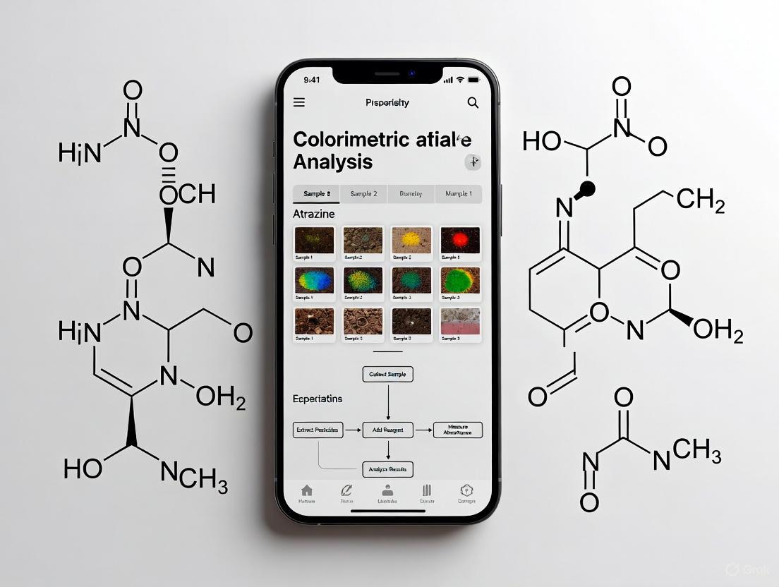
Detailed Experimental Protocol: Smartphone-Based Sensor Array for Multiple Pesticides
This protocol is adapted from a study that distinguished eight pesticides using a colorimetric sensor array of five different gold nanoparticles (AuNPs) and acetylcholinesterase (AChE) [3].
Research Reagent Solutions
Table 2: Essential Reagents and Materials
| Item | Function/Description | Source/Example |
|---|---|---|
| Gold Chloride (HAuCl₄) | Precursor for synthesis of AuNPs. | Aladdin Biochemical Technology [3]. |
| Acetylcholinesterase (AChE) | Enzyme inhibited by organophosphorus pesticides. | Commercial source (e.g., Sigma-Aldrich) [3]. |
| Acetylthiocholine Iodide (ATCh) | Enzyme substrate; hydrolyzed to produce thiocholine. | Sigma-Aldrich [3]. |
| Trisodium Citrate (TSC) | Reducing and stabilizing agent in AuNP synthesis. | Shanghai Macklin Biochemical Technology [3]. |
| Sodium Borohydride (NaBH₄) | Strong reducing agent for small AuNP synthesis. | Common commercial supplier [3]. |
| Target Pesticides | Analytes for detection (e.g., glyphosate, thiram). | Aladdin Biochemical Technology [3]. |
| Smartphone with Camera | Image acquisition device for colorimetric readout. | Standard smartphone (≥12 MP camera) [3] [4]. |
| Color Picking App | Application to extract RGB values from images. | e.g., "Color Name AR" or similar [3]. |
Step-by-Step Procedure
Part A: Synthesis of Diverse Gold Nanoparticles (AuNPs)
- Citrate-reduced AuNPs: Prepare AuNPs using the classical Turkevich-Frens method. Briefly, heat 100 mL of 1 mM HAuCl₄ solution under reflux. Rapidly add 3.5 mL of 38.8 mM trisodium citrate solution under vigorous stirring. Continue heating and stirring until the solution turns a deep wine-red, indicating NP formation [3].
- Borohydride-reduced AuNPs: For smaller AuNPs, mix 20 mL of 0.5 mM HAuCl₄ and 0.5 mL of 10 mM trisodium citrate in an ice bath. Then, add 1.0 mL of 10 mM ice-cold NaBH₄ dropwise under vigorous stirring. The solution will turn a transparent orange-red [3].
- Modification: To create a sensor array, synthesize up to five different types of AuNPs by varying the type and ratio of reducing agents (e.g., trisodium citrate, ascorbic acid) to produce NPs with slightly different sizes and surface properties [3]. Characterize the final NPs using UV-Vis spectroscopy (to confirm LSPR peak) and TEM (for size and morphology).
Part B: Sensor Operation and Smartphone Detection
- Sample Pre-treatment: Extract the pesticide residue from the soil sample using a suitable solvent (e.g., acetonitrile) and filter to remove particulates.
- Enzymatic Reaction: In a microcentrifuge tube, mix the following:
- 50 µL of soil sample extract (or standard pesticide solution for calibration).
- 50 µL of AChE solution (0.1 U/mL).
- Incubate at room temperature for 15 minutes.
- Add 50 µL of ATCh solution (1.0 mM) and incubate for another 10 minutes.
- Colorimetric Transduction: Add 100 µL of the reaction mixture to 100 µL of one type of synthesized AuNPs in a clear-bottom 96-well plate. Repeat this for each of the five different AuNP types. Incubate for 5-10 minutes at room temperature.
- Image Acquisition: Place the 96-well plate on a custom-built, light-shielded cradle with uniform LED white light illumination. Use a smartphone fixed in the cradle to capture an image of the plate, ensuring consistent focus and exposure across all wells [3] [4].
- Data Processing:
- Use a color-picking application on the smartphone to extract the Red, Green, and Blue (RGB) values from each well [3].
- Alternatively, transfer the image to a computer for analysis using image processing software (e.g., ImageJ) to define a region of interest (ROI) and extract average RGB values [4].
- The distinct color response patterns (RGB fingerprints) generated by the five AuNPs against the eight pesticides allow for differentiation. Analyze the data using multivariate statistical methods like Linear Discriminant Analysis (LDA) for pattern recognition and quantification [3].
The mechanism of the enzyme inhibition-based assay is detailed below.
Diagram 2: Mechanism of AChE inhibition assay.
Performance and Comparative Analysis
The performance of the described smartphone-based LSPR sensor is competitive with conventional techniques, offering a balance of sensitivity, speed, and portability.
Table 3: Performance Comparison of Pesticide Detection Methods
| Detection Method | Limit of Detection (LOD) | Analysis Time | Portability | Key Advantages |
|---|---|---|---|---|
| Smartphone-LSPR (This protocol) | < 1.5 × 10⁻⁷ M [3] | Minutes to hours | High | Rapid, on-site, multi-analyte distinguishment, cost-effective [3]. |
| Chromatography (HPLC, GC-MS) | Very Low (ppt-ppb) | Hours | Low | Gold standard; high accuracy and sensitivity for multi-residue analysis [1] [5]. |
| Traditional ELISA | Moderate (ppb) | Hours | Moderate | High specificity and throughput [5]. |
| Electrochemical Biosensors | Low to Moderate | Minutes | High | Highly sensitive, miniaturizable [5]. |
LSPR-based colorimetric transduction provides a powerful and versatile foundation for developing sophisticated yet accessible analytical tools. The integration of these nanosensors with smartphone technology, as demonstrated in the detailed protocol, paves the way for robust, portable, and highly sensitive systems for on-site pesticide monitoring in soil samples. Future advancements will likely involve the integration of machine learning for improved pattern recognition of colorimetric data and the development of more stable and selective nanoparticle probes to further enhance reliability in complex matrices [1] [6].
The accurate and sensitive on-site detection of pesticide residues in soil samples is a critical challenge in environmental monitoring and agricultural safety. Conventional techniques like chromatography and mass spectrometry, while highly sensitive and accurate, are often time-consuming, require expensive instrumentation and skilled personnel, and are unsuitable for field analysis [7] [8]. There is a pressing need for rapid, cost-effective, and portable detection methods.
Molecularly Imprinted Polymers (MIPs) and biomimetic recognition elements have emerged as powerful synthetic alternatives to natural receptors like antibodies. MIPs are polymer-based materials engineered to possess specific cavities that are complementary to a target molecule in shape, size, and functional groups, earning them the title "artificial antibodies" [9] [7]. Their advantages include high specificity, excellent physical and chemical stability, reusability, and relatively low cost [9] [10]. When integrated into sensors, particularly those coupled with smartphone-based colorimetric detection, MIPs enable the development of robust, portable, and highly selective platforms for on-site analysis of pesticides in complex matrices like soil [7] [8].
Fundamental Principles and Recognition Mechanisms
Molecular Imprinting Technology (MIT)
The creation of MIPs involves a process where functional monomers are assembled around a template molecule (e.g., a specific pesticide) and then copolymerized with a cross-linker. Subsequent removal of the template leaves behind cavities that are specifically tailored to recognize and rebind the target analyte [7] [10]. The general workflow is as follows:
- Pre-Complexation: The template molecule and functional monomers interact in a solvent to form a complex. Interactions can be covalent, non-covalent (e.g., hydrogen bonding, van der Waals forces, ionic interactions), or semi-covalent.
- Polymerization: A cross-linking monomer is added to form a highly cross-linked polymer network around the template-monomer complex, freezing the functional groups in their specific spatial arrangement.
- Template Extraction: The template molecules are removed from the polymer matrix using appropriate solvents, leaving behind specific recognition sites.
- Rebinding: The resulting MIP can now selectively rebind the target analyte from a complex sample mixture based on the complementary nature of the imprinted cavities [10].
The following diagram illustrates the logical workflow and key decisions involved in the molecular imprinting process for sensor development.
Biomimetic Sensing with Non-Antibody Probes
Beyond MIPs, other biomimetic recognition elements are being explored. A prominent example is the use of Odorant-Binding Proteins (OBPs). These are small, soluble proteins found in the antennae of insects that are involved in chemical sensing. Recombinant OBPs can be produced in high yield in E. coli, offering a cost-effective and stable alternative to monoclonal antibodies [11]. For instance, OBP2 from Diaphorina citri has been shown to exhibit a broad affinity for multiple neonicotinoid pesticides simultaneously, making it ideal for developing sensors for multi-analyte detection [11].
Performance Comparison of Recognition Elements
The selection of a recognition element is crucial for sensor design. The table below summarizes the key characteristics of MIPs and OBPs in comparison to traditional antibodies for pesticide detection.
Table 1: Comparison of Recognition Elements for Pesticide Sensing
| Feature | Molecularly Imprinted Polymers (MIPs) | Odorant-Binding Proteins (OBPs) | Traditional Antibodies |
|---|---|---|---|
| Specificity | High for target molecule | Broad affinity for a pesticide class | Very high for a single epitope |
| Stability | Excellent chemical & thermal stability; reusable [9] [7] | Good chemical stability | Susceptible to denaturation; limited shelf-life |
| Production Cost & Time | Low cost, relatively simple synthesis [7] | Low-cost, high-yield recombinant expression [11] | High cost, time-consuming production in animals |
| Advantages | "Artificial antibodies"; robust; customizable | Suitable for multi-analyte detection; biomimetic | Well-established technology; high specificity |
| Disadvantages | Occasional heterogeneity of binding sites | Broader specificity may not be desired for single targets | Stability issues; potential cross-reactivity |
Experimental Protocols
This section provides detailed methodologies for fabricating a MIP-based sensor and utilizing a smartphone for colorimetric detection.
Protocol 1: Synthesis of MIPs for Organophosphorus Pesticides via Bulk Polymerization
This protocol outlines the synthesis of MIPs specific to an organophosphorus pesticide (e.g., methyl parathion) using a non-covalent bulk polymerization method [9] [10].
Research Reagent Solutions:
- Template Molecule: Methyl parathion (1.0 mmol)
- Functional Monomer: Methacrylic acid (MAA, 4.0 mmol)
- Cross-linker: Ethylene glycol dimethacrylate (EGDMA, 20.0 mmol)
- Initiator: Azobisisobutyronitrile (AIBN, 0.1 mmol)
- Porogenic Solvent: Acetonitrile (10 mL)
Procedure:
- Pre-Assembly: Dissolve the template (methyl parathion) and functional monomer (MAA) in 5 mL of acetonitrile in a glass vial. Sonicate for 5 minutes and allow the mixture to pre-complex for 1 hour at room temperature.
- Polymerization Mixture: Add the cross-linker (EGDMA) and initiator (AIBN) to the pre-complexed solution. Dilute with the remaining 5 mL of acetonitrile and purge the solution with nitrogen or argon for 10 minutes to remove oxygen, which inhibits free-radical polymerization.
- Polymerization: Seal the vial and place it in a water bath at 60°C for 24 hours to initiate polymerization.
- Grinding and Sieving: After polymerization, break the monolithic polymer block and grind it into a fine powder using a mortar and pestle. Sieve the powder to obtain particles of a defined size range (e.g., 25-50 μm).
- Template Extraction: Wash the polymer particles thoroughly using a methanol-acetic acid (9:1, v/v) solution in a Soxhlet extractor for 24-48 hours to remove the template molecules. Finally, wash with pure methanol to remove residual acetic acid and dry the MIP particles under vacuum at 50°C.
- Control Polymer (NIP): Synthesize a non-imprinted polymer (NIP) following the identical procedure but in the absence of the template molecule. The NIP is used as a control to account for any non-specific adsorption.
Protocol 2: Smartphone-Based Colorimetric Detection of Pesticides using MIPs
This protocol describes how to use the synthesized MIPs as a pre-concentration and recognition element in a smartphone-based colorimetric sensor, adapting principles from nanoparticle-based assays [12] [8].
Research Reagent Solutions:
- Synthesized MIP/NIP particles
- Gold Chloride (HAuCl₄) solution: 1 mM
- Reducing/Stabilizing Agent: Sodium citrate (1%) or natural phenolic extract [12]
- Sample: Soil extract suspected to contain the target pesticide.
- Binding/Washing Buffer: Phosphate Buffered Saline (PBS, 10 mM, pH 7.4)
Procedure:
Solid-Phase Extraction (SPE):
- Pack a small column or a pipette tip with a bed of the synthesized MIP particles (e.g., 10 mg).
- Condition the MIP bed with 1 mL of methanol, followed by 1 mL of PBS buffer.
- Load the prepared soil sample extract (e.g., 1 mL) onto the MIP column.
- Wash the column with 1 mL of PBS buffer to remove unbound and interfering compounds.
- Elute the specifically captured pesticide from the MIP using 0.5 mL of a suitable eluent (e.g., methanol with 1% acetic acid). Collect the eluate.
Colorimetric Reaction (AuNP Growth Induction):
- In a 96-microwell plate, mix 100 μL of the eluate (containing the target pesticide) with 50 μL of 1 mM HAuCl₄ and 50 μL of the reducing agent (e.g., natural phenolic compound extract) [12].
- The presence of the pesticide can influence the in-situ growth of AuNPs, leading to a color change (e.g., from colorless to purple-red) or a change in the intensity of the colored product. Incubate the mixture for 10-15 minutes at room temperature for color development.
Smartphone Detection and Analysis:
- Place the 96-well plate inside a simple, 3D-printed light control box to ensure consistent and uniform illumination, eliminating ambient light interference [12].
- Capture an image of the well plate using a smartphone camera mounted in a fixed position. Do not use flash.
- Use a dedicated colorimetric analysis application (e.g., ColorGrab, ImageJ with a plugin, or a custom-developed app) to analyze the captured image [13]. Select the region of interest (the well) and measure the Green channel intensity or calculate the RGB ratio, which has been shown to provide excellent linearity with analyte concentration [12].
- Quantify the pesticide concentration by comparing the measured intensity/ratio against a calibration curve prepared with known standard concentrations.
The following diagram summarizes the integrated experimental workflow from sample preparation to smartphone-based detection.
Analytical Performance and Data
The integration of MIPs with smartphone colorimetry has demonstrated excellent analytical performance for pesticide detection. The following table summarizes reported data for different sensing strategies.
Table 2: Analytical Performance of Advanced Recognition Elements in Pesticide Sensing
| Recognition Element | Target Pesticide(s) | Detection Method | Limit of Detection (LOD) | Linear Range | Reference & Key Finding |
|---|---|---|---|---|---|
| Molecularly Imprinted Polymer (MIP) | Organophosphorus Pesticides (OPs) | Various (Electrochemical, Fluorescence) | Varies by specific sensor design (e.g., low ppb levels) | -- | [10] - MIPs offer high specificity and stability for OPs detection in complex samples. |
| Odorant-Binding Protein 2 (OBP2) | Imidacloprid | Digital Nanoplasmonometry (DiNM) | 1.4 ppb | -- | [11] - OBP2 allows direct, non-competitive detection of multiple neonicotinoids with high sensitivity. |
| Odorant-Binding Protein 2 (OBP2) | Acetamiprid | Digital Nanoplasmonometry (DiNM) | 1.5 ppb | -- | [11] - The LOD is significantly lower than the Maximum Residue Limits (MRLs) for most countries. |
| Odorant-Binding Protein 2 (OBP2) | Dinotefuran | Digital Nanoplasmonometry (DiNM) | 4.5 ppb | -- | [11] - The method showed high consistency with standard LC-ESI-MS/MS in blind tests. |
| Gold Nanoparticles (Colorimetric) | Tetracyclines (Model Assay) | Smartphone-based Colorimetric | 15 ng/mL (ppb) | 0.05 - 0.50 μg/mL | [12] - Smartphone-based digital image colorimetry provides a cost-effective and portable quantitative analysis. |
The Scientist's Toolkit: Essential Research Reagents
This table lists key materials and their functions for developing MIP-based biomimetic sensors.
Table 3: Key Research Reagent Solutions for MIP-based Sensor Development
| Reagent / Material | Function / Explanation | Example(s) |
|---|---|---|
| Functional Monomer | Provides interaction sites with the template molecule during polymerization. | Methacrylic acid (MAA), Acrylamide (AM) |
| Cross-linker | Creates a rigid polymer network to stabilize the imprinted cavities. | Ethylene glycol dimethacrylate (EGDMA), Trimethylolpropane trimethacrylate (TRIM) |
| Template Molecule | The target analyte or its analog, around which the specific cavity is formed. | Target pesticide (e.g., Methyl parathion, Imidacloprid) |
| Porogenic Solvent | Dissolves all components and creates pores in the polymer for template access. | Acetonitrile, Chloroform, Toluene |
| Initiator | Generates free radicals to start the polymerization reaction. | Azobisisobutyronitrile (AIBN), Ammonium persulfate (APS) |
| Gold Nanoparticles (AuNPs) | Act as colorimetric reporters; aggregation or growth induces visible color change. | Citrate-capped AuNPs, AuNP seeds for growth assays |
| Smartphone & App | Portable detection device and software for image capture and color intensity analysis. | Custom Android/iOS app, Open-source software (e.g., ImageJ) |
| Light Control Box | Provides uniform illumination, minimizing ambient light variation for reproducible imaging. | 3D-printed box with integrated LED lights [12] |
Gold and silver nanoparticles (AuNPs and AgNPs) are cornerstone materials in the development of modern colorimetric sensors, prized for their unique optical properties, particularly their localized surface plasmon resonance (LSPR) [14]. This Application Note details standardized protocols for the synthesis, functionalization, and application of AuNP and AgNP probes, specifically contextualized within a research framework aimed at the smartphone-based colorimetric analysis of pesticides in soil samples. These protocols are designed to produce highly stable and sensitive nanoparticle probes that can be deployed for on-site, rapid detection of pesticide residues, leveraging the ubiquity and analytical power of smartphones [3] [15].
Fundamental Properties of Gold and Silver Nanoparticles
The utility of AuNPs and AgNPs in colorimetric sensing stems from their intense LSPR bands in the visible spectrum. The LSPR is highly sensitive to changes in the local environment, including interparticle distance, size, shape, and composition of the nanoparticles. Aggregation of nanoparticles, induced by a specific target analyte, leads to a significant shift in the LSPR peak and a consequent visible color change, for instance, from red to blue for AuNPs [3] [14]. This phenomenon provides a direct, visually interpretable signal for detection.
Table 1: Key Properties of Gold and Silver Nanoparticles for Sensing
| Property | Gold Nanoparticles (AuNPs) | Silver Nanoparticles (AgNPs) |
|---|---|---|
| Characteristic Color | Wine red | Yellow |
| LSPR Wavelength | ~520-580 nm | ~400-450 nm |
| Extinction Coefficient | High | Very High |
| Common Reducing Agents | Sodium citrate, Sodium borohydride | Sodium borohydride, Sodium citrate |
| Common Stabilizing Agents | Citrate, PEG, Thiols | Citrate, Polymers, Surfactants |
| Key Advantage in Sensing | Excellent biocompatibility, facile functionalization | Higher sensitivity per unit volume |
Synthesis Protocols
Synthesis of Gold Nanoparticles (Turkevich Method)
This method produces spherical, citrate-capped AuNPs around 20 nm in diameter, ideal for further functionalization [16] [14] [17].
Reagents:
- Hydrogen tetrachloroaurate(III) trihydrate (HAuCl₄·3H₂O)
- Trisodium citrate dihydrate (C₆H₅Na₃O₇·2H₂O)
- Milli-Q water
Procedure:
- Glassware Cleaning: Thoroughly clean all glassware with aqua regia (3:1 HCl:HNO₃ by volume), followed by rinsing with copious amounts of deionized water and a final wash with Milli-Q water. This step is critical to remove metallic contaminants that can seed unintended nanoparticle formation [17].
- Prepare a 1 mM HAuCl₄ solution by dissolving the appropriate amount in 500 mL of Milli-Q water in a round-bottom flask.
- Heat the solution to a vigorous boil under reflux with constant stirring.
- Rapidly add 5 mL of a 38.8 mM trisodium citrate solution to the boiling solution.
- Continue heating and stirring for 15 minutes. The solution will change from pale yellow to deep red, indicating nanoparticle formation.
- Remove the solution from heat and continue stirring until it reaches room temperature.
- Purification (Optional): To remove excess citrate and other reagents, dialyze the final nanoparticle suspension against Milli-Q water using a cellulose membrane for 6-8 hours, changing the water at least three times [17].
- Characterize the synthesized AuNPs by UV-Vis spectroscopy (LSPR peak ~520 nm) and TEM for size and morphology confirmation.
Synthesis of Silver Nanoparticles (Sodium Borohydride Reduction)
This protocol yields spherical AgNPs with tunable optical properties and high stability, suitable for sensitive biosensing applications [18] [19].
Reagents:
- Silver nitrate (AgNO₃)
- Sodium borohydride (NaBH₄)
- A non-ionic surfactant (e.g., Triton X-100) or poly(vinyl pyrrolidone) (PVP)
- Milli-Q water
Procedure:
- Solution A: Dissolve AgNO₃ in Milli-Q water to a final concentration of 1 mM.
- Solution B: Prepare a freshly made, ice-cold solution of NaBH₄ (2 mM) in Milli-Q water. Note: NaBH₄ solution is unstable and must be prepared immediately before use and kept on ice.
- Stabilizer Solution: Add a non-ionic surfactant (e.g., 0.1% v/v Triton X-100) to Solution A to prevent aggregation [18].
- Under vigorous stirring, add Solution B (NaBH₄) dropwise to Solution A (AgNO₃ with stabilizer). The solution will turn pale yellow, indicating the formation of AgNPs.
- Continue stirring for 1 hour to ensure complete reduction and stabilization.
- Store the synthesized AgNPs at 4°C in the dark. Characterize by UV-Vis spectroscopy (LSPR peak ~400 nm) and TEM.
Table 2: Summary of Standard Nanoparticle Synthesis Methods
| Parameter | Turkevich AuNPs | Brust-Schiffrin AuNPs | Citrate/NaBH₄ AgNPs |
|---|---|---|---|
| Size Range | ~10-20 nm | ~1-5 nm | ~10-50 nm |
| Solvent | Water | Toluene (Organic) | Water |
| Reducing Agent | Sodium Citrate | Sodium Borohydride (NaBH₄) | NaBH₄ / Citrate |
| Stabilizing Agent | Citrate ions | Alkanethiols | Citrate / Polymers / Surfactants |
| Key Feature | Water-soluble, easy functionalization | Organic-soluble, very stable | High optical sensitivity, tunable |
Functionalization for Pesticide Sensing
A highly effective strategy for pesticide detection involves designing nanoparticle probes that respond to enzymatic activity inhibited by pesticides, such as acetylcholinesterase (AChE) [3].
Principle: The functionalized nanoparticle probe is integrated into an assay where AChE hydrolyzes acetylthiocholine (ATCh) to produce thiocholine. Thiocholine induces nanoparticle aggregation, causing a color shift. The presence of a pesticide inhibits AChE, reducing thiocholine production and thus altering the colorimetric response, which can be quantified [3].
Functionalization Protocol: Aptamer-Modified Probes
This protocol describes a modular approach to functionalize nanoparticles with DNA aptamers for specific pesticide recognition, using a PEG passivation layer for enhanced stability [20].
Reagents:
- Carboxylated AuNPs or AgNPs (synthesized as above)
- Amine-PEG-Azide (e.g., 1.6 kDa or 2 kDa)
- EDC (1-Ethyl-3-(3-dimethylaminopropyl)carbodiimide) and NHS (N-Hydroxysuccinimide)
- DBCO-modified oligonucleotide "handle"
- DNA aptamer with complementary sequence to the handle
- Buffers: MES buffer (0.1 M, pH 5.0), PBS buffer (0.1 M, pH 7.4)
Procedure:
- PEG Passivation:
- Activate carboxyl groups on nanoparticles (1 mL) by incubating with EDC (20 mM) and NHS (10 mM) in MES buffer for 20 minutes with gentle shaking.
- Purify the activated NPs from excess EDC/NHS using a centrifugal filter.
- Resuspend the NPs in PBS and add amine-PEG-azide at a ratio of ~10⁸ PEG molecules per 200 nm particle. React for 2-4 hours at room temperature.
- Purify the PEGylated nanoparticles. Successful passivation is confirmed by a shift in zeta potential towards neutrality (e.g., from -43 mV to -15 mV) [20].
Conjugation-Annealing Handle Attachment:
- Incubate the azide-functionalized NPs with a DBCO-modified oligonucleotide handle. The DBCO group reacts with the azide via a copper-free "click" chemistry.
- Purify the handle-conjugated nanoparticles.
Aptamer Attachment:
- Anneal the DNA aptamer (specific to the target pesticide, e.g., organophosphates) to the complementary sequence on the conjugation handle by heating the mixture to 90°C and slowly cooling to room temperature.
- Purify the final aptamer-functionalized nanoparticle probes and store in an appropriate buffer at 4°C.
The following diagram illustrates the core mechanism of a smartphone-based colorimetric sensor using functionalized nanoparticles for pesticide detection.
The Scientist's Toolkit: Essential Reagents and Materials
Table 3: Key Research Reagent Solutions for Nanoparticle-Based Sensors
| Reagent/Material | Function | Example Use Case |
|---|---|---|
| Chloroauric Acid (HAuCl₄) | Gold precursor for AuNP synthesis | Starting material for Turkevich and Brust methods [14] [17]. |
| Silver Nitrate (AgNO₃) | Silver precursor for AgNP synthesis | Starting material for chemical reduction of AgNPs [18] [19]. |
| Sodium Borohydride (NaBH₄) | Strong reducing agent | Rapid reduction of metal ions to form small, spherical NPs [18] [19] [17]. |
| Trisodium Citrate | Reducing and stabilizing agent | Mild reduction and electrostatic stabilization of NPs in water [3] [14]. |
| Amine-PEG-Azide | Passivating and functionalizing agent | Creates a biocompatible, inert layer and provides a handle for bio-conjugation [20]. |
| Acetylcholinesterase (AChE) | Enzyme for inhibition-based assays | Hydrolyzes ATCh to thiocholine; activity inhibited by pesticides [3]. |
| Acetylthiocholine (ATCh) | Enzyme substrate | Hydrolyzed by AChE to produce thiocholine, which triggers NP aggregation [3]. |
| DBCO-Modified DNA Handle | Modular conjugation linker | Facilitates easy attachment of various aptamers to PEGylated NPs [20]. |
Smartphone-Based Detection Workflow
The integration of functionalized nanoparticle probes with smartphone technology enables portable, quantitative point-of-care testing (POCT) [3] [15]. The following workflow details the process from sample preparation to data analysis.
Procedure:
- Sample Preparation: Extract pesticides from soil samples using a suitable solvent (e.g., methanol/water mixture). Filter and dilute the extract to a compatible pH.
- Assay Execution:
- In a microplate well or small vial, mix the soil extract (or standard), AChE, and ATCh. Incubate for 15-30 minutes.
- Add the functionalized AuNP or AgNP probe suspension. Incubate for another 5-10 minutes to allow the colorimetric reaction to proceed.
- Image Capture:
- Place the reaction vial in a simple, 3D-printed dark box to ensure consistent, uniform lighting.
- Use a smartphone camera to capture an image of the solution. Ensure the camera settings (white balance, exposure) are fixed or standardized.
- Colorimetric Analysis:
- Transfer the image to a color analysis application (e.g., ImageJ, Color Name AR, or a custom app) [3].
- Extract the Red, Green, Blue (RGB) values or convert to Hue, Saturation, Value (HSV) or Cyan, Magenta, Yellow, Black (CMYK) color spaces for analysis [15].
- The intensity of the RGB channels, particularly the Red/Green or Blue/Red ratio, can be correlated with the degree of nanoparticle aggregation and thus the pesticide concentration.
- Data Processing:
- Use statistical tools like Linear Discriminant Analysis (LDA) to distinguish between multiple pesticides based on their unique response patterns across different nanoparticle probes [3].
- Generate a calibration curve from standards to quantify the pesticide concentration in the unknown soil samples.
The synthesis and functionalization protocols outlined herein provide a robust foundation for developing highly sensitive and specific nanoparticle-based probes. When coupled with the smartphone-based detection platform, these probes form a powerful, low-cost, and portable system for the on-site monitoring of pesticide residues in environmental samples like soil. This integrated approach holds significant promise for enhancing food safety, environmental health, and public safety by enabling rapid, decentralized screening.
The integration of smartphone cameras with colorimetric sensors represents a transformative advancement in analytical chemistry, enabling portable, low-cost, and rapid quantification of analytes. This approach is particularly valuable for environmental monitoring, including the detection of pesticides in soil samples, where traditional lab equipment is inaccessible or impractical. The core principle relies on the RGB (Red, Green, Blue) color model, a device-dependent color space where the smartphone camera digitizes color information by assigning intensity values from 0 to 255 for each of the three primary color channels [21]. A color change in a chemical sensor, induced by the presence of a target analyte, alters the relative intensities of the R, G, and B values captured by the camera. By applying appropriate image processing algorithms and calibration models, these digital color signals can be quantified and correlated to analyte concentration [22] [23].
However, the use of consumer smartphones for analytical measurements introduces significant challenges. Ambient light conditions, variations in camera sensors, and built-in automatic image corrections (like auto-white balance and exposure) can substantially alter the recorded RGB values, leading to measurement inaccuracies [22] [24] [25]. Overcoming these hurdles is critical for developing reliable field-deployable methods for pesticide analysis. This document details the protocols and application notes for using smartphone cameras to quantify color changes, with specific considerations for research on pesticides in soil.
Key Principles of RGB Color Quantification
From Analog Color to Digital RGB Values
A smartphone camera sensor captures light reflected from a colorimetric sensor through filters sensitive to red, green, and blue wavelengths. The intensity of light in each channel is converted into a digital value, typically an 8-bit integer, resulting in the RGB triplet that defines the perceived color. In analytical applications, the reaction between an analyte and a chemical reagent on a sensor strip induces a color change. This change can be monitored as a shift in the RGB triplet. The relationship between the analyte concentration and the color intensity can be non-linear, often requiring sophisticated data processing models [23].
For pesticide analysis, the test strip might be functionalized with enzymes like acetylcholinesterase (AChE), which is inhibited by organophosphate and carbamate pesticides. The degree of inhibition reduces the enzymatic reaction that produces a colored product, leading to a quantifiable decrease in color intensity in a specific RGB channel [21] [22].
Color Space Selection and Correction Algorithms
Using the raw, device-dependent RGB values for concentration calculation is prone to error. Therefore, a common practice is to convert RGB values into a device-independent color space such as CIE L*a*b* or CIE 1976 u'v' [22] [24]. These spaces separate luminance (lightness) from chrominance (color), making the color information more robust against variations in ambient light intensity.
Color correction is a critical step to ensure data consistency across different smartphones and lighting environments. This is typically achieved using a reference color card captured in the same image as the sensor. Advanced algorithms, such as the Root Polynomial-based Correction Algorithm (RPCC) or a third-order polynomial model, map the distorted colors from the smartphone camera to their known reference values [22] [24]. One study demonstrated that this methodology could reduce inter-device and lighting-dependent color variations by 65-70%, significantly improving measurement reliability [25].
Table 1: Comparison of Color Spaces Used in Smartphone Colorimetry
| Color Space | Type | Key Components | Advantages for Colorimetry |
|---|---|---|---|
| RGB | Device-dependent | R (Red), G (Green), B (Blue) channels | Native to smartphone cameras; simple to access. |
| CIE L*a*b* | Device-independent | L* (Lightness), a* (Green-Red), b* (Blue-Yellow) | Perceptually uniform; separates intensity from color. |
| CIE 1976 u'v' | Device-independent | u' (chromaticity), v' (chromaticity) | Derived from CIE XYZ; useful for color adaptation models. |
Experimental Protocols
Protocol 1: Manufacturing of Paper-Based Colorimetric Sensors
This protocol is adapted from methods for soil pH and nutrient sensors, which can be extended to pesticide detection [26].
1. Materials and Reagents:
- Substrate: Chromatography paper (e.g., Whatman Grade 1).
- Patterning: Wax printer (e.g., Xerox ColorQube) or other hydrophobic barrier method.
- Chemical Reagents: Depending on the target pesticide. For AChE-based detection, this includes:
- Acetylcholinesterase (AChE) enzyme.
- Substrate (e.g., Acetylthiocholine iodide).
- Chromogenic compound (e.g., 5,5'-dithio-bis-(2-nitrobenzoic acid) - DTNB).
- Assembly: Cardboard cover, spray adhesive, vacuum sealer.
2. Procedure:
- Step 1: Design and Print Hydrophobic Barriers. Use design software to create the microfluidic pattern for the paper-based device (μPAD). Print the pattern onto the chromatography paper using a wax printer.
- Step 2: Melt the Wax. Heat the printed paper in an oven at 100°C for 1-2 minutes to allow the wax to permeate the paper, creating complete hydrophobic barriers and defining hydrophilic test zones [26].
- Step 3: Functionalize the Sensor. Deposit the chemical reagents onto the hydrophilic test zones. For an AChE sensor, this typically involves pre-immobilizing the enzyme and the chromogen in separate zones or in a specific sequence. Pre-storage stability can be improved by vacuum sealing [26].
- Step 4: Assemble the Sensor Card. Attach the functionalized paper sensor to a cardboard backing. Include a QR code for linking to calibration data and a reference color chart for post-processing color correction. Vacuum-seal the final sensor cards to prolong shelf life.
Protocol 2: Smartphone Imaging and Color Data Processing
This protocol ensures consistent image acquisition for quantitative analysis [26] [24].
1. Materials and Equipment:
- Smartphone: Any Android or iOS device with a camera.
- Lighting Control: A 3D-printed mini light box or a fixed, shaded environment to isolate the sensor from ambient light. Two white LED modules can provide consistent illumination [24].
- Reference Chart: A standardized color card with known color values (e.g., X-Rite ColorChecker).
2. Image Acquisition Procedure:
- Step 1: Setup. Place the reacted colorimetric sensor strip and the reference color chart inside the light box. Ensure the smartphone camera is fixed at a consistent distance and angle from the sensor, as oblique angles can introduce color errors (∆E increase up to 64%) [25].
- Step 2: Camera Settings. If possible, disable all automatic settings (auto-white balance, auto-exposure, auto-focus). Use the smartphone's "pro" or "manual" camera mode. If automatic settings cannot be disabled, ensure that the reference chart is included in every image for post-correction [22] [24].
- Step 3: Capture Image. Capture the image in the highest resolution possible without compression (e.g., saving in PNG format).
3. Image Processing and Data Extraction Procedure:
- Step 1: Image Segmentation. Use contour extraction algorithms (e.g., with OpenCV library) to automatically identify the regions of interest (ROI) for the sensor's test zones and the reference color chart patches [24].
- Step 2: Color Correction. Extract the average RGB values from each ROI. Apply a color correction algorithm (e.g., RPCC) using the known reference values from the color chart to convert the device-dependent RGB values to device-independent L*a*b* or standard RGB values [22].
- Step 3: Feature Extraction. Convert the corrected color values into an analytical signal. This could be the intensity of a single RGB channel, a ratio of channels (e.g., R/G, G/B), or the overall color difference (∆E) from a control. For pesticide detection based on AChE inhibition, the signal is often the intensity of the yellow product (e.g., in the blue channel).
The Scientist's Toolkit: Essential Research Reagent Solutions
Table 2: Key Reagents and Materials for Smartphone Colorimetry of Pesticides
| Item | Function/Description | Example in Protocol |
|---|---|---|
| Chromatography Paper | Cellulose-based substrate for microfluidic devices; wicks sample via capillary action. | Whatman Grade 1 paper as the base for μPADs [26]. |
| Acetylcholinesterase (AChE) | Enzyme inhibited by organophosphate/carbamate pesticides; the core biorecognition element. | Immobilized on paper test zone to detect pesticide presence [21]. |
| DTNB (Ellman's Reagent) | Chromogen that produces a yellow-colored anion when reacting with thiocholine. | Used to visualize AChE activity; color intensity inversely related to pesticide concentration. |
| Reference Color Chart | A card with patches of known color values; essential for color correction algorithms. | X-Rite ColorChecker captured in-frame to correct for lighting and camera variations [22] [24]. |
| 3D-Printed Light Box | An accessory to provide consistent, uniform illumination and block ambient light. | Custom box printed with black resin to hold phone, sensor, and LEDs [24]. |
Quantitative Data and Performance Metrics
The performance of smartphone-based colorimetric sensors is benchmarked against standard laboratory techniques like spectrophotometry. Key performance metrics include accuracy, precision, and the limit of detection (LOD).
Table 3: Performance Metrics from Representative Studies
| Analytical Target | Smartphone System & Color Model | Key Performance Metrics | Comparison to Standard Method |
|---|---|---|---|
| Soil pH [26] | Machine learning model on colorimetric data from μPADs with BCG/BCP indicators. | 97% correct classification (low/medium/high pH) in field tests. | Reduced analysis time from days (lab) to minutes (mobile). |
| Soil Nitrate-N [27] | Android app with commercial test strips (Quantofix), using smartphone as a reflectometer. | 87% of samples agreed with standard lab method within 10 mg kg⁻¹. | Effective as a screening tool; high-end phones showed less bias. |
| Color Correction [25] | Matrix-based color correction using a reference chart on various smartphones. | Reduced inter-device and lighting variation by 65-70% (ΔE). | Enabled consistent kinetic profiles from video analysis across devices. |
The RGB color model, when coupled with robust experimental protocols and advanced color correction algorithms, provides a powerful and accessible foundation for quantitative analytical chemistry using smartphones. The methodologies outlined in these application notes—from sensor fabrication and controlled imaging to sophisticated data processing—establish a reliable framework for developing field-deployable detection systems. For research focused on pesticides in soil, this approach promises to enable rapid, on-site screening, facilitating higher spatial resolution in mapping contamination and supporting more informed and sustainable agricultural decisions. Future advancements in machine learning and artificial intelligence will further enhance the accuracy and automate the interpretation of these smartphone-based colorimetric analyses [26] [23].
For researchers and agricultural professionals, monitoring pesticide residues in soil is essential for ensuring food safety and environmental sustainability [28]. Traditional laboratory-based methods, such as Gas Chromatography-Mass Spectrometry (GC-MS/MS) and High-Performance Liquid Chromatography (HPLC), have long been the gold standard for this analysis [29]. However, these techniques involve significant limitations, including extensive sample preparation, destructive analysis, and resource-intensive procedures, which hinder their application for rapid, on-site decision-making [21] [28]. This creates a critical need for alternative, field-deployable technologies. Smartphone-based colorimetric analysis is emerging as a powerful, cost-effective, and rapid alternative, enabling researchers to perform quantitative chemical analysis directly in the field and paving the way for more accessible environmental monitoring [21] [15].
Limitations of Traditional Analytical Methods
Traditional chromatographic methods, while highly sensitive and accurate, present several constraints that make them unsuitable for rapid, on-site analysis.
Operational and Resource Constraints
The following table summarizes the key limitations of GC-MS/MS, LC-MS/MS, and HPLC methods based on information from analytical service providers and scientific literature [29]:
| Method | Target Compounds | Key Limitations | Sample Suitability | Resource Intensity |
|---|---|---|---|---|
| GC-MS/MS | Volatile/Semi-volatile pesticides [29] | Not suitable for non-volatile or thermally unstable pesticides; may require derivatization [29]. | Dry, solid samples (e.g., grains, cannabis) [29] | High cost, time-consuming, requires skilled operators and laboratory infrastructure [21] [28] [29] |
| LC-MS/MS | Polar/Non-volatile pesticides (e.g., glyphosate, neonicotinoids) [29] | Requires precise sample preparation; resource-intensive [29]. | Oily, moist, delicate samples (e.g., fruits, plant extracts) [29] | High operational cost and complexity [29] |
| HPLC | Known UV-absorbing pesticides [29] | Reduced sensitivity for low-level or unknown residues; not ideal for broad-spectrum screening [29]. | Mostly liquids [29] | Lower sensitivity, though more cost-effective than MS methods [29] |
These methods are destructive, meaning the sample cannot be recovered for further analysis, and they often involve large time delays between sample collection and the availability of results, preventing prompt intervention [21] [28].
The Case for Quantitative NMR (qNMR)
In addition to colorimetric methods, other non-destructive techniques are being explored to overcome these limitations. Quantitative Nuclear Magnetic Resonance (qNMR) spectroscopy has been validated as an efficient method for detecting spinosad residues in agricultural soils [28]. It offers an 88% recovery rate and requires minimal sample preparation, establishing itself as a reliable, cost-effective alternative to chromatographic methods for specific applications [28].
Smartphone-Based Colorimetric Analysis as a Solution
Smartphone-based analysis leverages the powerful sensors and processing capabilities of ubiquitous mobile devices to create portable, inexpensive, and user-friendly analytical platforms [21] [15].
Core Principles and Sensing Modalities
The standard smartphone is equipped with a range of built-in hardware sensors, with the image sensor (camera) being the most frequently used for chemical analysis [21]. The core principle involves acquiring images of a sample and digitizing them for analysis.
- Color Spaces: The most common color model used is the RGB (Red, Green, Blue) model, where the intensity of each color component is expressed in a range of 0–255 [21] [15]. When analysis is affected by external factors like brightness, other color spaces such as HSV (Hue, Saturation, Value) are also utilized [15].
- Detection Platforms: Smartphones can be integrated with various platforms, including colorimetric, fluorescent, and microscopic imaging, demonstrating analytical performance comparable to traditional spectrometers [15].
Documented Applications in Soil Science
Research has demonstrated the successful application of smartphone-based detection for various soil parameters:
- Basic Soil Properties: Smartphone cameras have been applied to measure soil colour, a key property for classifying soil strata and inferring chemical composition and fertility [21]. The results show high agreement with traditional Munsell soil colour cards and spectrophotometry, especially under controlled lighting [21].
- Soil Contaminants and Nutrients: The technology has been developed for determining the content of organic matter, mineral fertilizers, and inorganic pollutants [21]. The general trend indicates a huge research interest in moving the technology into the field to provide cost-effective and rapid soil analysis [21].
Experimental Protocols
Protocol 1: General Workflow for Smartphone-Based Soil Color Analysis
This protocol outlines the steps for using a smartphone to determine soil color, a fundamental property, for classification and fertility assessment [21].
Research Reagent Solutions & Essential Materials
| Item | Function/Description |
|---|---|
| Smartphone | Analytical device with camera and processing software. Ideally has a high-resolution sensor. |
| Shading Device | A cover (e.g., black plastic tube) to prevent reflections and control external light. |
| Calibration Cards | Reference color standards for white balance and color calibration under consistent lighting. |
| Optical Lenses & Diaphragms | Adjust the size of the camera's field of view for consistent image capture. |
Step-by-Step Procedure:
- Sample Preparation: Collect and air-dry soil samples. Ensure they are finely ground and presented on a uniform surface [21].
- Setup: Place the sample within the shading device to eliminate external light interference. Include the calibration cards within the frame [21].
- Image Acquisition: Capture the photograph from a fixed distance (e.g., half a meter) using the smartphone camera. Ensure the flash is turned off and settings are consistent [21].
- Image Processing: Use software (e.g., ImageJ, MATLAB) to analyze the image. Select the region of interest (the soil) and extract the average RGB or HSV values [21] [15].
- Data Interpretation: Convert the extracted color values into relevant soil parameters using a pre-established calibration curve or a classification algorithm. The results can be exported to geographic information systems to create soil property maps [21].
Protocol 2: Workflow for an Intelligent Colorimetric Detection System
This protocol describes a generalized workflow for constructing an intelligent detection system for contaminants like pesticides, using prepared probes and a smartphone [15].
Implementation & Data Analysis
Overcoming Implementation Challenges
While promising, smartphone-based analysis faces hurdles that must be addressed for reliable results.
- Lighting Conditions: The accuracy of measurements is significantly influenced by ambient light. More accurate and precise results are obtained under consistent, bright conditions (e.g., sunny, or using a controlled light box) [21].
- Data Processing and AI: Advanced algorithms and artificial intelligence are crucial for improving the efficiency and accuracy of quality inspection. Machine learning classifiers can be constructed to identify patterns and predict soil properties or contamination levels from the extracted color data [15].
- System Integration: For widespread use, these systems can be integrated with the Internet of Things (IoT), enabling real-time monitoring and the creation of traceability systems for agricultural products from production to consumption [15].
Comparative Analysis: Traditional vs. Emerging vs. Smartphone Methods
The table below provides a direct comparison of the key analytical techniques discussed, highlighting the position of smartphone-based methods.
| Feature | Traditional GC-MS/LC-MS/HPLC | Emerging qNMR Method | Smartphone-Based Colorimetry |
|---|---|---|---|
| Portability | Low (laboratory-bound) | Low (laboratory-bound) | High (field-deployable) |
| Analysis Speed | Slow (hours to days) | Moderate to Fast | Very Fast (minutes) |
| Cost | High (equipment, reagents, labor) | Cost-effective after initial investment [28] | Very Low [21] |
| Sample Preparation | Extensive | Minimal [28] | Minimal to Moderate |
| Destructive | Yes | No [28] | No |
| User Skill Required | High (trained technician) | High (trained technician) | Low (minimal training) [21] |
| Primary Use Case | Regulatory compliance, definitive quantification | Accurate, non-destructive quantification [28] | Rapid screening, precision agriculture, field surveys |
Traditional methods like GC-MS and HPLC, while highly accurate, are hampered by their operational complexity, cost, and lack of portability, creating a significant gap for on-site analytical needs. Smartphone-based colorimetric analysis effectively addresses this gap by offering a rapid, cost-effective, and user-friendly platform for the on-site screening of soil properties and contaminants. When combined with robust experimental protocols, controlled imaging conditions, and advanced data processing algorithms, this technology holds immense potential to democratize environmental monitoring, empower farmers and researchers with real-time data, and significantly enhance the framework of precision and sustainable agriculture.
Building Your Sensor: Methodologies for Assay Development and Field Application
Paper-Based Analytical Devices (PADs) are low-cost, portable, and user-friendly platforms for chemical analysis, particularly suited for point-of-care testing (POCT) and on-site environmental monitoring [30]. Their development can be traced back to litmus paper in the 17th century, with significant milestones including the invention of paper chromatography in the mid-20th century and the groundbreaking introduction of microfluidic PADs (µPADs) by the Whitesides group in 2007 [30] [31]. The core principle of µPADs involves patterning hydrophobic barriers onto hydrophilic paper to create miniature channels that transport aqueous fluids via capillary action, eliminating the need for external pumps [32] [31]. This design enables the integration of sample introduction, analytical sensing, and signal output zones on a single, disposable substrate.
Within the context of a thesis focused on smartphone-based colorimetric analysis of pesticides in soil, PADs offer an ideal platform. Their portability allows for on-site sample collection and initial processing, while their compatibility with colorimetric assays enables visual detection that can be quantified using a smartphone camera [15]. The ability to functionalize paper with enzymes and nanomaterials specific to pesticide detection facilitates the development of highly selective and sensitive sensors, making advanced analytical techniques accessible in field settings [3].
Design and Fabrication of PADs
The design and fabrication of PADs are critical steps that determine their performance, including fluidic control, sensitivity, and reproducibility. Fabrication techniques primarily involve creating hydrophobic barriers to define hydrophilic channels and test zones.
Common Fabrication Techniques
A wide array of fabrication methods has been developed, balancing cost, resolution, and accessibility.
Table 1: Comparison of Common PAD Fabrication Techniques [32] [31]
| Fabrication Technique | Equipment | Reagents/Materials | Advantages | Typical Resolution |
|---|---|---|---|---|
| Photolithography | Lithography equipment, UV light source, hot plate | Photoresist (e.g., SU-8) | High resolution; first method for µPADs | ~80 µm [32] |
| Wax Printing | Solid ink printer, hot plate or oven | Solid wax | Simple, fast, low-cost; suitable for rapid prototyping | ~300-400 µm [33] |
| Inkjet Printing | Customized inkjet printer | Hydrophobic chemicals (e.g., AKD) | High resolution; digital process | Varies with printer |
| Laser Cutting | Laser cutting machine | None (subtractive process) | Rapid prototyping; no reagents required; high reproducibility | Susceptible to contamination [32] |
| Screen Printing | Screen mask, squeegee | Wax, UV-curable ink | Low cost; simple steps | Low resolution [32] |
| 3D Printing | 3D printer | PDMS, 3D printer resin | Rapid and accessible mass production; complex 3D structures | Depends on 3D printer [32] |
Advanced and Miniaturized Fabrication
Recent advancements focus on improving resolution and functionality. A notable method for creating high-resolution features involves the miniaturization of wax-printed PADs via periodate oxidation [33]. This technique involves immersing a wax-printed µPAD in an aqueous sodium periodate (NaIO₄) solution, which oxidizes cellulose and causes the paper to shrink up to 80% in surface area, thereby reducing the size of the printed features. This process can yield functional hydrophilic channels as narrow as 301 µm and hydrophobic barriers down to 387 µm, without the need for specialized microfabrication equipment [33].
For more complex fluidic control, optofluidic PADs can be fabricated using double-sided photolithography with solvents like perfluoropolyether dimethacrylate (PFPE-DMA) to create robust, three-dimensional microfluidic channels within a paper matrix [34]. These 3D devices are ideal for multi-step assays and complex pattern recognition tasks.
Figure 1: Generalized Workflow for Fabricating a PAD. The process begins with digital design, followed by a choice of patterning method to create hydrophobic barriers. Optional miniaturization can enhance feature resolution before functional reagents are integrated.
In PADs, sample introduction and subsequent fluid transport are governed by the capillary action of the cellulose network. Fluid movement is passive and can be described by the Washburn equation: ( l = \sqrt{\frac{\gamma r \cos\theta}{2\eta} t} ), where ( l ) is the wicking distance, ( \gamma ) is the surface tension, ( r ) is the average pore radius, ( \theta ) is the contact angle, ( \eta ) is the fluid viscosity, and ( t ) is time [31]. This equation highlights that the wicking rate and distance depend on the properties of both the paper substrate and the liquid sample.
For soil samples, a liquid extract must typically be prepared before introduction to the PAD. A common protocol involves mixing soil with deionized water (e.g., a 1:2 soil-to-water ratio), vigorous shaking for 1-2 minutes, and allowing the mixture to settle or filtering it to obtain a clear supernatant [35]. This liquid extract is then pipetted directly onto the sample inlet zone of the PAD.
Advanced PADs incorporate sophisticated mechanisms for fluidic control. For instance, Macromolecule-Driven Flow (MDF) gates can be created by impregnating paper channels with different polymers (e.g., PDMS, PVA, PCL) [34]. The interaction between the liquid analyte and the polymer dictates the flow behavior—allowing the liquid to pass, slowing it down (intermediate pass), or completely stopping it. This provides a powerful mechanism for discriminating between different chemical substances based on their physicochemical properties, mimicking an olfactory system [34].
Application Protocol: Smartphone-Based Colorimetric Detection of Pesticides
This protocol details the application of a gold nanoparticle (AuNP)-based colorimetric sensor array on a PAD for distinguishing multiple pesticides, integrating smartphone-based readout.
The Scientist's Toolkit: Research Reagent Solutions
Table 2: Essential Materials and Reagents for Pesticide Detection PAD [3]
| Reagent/Material | Function/Description | Role in the Assay |
|---|---|---|
| Gold Nanoparticles (AuNPs) | Colorimetric sensing element; ~20-40 nm diameter, synthesized via Turkevich-Frens method. | Signal transducer; aggregation causes visible color change from red to blue. |
| Acetylcholinesterase (AChE) | Enzyme. | Hydrolyzes ATCh; activity inhibited by specific pesticides. |
| Acetylthiocholine Iodide (ATCh) | Enzyme substrate. | Hydrolyzed by AChE to produce thiocholine. |
| Thiocholine | Product of ATCh hydrolysis. | Induces AuNP aggregation via Au-S covalent bonds. |
| Pesticide Standards | Target analytes (e.g., glyphosate, thiram). | Inhibit AChE, reducing thiocholine production and altering color response. |
| Chromatography Paper | Substrate (e.g., Whatman No. 1). | Porous cellulose matrix for fluid transport and reagent immobilization. |
| Smartphone with Color Picking App | Analytical instrument. | Captures image and extracts RGB values from detection zones for quantification. |
Experimental Workflow and Detection Mechanism
The detection principle relies on the enzyme inhibition of AChE by pesticides, which modulates the production of thiocholine and subsequently controls the aggregation of AuNPs [3].
Figure 2: Signaling Pathway for AChE-AuNP Pesticide Detection. Pesticides inhibit AChE, reducing thiocholine production. Thiocholine induces AuNP aggregation, causing a color change. The degree of color change is inversely related to pesticide concentration.
Step-by-Step Experimental Protocol
Part A: Device Fabrication
- Design: Design a µPAD with a sample inlet zone connected to multiple detection zones using CAD software.
- Fabricate: Print the design onto chromatography paper using a wax printer.
- Reflow: Heat the printed paper in a convection oven at 195°C for 2 minutes to allow the wax to melt and penetrate the paper, forming complete hydrophobic barriers.
- Functionalize: Spot different types of synthesized AuNPs (e.g., varying in size or surface chemistry) into separate detection zones. Allow to dry.
- Immobilize Enzyme: Co-immobilize AChE and its substrate ATCh in the detection zones or in a pre-zone leading to them.
Part B: Sample Preparation and Assay Execution
- Soil Extraction: Prepare a soil extract by shaking soil with a suitable buffer or water, followed by centrifugation or filtration to obtain a clear supernatant.
- Apply Sample: Pipette the soil extract supernatant onto the sample inlet of the µPAD.
- Capillary Flow: Allow the sample to wick through the device via capillary action, dissolving and carrying the reagents (AChE, ATCh).
- Reaction and Incubation: As the sample reaches the AuNP-functionalized detection zones, the enzymatic reaction occurs. If pesticides are absent, AChE hydrolyzes ATCh to produce thiocholine, which aggregates the AuNPs, causing a color change from red to blue. If pesticides are present, AChE is inhibited, less thiocholine is produced, and the red color of dispersed AuNPs is preserved. Incubate for a fixed time (e.g., 10-15 minutes) at room temperature.
Part C: Smartphone Readout and Data Analysis
- Image Capture: Place the µPAD in a light-controlled box to ensure consistent illumination. Capture an image of the detection zones using a smartphone camera.
- Color Value Extraction: Use a color picker application or custom software (e.g., ImageJ, MATLAB, or a dedicated mobile app) to extract the Red, Green, and Blue (RGB) values from each detection zone.
- Data Processing: Normalize the RGB values and use pattern recognition algorithms, such as Linear Discriminant Analysis (LDA), to differentiate between multiple pesticides based on their unique color response patterns [3].
- Quantification: Construct a calibration curve by plotting the RGB values (or a derived value like grayscale intensity) against the logarithm of pesticide concentration for quantitative analysis.
This integrated approach, combining specific biochemical reactions with nanomaterials on a portable PAD and a smartphone reader, provides a powerful, low-cost tool for on-site pesticide monitoring, directly supporting the objectives of advanced research in agricultural and environmental analytics.
The need for on-site detection of pesticides in soil samples has driven the development of portable, robust, and cost-effective sensor platforms. Smartphone-based colorimetric analysis presents a powerful solution, leveraging the ubiquitous presence of smartphones to provide rapid, in-field analytical capabilities. A significant challenge, however, lies in the fabrication and housing of the sensors themselves, which must be durable enough for field use while maintaining analytical precision. Additive manufacturing, or 3D printing, is revolutionizing this space by enabling the rapid prototyping and production of custom sensor housings, microfluidic cells, and even functional electrode components. These 3D-printed platforms ensure that sensors are properly aligned, protected from environmental variables, and integrated with smartphone detectors, forming complete field-deployable kits [36] [37]. This document outlines the application of 3D printing in creating robust sensor integration platforms, with specific protocols and guidelines for researchers developing smartphone-based colorimetric kits for pesticide analysis in soil.
The 3D-Printing Advantage in Sensor Fabrication and Integration
Traditional manufacturing methods for sensor housings and components are often time-consuming, expensive, and ill-suited for custom, low-volume production required in research and specialized field applications. 3D printing addresses these limitations through several key advantages:
- Rapid Prototyping and Customization: Additive manufacturing allows for the quick iteration of designs, enabling researchers to optimize the sensor housing for specific smartphone models, optical paths, and soil sample extraction volumes [36] [38]. Complex geometries, such as light-tight chambers and integrated fluidic channels, can be produced in a single print.
- Multi-Material and Integrated Printing: Advanced 3D printers can utilize multiple materials within a single print job. This allows for the co-printing of rigid structural elements (for the housing) and flexible, sealing gaskets, or even the integration of conductive traces for electrochemical sensors [36]. Techniques like conductor infusion can create networks of channels for conductive inks post-printing [36].
- Cost-Effectiveness and Accessibility: Fused Deposition Modeling (FDM) printers and materials are relatively low-cost, making this technology accessible to most research laboratories. This democratizes the development of custom sensor platforms without the need for expensive tooling or outsourcing [37] [38].
- Robustness for Field Deployment: 3D-printed parts can be fabricated from high-performance, chemically resistant polymers (e.g., PETG, Nylon, ULTEM), capable of withstanding harsh environmental conditions. Expeditionary 3D printers, certified to military standards for shock, vibration, and extreme temperatures, can even produce replacement parts or entire sensor housings in the field, ensuring operational continuity [39].
Table 1: Comparison of 3D-Printing Techniques for Sensor Platform Fabrication
| Printing Technique | Typical Materials | Advantages | Limitations | Best Suited For |
|---|---|---|---|---|
| Fused Deposition Modeling (FDM) | PLA, ABS, PETG, Nylon, TPU, ULTEM [39] | Low cost, wide material selection, fast prototyping, high strength | Lower resolution, visible layer lines | Sensor housings, light-tight boxes, structural components |
| Stereolithography (SLA) | Photocurable resins | High resolution, smooth surface finish | Less chemical resistant materials, often more brittle | Microfluidic chips, high-precision components |
| Direct Ink Writing (DIW) | Functional inks (conductive, semiconductive) | Ability to print functional sensor elements directly | Specialized equipment, post-processing may be required | Printed electrodes, conductive traces |
Experimental Protocols for a 3D-Printed Smartphone Colorimetric Kit
The following protocols describe the fabrication, assembly, and use of a field-deployable kit for pesticide detection, inspired by state-of-the-art research [40].
Protocol 1: Design and Fabrication of a 3D-Printed Light-Tight Sensor Housing
Objective: To create a custom housing that aligns a smartphone camera with a multi-well sample plate under controlled lighting conditions.
Materials and Equipment:
- CAD software (e.g., Fusion 360, SolidWorks)
- FDM 3D printer
- Black PETG or ABS filament (to prevent internal light reflection)
- 3D printable model of housing (see Appendix A for design concepts)
Procedure:
- Design the Housing: Using CAD software, design a two-part housing consisting of:
- A base to securely hold a standard multi-well plate or microfluidic chip.
- A lid with an integrated, precisely positioned slot to align the smartphone's camera and flash directly above the sample wells.
- Light-blocking baffles around the camera slot and a snug fit between base and lid to eliminate ambient light.
- 3D Printing Parameters:
- Material: Black PETG.
- Infill: 40-60% for structural rigidity.
- Layer Height: 0.2 mm for a balance of speed and surface quality.
- Nozzle Temperature: As per filament manufacturer specifications (e.g., 230-250°C for PETG).
- Build Plate Temperature: 70-80°C for PETG.
- Post-Processing: Remove support structures. Lightly sand mating surfaces to ensure a tight, light-proof seal.
Protocol 2: Sensor Integration and Smartphone-Based Colorimetric Detection
Objective: To integrate nanozyme-based colorimetric sensors into the platform and perform quantitative analysis of pesticide residues.
Materials and Reagents:
- Sensing Units: Cu-amino acid self-assembled nanozymes (Cu-Leu, Cu-Ile, Cu-Phe) [40].
- Substrate Solution: 2,4-dichlorophenol (2,4-DP) and 4-aminoantipyrine (4-AP) in a suitable buffer [40].
- Pesticide Standards: Target analytes (e.g., organochlorines, carbamates).
- Sample: Soil extract in a compatible aqueous buffer.
- 3D-Printed Housing: From Protocol 1.
- Smartphone: With a high-resolution camera and a color analysis app (e.g., Color Grab, or a custom-developed application).
Procedure:
- Soil Sample Preparation: Extract pesticides from a 10 g soil sample using 20 mL of acetonitrile or a buffered solution. Shake for 2 minutes, then allow solids to settle or centrifuge.
- Colorimetric Reaction:
- In each well of the plate, mix 100 µL of the soil extract (or pesticide standard) with 100 µL of the substrate solution (2,4-DP/4-AP).
- Add 50 µL of one of the three Cu-AC nanozyme suspensions (Cu-Leu, Cu-Ile, Cu-Phe) to different wells to create the sensor array.
- Incubate for 10 minutes at room temperature to allow for the color development reaction. The nanozymes catalyze the oxidation of the substrate, producing a colored product whose intensity is inhibited by the presence of pesticides.
- Image Acquisition:
- Place the reaction plate into the base of the 3D-printed housing.
- Secure the lid, ensuring the smartphone camera is aligned over the wells.
- Capture an image of the plate using the smartphone's camera. Ensure the flash is set to a consistent mode for all measurements.
- Data Analysis with AI:
- Extract the RGB, HSV, or CMYK color values from each well using image processing software (e.g., ImageJ, MATLAB, or a custom Python script).
- Input the color values from the three sensing units into a trained machine learning model. As demonstrated in recent work, the YOLOv8 algorithm can be trained to automatically classify and quantify pesticides from such colorimetric data with high confidence (mAP > 0.98) [40].
- Quantify the pesticide concentration in the unknown sample by comparing the array's fingerprint to the calibration model.
Table 2: Research Reagent Solutions for Nanozyme-Based Pesticide Detection
| Reagent/Material | Function/Description | Role in the Experiment |
|---|---|---|
| Cu-Amino Acid Nanozymes (Cu-Leu, Cu-Ile, Cu-Phe) | Self-assembled materials with laccase-mimic activity [40] | Sensing units that catalyze a color change; different amino acids provide cross-reactive signals for array-based detection. |
| 2,4-Dichlorophenol (2,4-DP) & 4-Amino-antipyrine (4-AP) | Enzyme substrates and chromogenic agents [40] | React with the nanozyme to produce a colored product (quinoneimine dye). The reaction rate, visually apparent as color intensity, is inhibited by pesticides. |
| Smartphone with Color Analysis App | Portable detector and data processor [15] | Captures the colorimetric signal and converts it into digital RGB/HSV values for quantitative analysis. |
| 3D-Printed Light-Tight Housing | Custom sensor integration platform [37] | Provides a controlled, reproducible optical environment by aligning the camera and excluding ambient light, critical for accurate color measurement. |
| YOLOv8 Deep Learning Model | Artificial intelligence algorithm for object detection and classification [40] | Automates the analysis of colorimetric patterns, reducing detection time and improving classification accuracy of multiple pesticides. |
Visualization of Workflows and Signaling Pathways
The following diagrams, generated using Graphviz DOT language, illustrate the core experimental workflow and the signaling logic of the colorimetric sensor array.
Figure 1: Pesticide Detection Workflow
Figure 2: Sensor Array Signaling Logic
The integration of 3D printing with smartphone-based colorimetric sensing creates a powerful, synergistic platform for field-deployable pesticide detection. 3D printing provides the physical backbone for these kits, enabling the creation of custom, robust, and repeatable optical environments that are essential for reliable quantitative analysis outside the laboratory. By following the detailed protocols for housing fabrication and sensor integration, researchers can rapidly develop and deploy their own customized detection systems. The combination of nanozyme-based sensor arrays, 3D-printed hardware, smartphone imaging, and AI-powered data analysis represents a state-of-the-art, accessible, and highly effective approach to addressing the critical need for on-site environmental monitoring.
This application note details a standardized protocol for the extraction and colorimetric detection of pesticides in soil samples, culminating in a smartphone-based quantitative readout. This methodology is designed for researchers and scientists developing analytical methods for environmental monitoring and aligns with the broader research on in-field, smartphone-based colorimetric analysis [26] [41]. The protocol leverages paper-based analytical devices (PADs) to provide a cost-effective, rapid, and field-deployable alternative to conventional laboratory techniques, such as UV-Vis spectrophotometry or chromatography [41] [42]. The entire process, from soil sample to digital result, can be completed in approximately 20 minutes.
Experimental Workflow
The following diagram illustrates the complete end-to-end experimental procedure.
Diagram 1: Complete Experimental Workflow
Materials and Reagents
The Scientist's Toolkit: Essential Research Reagents & Materials
Table 1: Key research reagents and materials for soil extraction and colorimetric sensing.
| Item | Function/Description |
|---|---|
| Colorimetric Paper Sensor (μ-PAD) | A paper-based analytical device with wax-printed hydrophobic barriers defining detection zones; often contains embedded colorimetric reagents and a QR code for calibration [26]. |
| Soil Extraction Solvent | Aqueous solution (e.g., deionized water) for dissolving and extracting target analytes (nutrients, pesticides) from the soil matrix [42]. |
| Cafetière (French Press) | A standard coffee plunger used as a rapid, effective, and low-cost tool for soil nutrient extraction, achieving high recovery in minutes [42]. |
| Smartphone with Camera & App | A standard smartphone equipped with a camera for image capture and a dedicated application for image analysis, color value (RGB) extraction, and data processing [26] [41]. |
| Colorimetric Reagents | Chemical indicators that undergo a visible color change upon reaction with the target analyte (e.g., Bromocresol Green for pH [26]; specific reagents for paraquat [41] or organophosphorus pesticides [43]). |
| Color Reference Card | A card with standardized color patches included in the imaging frame to correct for variable ambient lighting conditions during smartphone image capture [26]. |
Step-by-Step Protocol
Stage 1: Soil Sample Extraction
This stage details the rapid extraction of analytes from soil using a cafetière-based method [42].
Step 1.1: Sample Preparation
- Collect a representative soil sample from the field.
- Air-dry and homogenize the sample, removing large debris and stones.
- Weigh out a defined mass of soil (e.g., 5 grams) and place it into the beaker of the cafetière.
Step 1.2: Solvent Addition and Extraction
- Add a defined volume of extraction solvent (e.g., 50 mL of deionized water) to the cafetière.
- Securely attach the plunger lid.
- Vigorously push and pull the plunger repeatedly for a defined extraction time (e.g., 3 minutes) [42].
- Let the mixture settle for approximately 1 minute to allow soil particulates to settle.
Step 1.3: Extract Collection
- Slowly press the plunger down to filter the extract.
- Carefully pour or pipette the clarified supernatant for analysis. This liquid extract contains the dissolved target analytes.
Stage 2: Sensor Preparation and Colorimetric Reaction
This stage involves using a paper-based sensor to detect the target analyte in the extract.
Step 2.1: Sensor Preparation
- Remove a vacuum-sealed paper-based sensor card from its packaging [26].
- Note the QR code, which links to the appropriate calibration data for the test.
Step 2.2: Sample Loading and Reaction
- Pipette a controlled volume (e.g., 10–20 µL) of the clarified soil extract onto the designated sample inlet zone of the PAD.
- Capillary action wicks the liquid through the paper substrate to the detection zones containing the colorimetric reagents.
- Allow the colorimetric reaction to proceed for a defined development time (e.g., 15 minutes) under stable lighting conditions [42].
- A stable, specific color change will develop in the detection zones based on the concentration of the target analyte.
Stage 3: Smartphone Readout and Data Analysis
This stage covers the digital capture and analysis of the colorimetric signal.
Step 3.1: Image Capture Setup
- Place the reacted PAD on a neutral, non-reflective background alongside the color reference card.
- Ensure consistent, diffuse ambient lighting. Avoid shadows and direct glare.
- Use a smartphone mounting stand to maintain a fixed distance and angle (e.g., 90 degrees) relative to the sensor.
Step 3.2: Image Acquisition
- Open the dedicated analysis application on the smartphone.
- Use the app to capture an image of the PAD within the frame. The app may automatically identify the detection zones and color reference patches via the QR code or shape recognition [26].
Step 3.3: Data Processing and Quantification
- The application automatically performs image segmentation, extracting the average Red, Green, and Blue (RGB) values from each detection zone [41].
- The app corrects for ambient light variations using the reference card data.
- A pre-loaded machine-learning model or calibration curve converts the corrected RGB values into a quantitative concentration for the analyte [26] [43].
- Results are displayed on the smartphone screen and can be tagged with GPS coordinates and timestamp for geospatial mapping.
Performance Data and Validation
The following table summarizes typical performance metrics for smartphone-based colorimetric sensors, as reported in the literature for various analytes.
Table 2: Analytical performance of smartphone-based colorimetric sensors for environmental contaminants.
| Analyte | Sample Matrix | Detection Range | Limit of Detection (LOD) | Recovery (%) | Analysis Time | Citation |
|---|---|---|---|---|---|---|
| Soil pH | Soil Extract | 3 – 9 (pH units) | N/A (Classification) | 97% Classification Accuracy | Minutes | [26] |
| Paraquat | Fruit/Vegetable Juice | Not Specified | 0.127 – 0.164 mg kg⁻¹ | 95 – 107% | Not Specified | [41] |
| Dichlorvos | Coastal Water | 10⁻⁴ – 10 ng·mL⁻¹ | 1.37 × 10⁻⁶ ng·mL⁻¹ | 94.5 – 103% | 10 – 15 min | [43] |
| Phosphate, Nitrate, pH | Soil Extract | 1 – 22.5 mg L⁻¹ (PO₄)10 – 100 mg L⁻¹ (NO₃)5.0 – 8.5 (pH) | Not Specified | Excellent Agreementwith Standard Methods | < 20 min Total | [42] |
Sensing Mechanism
The fundamental principle of detection is based on a biochemical reaction that produces a color change, which is then digitized.
Diagram 2: Colorimetric Sensing and Smartphone Quantification Mechanism
Smartphone-based colorimetric analysis is emerging as a powerful, portable, and cost-effective alternative to traditional laboratory techniques for environmental monitoring. This approach is particularly valuable for detecting pesticide residues in soil samples, enabling on-site analysis that is accessible to researchers and agricultural professionals in field settings. The ubiquity of smartphones, with their advanced cameras and processing capabilities, makes them an ideal platform for developing decentralized analytical tools. This document outlines detailed application notes and protocols for implementing RGB color analysis workflows, specifically within the context of a broader thesis on detecting pesticides in soil.
The core principle involves using the smartphone's camera to capture color changes in a chemosensor or assay. These color changes, expressed as variations in Red, Green, and Blue (RGB) values, are quantitatively correlated with the concentration of a target analyte. When properly calibrated, this method provides a reliable means of detection without the need for complex, stationary instrumentation [44]. The following sections provide a comprehensive guide to the methodologies, materials, and data processing techniques required to establish a robust smartphone-based colorimetric analysis system.
Key Methodologies in Smartphone-Based Colorimetry
The search results reveal two primary methodological approaches for detection that are relevant to a pesticide analysis framework: direct vapor sensing and enzyme inhibition assays.
Paper-Based Vapochromic Sensor Detection
This method utilizes a solid-state sensor that changes color upon exposure to specific vapor-phase analytes.
- Principle: A paper-based film is coated with a zinc-Schiff base (salen) complex. This complex acts as a chemosensor, selectively changing its color (vapochromism) when exposed to pyridine vapors. The color change is then captured by a smartphone camera and analyzed using an RGB model [45].
- Detection Range: This method has been demonstrated to detect pyridine vapor concentrations over a wide range, from tens to thousands of parts per million (ppm) [45].
- Role in Pesticide Context: While this specific study detected pyridine, the methodology is significant for pesticide research as many organophosphorus pesticides can degrade or be processed into volatile compounds that could be detected using a similar vapochromic sensor design.
Enzyme Inhibition-Based Detection of Organophosphorus Pesticides
This is a more direct method for detecting pesticide residues, leveraging their biochemical mechanism of action.
- Principle: Organophosphorus pesticides (e.g., trichlorfon) inhibit the enzyme acetylcholinesterase (AChE). In a typical assay cascade:
- AChE normally catalyzes the hydrolysis of acetylcholine (ACh) into choline.
- Choline oxidase (ChOx) then oxidizes choline to produce hydrogen peroxide (H₂O₂).
- The generated H₂O₂ etches the silver shell of a core-shell nanoparticle (e.g., silver-coated gold nanostars, AuNS@Ag), causing a measurable color shift due to a change in its Localized Surface Plasmon Resonance (LSPR).
- When pesticide is present, AChE is inhibited, less H₂O₂ is produced, and the etching effect (and consequent color change) is diminished [46].
- Performance: This method can be highly sensitive. One study reported a detection limit for trichlorfon as low as 0.098 μg mL⁻¹ (or 0.098 ppm), with a linear detection range of 0.1–5.0 μg mL⁻¹ [46]. Another study using an alternative enzyme source reported detection limits for various organophosphates and carbamates in the range of 0.002–0.877 ppm [47].
Experimental Protocols
Below are detailed step-by-step protocols for key experiments, adapted from the literature for a soil pesticide analysis context.
Protocol 1: Paper-Based Vapochromic Sensor for Volatile Analytes
This protocol is adapted from the pyridine vapor detection method for use with volatile pesticide derivatives [45].
Workflow Diagram:
Materials:
- Zn(salen)-type complex solution (e.g., 1 mM in suitable solvent)
- Filter paper or chromatography paper (cut into strips)
- Dip-coating apparatus (or simple clamps)
- Sampling chamber (e.g., sealed vial or jar)
- Smartphone with color recognition application
Procedure:
- Sensor Fabrication:
- Immerse the paper strips in the Zn(salen) complex solution for a defined period (e.g., 30 seconds).
- Withdraw the strips slowly and consistently to ensure uniform coating.
- Allow the coated strips to dry completely in a clean environment at room temperature.
Sample Exposure:
- Place the dried sensor strip in a sealed sampling chamber.
- Introduce the soil sample or its headspace (vapor) into the chamber.
- Allow the sensor to be exposed to the vapor for a fixed duration (e.g., 5-15 minutes).
Image Acquisition:
- Place the sensor strip on a neutral, non-reflective background under consistent lighting conditions. A simple light-diffusing enclosure is recommended.
- Using a smartphone mounted on a stand, capture an image of the sensor strip. Ensure the camera flash is off and settings (white balance, focus) are fixed or manually controlled.
RGB Data Extraction:
- Use a mobile app or script to select a consistent Region of Interest (ROI) on the sensor from the image.
- Extract the average R, G, and B values (each typically 0-255) from the ROI.
Data Processing:
- Calculate the normalized RGB values (r, g, b) where r = R/(R+G+B), and so forth.
- Compute the color difference, ΔE, between the exposed sensor and an unexposed control using the formula:
ΔE = √[(Δr)² + (Δg)² + (Δb)²] - Plot ΔE against the concentration of the standard analyte to generate a calibration curve.
Protocol 2: Enzyme Inhibition Assay for Trichlorfon in Soil Extracts
This protocol is based on the H₂O₂ etching of AuNS@Ag nanoparticles for the detection of organophosphorus pesticides like trichlorfon [46].
Workflow Diagram:
Materials:
- Acetylcholinesterase (AChE)
- Choline Oxidase (ChOx)
- Acetylcholine chloride (ACh)
- Silver-coated Gold Nanostars (AuNS@Ag) suspension
- Buffer solution (e.g., Tris buffer, pH 8.0)
- Soil extraction solvents (e.g., methanol/water mixture)
Procedure:
- Soil Sample Preparation:
- Weigh 10 g of soil.
- Extract pesticides by shaking with 20 mL of a suitable solvent (e.g., 50:50 methanol/water) for 30 minutes.
- Centrifuge or filter the mixture to obtain a clear extract.
Enzymatic Reaction:
- In a reaction vial, mix the following:
- 500 μL of buffer (pH 8.0)
- 50 μL of soil extract (or standard pesticide solution for calibration)
- 20 mU of AChE
- Incubate for 15 minutes at 25°C to allow pesticide inhibition of the enzyme.
- Add the following to the mixture:
- 10 μL of ACh (100 mM)
- 20 mU of ChOx
- Incubate for another 20 minutes at 25°C to allow H₂O₂ generation.
- In a reaction vial, mix the following:
Nanoparticle Etching and Detection:
- Add 100 μL of the synthesized AuNS@Ag nanoparticle suspension to the reaction mixture.
- Allow the etching reaction to proceed for 10 minutes. The solution color will shift depending on the amount of H₂O₂ generated.
- Transfer a drop of the solution to a white background or a microfluidic chip. Capture an image using the smartphone under controlled lighting.
Data Processing:
- Extract RGB values from the image. The blue channel intensity or the ratio of intensity at two different color channels (e.g., Red/Green) often shows a strong correlation with the degree of etching.
- The signal is inversely proportional to pesticide concentration. Generate a calibration curve with standard solutions to quantify the pesticide in the unknown sample.
The Scientist's Toolkit: Research Reagent Solutions
Table 1: Essential Reagents and Materials for Smartphone-Based Pesticide Analysis
| Item | Function/Description | Example Application |
|---|---|---|
| Zn(Salen) Complex | A vapochromic material that selectively changes color upon binding with specific volatile analytes [45]. | Paper-based vapor sensing |
| Gold Nanostars @ Silver (AuNS@Ag) | Bimetallic nanoparticles whose Localized Surface Plasmon Resonance (LSPR) shifts upon H₂O₂ etching, causing a visible color change [46]. | Enzyme inhibition assays |
| Acetylcholinesterase (AChE) | A key enzyme in the nervous system; its inhibition is the primary mode of action for organophosphorus and carbamate pesticides, making it the recognition element in biosensors [46] [47]. | Enzyme inhibition assays |
| Choline Oxidase (ChOx) | An enzyme that oxidizes choline to produce betaine and H₂O₂, linking AChE activity to a measurable signal (H₂O₂) [46]. | Enzyme inhibition assay cascade |
| Cricket Cholinesterase | A low-cost, in-house alternative source of cholinesterase enzyme, enabling affordable test development [47]. | Low-cost colorimetric tests |
| 5,5'-Dithiobis(2-nitrobenzoic) acid (DTNB) | Known as Ellman's reagent, it reacts with thiocholine (a product of AChE activity) to produce a yellow-colored compound, allowing for direct activity measurement [47]. | Standard cholinesterase activity assay |
RGB Data Acquisition and Processing Protocol
A rigorous and standardized approach to image-based color data acquisition is critical for obtaining reliable and reproducible results.
Image Acquisition Best Practices
- Consistent Lighting: Perform analyses in a controlled lighting environment or use a custom, portable enclosure to shield from ambient light variations. Studies show that lighting conditions can introduce significant bias in color measurements [48].
- Fixed Camera Settings: Use a smartphone app that allows manual control over camera settings. Lock the focus, exposure, white balance, and ISO sensitivity to prevent automatic adjustments between shots.
- Color Reference Chart: Include a standard color reference chart (e.g., X-Rite ColorChecker) in every image. This allows for post-acquisition color correction to account for device-to-device variability and lighting imperfections. One study implemented a matrix-based correction method that reduced inter-device and lighting-dependent variations by 65-70% [48].
- Stable Positioning: Mount the smartphone on a stand to ensure a consistent distance and angle to the sample. Viewing angles can introduce substantial color bias, with the color change metric (ΔE) increasing by up to 64% at oblique angles [48].
Data Processing and Analysis
- Color Space and Normalization: Work with normalized RGB values to minimize the effects of non-uniform illumination and shadowing. Simple raw RGB values can lead to "unreliable and hardly repeatable results" [45].
- Quantitative Analysis: Use the color difference metric (ΔE) for vapochromic sensors. For enzyme assays, establish a calibration curve by plotting the normalized RGB values (or a ratio of channels) against the logarithm of the analyte concentration.
- Avoiding Gamut Limitations: Be aware that highly saturated colors can sometimes exceed the standard RGB (sRGB) gamut of smartphone displays, which can create artificial discontinuities in data. This manifests as a "shouldering" effect in kinetic profiles that is not present in spectrophotometric data [48].
The integration of smartphone RGB analysis with robust chemical assays presents a formidable toolkit for advancing field-deployable pesticide detection. The protocols detailed herein—ranging from vapochromic film sensors to sophisticated enzyme inhibition assays—provide a concrete foundation for research in this area. Key to success is the meticulous application of color correction techniques and standardized data processing to overcome the inherent challenges of using consumer-grade cameras for analytical science. As smartphone technology continues to evolve and assay designs become more sophisticated, the potential for these systems to provide accurate, affordable, and immediate soil health diagnostics will only increase, ultimately contributing to safer and more sustainable agricultural practices.
The extensive use of organophosphorus pesticides (OPPs) in modern agriculture has significantly improved crop yields but introduced serious concerns regarding environmental contamination and food safety. OPPs are toxic organic compounds designed to control undesirable pests; however, only about 0.1% effectively reaches target organisms, while the remainder contaminates the surrounding environment, leaving detectable residues in soil, water, air, and food crops [49]. These residues pose significant threats to ecosystems and human health through potential bioaccumulation in the food chain [50]. Monitoring OPP residues in complex matrices like soil is particularly challenging due to the intricate composition of these samples, which can interfere with analytical detection [51]. This application note focuses on advanced detection methodologies, particularly smartphone-based colorimetric sensors, which offer rapid, cost-effective, and on-site detection capabilities suitable for resource-limited environments [52]. Framed within a broader thesis on smartphone-based colorimetric analysis, this document provides detailed protocols and case studies for detecting OPPs in soil samples, emphasizing practical applications for researchers and scientists engaged in environmental monitoring and analytical method development.
Current Landscape of Pesticide Detection Technologies
The evolution of pesticide detection technology has progressed from early chemical analysis and chromatographic separation techniques to sophisticated hyphenated instruments and portable sensors [50]. Conventional methods like gas chromatography-mass spectrometry (GC-MS) and liquid chromatography-tandem mass spectrometry (LC-MS/MS) remain the gold standard for laboratory-based pesticide residue analysis, offering high sensitivity, specificity, and multi-residue detection capabilities [51] [53]. These techniques effectively overcome matrix interference in complex samples like soil, providing reliable quantitative data with detection limits often reaching parts-per-billion (ppb) levels [50]. However, they require expensive instrumentation, specialized training, and extensive sample preparation, making them impractical for rapid on-site screening [5].
Table 1: Comparison of Major Pesticide Detection Technologies
| Technology | Detection Principle | Sensitivity | Analysis Time | Portability | Best Use Cases |
|---|---|---|---|---|---|
| GC-MS/LC-MS/MS | Chromatographic separation with mass spectrometry | ppt-ppb level [50] | Hours (including sample prep) | No | Laboratory confirmation, multi-residue analysis [51] |
| SERS | Raman signal enhancement by nanostructures | ppb level [5] | Minutes | Portable systems available | Molecular fingerprinting, trace detection [50] |
| Hyperspectral Imaging | Spatial and spectral analysis | Moderate | Minutes | Field-deployable | Surface mapping, non-destructive screening [50] |
| Nanozyme-based Sensors | Enzyme-mimicking catalysis | nM-μM range [52] | <30 minutes | Yes | On-site rapid detection, resource-limited settings [54] |
In recent years, biosensing technologies have emerged as promising alternatives, offering high specificity, portability, and real-time detection capabilities [5]. Notable advancements include enzyme biosensors, immunosensors, aptamer sensors, and microbial sensors, which convert biological interactions into measurable electrical or optical signals [5]. These technologies have been further enhanced through integration with nanomaterials, artificial intelligence, and microfluidic systems, driving detection toward miniaturization, intelligence, and operational simplicity [50]. Among these, smartphone-based colorimetric sensors represent a particularly innovative approach, combining the ubiquitous nature of mobile technology with advanced biochemical sensing for practical field deployment [52] [54].
Case Study 1: PANI-MnO₂ Nanozyme-Based Smartphone Colorimetric Platform
Experimental Protocol
Research Reagent Solutions
Table 2: Essential Materials for PANI-MnO₂ Nanozyme Experiment
| Reagent/Material | Function | Specifications |
|---|---|---|
| Aniline monomer | Precursor for PANI synthesis | Purified by distillation |
| Potassium permanganate (KMnO₄) | Oxidizing agent for MnO₂ formation | Analytical grade |
| 3,3',5,5'-Tetramethylbenzidine (TMB) | Chromogenic substrate | ≥99% purity |
| Alkaline phosphatase (ALP) | Enzymatic catalyst for signal generation | Activity >1000 U/mg |
| Ascorbic acid 2-phosphate (AAP) | Enzyme substrate | ≥95% purity |
| Organophosphorus pesticide standards | Analytes for detection | Certified reference materials |
| Smartphone with color picking app | Detection and quantification | Pre-installed with SpotxelReader or equivalent |
Step-by-Step Procedure
Synthesis of PANI-MnO₂ Nanozyme:
- Prepare polyaniline (PANI) nanofibers by rapidly mixing aniline monomer (0.1 M) with an oxidizing agent in acidic conditions (pH 1-3) at room temperature for 2 hours [52].
- Centrifuge the resulting green precipitate at 10,000 rpm for 10 minutes and wash repeatedly with deionized water until the supernatant reaches neutral pH.
- Redisperse the PANI nanofibers in deionized water at a concentration of 1 mg/mL.
- Add 5 mL of 10 mM KMnO₄ dropwise to 20 mL of the PANI dispersion under constant stirring at room temperature.
- Continue stirring for 4 hours until the color changes from green to dark brown, indicating the formation of PANI-MnO₂ nanocomposites.
- Characterize the resulting nanozyme using scanning electron microscopy (SEM) and transmission electron microscopy (TEM) to confirm the rod-shaped structure with a diameter of 70-80 nm and a thin layer of MnO₂ nanoparticles (10-25 nm thickness) wrapped around the PANI core [52].
Sensor Operation and Detection:
- Prepare the reaction mixture containing 100 μL of PANI-MnO₂ nanozyme (0.1 mg/mL), 50 μL of ALP (1 U/mL), and 50 μL of AAP (1 mM) in phosphate buffer (0.1 M, pH 7.4).
- Add 100 μL of soil extract (prepared using acetonitrile extraction and filtration) or standard OPP solution to the reaction mixture.
- Incubate at 37°C for 15 minutes to allow the enzymatic reaction to proceed.
- Add 50 μL of TMB solution (2 mg/mL in DMSO) and incubate at room temperature for 2 minutes.
- Observe the color development from colorless to blue, which indicates the oxidase-mimetic activity of PANI-MnO₂ on TMB.
- Capture the image using a smartphone camera under controlled lighting conditions.
- Analyze the blue color intensity using a color picking application to determine the RGB values, with the B value (blue channel) providing the quantitative measurement for OPP concentration [52].
Figure 1: PANI-MnO₂ Smartphone Detection Workflow
Signaling Mechanism and Performance
The detection mechanism relies on the inhibition of alkaline phosphatase (ALP) by organophosphorus pesticides. In the absence of OPPs, ALP hydrolyzes AAP to produce ascorbic acid (AA), which inhibits the oxidase-mimetic activity of PANI-MnO₂ nanozyme, preventing the oxidation of TMB and resulting in no color change. However, when OPPs are present, they inhibit ALP activity, reducing AA production and allowing the nanozyme to catalyze TMB oxidation, generating a blue color (OxTMB) [52]. The intensity of this color is inversely proportional to the OPP concentration in the sample.
This method demonstrated excellent detection performance for glyphosate as a model OPP, with a linear range from 0.50 to 50 μM and a detection limit of 0.39 μM (S/N = 3) [52]. The smartphone-based platform achieved instrument-free detection and overcame uneven color distribution issues common in traditional paper-based chips, providing an effective strategy for semi-quantitative on-site analysis of OPs in environmental samples.
Case Study 2: Pt@Au Nanozyme Immunosensor for Omethoate Detection
Experimental Protocol
Research Reagent Solutions
Table 3: Essential Materials for Pt@Au Nanozyme Immunosensor
| Reagent/Material | Function | Specifications |
|---|---|---|
| K₂PtCl₄ and HAuCl₄ | Precursors for Pt@Au nanozyme synthesis | ≥99.9% metal basis |
| Pluronic F127 | Stabilizing agent for nanoparticles | Molecular biology grade |
| Anti-omethoate antibody | Recognition element for target pesticide | Monoclonal, specific binding |
| Magnetic polystyrene microspheres (MPMs) | Solid support for separation | Carboxyl-functionalized, 1-2 μm diameter |
| Omethoate coating antigen (OCA) | Competitive binding agent | Hapten-protein conjugate |
| EDC/NHS | Cross-linking agents for conjugation | ≥98% purity |
| Tetramethylbenzidine (TMB) | Chromogenic substrate | Ready-to-use solution |
Step-by-Step Procedure
Preparation of Pt@Au Nanozymes:
- Mix 9.0 mL of 20 mM K₂PtCl₄ solution, 1.0 mL of 20 mM HAuCl₄ solution, and 104.0 mg of Pluronic F127 in a 25 mL round-bottomed flask.
- Sonicate the mixture until completely dissolved.
- Add 1.0 mL of 100 mM ascorbic acid (Vc) to the solution.
- Sonicate the reaction mixture in a water bath at 30°C for 3 hours.
- Separate the product by centrifugation at 10,000 rpm for 10 minutes.
- Wash the precipitate with ethanol and deionized water to remove excess reagents.
- Reconstitute the Pt@Au nanozyme precipitate in 0.9 mL of deionized water and store at 4°C until use [54].
Fabrication of Signal and Capture Probes:
- Prepare the signal probe (Pt@Au@Antibody) by adding 50 μg of anti-omethoate antibody to 3.0 mL of 15-fold-diluted Pt@Au solution (pH 9.0).
- Incubate at 4°C for 4 hours to allow antibody conjugation.
- Add 30 μL of PBS (0.01 M, pH 7.4, containing 10% BSA) to block excess sites on the nanozyme surface.
- Centrifuge at 10,000 rpm for 20 minutes to remove unconjugated antibody and BSA.
- Reconstitute the Pt@Au@Antibody signal probe in 1.0 mL of PBS (0.01 M, pH 7.4, 10% BSA) and store at 4°C.
- Prepare the capture probe by activating 1.0 mL of 5.0 mg/mL MPMs with 10.0 mg EDC and 5.0 mg NHS for 30 minutes.
- Add 50 μg of omethoate coating antigen (OCA) and incubate for 1 hour.
- Block excess MPM epitopes with 30 μL PBS (0.01 M, pH 7.4, containing 10% BSA).
- Separate the OCA@MPMs conjugate using magnetic force and wash thoroughly.
- Reconstitute the capture probe in 5.0 mL PBS (0.01 M, pH 7.4) [54].
Detection Protocol:
- Mix 50 μL of the capture probe with 50 μL of omethoate standard solution or soil extract in a 1.5 mL tube.
- Add 50 μL of the catalytic signal probe and incubate for 20 minutes at room temperature.
- Discard the supernatant by magnetic separation and wash the immunoprecipitated complex (Pt@Au@Antibody-OCA@MPMs) with PBS.
- Add 200 μL of TMB chromogenic solution to the complex and incubate for 10 minutes.
- Observe the color development, where visible blue color appears in inverse proportion to omethoate concentration.
- Capture the color image using a smartphone and analyze the B value using a colorimetric application [54].
Figure 2: Pt@Au Nanozyme Immunosensor Mechanism
Analytical Performance and Validation
This sensor demonstrated excellent performance for omethoate detection with a linear range of 0.5-50 μg/L (R² = 0.9965) and a detection limit of 0.01 μg/L [54]. The method showed high specificity for omethoate with minimal cross-reactivity to other pesticides such as dimethoate, dichlorvos, methyl parathion, glyphosate, malathion, profenofos, acetamiprid, and trichlorfon. The accuracy and reliability of the colorimetric sensor were successfully confirmed through comparison with enzyme-linked immunosorbent assay (ELISA) and gas chromatography, demonstrating its potential for practical application in resource-scarce laboratories [54]. The integration of smartphone technology enabled visual and quantitative detection without sophisticated instruments, making it suitable for on-site analysis of agricultural products.
Advanced Materials and Emerging Technologies
Innovative Sensing Platforms
Recent advancements in material science have led to the development of various novel sensing platforms for pesticide detection. Triphenylamine (TPA)-enriched π-conjugates have been designed as dual-state emitters for rapid visual detection of pesticides like trifluralin and fenitrothion [55]. These small molecules display intense, visually detectable emission in both solution and solid states due to extensive π-conjugations and multiple twisted sites in their molecular structures. The detection mechanism involves photo-induced electron transfer (PET) and inner-filter effect (IFE), enabling recognition through the naked eye with a detection limit up to 180 nM [55]. Such inexpensive protocols can help common people test household items before handling them.
Similarly, multimodal fusion approaches that combine multiple detection technologies have shown promise in enhancing analytical performance. For instance, a dual-mode analytical platform integrating GC-MS with surface-enhanced Raman spectroscopy (SERS) establishes a synergistic framework for comprehensive pesticide residue analysis [50]. Such systems combine "laboratory-grade precision with on-site rapid screening," providing innovative solutions for agricultural product safety monitoring.
Sample Preparation Techniques for Complex Matrices
Analysis of pesticides in complex matrices like soil requires efficient sample preparation to extract target analytes while minimizing interfering substances. Modern extraction techniques have significantly improved the efficiency of analyzing complex food and environmental matrices [51]. Methods such as solid-phase extraction (SPE) and its miniaturized forms, QuEChERS (Quick, Easy, Cheap, Effective, Rugged, and Safe), and dispersive liquid-liquid extraction have been widely adopted for pesticide residue analysis [51].
The QuEChERS method, in particular, has revolutionized sample preparation for pesticide analysis in complex matrices. It involves an acetonitrile-based extraction followed by a dispersive solid-phase extraction clean-up step using primary secondary amine (PSA) and other sorbents to remove interfering compounds [50]. This approach has been successfully applied to soil samples, enabling efficient extraction and purification of multiple pesticide residues simultaneously. Innovations in extraction, such as solid-phase microextraction (SPME) and microwave-assisted extraction, provide rapid, cost-effective, and environmentally friendly alternatives to conventional methods [53].
This application note has detailed advanced methodologies for detecting organophosphates and other pesticide classes in complex matrices, with particular emphasis on smartphone-based colorimetric platforms suitable for soil analysis. The case studies presented demonstrate that nanozyme-based sensors, particularly those utilizing PANI-MnO₂ and Pt@Au nanocomposites, offer viable alternatives to conventional chromatographic methods for rapid on-site screening. These technologies provide satisfactory sensitivity with detection limits in the nM to μg/L range, adequate for monitoring regulatory compliance where maximum residue limits typically range from 0.01 to 0.5 mg/kg for most OPPs in agricultural products [54].
The integration of smartphone technology with colorimetric sensing represents a significant advancement in field-deployable analytical tools, enabling visual and quantitative detection without sophisticated instruments. These platforms leverage the ubiquitous nature of mobile technology and increasingly sophisticated camera systems to provide practical solutions for environmental monitoring in resource-limited settings. As research continues, further improvements in sensitivity, specificity, and multiplexing capabilities are expected through the integration of advanced materials, artificial intelligence, and microfluidic technologies, ultimately strengthening food safety systems from farm to table while protecting ecological balance [50].
Optimizing Performance and Overcoming Field Deployment Challenges
Soil matrix effects pose a significant challenge for the accurate quantification of pesticides, particularly in smartphone-based colorimetric analysis. The complex soil composition, comprising organic matter, minerals, and moisture, can interfere with analytical signals, leading to inaccurate results. This application note details effective sample preparation and clean-up strategies to mitigate these effects, enabling reliable integration of soil analysis with rapid colorimetric biosensors. These methodologies support the advancement of field-deployable, smartphone-based detection systems for organophosphorus pesticides (OPs) and other contaminants, bridging the gap between laboratory-grade precision and on-site testing convenience.
Soil Sample Preparation: A Critical First Step
Proper sample preparation is fundamental to minimizing matrix interference and ensuring the efficacy of subsequent colorimetric detection. The primary goals are to efficiently extract target analytes from the complex soil matrix while co-extracting the minimal amount of interfering substances that could affect color development and detection in the biosensor.
Comparison of Primary Extraction Techniques
Three wide-scope sample preparation methods have been systematically evaluated for the determination of organic micropollutants in soil samples using high-resolution mass spectrometry, and their principles are directly applicable to preparing samples for colorimetric biosensors [56]. The table below summarizes the key characteristics of these methods:
Table 1: Comparison of Soil Extraction Methods
| Method | Key Features | Typical Solvents | Considerations for Colorimetric Analysis |
|---|---|---|---|
| Modified QuEChERS (mQuEChERS) | Utilizes 5 mL water and 10 mL acetonitrile with shaking and ultrasonic bath; involves solvent exchange to hexane/acetone for clean-up [56]. | Acetonitrile, Water | The solvent exchange step is crucial to ensure compatibility with enzyme-based or nanozyme-based colorimetric sensors. |
| Accelerated Solvent Extraction (ASE) | Uses elevated temperatures and pressures to achieve efficient extraction; can be combined with in-cell clean-up [56]. | Various (method-dependent) | High temperature must be controlled to prevent degradation of sensitive biological components (enzymes, aptamers) used in biosensors. |
| Ultrasonic Assisted Extraction (UAE) | Employs ultrasonic energy to facilitate analyte transfer from soil into the solvent [56]. | Various (method-dependent) | Simple setup but may require additional clean-up steps to remove fine particulates that could scatter light in colorimetry. |
Among these, the modified QuEChERS method was identified as the most effective for comprehensive screening, demonstrating recoveries of 70-120% for 75 diverse analytes, including pesticides, with optimal precision (RSD < 11%) [56]. Its flexibility for modification makes it highly suitable for preparing samples for smartphone-based detection.
Advances in Miniaturized and Green Preparation
Recent trends align with the need for field-deployable analysis. Miniaturized liquid-phase techniques offer advantages rooted in green analytical chemistry, such as reduced sample and solvent consumption [57]. This is particularly beneficial for onsite analysis, as it minimizes waste and simplifies logistics. Furthermore, the exploration of green solvents in these miniaturized systems can open new horizons for developing more environmentally friendly and safer sample preparation workflows for environmental monitoring [57].
Clean-up Strategies for Colorimetric Analysis
Following extraction, clean-up is imperative to remove co-extracted interferents like humic acids, pigments, and lipids that can quench color reactions, inhibit enzyme/nanozyme activity, or cause non-specific signal changes.
Purification Techniques
- Solid-Phase Extraction (SPE): The modified QuEChERS protocol employs clean-up using Florisil cartridges after a solvent change from acetonitrile to a mixture of hexane and acetone (4 mL of 20% acetone in hexane) [56]. Florisil is effective in retaining polar pigments and fatty acids.
- Dispersive SPE (dSPE): Traditional dSPE often uses graphitized carbon black (GCB) to remove pigments. However, GCB can also planar pesticides, necessitating the use of toluene for satisfactory recoveries—a solvent less desirable for green and safe field applications [56].
The choice of clean-up sorbent and solvent should be optimized to maximize the removal of interferents specific to the colorimetric system while maintaining high recovery of the target pesticides.
Integration with Smartphone-Based Colorimetric Detection
The prepared soil extracts must be compatible with the detection platform. Smartphone-based colorimetric biosensors for OPs often rely on enzyme inhibition or nanozyme catalytic activity.
Sensor Principles and Workflows
A prominent detection strategy utilizes the oxidase-like activity of nanozymes. For instance, Fe-N/C single-atom nanozymes (SAzymes) can catalyze the oxidation of a colorless substrate like 3,3',5,5'-tetramethylbenzidine (TMB) to a blue product, generating a color signal without the need for unstable hydrogen peroxide [58]. In a typical assay:
- Acid phosphatase (ACP) hydrolyzes L-ascorbic acid-2-phosphate (AAP) to ascorbic acid (AA).
- AA reduces oxTMB, diminishing the blue color.
- The presence of OPs inhibits ACP, reducing AA production and allowing the full development of the blue color, the intensity of which is proportional to the OPs concentration [58].
Another approach uses octahedral Ag₂O particles with aptamer-enhanced oxidase activity. The aptamer accelerates the generation of oxygen radicals from dissolved oxygen, significantly boosting the oxidase activity and leading to a color change in the solution for RGB quantification [59].
The following workflow diagram illustrates the integrated process from soil sample to result:
Smartphone Quantification and Workflow Logic
The color change is digitized using a smartphone's camera and RGB (Red, Green, Blue) analysis. The B/(R+G) value has been shown to provide a good linear detection range for OPs like dimethoate (1–100 nM), with a low detection limit of 0.4177 nM [58]. This integration of smartphone-based RGB analysis allows for real-time and in-situ quantification, making the entire system highly portable and user-friendly. The logical flow of the colorimetric sensing principle is detailed below:
Essential Research Reagent Solutions
Successful implementation of this integrated protocol relies on key materials and reagents. The following table lists essential components and their functions.
Table 2: Key Research Reagent Solutions for Sample Prep and Sensing
| Item | Function/Description | Application Note |
|---|---|---|
| Florisil Cartridges | Solid-phase extraction sorbent for clean-up; retains polar interferents like pigments and fatty acids from soil extracts [56]. | Critical for reducing matrix effects prior to colorimetric analysis to prevent false signals. |
| Fe-N/C Single-Atom Nanozymes | Nanozymes with high oxidase-like activity; catalyze TMB oxidation without H₂O₂ [58]. | Offers superior stability over natural enzymes and high catalytic activity for signal generation. |
| Octahedral Ag₂O Particles | Particles with aptamer-enhanced oxidase activity for colorimetric reaction [59]. | Aptamer enhancement improves sensitivity and specificity for target OPs. |
| 3,3',5,5'-Tetramethylbenzidine (TMB) | Colorimetric substrate; changes from colorless to blue upon oxidation [58]. | The blue product is ideal for RGB analysis with smartphones due to strong contrast. |
| Acid Phosphatase (ACP) | Enzyme that hydrolyzes AAP to ascorbic acid; its activity is inhibited by OPs [58]. | Serves as the biological recognition element in the inhibition-based sensor. |
| L-ascorbic acid-2-phosphate (AAP) | Enzyme substrate for ACP [58]. | Hydrolyzed to ascorbic acid, which acts as a reducing agent in the system. |
The analysis of specific pesticides in soil via smartphone-based colorimetric methods is fundamentally challenged by cross-reactivity from co-existing contaminants. These interferents, which can include other pesticides, heavy metals, or organic acids, compete for binding sites on the sensor, leading to false positives or an overestimation of concentration. This document provides detailed application notes and protocols, framed within a broader thesis on smartphone-based colorimetry, to equip researchers with strategies to enhance sensor selectivity. The approaches detailed herein focus on advanced probe design, array-based sensing, and data analytics to effectively minimize interference and ensure reliable in-field detection.
Theoretical Background and Key Challenges
Smartphone-based colorimetric detection typically relies on a biochemical reaction between a sensing probe and the target pesticide, producing a color change quantified via the smartphone's camera using RGB (Red, Green, Blue) or HSV (Hue, Saturation, Value) analysis [60] [35]. The primary optical mechanisms include:
- Localized Surface Plasmon Resonance (LSPR): Employing nanomaterials like gold nanoparticles (AuNPs), where analyte-induced aggregation or dispersion causes a visible color shift (e.g., red to blue) [61].
- Nanozyme Catalysis: Using nanomaterials with enzyme-like activity (e.g., peroxidase-mimics) that catalyze a chromogenic reaction, the kinetics of which are inhibited or enhanced by the target [62].
In complex soil matrices, cross-reactivity arises because non-target species may also induce similar optical changes. Common interferents in agricultural soils include:
- Other Pesticides: Especially those with similar functional groups or modes of action.
- Heavy Metal Ions: Such as Cd²⁺, Pb²⁺, and Cu²⁺, which can quench signals or bind nonspecifically to probes [63] [64].
- Soil Organic Matter and Ions: Humic acids and common anions/cations (e.g., CO₃²⁻, PO₄³⁻) can interfere with color development [65].
Strategies and Experimental Protocols to Minimize Cross-Reactivity
The following strategies can be systematically implemented to enhance selectivity.
Strategy 1: Probe Engineering for Target-Specific Recognition
Functionalizing colorimetric probes with highly specific recognition elements is the most direct method to circumvent cross-reactivity.
Protocol 1.1: Functionalizing AuNPs with DNA Aptamers for Specific Pesticide Detection
This protocol details the creation of a selective probe for a target pesticide (e.g., tetracycline) using thiol-modified DNA aptamers on AuNPs [61].
- Principle: The DNA aptamer folds into a unique 3D structure that binds its target with high affinity and specificity. Upon binding, it induces AuNP aggregation or stabilizes them against salt-induced aggregation, yielding a target-specific colorimetric response.
- Materials:
- Citrate-capped AuNPs (e.g., 15 nm diameter, synthesized via the Turkevich method) [61].
- Thiol-modified DNA aptamer (specific to your target pesticide).
- Tris(2-carboxyethyl)phosphine (TCEP) hydrochloride.
- Phosphate Buffered Saline (PBS), pH 7.4.
- NaCl.
- Ultrapure water.
- Procedure:
- Aptamer Reduction: Incubate the thiol-modified aptamer (100 µM) with TCEP (5 mM) in PBS for 1 hour at room temperature to reduce disulfide bonds. Purify the aptamer using a desalting column.
- Functionalization: Mix the reduced aptamer solution (final conc. 2 µM) with citrate-capped AuNPs. Allow the mixture to incubate overnight at room temperature with gentle shaking.
- Salting and Aging: Slowly add PBS containing NaCl (final NaCl conc. 0.1 M) over 4-6 hours. Incubate the solution for another 24 hours to allow for maximum aptamer loading on the AuNP surface.
- Purification: Centrifuge the functionalized AuNPs (14,000 rpm, 20 minutes) to remove unbound aptamers. Resuspend the red pellet in an appropriate storage buffer (e.g., 10 mM PBS, pH 7.4).
- Validation: Confirm functionalization by measuring the shift in the LSPR peak using UV-Vis spectroscopy and a change in hydrodynamic diameter via Dynamic Light Scattering (DLS).
Protocol 1.2: Developing a Molecularly Imprinted Polymer (MIP)-Based Sensor
MIPs provide synthetic, robust recognition sites complementary to the shape and functionality of the target molecule [66] [67].
- Principle: The target pesticide (template) is copolymerized with functional monomers and a cross-linker. Subsequent template removal leaves behind cavities that selectively rebind the target.
- Materials:
- Target pesticide (template molecule).
- Methacrylic acid (functional monomer).
- Ethylene glycol dimethacrylate (cross-linker).
- Azobisisobutyronitrile (AIBN, initiator).
- Acetonitrile (porogen).
- Acetic acid/Methanol (9:1 v/v) for template washing.
- Procedure:
- Pre-polymerization Complex: Dissolve the template (0.1 mmol), methacrylic acid (0.4 mmol), and ethylene glycol dimethacrylate (2.0 mmol) in acetonitrile (5 mL) in a glass vial. Add AIBN (10 mg). Sonicate and purge with nitrogen for 10 minutes.
- Polymerization: Seal the vial and incubate in a water bath at 60°C for 24 hours to complete the polymerization.
- Template Removal: Grind the resulting bulk polymer and wash it repeatedly with the acetic acid/methanol solution until the target pesticide is no longer detectable in the washings (via HPLC). Dry the MIP particles under vacuum.
- Sensor Integration: The MIP particles can be embedded into a paper-based sensor or used as a pre-concentration medium in a microfluidic chip. Colorimetric readout can be achieved by coupling the MIP with a chromogenic reagent.
Strategy 2: Cross-Reactive Sensor Array (Electronic Tongue)
This strategy turns the selectivity problem into a pattern recognition problem, ideal for discriminating a target within a complex mixture of interferents [62] [65].
Protocol 2: Constructing a Trimetallic Nanozyme Sensor Array for Pesticide Discrimination
This protocol adapts a high-throughput method for discriminating antioxidants to the context of pesticide analysis [62].
- Principle: A single nanozyme (e.g., FeCeCu-Metal-Organic Coordination Polymer, FeCeCu-MOP) with peroxidase-like activity catalyzes the oxidation of multiple chromogenic substrates (TMB, OPD, ABTS) to produce different colored products. Various pesticides will inhibit this catalytic activity to different degrees, generating a unique "fingerprint" response pattern for each analyte.
- Materials:
- Trimetallic nanozyme (e.g., FeCeCu-MOPs suspension, 0.25 mg/mL).
- Chromogenic substrates: TMB (3,3',5,5'-Tetramethylbenzidine), OPD (o-Phenylenediamine), ABTS (2,2'-Azinobis (3-ethylbenzothiazoline-6-sulfonic acid)).
- Hydrogen peroxide (H₂O₂, 100 mM).
- Acetate buffer (0.2 M, pH 4.0).
- Pesticide standards and soil samples.
- 96-well plate and a smartphone with a colorimetry app (e.g., PhotoMetrix PRO).
- Procedure:
- Sample Preparation: Extract pesticides from soil using a standard QuEChERS (Quick, Easy, Cheap, Effective, Rugged, Safe) method. Reconstitute the extract in acetate buffer.
- Array Setup: In a 96-well plate, for each sample, prepare three separate reaction mixtures:
- Well 1 (TMB Channel): 50 µL nanozyme + 20 µL H₂O₂ + 20 µL TMB + 10 µL sample.
- Well 2 (OPD Channel): 50 µL nanozyme + 20 µL H₂O₂ + 20 µL OPD + 10 µL sample.
- Well 3 (ABTS Channel): 50 µL nanozyme + 20 µL H₂O₂ + 20 µL ABTS + 10 µL sample.
- Incubation and Imaging: Incubate the plate at room temperature for 15 minutes. Capture an image of the plate under controlled lighting using a smartphone mounted on a stand.
- Data Extraction: Use the smartphone app to extract the RGB values from each well. Calculate the differential RGB values (ΔR, ΔG, ΔB) by subtracting the values of a blank (no pesticide) control.
- Pattern Recognition: The 9-dimensional vector (ΔR, ΔG, ΔB for each of the 3 channels) for each sample is analyzed using multivariate statistics like Linear Discriminant Analysis (LDA) to generate a clustering plot.
The workflow for this sensor array is illustrated in the diagram below.
Diagram 1: Workflow of a cross-reactive sensor array for pesticide discrimination.
Strategy 3: Sample Pre-Treatment and Cleanup
A simple pre-treatment step can significantly reduce matrix interference before colorimetric analysis.
Protocol 3: Integrated Solid-Phase Extraction (SPE) Cleanup
- Principle: An SPE cartridge packed with a selective sorbent (e.g., C18, PSA) is used to retain the target pesticide while allowing interfering substances (e.g., organic acids, pigments) to pass through. The pesticide is then eluted with a small volume of organic solvent.
- Procedure:
- Conditioning: Condition a C18/PSA SPE cartridge with 5 mL of methanol, followed by 5 mL of ultrapure water.
- Loading: Load the soil extract (in water or a weak aqueous solvent) onto the cartridge.
- Washing: Wash the cartridge with 5 mL of a weak solvent (e.g., 5% methanol in water) to remove polar interferents.
- Elution: Elute the target pesticide with 2-5 mL of a strong solvent (e.g., acetonitrile or methanol).
- Analysis: Evaporate the eluent under a gentle nitrogen stream and reconstitute the residue in the buffer compatible with the colorimetric assay.
Data Analysis and Validation
Smartphone Colorimetry: Ensure consistent imaging conditions (lighting, distance, no flash). Use apps that can perform RGB analysis and export numerical data. The HSV color space is often more robust to ambient light variations than RGB [35] [65]. Multivariate Analysis: For sensor arrays, use software like R or Python with packages for LDA and Principal Component Analysis (PCA). The goal is to achieve clear clustering of the target pesticide pattern distinct from interferents.
Table 1: Summary of Strategies to Minimize Cross-Reactivity
| Strategy | Mechanism | Key Advantage | Potential Limitation |
|---|---|---|---|
| Aptamer-Functionalized Probes [61] | High-affinity biomolecular recognition | Exceptional specificity for a single target | Sensitivity to degradation; complex probe synthesis |
| Molecularly Imprinted Polymers (MIPs) [66] [67] | Synthetic, physicochemically complementary cavities | High chemical/thermal stability; reusable | Incomplete template removal; batch-to-batch variability |
| Cross-Reactive Sensor Array [62] [65] | Pattern-based discrimination using multiple sensing elements | Can identify multiple analytes simultaneously; no need for highly specific probes | Requires sophisticated data analysis and a training dataset |
| Sample Pre-Treatment (SPE) | Physical separation of target from interferents | Broadly applicable; significantly reduces background noise | Adds a step to the protocol; potential for analyte loss |
Table 2: Example Research Reagent Solutions
| Reagent / Material | Function in Experiment | Example Application |
|---|---|---|
| Gold Nanoparticles (AuNPs) [61] | Colorimetric probe; signal transduction via LSPR shift | Core sensing element in aggregation-based assays |
| DNA Aptamers [61] [66] | Biological recognition element; provides selectivity | Functionalized on AuNPs or other transducers for specific binding |
| Trimetallic Nanozyme (FeCeCu-MOP) [62] | Peroxidase-mimic catalyst; amplifies signal | Used in sensor arrays to generate multiple colorimetric signals |
| Chromogenic Substrates (TMB, OPD, ABTS) [62] | Enzyme substrate; produces measurable color change | Oxidized by nanozymes in the presence of H₂O₂ to yield colored products |
| Molecularly Imprinted Polymers (MIPs) [66] [67] | Synthetic recognition element; pre-concentrates target | Used as a selective filter or coating on sensor surfaces |
The relationship between the core components of a selective sensing system is summarized below.
Diagram 2: Decision logic for selecting a cross-reactivity mitigation strategy.
Minimizing cross-reactivity is paramount for the accuracy of in-field smartphone-based colorimetric sensors. No single strategy is universally superior; the choice depends on the specific application. For detecting a single, well-defined pesticide, probe engineering with aptamers or MIPs is highly effective. For discriminating a target from a known set of similar pesticides or monitoring multiple residues, a sensor array coupled with multivariate analysis is the most powerful approach. In all cases, a robust sample pre-treatment protocol forms the foundational step for reliable analysis. By implementing these protocols, researchers can significantly enhance the selectivity and real-world applicability of their pesticide detection platforms.
In smartphone-based colorimetric analysis for pesticides in soil samples, consistent and standardized illumination is not merely a best practice—it is a fundamental requirement for quantitative data integrity. Variations in lighting conditions represent a significant source of error, directly impacting color perception and the accuracy of pesticide concentration measurements. This document establishes Application Notes and Protocols for controlling illumination to ensure reproducible image capture, thereby enhancing the reliability of analytical results within research and development settings. The procedures outlined herein are contextualized within a broader methodology for smartphone-based environmental analysis, addressing a critical gap between laboratory-grade instrumentation and field-deployable screening tools.
Quantitative Lighting Standards and Specifications
Adherence to defined quantitative standards is essential for minimizing instrumental variation in colorimetric analysis. The following tables summarize key parameters for illumination control.
Table 1: Summary of Key Illumination Parameters for Reproducible Image Capture
| Parameter | Target Specification | Tolerance | Measurement Instrument |
|---|---|---|---|
| Color Temperature | 5000 K (D50 Standard) | ± 200 K | Spectrometer / Colorimeter |
| Illuminance | 1000 Lux | ± 100 Lux | Lux Meter |
| Color Rendering Index (CRI) | ≥ 95 | - | Spectrometer |
| Ambient Light Exclusion | < 10 Lux | - | Lux Meter |
| Viewing Angle | 90° (perpendicular to sensor) | ± 5° | Goniometer / Protractor |
Table 2: WCAG 2.1 Color Contrast Guidelines for Diagrammatic Visualization [68] [69]
| Element Type | Minimum Contrast Ratio | Applicable Standard | Example Use Case |
|---|---|---|---|
| Normal Text | 4.5:1 | WCAG 2.0 Level AA | Labels, annotations |
| Large Text (18pt+ or 14pt+ Bold) | 3:1 | WCAG 2.0 Level AA | Diagram titles, headings |
| User Interface Components | 3:1 | WCAG 2.1 Level AA | Buttons, input borders |
| Graphical Objects | 3:1 | WCAG 2.1 Level AA | Chart elements, diagram nodes |
| Enhanced Contrast (Normal Text) | 7:1 | WCAG 2.0 Level AAA | High-precision diagnostic displays |
Experimental Protocols for Illumination Control
Protocol: Construction of a Standardized Image Capture Chamber
Objective: To build a low-cost, portable enclosure that standardizes lighting conditions and eliminates ambient light interference for capturing colorimetric soil sensor images.
Materials Required:
- Rigid cardboard box or plastic container (minimum 30cm x 30cm x 30cm)
- Matte black spray paint
- Two 5000 K, CRI ≥ 95 LED light strips
- DC power supply for LED strips
- Smartphone holder or mounting bracket
- Ruler, cutter, and double-sided tape
- Standardized color reference card (e.g., X-Rite ColorChecker Classic)
- Pre-defined sample positioning stage
Methodology:
- Preparation: Remove all internal hardware from the box. Apply multiple coats of matte black spray paint to the entire interior surface and allow to dry completely. This minimizes light reflection.
- Light Source Installation: Affix the two LED light strips to the left and right interior top edges of the box, angled at 45 degrees towards the center of the box floor. Ensure the light strips are connected to a stable power supply to prevent flickering.
- Smartphone Mounting: Cut a circular hole on the top-center of the box to serve as the camera aperture. Securely mount the smartphone holder over this hole, ensuring the phone's camera is centered and can view the entire sample stage area without obstruction.
- Calibration and Validation: Place the standardized color reference card on the sample stage. Using the smartphone camera application, capture an image of the card. Use a color analysis application (e.g., ColorGrab) to verify that the captured RGB values for neutral gray patches are approximately equal (R ≈ G ≈ B), confirming neutral white balance. Re-adjust light positioning if shadows or hotspots are observed.
Protocol: Pre-Acquisition Calibration and Image Capture Workflow
Objective: To establish a repeatable routine for calibrating the smartphone camera and capturing images of colorimetric paper sensors used for pesticide detection.
Materials Required:
- Smartphone with a manual camera mode application
- Standardized image capture chamber
- Colorimetric paper sensors for pesticides
- Standardized color reference card
- Timer
Methodology:
- Camera Settings Lock:
- Open the manual camera application on the smartphone.
- Set the ISO to its lowest native value (e.g., 50 or 100).
- Set a fixed aperture if adjustable.
- Set the shutter speed to a value that produces a correctly exposed image of the color reference card under the chamber's fixed lighting.
- Set the focus to manual and lock it on the sample plane.
- Set the White Balance to a manual "Custom" mode and calibrate it using the white patch of the reference card. Once all settings are fixed, do not change them for a given experimental batch.
- Image Capture Procedure:
- Position the color reference card and the developed colorimetric paper sensor side-by-side on the sample stage within the capture chamber.
- Ensure the smartphone is securely mounted and the camera's field of view frames both the sensor and the reference card.
- Initiate image capture using a timer or remote shutter to prevent camera shake.
- Capture a minimum of three images per sample.
- Data Annotation: For each image, record the unique sample identifier, date, time, and the specific smartphone model used, as sensor characteristics vary between devices [21].
Visual Workflow for Standardized Image Analysis
The following diagram illustrates the complete experimental workflow from sample preparation to data analysis, highlighting critical control points for illumination.
The Scientist's Toolkit: Essential Research Reagents and Materials
The following table details key materials required for implementing smartphone-based colorimetric analysis with controlled illumination.
Table 3: Key Research Reagent Solutions for Smartphone-Based Soil Analysis
| Item Name | Function / Rationale | Specification Notes |
|---|---|---|
| Colorimetric Paper Sensor | Solid-phase substrate impregnated with reagents that change color upon reaction with target pesticides. | Functionalized with cholinesterase enzyme & chromogenic substrate (e.g., indoxyl acetate) for organophosphate detection [70]. |
| Nanozyme Signal Recognition Elements | Synthetic nanomaterials with enzyme-like activity for catalytic signal amplification. | AuPt@Fe-N-C SAzymes offer high stability and POD-like activity for enhanced sensitivity [70]. |
| Soil Extraction Buffer | Liquid medium for extracting pesticide residues from soil matrices. | Phosphate buffer saline (PBS) at neutral pH (7.4) to maintain enzyme/nanozyme activity [35]. |
| Standardized Color Reference Card | Provides benchmark for white balance correction and color calibration during image processing. | Must contain neutral white, black, and primary color patches (e.g., X-Rite ColorChecker). |
| Chromogenic Substrate | Colorless compound that produces a colored product upon enzymatic reaction. | 3,3',5,5'-Tetramethylbenzidine (TMB) is a common substrate for peroxidase-like nanozymes [70]. |
| D50 Standard LED Light | Provides consistent, full-spectrum illumination that mimics daylight, standardizing the light source. | 5000 K color temperature, CRI ≥ 95 [21]. |
| Smartphone with Manual Camera App | The primary detector for capturing colorimetric data. Requires control over key settings. | Must allow manual control of ISO, shutter speed, focus, and white balance. |
Standardization of illumination conditions is a critical, non-negotiable component of robust smartphone-based colorimetric analysis for pesticides in soil. By implementing the protocols for controlled lighting environments, camera calibration, and systematic workflow execution detailed in these Application Notes, researchers can significantly reduce pre-analytical variability. This rigorous approach to image capture ensures that the resulting data is of high quality, reproducible, and fit for purpose, thereby strengthening the validity of conclusions drawn in both research and applied drug development contexts.
The accurate detection of pesticide residues in soil samples represents a critical challenge in environmental monitoring and agricultural sustainability. Traditional methods for pesticide analysis, such as chromatography and spectrometry, provide high accuracy but are often limited by complex operational procedures, lengthy detection cycles, and substantial costs [71]. In recent years, smartphone-based colorimetric analysis has emerged as a promising alternative, combining accessibility with rapidly advancing computational capabilities. This approach leverages the sophisticated cameras and processing power of modern mobile devices to capture and analyze color changes in chemical indicators that respond to the presence of target pesticides.
The integration of machine learning algorithms for color classification significantly enhances the potential of these smartphone-based systems. Where traditional colorimetric analysis relied on subjective visual assessment or basic RGB value thresholds, machine learning enables precise quantification of complex color patterns under varying environmental conditions [71] [72]. This technical advancement is particularly valuable for detecting pesticides in soil samples, where complex matrices and heterogeneous compositions present analytical challenges. By employing sophisticated color spaces, feature extraction techniques, and classification algorithms, researchers can now achieve laboratory-grade accuracy with field-deployable devices.
This application note details the methodological framework and experimental protocols for implementing machine learning-enhanced color classification in smartphone-based pesticide detection systems. We focus specifically on the algorithmic components that transform raw image data into reliable quantitative measurements of pesticide concentrations in soil samples, addressing the unique challenges posed by environmental testing conditions.
Mathematical Foundations of Color Detection
Color Space Transformations for Robust Feature Extraction
The accurate interpretation of color in chemical indicators requires a fundamental understanding of color spaces and their mathematical representations. The RGB color space, while directly obtainable from most imaging sensors, presents significant limitations for scientific analysis due to its inherent coupling of color information with illumination intensity [72]. For pesticide detection applications, we instead prioritize color spaces that separate chromatic information from brightness components.
The HSV color space provides a particularly valuable representation for colorimetric analysis in pesticide detection. The transformation from RGB to HSV occurs through the following mathematical operations:
Hue Calculation:
Saturation Calculation:
Value Calculation:
Where R, G, and B represent the normalized red, green, and blue values, respectively, and Cmax and Cmin correspond to the maximum and minimum values among these three channels [72].
The Lab color space offers an alternative representation designed to be perceptually uniform, meaning that Euclidean distances in this space correspond more closely to human perception of color differences. This characteristic proves particularly valuable when developing automated systems intended to replicate expert visual assessment [72].
Table 1: Color Space Characteristics and Applications in Pesticide Detection
| Color Space | Components | Advantages | Limitations | Application in Pesticide Detection |
|---|---|---|---|---|
| RGB | Red, Green, Blue | Direct sensor output, simple processing | Illumination-dependent, coupled channels | Initial image capture, basic processing |
| HSV | Hue, Saturation, Value | Illumination invariance, intuitive parameters | Non-linear transformation, discontinuous hue | Primary analysis, feature extraction |
| Lab | Lightness, a, b | Perceptual uniformity, device-independent | Computational complexity, less intuitive | Fine discrimination, expert assessment emulation |
Feature Extraction Methodologies
The transformation from raw pixel values to meaningful features represents a critical step in the color classification pipeline. Effective feature extraction reduces data dimensionality while preserving discriminative information essential for accurate pesticide concentration estimation.
Color histograms provide a statistical representation of color distribution within a defined region of interest. For a given image region I and color channel c, the histogram Hc can be represented as:
where bin(k) represents the k-th bin in the histogram and δ is the Kronecker delta function [72].
Color moments offer a compact representation of color distribution through statistical measures:
Mean (First Moment):
Variance (Second Moment):
Skewness (Third Moment):
where xi represents the color value, μ is the mean, and σ is the standard deviation [72].
For complex color patterns that may emerge in heterogeneous soil samples, Color Histograms of Oriented Gradients combine color information with textural features. The computation involves:
Gradient Calculation:
Orientation Binning: Gradient orientations are quantized into histogram bins within localized image cells
- Block Normalization: Histograms are normalized across overlapping blocks to enhance illumination invariance [72]
Machine Learning Approaches for Color Classification
Algorithm Selection and Implementation
The selection of appropriate machine learning algorithms for color classification depends on multiple factors, including dataset size, computational constraints, and required precision. For smartphone-based pesticide detection systems, we focus on algorithms that balance performance with computational efficiency.
K-Nearest Neighbors provides a straightforward yet effective approach for color classification, particularly valuable for its simplicity and interpretability. The classification process involves:
Distance Calculation:
Neighbor Identification: Selection of the K training samples with minimal distance to the query point
- Majority Voting: Assignment of the class label most frequently represented among the K neighbors [72]
Support Vector Machines construct optimal hyperplanes to separate different color classes in the feature space. The fundamental decision function for a linear SVM can be expressed as:
where w represents the weight vector, b is the bias term, and x is the feature vector [72].
For complex, non-linear relationships between color features and pesticide concentrations, Neural Networks offer powerful modeling capabilities. These networks learn hierarchical representations through multiple layers of processing, with each layer transforming its inputs according to:
where W and b represent the weight matrices and bias vectors, respectively, and σ denotes the activation function [72].
Model Evaluation and Optimization
Rigorous evaluation of color classification models ensures reliable performance in real-world pesticide detection scenarios. Standard evaluation metrics include:
Accuracy:
Precision:
Recall:
F1 Score:
where TP, TN, FP, and FN represent true positives, true negatives, false positives, and false negatives, respectively [72].
Table 2: Performance Comparison of Classification Algorithms for Color-Based Pesticide Detection
| Algorithm | Accuracy Range | Precision | Recall | Computational Demand | Recommended Application Context |
|---|---|---|---|---|---|
| K-Nearest Neighbors | 85-92% | 0.87 | 0.84 | Low | Rapid screening, resource-constrained implementations |
| Support Vector Machines | 90-95% | 0.93 | 0.91 | Medium | Standard laboratory analysis, balanced performance |
| Neural Networks | 94-99% | 0.97 | 0.96 | High | High-precision requirements, complex color patterns |
| Random Forest | 92-96% | 0.94 | 0.93 | Medium | Field deployment with varied environmental conditions |
Experimental Protocols
Sample Preparation and Image Acquisition
Materials Required:
- Soil samples from target locations
- Pesticide residue rapid test cards [71]
- Smartphone with camera (minimum 12MP resolution)
- Controlled lighting environment (standardized illumination box recommended)
- Temperature control component (for maintaining consistent reaction conditions) [71]
Sample Processing Protocol:
- Collect soil samples from predetermined locations and depths
- Prepare soil extract by mixing 10g soil with 20mL distilled water, vortex for 60 seconds
- Centrifuge mixture at 3000rpm for 5 minutes to obtain clear supernatant
- Apply 100μL supernatant to the test region of pesticide rapid test card
- Incubate test card at constant temperature (35°C) for 10 minutes [71]
- Position test card in standardized imaging chamber with consistent lighting
- Capture image using smartphone camera with fixed distance and settings
Image Processing and Feature Extraction Workflow
Software Requirements:
- Image processing library (OpenCV recommended)
- Machine learning framework (TensorFlow, PyTorch, or scikit-learn)
- Custom Python or MATLAB scripts for feature extraction
Processing Protocol:
- Image Preprocessing:
- Apply Gaussian blur to reduce noise (σ=1.5)
- Correct for non-uniform illumination using flat-field normalization
- Identify reference color standards for calibration
Region of Interest Identification:
- Locate test card using predefined markers
- Segment reaction zone based on predetermined coordinates
- Validate segmentation quality through contrast verification
Color Space Transformation:
- Convert image from RGB to HSV color space
- Extract H, S, and V channels for separate analysis
- Apply quantization to hue channel (8-bit representation)
Feature Extraction:
- Compute color histograms for each channel (16 bins per channel)
- Calculate first-order statistics (mean, variance, skewness) for each channel
- Extract spatial distribution features through grid-based analysis
Model Training and Validation Protocol
Data Preparation:
- Curate dataset of at least 500 annotated images with known pesticide concentrations
- Implement data augmentation through rotation, brightness adjustment, and slight color variations
- Partition dataset into training (70%), validation (15%), and test (15%) subsets
Model Training:
- Initialize model with appropriate architecture for selected algorithm
- Set hyperparameters through grid search or Bayesian optimization
- Train model using mini-batch gradient descent with early stopping
- Validate performance after each epoch using separate validation set
- Apply regularization techniques to prevent overfitting
Performance Validation:
- Evaluate final model on withheld test set
- Assess cross-reactivity with common soil interferents
- Test robustness across different smartphone models and lighting conditions
- Determine limit of detection and quantitative range for target pesticides
Advanced Sensing Materials and Their Integration
The development of advanced sensing materials has significantly expanded the capabilities of colorimetric pesticide detection. Recent innovations include multi-color sensing probes that provide distinctive color response patterns for enhanced specificity and sensitivity.
Quantum Dot-Based Sensing Probes represent a particularly promising approach. These nanomaterial-based sensors exhibit tunable optical properties that can be engineered for specific pesticide targets. A notable example includes:
- Blue-emitting carbon quantum dots (B-CQDs)
- Green-emitting cadmium telluride quantum dots (G-CdTe QDs)
- Red-emitting cadmium telluride quantum dots (R-CdTe QDs)
When combined in specific ratios, these quantum dots create a three-color sensing probe that produces distinctive color shift patterns in the presence of target pesticides such as thiram and copper ions [73]. The integration of these advanced materials with smartphone-based detection creates a powerful platform for field-deployable pesticide monitoring.
Table 3: Research Reagent Solutions for Colorimetric Pesticide Detection
| Material/Reagent | Composition | Function | Target Pesticides | Detection Mechanism |
|---|---|---|---|---|
| Pesticide Rapid Test Card | Cholinesterase enzyme, indicator substrate | Biological recognition element | Organophosphates, carbamates | Enzyme inhibition color change |
| Three-Color Sensing Probe | Carbon QDs, CdTe QDs (green/red) | Fluorescent colorimetric probe | Thiram, copper ions | Fluorescence quenching |
| Chromogenic Substrate | Indoxyl acetate, 2,6-dichloroindophenol | Color development agent | Multiple classes | Enzymatic conversion to colored product |
| Buffer Solution | Phosphate buffer (pH 7.4) | Reaction environment maintenance | All | pH stabilization for enzymatic activity |
| Reference Standards | Pesticide analytical standards | Quantification calibration | Target-specific | Standard curve generation |
Implementation Workflow and System Integration
The complete integration of algorithmic color classification within a smartphone-based pesticide detection system requires careful orchestration of multiple components. The following workflow diagram illustrates the information flow from sample collection to result interpretation:
Challenges and Future Directions
Despite significant advances in algorithmic color classification for pesticide detection, several challenges remain. Lighting variability across different measurement environments continues to impact color consistency, necessitating robust normalization techniques [72]. Device variability between different smartphone models introduces another source of variation that must be addressed through calibration protocols [72]. Additionally, complex backgrounds and heterogeneous soil matrices can interfere with accurate color interpretation.
Future research directions should focus on several key areas:
- Development of deep learning architectures specifically optimized for colorimetric analysis
- Implementation of transfer learning approaches to adapt models to new environmental conditions
- Integration of multi-modal data (including texture and spatial patterns) with color information
- Creation of standardized reference materials for cross-platform calibration
- Exploration of advanced nanomaterials with more distinctive colorimetric responses [73]
The continued refinement of machine learning approaches for color classification in pesticide detection holds significant promise for environmental monitoring and agricultural safety. By enhancing the accuracy, reliability, and accessibility of these detection systems, we can empower broader monitoring efforts and contribute to more sustainable agricultural practices worldwide.
The integration of smartphone-based colorimetric analysis for detecting pesticides in soil samples represents a significant advancement in field-deployable environmental monitoring. However, the transition from controlled laboratory settings to variable field conditions hinges on a critical, often overlooked factor: the stability and shelf-life of the nanoparticle probes and paper sensors at the heart of these systems. Environmental fluctuations in temperature, humidity, and light can degrade sensor components, leading to diminished analytical performance, false readings, and unreliable data. This Application Note provides a detailed examination of stability-influencing factors and offers standardized protocols to preserve sensor integrity, ensuring reliable in-field analysis for pesticide detection in soil.
Stability Profiles of Sensor Components
The performance degradation of sensor components under environmental stress directly impacts the accuracy and reliability of pesticide detection. The stability of key materials varies significantly, as summarized in Table 1.
Table 1: Stability and Performance Profiles of Key Sensor Components
| Material/Component | Key Stability Findings | Test Conditions | Impact on Performance | Citation |
|---|---|---|---|---|
| MnO₂/rGO Nanozyme | Stable for 30 days; Recovery rates of 94.5–103% in seawater. | Field validation in Beibu Gulf seawater. | Maintains high detection accuracy for dichlorvos over time. | [43] |
| CF/GO/Cellulose Electrode | No swelling/leaching after 60 hours in water; <3.5% RSD for electrode repeatability. | Immersion in water with CV measurements every 12 hours. | Retains electrochemical properties; ensures reproducible sensor signals. | [74] |
| PEO/PVA Polymer Composite | Long-term stability for 90 days; Temperature-independent performance (0–60% RH). | Electrical characterization at 30°C, 35°C, and 40°C. | Reliable humidity response unaffected by temperature fluctuations. | [75] |
| Origami Paper Sensor (NitriPad) | Reagents are light-sensitive; require protection from light during incubation. | Incubation at room temperature (~22°C) for up to 2 hours. | Prevents reagent degradation and background interference in colorimetric signal. | [76] |
| Plasmonic Pd₇₀Au₃₀ H₂ Sensor | Performance loss due to H₂O-induced deactivation; stable for 140 h with protective coating. | Operation in 80% relative humidity at elevated temperatures. | Surface blocking prevents H₂ adsorption, leading to false readings. | [77] |
Experimental Protocols for Stability Assessment
Standardized experimental protocols are essential for quantitatively evaluating the shelf-life and environmental resilience of sensors. The following sections provide detailed methodologies for key stability tests.
Protocol for Long-Term Stability and Shelf-Life Testing
This protocol assesses the performance degradation of nanoparticle probes and paper sensors over extended periods under controlled storage conditions.
- Objective: To determine the operational shelf-life of fabricated sensors by monitoring key performance metrics over time.
- Materials:
- Batch of fabricated sensors (e.g., MnO₂/rGO nanozyme strips, origami paper sensors).
- Controlled environment chambers (for temperature, humidity).
- Light-proof storage containers.
- Standard analyte solutions (e.g., dichlorvos in water at known concentrations).
- Smartphone-based readout system or reference spectrophotometer.
- Procedure:
- Initial Calibration: Characterize the performance of all sensors in the batch (n ≥ 5) using standard solutions to establish a baseline for sensitivity, limit of detection (LOD), and colorimetric response.
- Storage Conditions: Divide the sensors into groups and store them under different conditions:
- Group A (Control): -20°C, desiccated, and dark.
- Group B (Recommended): 4°C, desiccated, and dark.
- Group C (Stress): 40°C, 80% Relative Humidity (RH), with light exposure.
- Periodic Testing: At predetermined intervals (e.g., 1 day, 7 days, 30 days, 90 days), retrieve a subset of sensors (n=3) from each storage group.
- Performance Measurement: Test the retrieved sensors against the same standard solutions used in initial calibration.
- Data Analysis: Calculate the recovery rates and signal deviation from the baseline. The shelf-life is defined as the period during which the sensor maintains recovery rates within 90-110% and a relative standard deviation (RSD) of <5% [43].
Protocol for Testing Humidity and Temperature Resilience
Fluctuations in humidity and temperature are inevitable in field conditions. This protocol evaluates their specific impact on sensor function, as demonstrated in studies on humidity sensors [75] and plasmonic H₂ sensors [77].
- Objective: To evaluate the resilience of nanoparticle probes and paper sensors to combined humidity and temperature stress.
- Materials:
- Fabricated sensors.
- Environmental chamber with precise control of temperature and humidity.
- Impedance analyzer or LCR meter.
- Standard analyte solutions.
- Procedure:
- Baseline Establishment: Measure the sensor's initial response (e.g., impedance, colorimetric signal) to a standard analyte at a baseline condition (e.g., 25°C, 30% RH).
- Environmental Stress Application: Place the sensor in the environmental chamber. Systematically vary the temperature (e.g., 30°C, 35°C, 40°C) and humidity (e.g., 20% RH, 50% RH, 80% RH) [75].
- In-Situ Monitoring: At each environmental set-point, record the sensor's baseline signal (in a neutral gas like N₂ for gas sensors) and its response upon exposure to the standard analyte.
- Hysteresis and Recovery: Cycle the humidity from low to high and back to low at a constant temperature to measure response/recovery times and hysteresis.
- Data Analysis: Plot sensor response versus %RH at different temperatures. A temperature-independent sensor will show overlapping curves. Calculate response (τres) and recovery (τrec) times, targeting values as low as 9s and 16s, respectively [75].
The following workflow visualizes the key stages of sensor stability assessment:
Mechanisms of Sensor Degradation and Preservation Strategies
Understanding the fundamental mechanisms behind sensor degradation is crucial for developing effective preservation strategies. The primary pathways are illustrated below:
Preservation Strategies Based on Degradation Mechanisms
- Controlled Atmosphere Storage: Storing sensors in a dry, inert atmosphere (e.g., nitrogen-filled packages with desiccants) is paramount. This prevents humidity-induced deactivation of catalytic nanoparticle surfaces (e.g., Pd-based sensors [77]) and mitigates the oxidation of sensitive materials.
- Light-Sensitive Packaging: As colorimetric reagents like Sulfanilamide and NED in the Griess reaction are light-sensitive [76], sensors must be stored in light-proof, opaque containers or amber vials to prevent photodegradation and background signal interference.
- Stable Substrate Engineering: Using composite materials can significantly enhance stability. The CF/GO/cellulose composite paper electrode, for instance, shows no swelling or leaching of components after 60 hours in water, ensuring mechanical and electrochemical integrity [74].
- Protective Coatings and Material Design: Applying protective polymer coatings (e.g., PMMA) can shield active sensor components from direct exposure to harsh environments [77]. Furthermore, designing composite active layers, such as the PEO/PVA polymer composite, can yield sensors whose performance is independent of ambient temperature fluctuations, a key advantage for field use [75].
The Scientist's Toolkit: Essential Reagents and Materials
Table 2: Key Research Reagent Solutions for Sensor Fabrication and Testing
| Reagent/Material | Function/Application | Key Stability Consideration | Citation |
|---|---|---|---|
| Graphene Oxide (GO) | Precursor for conductive, electrocatalytic electrodes (e.g., reduced GO). | Requires electrochemical reduction; stable composite formation with cellulose prevents agglomeration. | [74] |
| MnO₂/rGO Nanozyme | Catalytic material for colorimetric detection of organophosphates like dichlorvos. | Stable for at least 30 days; enables ultra-sensitive, low-LOD detection. | [43] |
| Sulfanilamide & NED | Griess reaction reagents for nitrite detection; model for colorimetric systems. | Highly light-sensitive; requires immobilization on paper and storage in the dark. | [76] |
| PEO/PVA Polymer Composite | Humidity-sensitive material for environmental monitoring. | Offers temperature-independent performance and 90-day stability. | [75] |
| Pd₇₀Au₃₀ Nanoparticles | Plasmonic transducer for hydrogen gas sensing. | Susceptible to H₂O-induced deactivation; requires protective coatings or operation at elevated T. | [77] |
| CF/GO/Cellulose Paper | Scalable, robust substrate for electrochemical sensors. | Exhibits excellent water stability and reproducible electrode properties. | [74] |
The successful deployment of smartphone-based colorimetric sensors for pesticide analysis in soil is intrinsically linked to the robust stability and defined shelf-life of their constituent nanoparticle probes and paper substrates. By understanding the degradation mechanisms induced by humidity, temperature, and light, and by implementing the standardized testing protocols and preservation strategies outlined in this document, researchers can significantly enhance the reliability and field-readiness of their sensing platforms. Proactive stability engineering is not merely a supplementary step but a foundational requirement for transforming innovative sensor designs into trustworthy analytical tools for environmental protection.
Benchmarking Performance: Validation, Comparative Analysis, and Regulatory Considerations
In the development of analytical methods, particularly for emerging fields like smartphone-based colorimetric analysis for pesticide detection in soil, establishing key validation parameters is paramount to ensuring data reliability and scientific rigor. This document details standardized protocols for determining the Limit of Detection (LOD), Limit of Quantification (LOQ), and the Linear Range of an analytical method. These parameters are foundational for characterizing method sensitivity, precision at low concentrations, and the working range for accurate quantification. The protocols are framed within the context of a broader thesis on smartphone-based colorimetric analysis, a technology that offers a portable, cost-effective alternative to traditional lab-based analysis for environmental monitoring [21] [3]. Such methods are particularly relevant for on-site pesticide screening, enabling rapid soil health assessment and promoting sustainable agricultural practices [21] [35].
Theoretical Foundations
Definitions and Significance
Limit of Detection (LOD): The lowest concentration of an analyte that can be reliably distinguished from a blank sample but not necessarily quantified as an exact value [78] [79]. It represents a threshold for detection with a defined confidence level. In practice, for a smartphone-based colorimetric method, it is the smallest pesticide concentration that produces a perceptible and reproducible change in the color signal of the sensor compared to a blank soil extract.
Limit of Quantification (LOQ): The lowest concentration of an analyte that can be quantified with acceptable precision and accuracy under stated experimental conditions [78] [79]. The signal at the LOQ is sufficiently high and precise to be used in a calibration curve for reliable concentration determination.
Linear Range: The interval of analyte concentrations over which the analytical response is directly proportional to concentration, as demonstrated by a linear calibration model. It is bounded at the lower end by the LOQ and at the upper end by a deviation from linearity [80]. This range defines the span within which quantitative results can be obtained without sample dilution or alternative modeling.
The relationship between these parameters is hierarchical: for any validated method, LOD ≤ LOQ, and the LOQ resides within the established linear range [78].
Statistical Basis for LOD and LOQ
The following principles underpin the standard calculations for LOD and LOQ.
Signal and Noise: The LOD is fundamentally based on the signal-to-noise ratio (S/N), where the "signal" is the analytical response from a low-concentration analyte and "noise" is the variability of the blank response [79] [81]. A generally accepted S/N for LOD is 3:1, while for LOQ it is 10:1 [79] [82] [81].
Standard Deviation and Slope: A more robust approach, recommended by guidelines like ICH Q2(R1), uses the standard deviation of the response (σ) and the slope (S) of the calibration curve [79] [82] [83]. The formulas are:
- LOD = 3.3 * σ / S
- LOQ = 10 * σ / S
The factor 3.3 (approximately 2*t-value for a 95% confidence level with a large number of degrees of freedom) and 10 are expansion factors that account for the statistical risk of false detection and the requirement for quantitation precision, respectively [79] [82] [83]. The standard deviation (σ) can be derived from the response of blank samples, the y-intercepts of regression lines, or the standard error of the regression [79] [82].
Experimental Protocols
This section provides detailed methodologies for determining LOD, LOQ, and linear range, adapted for a smartphone-based colorimetric analysis of pesticides in soil samples.
Protocol for Determining LOD and LOQ via Calibration Curve
This method is highly suited for instrumental techniques, including smartphone-based colorimetry, where a calibration curve is readily generated [82] [83].
Materials and Reagents
- Analyte Standards: High-purity pesticide standards for preparing calibration solutions.
- Soil Matrix: Analyte-free soil for preparing blank and spiked samples.
- Extraction Solvent: Appropriate solvent for extracting pesticides from soil (e.g., methanol, acetonitrile).
- Colorimetric Sensor: Paper-based sensor or solution-based assay (e.g., functionalized gold nanoparticles) that produces a color change upon pesticide interaction [3] [35].
- Smartphone Imaging System: A smartphone with a high-resolution camera, equipped with a custom or commercial application for capturing and analyzing color values (e.g., RGB, HSV) [21] [35]. A controlled lighting environment or light-shading device is critical for reproducibility [21].
Step-by-Step Procedure
Step 1: Sample Preparation and Data Collection
- Prepare a blank sample (soil extract without pesticide) and a series of at least 5 standard solutions with concentrations expected to be in the low, linear range of the assay.
- For each standard and the blank, perform the sample processing and colorimetric analysis as per the developed method. This includes soil extraction, mixing with the colorimetric reagent, and reaction incubation.
- Using the smartphone setup, capture an image of each sensor under consistent lighting conditions. Extract the numerical color value (e.g., R, G, B, or a combined value like grayscale intensity) for each standard and the blank. A minimum of three replicates per concentration is recommended [78].
Step 2: Calibration Curve and Regression Analysis
- Plot the analytical response (e.g., G-channel intensity or calculated absorbance) against the analyte concentration.
- Perform a linear regression analysis on the data. Microsoft Excel's Data Analysis ToolPak or more advanced statistical software can be used. From the regression output, record the slope (S) and the standard error of the regression (σ), which serves as the estimate of the standard deviation of the response [82] [83].
Step 3: Calculation of LOD and LOQ
- Calculate the LOD and LOQ using the formulas:
Step 4: Experimental Verification
- Prepare and analyze multiple replicates (n ≥ 6) of samples spiked at the calculated LOD and LOQ concentrations.
- For the LOD concentration, at least 95% of the samples should produce a detectable signal distinguishable from the blank.
- For the LOQ concentration, the analysis should demonstrate acceptable precision (typically ≤ 15% relative standard deviation) and accuracy (e.g., 80-120% recovery) [82] [81]. If these criteria are not met, the estimated LOD/LOQ must be re-evaluated at a higher concentration.
The workflow for this protocol is summarized in the diagram below.
Protocol for Determining the Linear Range
Materials and Reagents
(Same as Section 3.1.1)
Step-by-Step Procedure
Step 1: Wide-Range Calibration
- Prepare a series of standard solutions covering a wide concentration range, from below the expected LOQ to above the expected saturation point of the sensor.
- Process each standard through the full analytical method and measure the analytical response via smartphone imaging, as described in Section 3.1.2.
Step 2: Linear Regression and Evaluation
- Plot the analytical response against concentration.
- Perform a linear regression on a progressively wider range of data points, starting from the lowest concentrations.
- The linear range is defined as the concentration interval over which the regression coefficient (R²) is sufficiently high (e.g., R² ≥ 0.990 or 0.995) and the residuals (the differences between the observed and fitted values) are randomly distributed without a systematic trend [80].
- The lower end of the linear range is typically the experimentally verified LOQ. The upper end is the highest concentration before the response curve significantly deviates from linearity, which may manifest as plateauing in color intensity.
Data Presentation and Analysis
The following table compares the primary approaches for determining LOD and LOQ.
Table 1: Comparison of LOD and LOQ Determination Methods
| Method | Description | Typical Application | Advantages | Disadvantages |
|---|---|---|---|---|
| Signal-to-Noise (S/N) [79] | Measures the ratio of the analyte signal to the background noise. LOD: S/N ≈ 3, LOQ: S/N ≈ 10. | Instrumental methods with a stable baseline (e.g., chromatography). | Quick and intuitive. | Can be subjective; requires a defined noise region; less suitable for methods without a continuous baseline. |
| Standard Deviation & Slope [79] [82] | Uses statistical parameters from the calibration curve. LOD = 3.3σ/S, LOQ = 10σ/S. | Most quantitative methods, including smartphone colorimetry. | Statistically rigorous; widely accepted by regulatory bodies. | Requires a properly constructed calibration curve in the low concentration range. |
| Visual Evaluation [79] | The lowest concentration that can be reliably detected or quantified by visual inspection. | Non-instrumental, qualitative, or semi-quantitative methods (e.g., lateral flow tests). | Does not require instrumentation. | Highly subjective and operator-dependent; not suitable for rigorous quantitative work. |
Example Data Set and Calculations
The following table illustrates a hypothetical data set from a smartphone-based colorimetric assay for a pesticide.
Table 2: Example Calibration Data for a Smartphone-Based Pesticide Assay
| Concentration (µM) | G-Channel Intensity (Mean ± SD, n=3) | Signal-to-Blank Ratio |
|---|---|---|
| 0 (Blank) | 125.5 ± 4.2 | 1.00 |
| 0.05 | 135.8 ± 5.1 | 1.08 |
| 0.10 | 148.2 ± 4.8 | 1.18 |
| 0.25 | 180.6 ± 5.5 | 1.44 |
| 0.50 | 225.3 ± 6.2 | 1.80 |
| 1.00 | 310.1 ± 8.1 | 2.47 |
Assumptions for calculation: A linear regression of Concentration vs. G-Channel Intensity for all data points yields: Slope (S) = 180.2, Standard Error (σ) = 5.8.
- LOD Calculation: LOD = 3.3 * 5.8 / 180.2 ≈ 0.11 µM
- LOQ Calculation: LOQ = 10 * 5.8 / 180.2 ≈ 0.32 µM
In this example, the LOQ (0.32 µM) falls within the tested concentration range, and its precision and accuracy would need to be verified experimentally. The linear range would be determined by assessing the linearity across all concentrations, likely from the LOQ (0.32 µM) up to at least 1.00 µM.
The Scientist's Toolkit
Table 3: Essential Research Reagent Solutions for Smartphone-Based Colorimetric Analysis
| Item | Function/Description | Application Example |
|---|---|---|
| Gold Nanoparticles (AuNPs) [3] | Colorimetric sensing element; aggregation or anti-aggregation induced by analytes causes a visible color shift from red to blue. | Detection of pesticides via enzyme inhibition (AChE-acetylthiocholine system) [3]. |
| Enzymes (e.g., AChE) [3] | Biocatalyst whose activity is inhibited by specific analytes (e.g., organophosphates), providing a highly specific recognition mechanism. | Core element in enzymatic assays for pesticide detection [3]. |
| Paper-Based Sensors [35] | Cellulose substrate containing embedded reagents; enables low-cost, portable, and disposable microfluidic analysis. | On-site soil pH classification and nutrient detection [35]. |
| Color Calibration Cards [21] [35] | Reference standards with known color values; used to normalize and correct for varying lighting conditions during smartphone imaging. | Essential for achieving reproducible RGB values in field analysis [21]. |
| Smartphone with RGB Analysis App [21] [35] | The detection platform; camera captures color data, and software converts it to quantitative RGB values or other color models for analysis. | Core instrument for all image-based colorimetric analysis in the field [21]. |
| Soil Extraction Kits | Simplified kits for extracting target analytes (e.g., pesticides) from the complex soil matrix into a liquid phase compatible with the colorimetric assay. | Sample preparation for any soil analysis, crucial for removing interferents and concentrating analytes. |
Advanced Considerations
Method Validation in Context
Determining LOD, LOQ, and linear range is not an isolated activity but part of a comprehensive method validation process. This process also includes establishing specificity, accuracy, precision, and robustness [81] [80]. For smartphone-based methods, particular attention must be paid to matrix effects, as soil components can interfere with color development or extraction efficiency. Techniques such as standard addition or analysis of certified reference materials should be employed to demonstrate accuracy [80].
The Role of Machine Learning
Advanced data processing can significantly enhance smartphone-based sensors. Machine learning (ML) models, such as linear discriminant analysis (LDA) or artificial neural networks, can be trained on RGB data to not only quantify a single analyte but also to distinguish between multiple pesticides based on their unique color response patterns [3] [35]. This transforms a simple colorimetric sensor into a powerful multi-analyte detection array. The workflow for integrating ML is shown below.
In the field of environmental and agricultural science, the accurate detection of pesticide residues in soil is critical for ensuring food safety and environmental health. While gas chromatography-mass spectrometry (GC-MS) and liquid chromatography-tandem mass spectrometry (LC-MS/MS) represent the reference standard for confirmatory laboratory analysis, smartphone-based colorimetric analysis is emerging as a powerful, portable alternative for rapid on-site screening. This Application Note provides a detailed comparative performance analysis between these methodologies, establishing benchmark data for validation and offering standardized protocols for researchers and analytical professionals. The context is framed within a broader thesis on advancing smartphone-based colorimetric techniques, with a specific focus on their applicability for pesticide analysis in soil samples.
Performance Benchmarking: Capabilities and Limitations
The selection of an appropriate analytical method depends on the specific pesticides targeted, the required sensitivity, and the operational context (e.g., high-throughput laboratory vs. rapid field screening). The table below summarizes the key performance characteristics of GC-MS, HPLC-MS/MS, and smartphone-based colorimetric methods.
Table 1: Comparative Analysis of Analytical Techniques for Pesticide Detection
| Criterion | GC-MS/MS | HPLC-MS/MS | Smartphone-Based Colorimetry |
|---|---|---|---|
| Target Compounds | Volatile and semi-volatile pesticides (e.g., organophosphates) [29] [84] | Non-volatile, polar, and thermally labile pesticides (e.g., glyphosate, neonicotinoids) [29] [84] | Wide range, including organophosphorus pesticides (e.g., malathion, glyphosate) [3] [85] |
| Sample Suitability | Dry, solid matrices (e.g., grains, soil) [29] | Oily, moist, and delicate samples (e.g., fruits, plant extracts) [29] | Liquid extracts from soil, food surfaces, and environmental water [3] [86] |
| Sensitivity | Very High (LODs in the low µg/kg range) [87] | Very High (LODs in the low µg/kg range) [88] | Moderate to High (LODs reported at ≤ 1.5 x 10⁻⁷ M for many pesticides) [3] |
| Analysis Speed | Longer run times, extensive sample preparation [89] | Shorter run times than GC-MS, but still requires preparation [89] | Very rapid (minutes after extraction), minimal sample preparation [85] |
| Cost & Portability | High cost, laboratory-bound [29] | High cost, laboratory-bound [29] | Low cost, highly portable for on-site use [85] [15] |
| Primary Use Case | Confirmatory analysis and regulatory compliance [29] | Confirmatory analysis and regulatory compliance [29] [88] | Rapid screening, point-of-care testing (POCT), and field deployment [3] [85] |
Detailed Experimental Protocols
Reference Method: GC-MS/MS Analysis for Penicillin G (Adaptable for Pesticides)
This protocol, adapted from a validated method for antibiotic analysis in complex matrices, outlines a robust procedure suitable for pesticide analysis using GC-MS/MS [87].
3.1.1 Materials and Reagents
- GC-MS/MS System: Equipped with a Triplus RSH autosampler and TSQ 8000 triple quadrupole mass spectrometer.
- Chromatography Column: TG-1MS capillary column (30 m × 0.25 mm i.d., 0.25 µm film thickness).
- Extraction: ASE 350 system for accelerated solvent extraction.
- Purification: Oasis HLB solid-phase extraction (SPE) cartridges (60 mg/3 mL).
- Derivatization Reagent: Trimethylsilyl diazomethane (TMSD) in hexane.
- Solvents: HPLC-grade acetonitrile, methanol, n-hexane, and phosphate buffers.
3.1.2 Step-by-Step Procedure
- Sample Preparation: Homogenize the soil sample. Precisely weigh 5.0 g into an ASE extraction cell mixed with infusorial earth.
- Accelerated Solvent Extraction (ASE): Extract the sample using acetonitrile as the solvent at a temperature of 80°C and a pressure of 1500 psi. Perform two static cycles of 5 minutes each.
- Extract Purification: Evaporate the combined extracts to near dryness under a gentle nitrogen stream. Reconstitute in 5 mL of phosphate buffer (0.1 M, pH 7). Load onto a pre-conditioned HLB SPE cartridge. Wash with 5 mL of ultrapure water and 5 mL of water-methanol (90:10, v/v). Elute the target analytes with 6 mL of acetonitrile.
- Derivatization: Evaporate the eluent to complete dryness. Add 100 µL of methanol and 50 µL of TMSD derivatization reagent. Vortex and allow the reaction to proceed at room temperature for 15 minutes. Evaporate the mixture and reconstitute in 1 mL of n-hexane for instrumental analysis.
- GC-MS/MS Analysis:
- Injector: Splitless mode at 280°C.
- Oven Program: Initial 100°C (hold 1 min), ramp to 220°C at 30°C/min (hold 1 min), then to 280°C at 30°C/min (hold 5 min).
- Carrier Gas: Helium at a constant flow of 1.0 mL/min.
- MS Detection: Electron Impact (EI) ionization in selected reaction monitoring (SRM) mode.
Reference Method: LC-MS/MS Analysis for Chlordecone (Adaptable for Pesticides)
This QuEChERS-based LC-MS/MS protocol exemplifies a high-throughput method for sensitive pesticide quantification in complex biological and environmental samples [88].
3.2.1 Materials and Reagents
- LC-MS/MS System: High-performance liquid chromatography system coupled to a tandem mass spectrometer.
- Chromatography Column: C18 reversed-phase column (e.g., 100 mm x 2.1 mm, 1.7 µm).
- Extraction Salts: QuEChERS extraction packet containing MgSO4 and NaCl.
- Solvents: HPLC-grade acetonitrile and formic acid.
- Clean-up: dispersive SPE (d-SPE) sorbents.
3.2.2 Step-by-Step Procedure
- Sample Extraction: Weigh 2.0 g of soil into a 50 mL centrifuge tube. Add 10 mL of acetonitrile and 1% formic acid. Shake vigorously for 1 minute.
- Partitioning: Add a commercial QuEChERS salts packet (e.g., containing MgSO4 and NaCl). Immediately shake for another minute and centrifuge at 4000 rpm for 5 minutes.
- Clean-up: Transfer 1 mL of the upper acetonitrile layer to a d-SPE tube containing clean-up sorbents. Vortex for 30 seconds and centrifuge.
- LC-MS/MS Analysis:
- Injection Volume: 5 µL.
- Mobile Phase: (A) Water with 0.1% formic acid and (B) acetonitrile with 0.1% formic acid.
- Gradient Program: Initial 5% B, increase to 95% B over 10 minutes, hold for 2 minutes.
- MS Detection: Electrospray Ionization (ESI) in positive or negative mode, with multiple reaction monitoring (MRM).
Smartphone-Based Colorimetric Protocol for Organophosphorus Pesticides
This protocol details a sensitive sensor array using gold nanoparticles (AuNPs) and acetylcholinesterase (AChE) inhibition for distinguishing multiple pesticides [3].
3.3.1 Materials and Reagents
- Sensing Elements: Five different types of gold nanoparticles (AuNPs), synthesized by varying reducing agents [3].
- Enzyme: Acetylcholinesterase (AChE).
- Enzyme Substrate: Acetylthiocholine iodide (ATCh).
- Pesticide Standards: Glyphosate, thiram, imidacloprid, etc.
- Detection Platform: Smartphone with a high-resolution camera and a color analysis application (e.g., Color Name AR for RGB values).
- Light Control: A light-tight box with LED illumination to ensure consistent imaging conditions [86] [15].
3.3.2 Step-by-Step Procedure
- Sample Extraction: Extract pesticides from 10 g of soil with 20 mL of a suitable solvent (e.g., acetonitrile or buffer). Filter the extract for analysis.
- Microfluidic Chip Preparation: Use a transparent polymer (e.g., PDMS) to fabricate a microchannel chip. The design includes separate channels for the sample and reagent mixture [90].
- Reaction Incubation:
- In a microplate or reaction tube, mix 50 µL of the soil extract with 50 µL of AChE solution. Incubate for 10 minutes at room temperature.
- Add 50 µL of ATCh substrate solution and incubate for another 10 minutes.
- The enzymatic reaction produces thiocholine, which induces aggregation of the AuNPs.
- Color Development: Add 100 µL of each of the five different AuNP solutions to the reaction mixture. A distinct color change from red to blue/purple occurs due to the aggregation of AuNPs, the degree of which is inversely proportional to pesticide concentration.
- Image Capture and Analysis:
- Place the reaction solution or microfluidic chip into the light-controlled box.
- Capture an image using the smartphone camera, ensuring consistent focus and no flash.
- Use the smartphone application to analyze the RGB (Red, Green, Blue) values, HSV (Hue, Saturation, Value), or CMYK color values from the image.
- The concentration of the pesticide is quantified by comparing the color values to a pre-established calibration curve.
Workflow and Signaling Pathway Visualization
The following diagrams illustrate the logical workflow for the comparative analysis and the mechanistic pathway for the smartphone-based colorimetric sensor.
Comparative Method Workflow
Smartphone Colorimetric Sensing Mechanism
The Scientist's Toolkit: Essential Research Reagents and Materials
Successful implementation of the described protocols requires specific reagents and materials. The following table lists key items and their functions.
Table 2: Essential Research Reagent Solutions for Pesticide Analysis
| Item | Function/Description | Application in Protocol |
|---|---|---|
| Gold Nanoparticles (AuNPs) | Colorimetric sensing elements; color change from red (dispersed) to blue (aggregated) indicates analyte presence [3]. | Smartphone Colorimetry |
| Acetylcholinesterase (AChE) | Enzyme inhibited by organophosphorus pesticides; its activity level is measured to determine pesticide concentration [3] [85]. | Smartphone Colorimetry |
| Acetylthiocholine Iodide (ATCh) | Substrate for AChE; hydrolysis produces thiocholine, which triggers AuNP aggregation [3]. | Smartphone Colorimetry |
| Trimethylsilyl Diazomethane (TMSD) | Derivatizing agent; converts non-volatile analytes (e.g., penicillin G, acidic pesticides) into volatile derivatives for GC analysis [87]. | GC-MS/MS |
| QuEChERS Extraction Kits | Quick, Easy, Cheap, Effective, Rugged, Safe; standardized kits for sample extraction and clean-up from complex matrices [88]. | LC-MS/MS |
| Oasis HLB Solid-Phase Extraction Cartridges | Reversed-phase polymer sorbent for purifying a wide range of analytes from aqueous samples or extracts [87]. | GC-MS/MS |
| Mobile Phone Color Picking App | Software application that captures and quantifies RGB/HSV/CMYK values from images for quantitative analysis [3] [86]. | Smartphone Colorimetry |
This Application Note provides a rigorous performance benchmark and detailed protocols for comparing established chromatographic methods with emerging smartphone-based colorimetry for pesticide detection. While GC-MS/MS and LC-MS/MS remain the undisputed champions of sensitivity, specificity, and regulatory compliance for confirmatory analysis, smartphone-based colorimetric sensors offer a transformative approach for rapid, on-site screening. Their portability, low cost, and connectivity potential make them exceptionally suited for widespread monitoring, preliminary testing, and applications in resource-limited settings. The future of pesticide residue analysis lies in the strategic integration of these field-deployable screening tools with confirmatory laboratory techniques, creating a robust, efficient, and comprehensive analytical ecosystem.
Smartphone-based colorimetric analysis has emerged as a powerful tool for the on-site detection of pesticides in soil, offering a practical alternative to traditional laboratory methods. This application note consolidates quantitative results from real-world deployments, validating the accuracy of these portable systems against established laboratory benchmarks. The data presented herein provides researchers and scientists with a clear understanding of the performance, limitations, and operational protocols for implementing smartphone colorimetry in field conditions for pesticide residue analysis.
Data from multiple field studies demonstrate that smartphone-based colorimetric sensors achieve a high degree of accuracy and sensitivity when compared to standard laboratory techniques such as HPLC, GC-MS, and laboratory spectrophotometry.
Table 1: Performance Summary of Smartphone-Based Colorimetric Analysis for Pesticides
| Detection Target | Sensor Platform / Mechanism | Correlation with Lab Methods (R²) | Reported Accuracy | Limit of Detection (LOD) | Real-World Sample Matrix | Citation |
|---|---|---|---|---|---|---|
| Multiple Pesticides (e.g., glyphosate, thiram) | AuNP + AChE inhibition & smartphone RGB | Validated by LDA | LOD < 1.5 x 10⁻⁷ M for all 8 pesticides | < 1.5 x 10⁻⁷ M | Fruits, vegetables, traditional Chinese herbs | [3] |
| Soil pH | Colorimetric paper sensor + smartphone & ML classification | Benchmarked against standard soil lab | 97% correct classification | N/A (Classification) | Tropical field soil | [35] |
| Soil Nutrients (NO₃⁻, P) | Test strips (Quantofix) + smartphone app (Akvo Caddisfly) | Correlation with yield response | Sufficient for fertilizer recommendation | N/A | Vegetable farm soil | [91] |
| Pesticide Residues | Paper-based device (PAD) + smartphone & Machine Learning | N/A | 90.8% concentration class prediction | N/A (Classification) | N/A | [92] |
The high sensitivity of these methods is exemplified by a sensor array using gold nanoparticles (AuNPs) and acetylcholinesterase (AChE), which detected eight different pesticides at limits of detection (LOD) lower than 1.5 × 10⁻⁷ M, surpassing the sensitivity thresholds set by the U.S. Environmental Protection Agency [3]. For non-enzymatic detection, such as soil property testing, a mobile system using paper sensors and machine learning correctly classified soil pH in 97% of cases when benchmarked against standard laboratory analysis, significantly reducing the analysis time from days to minutes [35].
Table 2: Key Advantages and Validated Performance Metrics
| Performance Metric | Description | Evidence from Field Studies |
|---|---|---|
| Analytical Sensitivity | Ability to detect low analyte concentrations. | LOD for pesticides < 1.5 x 10⁻⁷ M [3]. |
| Classification Accuracy | Correct categorization of samples (e.g., low/medium/high). | 97% accuracy for soil pH classification [35]. |
| Operational Efficiency | Time from sample collection to result. | Turnaround time reduced from days (lab) to minutes (mobile) [35]. |
| Spatial Resolution | Density of sampling possible in a given area. | 9-fold increase in spatial resolution of soil pH mapping [35]. |
| Correlation with Reference | Statistical agreement with standard lab methods. | Strong correlation with crop yield response for nutrient analysis [91]. |
Detailed Experimental Protocols
The transition from laboratory validation to reliable field deployment requires robust and well-defined protocols. The following sections detail the methodologies cited in the performance summary.
Protocol 1: Distinguishing Multiple Pesticides via AuNP-AChe Sensor Array
This protocol is adapted from the study that successfully distinguished eight pesticides using a colorimetric sensor array of five different gold nanoparticles (AuNPs) and the enzyme acetylcholinesterase (AChE) [3].
- Principle: Different pesticides inhibit AChE to varying degrees. The active enzyme hydrolyzes acetylthiocholine iodide (ATCh) to produce thiocholine, which causes the aggregation of AuNPs via an Au-S bond. This aggregation induces a color change from red to blue. The pattern of color changes across a diverse array of AuNPs creates a unique fingerprint for each pesticide [3].
- Workflow:
- Key Reagents and Materials:
- Gold Chloride Trihydrate (HAuCl₄·3H₂O): Precursor for synthesizing AuNPs [3].
- Acetylcholinesterase (AChE): The inhibition of this enzyme is the core detection mechanism [3].
- Acetylthiocholine Iodide (ATCh): Enzyme substrate; its hydrolysis product triggers AuNP aggregation [3].
- Diverse Gold Nanoparticles (AuNPs): Synthesized with different properties (e.g., size, shape, capping agents) to create a cross-reactive sensor array [3].
- Validation and Data Analysis:
- Color changes are quantified by extracting RGB (Red, Green, Blue) values from smartphone images [3].
- Linear Discriminant Analysis (LDA) is used to process the multi-dimensional color response data and successfully distinguish between different pesticides [3].
- The method's accuracy was confirmed in real samples, including fruits, vegetables, and traditional Chinese herbs [3].
Protocol 2: Field-Based Soil pH Classification Using Paper Sensors and Machine Learning
This protocol is based on a system that uses a standard smartphone and colorimetric paper sensors to classify soil pH into low, medium, or high categories under tropical field conditions [35].
- Principle: Chemical reagents on a paper sensor change color upon contact with a soil extract, where the hue is dependent on the soil pH. A smartphone app captures an image of the sensor, and a machine learning model classifies the color change into a predefined pH category, factoring out variations in ambient lighting [35].
- Workflow:
- Key Reagents and Materials:
- Validation and Data Analysis:
- The system was benchmarked against standard laboratory soil analysis on a 9-hectare test site.
- The machine learning model, trained on the colorimetric indicators, achieved 97% classification accuracy in the field [35].
- This mobile approach allowed for a 9-fold increase in spatial sampling resolution compared to the standard compound lab mapping, revealing pH variations that were otherwise undetectable [35].
The Scientist's Toolkit: Essential Research Reagents and Materials
Table 3: Key Research Reagent Solutions for Smartphone-Based Colorimetric Analysis
| Reagent / Material | Function in the Assay | Example Application |
|---|---|---|
| Gold Nanoparticles (AuNPs) | Colorimetric sensing element; aggregation causes visible color shift from red to blue. | Detection of pesticides via enzyme inhibition [3]. |
| Acetylcholinesterase (AChE) & ATCh | Enzyme-substrate system; pesticide inhibition reduces thiocholine production, modulating AuNP aggregation. | Detection of organophosphates and other AChE-inhibiting pesticides [3] [92]. |
| Colorimetric Paper Sensors | Low-cost, disposable substrate containing embedded reagents that react with the target analyte. | Soil pH and nutrient testing [35] [92]. |
| Quantofix Test Strips | Commercial test strips for nutrients like nitrate and phosphate; color change is read by smartphone. | Soil nutrient analysis (NO₃⁻, P) [91]. |
| Machine Learning Models (e.g., SVM, ANN) | Data analysis algorithm that interprets colorimetric data (RGB, HSV, LAB) from images for robust concentration prediction under variable field conditions. | Improving accuracy of pesticide and pH analysis from paper sensors [35] [92]. |
The consolidated data from independent field studies provides compelling evidence that smartphone-based colorimetric analysis is a highly accurate and reliable method for detecting pesticides and analyzing soil properties. The strong correlation with laboratory standards, coupled with the advantages of portability, speed, and cost-effectiveness, establishes this technology as a mature tool for researchers and agronomists. Its successful deployment in real-world agricultural settings paves the way for widespread adoption in precision farming and environmental monitoring, enabling data-driven decisions directly in the field.
The analysis of environmental samples, such as soil for pesticide contamination, has traditionally been the domain of centralized laboratories. While these labs offer high data fidelity, the process can be time-consuming and costly, hindering rapid decision-making. The emergence of smartphone-based colorimetric analysis presents a paradigm shift, offering a portable, rapid, and cost-effective alternative. This application note provides a structured cost-benefit analysis and detailed experimental protocols to guide researchers and scientists in evaluating and implementing portable testing solutions for the colorimetric detection of pesticides, such as chlorpyrifos, in soil samples.
Comparative Analysis: Portable vs. Laboratory Testing
The choice between portable and laboratory testing involves trade-offs between analytical performance, operational efficiency, and cost. The following tables summarize the key comparative metrics.
Table 1: Operational and Performance Metrics
| Metric | Portable Smartphone-Based Analysis | Traditional Laboratory Analysis |
|---|---|---|
| Analysis Turnaround Time | Minutes from sample to result [35] [26] | Days, due to transport and processing queues [93] [35] |
| Cost per Test | Low (minimal equipment, no transport) [93] [94] | High (lab fees, technician time, transport) [93] [95] |
| Accuracy/Precision | High accuracy for classification (e.g., 97% for soil pH) [26]; Sufficient for threshold-based decision making [35] | Very High; Gold standard for precise quantification [93] |
| Spatial Resolution | High (enables dense, on-the-spot sampling) [26] | Lower (often limited by cost and logistics of sample collection) [26] |
| Equipment Cost | Low (smartphone and low-cost sensors) [94] [26] | Very High (HPLC, GC-MS, dedicated lab space) [93] [94] |
| Expertise Required | Moderate training for sample prep and device operation [93] | High (requires trained technicians and scientists) [93] [94] |
Table 2: Economic Cost-Benefit Analysis
| Factor | Portable Smartphone-Based Analysis | Traditional Laboratory Analysis |
|---|---|---|
| Capital Expenditure | Primarily smartphones and sensor manufacturing. Low cost [94]. | Multi-million dollar equipment (e.g., HPLC, GC-MS) [93] [94]. |
| Operational Expenditure | Low-cost consumables (paper sensors, reagents) [26]. Minimal transport costs [93]. | High reagent costs, skilled staff salaries, sample transport, and maintenance [93] [95]. |
| Value for Screening | Excellent. Ideal for rapid, widespread screening and threshold-based classification (e.g., low/medium/high) [35] [26]. | Inefficient. High cost and slow turnaround make it impractical for large-scale initial screening [93]. |
| Return on Investment (ROI) | High for applications requiring high spatial/temporal data density and immediate results (e.g., precision agriculture) [26]. | High for applications requiring definitive, court-admissible data or maximum analytical precision [93]. |
| Hidden/External Costs | Potential for operator error; less controlled environment [93]. | Costs of delayed decisions; operational downtime while awaiting results [93]. |
Experimental Protocol: Smartphone-Based Chlorpyrifos Detection
The following protocol is adapted from a recent study on the cost-effective detection of chlorpyrifos using smartphone-based colorimetry and a microextraction method [94].
Principle
The method involves the derivatization of chlorpyrifos (CPF) with anthranilic acid in an acidic medium to form a yellow-colored product, 6,8-dichloro-9-hydroxy-11H-pyrido[2,1-b] quinazolin-11-one (DHPQ). This compound is then extracted using a green, hydrophobic Deep Eutectic Solvent (DES) and the color intensity is quantified using a smartphone's camera and a dedicated application. The intensity is proportional to the concentration of CPF.
Reagents and Materials
- Research Reagent Solutions
- Chlorpyrifos standard: Analytical grade for calibration curves.
- Anthranilic acid: Derivatization reagent.
- Choline Chloride and Decanoic Acid: Components for synthesizing the hydrophobic Deep Eutectic Solvent (DES).
- Sulfuric Acid (H₂SO₄): Provides the acidic medium for derivatization.
- Acetone: Used in sample preparation.
- Deionized water: Used for preparing all aqueous solutions.
Equipment
- Smartphone with a high-resolution camera and a colorimetry application.
- Centrifuge tubes.
- Vortex mixer.
- Centrifuge.
- Micropipettes.
Step-by-Step Procedure
Step 1: Sample Preparation and Derivatization
- Extract the pesticide from the soil sample using a suitable solvent (e.g., acetone).
- In a centrifuge tube, mix 1 mL of the soil extract with 1 mL of anthranilic acid reagent.
- Add 0.5 mL of concentrated H₂SO₄ to the mixture.
- Heat the mixture at 75°C for 15 minutes to allow for complete derivatization and formation of the yellow-colored DHPQ product.
- Cool the solution to room temperature.
Step 2: Microextraction with DES
- Synthesize the DES by mixing choline chloride and decanoic acid in a 1:2 molar ratio at 80°C until a clear liquid forms.
- Add 100 μL of the synthesized DES to the cooled derivatized sample.
- Vortex the mixture vigorously for 1 minute to ensure complete extraction of the DHPQ into the DES droplets.
- Centrifuge the mixture at 4000 rpm for 5 minutes to separate the phases. The DES phase, containing the extracted yellow complex, will form a separate layer at the bottom of the tube.
Step 3: Colorimetric Detection and Analysis
- Extract the DES layer using a micropipette.
- Place a drop of the extract on a white background or into a microcuvette.
- Use the smartphone application to capture an image of the droplet under controlled lighting conditions.
- The application analyzes the RGB (Red, Green, Blue) values of the image, typically converting them to a grayscale or specific channel intensity.
- The intensity value is compared against a pre-established calibration curve to determine the concentration of chlorpyrifos in the sample.
Analytical Performance
- Limit of Detection (LOD): 25.4 ng mL⁻¹ [94].
- Precision: Relative standard deviation (RSD) below 3.3% [94].
- Recovery: Rates between 94.4% and 106.0% in real-world samples [94].
Workflow Visualization
The following diagram illustrates the logical workflow for the smartphone-based analysis, from sample collection to result interpretation.
Smartphone Analysis Workflow
The Scientist's Toolkit: Essential Research Reagents
Table 3: Key Reagents for Smartphone-Based Chlorpyrifos Detection
| Reagent/Material | Function in the Experimental Protocol |
|---|---|
| Hydrophobic Deep Eutectic Solvent (DES) | A green solvent for the efficient extraction and pre-concentration of the target analyte from the sample matrix, minimizing environmental impact [94]. |
| Anthranilic Acid | Derivatization agent that reacts with chlorpyrifos under acidic conditions to form a colored compound (DHPQ) suitable for colorimetric analysis [94]. |
| Colorimetric Paper Sensor | A low-cost, cellulose-based platform that incorporates chemical reagents to produce a color change in the presence of a target analyte; can be used as an alternative to solution-based tests [35] [26]. |
| Smartphone with Analysis App | The core analytical instrument. The camera acts as a optical sensor, while the application handles image analysis, data processing, and classification using built-in calibration models [94] [26]. |
The economic and practical advantages of portable, smartphone-based colorimetric analysis are compelling for a wide range of field-deployable applications. While traditional laboratory methods remain the gold standard for ultimate precision, the significant reductions in cost and analysis time, coupled with classification accuracies exceeding 97% in benchmarked studies, make portable testing a powerful tool for researchers and environmental professionals. The provided protocol for chlorpyrifos detection serves as a replicable model that can be adapted for the detection of other pesticides and environmental contaminants, enabling smarter, faster, and more sustainable monitoring practices.
The increasing use of pesticides in agriculture has raised significant concerns regarding food safety, environmental health, and regulatory compliance [96]. Uncontrolled pesticide application can lead to residue levels exceeding established Maximum Residue Limits (MRLs), posing potential risks to human health through dietary exposure and bioaccumulation [96]. For researchers and agricultural professionals, verifying compliance with MRLs has traditionally relied on laboratory-based chromatographic techniques, which, while highly accurate, are costly, time-consuming, and impractical for rapid, on-site decision-making [97] [98].
The emergence of smartphone-based colorimetric analysis presents a transformative opportunity for rapid, on-site pesticide screening. These methods leverage the ubiquity and computational power of smartphones to quantify pesticide concentrations by analyzing color changes in chemical reactions [12]. This application note, framed within broader thesis research on smartphone-based analysis for soil samples, provides a standardized framework for validating these rapid on-site results against regulatory MRLs. We detail experimental protocols, data comparison methodologies, and provide a critical assessment of the capabilities and limitations of this innovative technology for compliance assessment.
Technical Background: MRLs and Analytical Principles
Understanding Maximum Residue Limits (MRLs)
Maximum Residue Limits (MRLs) are the highest legally permissible concentrations of pesticide residues in or on food commodities and animal feeds [99] [100]. Established by national and international regulatory bodies, these limits are based on comprehensive toxicological data and represent a level that is not expected to pose a risk to human health under conditions of normal consumption [96]. Adherence to MRLs is non-negotiable for market access, and failure to comply can result in legal action, shipment rejections, and damage to brand reputation [99] [100].
MRLs are not uniform and can vary significantly between different countries and regions, adding a layer of complexity for exporters [99] [100]. For instance, European standards are often more numerous and stringent than those in other markets [100]. This variability necessitates access to up-to-date, reliable MRL databases for target markets, a service now offered by various specialized providers [101].
Fundamentals of Smartphone-Based Colorimetric Analysis
Smartphone-based colorimetry for pesticide detection typically relies on enzyme inhibition assays or nanozyme-catalyzed reactions that produce a color change proportional to the pesticide concentration [102] [97].
- Enzyme Inhibition Assays: These methods exploit the ability of certain pesticides (e.g., organophosphates and carbamates) to inhibit the activity of enzymes like acetylcholinesterase. The degree of inhibition reduces the rate of a color-producing enzymatic reaction, allowing for quantification of the pesticide [103].
- Nanozyme-Catalyzed Reactions: Nanozymes are nanomaterial-based artificial enzymes. For example, a copper-based metal-organic framework (MOF) composite with peroxidase-like and laccase-like dual-enzyme mimetic activities can catalyze various chromogenic substrates [97]. The presence of specific pesticides can selectively enhance or inhibit this catalytic activity, generating a unique colorimetric fingerprint for each analyte [97].
In these assays, the smartphone's camera captures an image of the colorimetric reaction under controlled lighting conditions. Dedicated applications then analyze the RGB (Red, Green, Blue) values or other color space components of the image, which are correlated with the analyte concentration through a pre-established calibration curve [12].
Comparative Analytical Data: On-Site vs. Laboratory Benchmarks
To assess the viability of on-site methods for compliance screening, their analytical performance must be benchmarked against established laboratory techniques. The following table summarizes key performance metrics reported in recent literature for emerging on-site methods alongside typical benchmarks from standard chromatographic methods.
Table 1: Comparative Analytical Performance of Pesticide Detection Methods
| Methodology | Representative Analytes | Linear Range | Limit of Detection (LOD) | Key Advantages | Primary Limitations |
|---|---|---|---|---|---|
| Smartphone Colorimetry (Nanozyme) [97] | Carbendazim, Acetamiprid | Not Specified | (Differentiation, not quantification) | Multi-pesticide discrimination, cost-effective, rapid | Qualitative/Semi-quantitative for complex mixtures |
| Smartphone Colorimetry (AuNPs) [12] | Tetracyclines (Model System) | 0.05–0.50 μg mL⁻¹ | 15 ng mL⁻¹ | High sensitivity for target, green chemistry, portable | Demonstrated for antibiotics; validation needed for pesticides |
| LC-MS/MS / GC-MS/MS [96] [98] [100] | Multi-residue (100+ compounds) | Broad dynamic range | < 10 ng g⁻¹ (typically) | High sensitivity, accuracy, regulatory gold standard | Costly, slow, requires expert operators, lab-bound |
| Colorimetric Sensor Array [97] | 5 Pesticides in tobacco | Not Specified | (Pattern-based identification) | Rapid, multi-analyte discrimination | Requires statistical analysis (LDA, HCA), semi-quantitative |
The data indicates that while advanced laboratory methods like LC-MS/MS provide unparalleled sensitivity and multi-residue capability, smartphone-based methods are evolving to offer compelling advantages in speed, cost, and portability. The key distinction lies in their application: on-site methods are ideal for rapid screening and semi-quantitative assessment, whereas laboratory methods remain essential for definitive, quantitative compliance verification.
Experimental Protocols
Protocol 1: On-Site Analysis Using a Smartphone-Based Colorimetric Sensor
This protocol outlines the procedure for detecting pesticides using a smartphone-based digital image colorimetric sensor, adapted from methods used for antibiotic detection [12] and nanozyme sensor arrays [97].
Research Reagent Solutions Table 2: Essential Materials and Reagents
| Item | Function / Description |
|---|---|
| Smartphone with Camera | Image capture device; requires a consistent setup and a color analysis app. |
| Light Control Box [12] | Standardizes lighting conditions to eliminate ambient light interference, critical for reproducible color intensity measurements. |
| 96-well Microwell Plate | Reaction vessel for holding multiple samples and standards simultaneously. |
| Chromogenic Substrate (e.g., TMB, ABTS) [97] | Compound that undergoes a color change in the presence of the catalyst (enzyme or nanozyme). |
| Nanozyme Catalyst (e.g., Co,Zn@Cu-MOF(VB2)) [97] | Artificial enzyme that catalyzes the color-producing reaction; its activity is modulated by the target pesticide. |
| Pesticide Standards | Analytical standards of target pesticides for preparing calibration curves. |
| Image Analysis Software (e.g., ImageJ, custom app) | Software to convert the color intensity of the digital image (e.g., Green channel value [12]) into an analytical signal. |
Procedure:
- Calibration Curve Preparation: Prepare a series of standard solutions of the target pesticide across a concentration range encompassing the relevant MRL.
- Reaction Setup: In each well of the microwell plate, mix a fixed volume of the standard (or unknown sample) with the nanozyme catalyst and chromogenic substrate.
- Incubation and Reaction: Allow the reaction to proceed for a predetermined time under controlled conditions (e.g., room temperature, neutral pH [97]).
- Image Capture: Place the microwell plate inside the light control box. Using a smartphone mount to maintain a fixed distance and angle, capture an image of the plate without using flash.
- Colorimetric Analysis: Transfer the image to analysis software. Measure the intensity of the selected color channel (e.g., Green) for each well.
- Data Processing: Plot the measured color intensity (or a derived value) against the known pesticide concentrations to generate a calibration curve. Use this curve to interpolate the concentration of pesticides in unknown samples.
Protocol 2: Laboratory Confirmatory Analysis using LC-MS/MS
This protocol summarizes the standard laboratory method for definitive pesticide residue quantification and MRL compliance verification [98] [100] [104].
Procedure:
- Sample Preparation: Homogenize the soil or food sample. For complex matrices, a QuEChERS (Quick, Easy, Cheap, Effective, Rugged, Safe) approach is commonly used for extraction and clean-up to remove interferents like chlorophyll [98] [104].
- Instrumental Analysis:
- Chromatography: Separate the extract components using Liquid Chromatography (LC) with a suitable column and gradient elution.
- Mass Spectrometry: Detect and quantify the pesticides using Tandem Mass Spectrometry (MS/MS) in multiple reaction monitoring (MRM) mode. This provides high selectivity and sensitivity by monitoring specific precursor and product ion transitions.
- Quantification: Quantify residues by comparing the analyte signal to a calibration curve of matrix-matched standards. Results are reported in mg/kg or ppm and directly compared against the established MRL for the commodity [100].
Workflow Visualization for Compliance Assessment
The following diagram illustrates the integrated workflow for using on-site screening in conjunction with laboratory confirmation to assess MRL compliance efficiently.
Discussion & Concluding Remarks
The integration of smartphone-based colorimetric analysis into a compliance assessment strategy offers a paradigm shift from purely laboratory-based monitoring to a more agile, two-tiered system. The primary strength of on-site methods lies in their role as a high-throughput screening tool. They enable rapid decision-making at the point of sampling—be it in a field, at a border inspection, or in a food processing plant—allowing for the quick identification of potential compliance issues [102] [97]. This screening function can significantly optimize resource allocation by ensuring that only samples exceeding a pre-defined action threshold are sent for costly and time-consuming confirmatory analysis in the laboratory [98].
However, several challenges remain. The complexity of food and soil matrices can interfere with colorimetric reactions, leading to potential false positives or negatives [100] [104]. While techniques like the QuEChERS method can mitigate this, they add a step of complexity to on-site testing [104]. Furthermore, current smartphone methods often lack the multi-residue capability and ultra-low detection limits of LC-MS/MS, making them less suitable for pesticides with very low MRLs or for comprehensive scanning of unknown chemical cocktails [96] [98].
In conclusion, while smartphone-based colorimetric analysis is not yet a replacement for standardized laboratory methods for definitive MRL compliance, it represents a powerful and rapidly advancing screening technology. Its value is maximized when integrated into a structured workflow, as depicted in this application note. Future research should focus on improving sensitivity, expanding multi-residue discrimination capabilities using sensor arrays and machine learning [97] [103], and simplifying sample preparation to fully realize the potential of this technology in ensuring food safety and environmental health.
Conclusion
Smartphone-based colorimetric analysis represents a paradigm shift in environmental monitoring, offering a powerful, portable, and cost-effective alternative to conventional laboratory techniques for pesticide detection in soil. The integration of advanced materials like MIPs and functionalized nanoparticles with the ubiquitous smartphone platform enables highly selective and sensitive on-site analysis. While challenges remain in standardizing field protocols and ensuring robustness across diverse soil types, the successful validation of these systems against gold-standard methods underscores their immense potential. Future directions should focus on developing multi-analyte arrays for simultaneous pesticide screening, deeper integration with IoT and cloud data systems for large-scale surveillance, and adapting these biosensing principles to address broader challenges in clinical diagnostics and therapeutic drug monitoring, ultimately bridging the gap between the field and the laboratory.
References
- 1. Recent advances in localized surface plasmon resonance ... [https://www.sciencedirect.com/science/article/abs/pii/S0889157525004685]
- 2. Basics of the LSPR Sensors for Soft Matter at Interfaces [https://research.chalmers.se/en/publication/535257]
- 3. Smartphone-based colorimetric sensor array using gold ... [https://www.sciencedirect.com/science/article/abs/pii/S0308814622027303]
- 4. Colorimetric Detection - an overview [https://www.sciencedirect.com/topics/physics-and-astronomy/colorimetric-detection]
- 5. Recent advances in rapid detection techniques for pesticide ... [https://pmc.ncbi.nlm.nih.gov/articles/PMC10584040/]
- 6. Emerging trends in AI-integrated optical biosensors for point ... [https://pubs.rsc.org/en/content/articlehtml/2025/cc/d5cc04899k]
- 7. Emerging trends in sensors based on molecular imprinting ... [https://www.sciencedirect.com/science/article/abs/pii/S0039914023010342]
- 8. Colorimetric based on-site sensing strategies for the rapid ... [https://www.sciencedirect.com/science/article/abs/pii/S0010854521003350]
- 9. Molecularly imprinted polymers' application in pesticide ... [https://pubs.rsc.org/en/content/articlelanding/2018/an/c8an00907d]
- 10. Research progress on molecularly imprinted polymers (MIPs) [https://www.sciencedirect.com/science/article/abs/pii/S030881462502388X]
- 11. Biomimetic affinity sensor for the ultrasensitive detection of ... [https://www.sciencedirect.com/science/article/abs/pii/S0956566323005729]
- 12. A facile smartphone-based digital image colorimetric ... [https://pmc.ncbi.nlm.nih.gov/articles/PMC11917209/]
- 13. Smartphone-based colorimetric sensor application for ... [https://www.sciencedirect.com/science/article/pii/S221418042100009X]
- 14. Gold and Silver Nanoparticle-Based Colorimetric Sensors [https://www.mdpi.com/2227-9040/9/11/305]
- 15. Smartphone-Integrated Visual Inspection for Enhancing ... [https://pmc.ncbi.nlm.nih.gov/articles/PMC11876467/]
- 16. Gold nanoparticles: Synthesis properties and applications [https://www.sciencedirect.com/science/article/pii/S1018364721002214]
- 17. A visual tutorial on the synthesis of gold nanoparticles - PMC [https://pmc.ncbi.nlm.nih.gov/articles/PMC3097789/]
- 18. Protocol for synthesis of spherical silver nanoparticles with ... [https://www.sciencedirect.com/science/article/pii/S2666166724000856]
- 19. Synthesis of silver nanoparticles: chemical, physical and ... [https://pmc.ncbi.nlm.nih.gov/articles/PMC4326978/]
- 20. Modular fluorescent nanoparticle DNA probes for detection ... [https://www.nature.com/articles/s41598-021-99084-4]
- 21. Analytical applications of smartphones for agricultural soil ... [https://pmc.ncbi.nlm.nih.gov/articles/PMC10328891/]
- 22. Smartphone-based colorimetric detection platform using ... [https://www.sciencedirect.com/science/article/abs/pii/S138614252400516X]
- 23. Smartphone-Assisted Colorimetric Sensors: Innovations, ... [https://www.sciencedirect.com/science/article/abs/pii/S3050475925008371]
- 24. A Smartphone-Based Automatic Measurement Method for ... [https://pmc.ncbi.nlm.nih.gov/articles/PMC5539506/]
- 25. RGB Color Correction and Gamut Limitations in ... [https://chemrxiv.org/engage/chemrxiv/article-details/681e85de50018ac7c53d7709]
- 26. AI enabled, mobile soil pH classification with colorimetric paper... [https://pmc.ncbi.nlm.nih.gov/articles/PMC11753690/]
- 27. Adapting smartphone app used in water testing, for soil ... [https://www.sciencedirect.com/science/article/pii/S016816991931943X]
- 28. Quantitative NMR for detection of spinosad residues in ... [https://pubs.rsc.org/en/content/articlehtml/2025/ra/d5ra00356c]
- 29. Pesticide Residue Testing: GC-MS/MS vs LC ... [https://resolvemass.ca/pesticide-residue-testing-gc-ms-ms-vs-lc-ms-ms-vs-hplc/]
- 30. Advances in paper-based analytical devices relying on ... [https://link.springer.com/article/10.1007/s42114-025-01333-x]
- 31. Advancements and challenges in microfluidic paper-based ... [https://www.frontiersin.org/journals/lab-on-a-chip-technologies/articles/10.3389/frlct.2024.1467423/full]
- 32. Fabrication, Flow Control, and Applications of Microfluidic ... [https://pmc.ncbi.nlm.nih.gov/articles/PMC6721703/]
- 33. Fabrication of Miniaturized Paper-Based Microfluidic ... [https://www.nature.com/articles/s41598-018-37029-0]
- 34. Optofluidic paper-based analytical device for discriminative ... [https://www.nature.com/articles/s41378-024-00865-4]
- 35. AI enabled, mobile soil pH classification with colorimetric ... [https://journals.plos.org/plosone/article?id=10.1371/journal.pone.0317739]
- 36. 3D Printed Integrated Sensors: From Fabrication to ... [https://pmc.ncbi.nlm.nih.gov/articles/PMC10745374/]
- 37. Portable Electrochemiluminescent Detection System with ... [https://www.sciencedirect.com/science/article/pii/S2666831925001377]
- 38. 3D Printing in the Design of Potentiometric Sensors [https://www.mdpi.com/1424-8220/25/16/4986]
- 39. FieldFab Expeditionary 3D Printer [https://deployabletechnologies.com/fieldfab-expeditionary-3d-printer/]
- 40. Artificial intelligence-assisted colorimetric sensor array ... [https://www.sciencedirect.com/science/article/abs/pii/S0925400525012699]
- 41. Colorimetric Paraquat detection with a mobile-based green ... [https://www.sciencedirect.com/science/article/abs/pii/S0963996925017879]
- 42. A paper-based analytical device for the on-site multiplexed monitoring of soil nutrients extracted with a cafetière - ScienceDirect [https://www.sciencedirect.com/science/article/pii/S0925400524016113]
- 43. Field-deployable smartphone colorimetric sensor for rapid ... [https://link.springer.com/article/10.1007/s10661-025-14830-9]
- 44. Smartphones as a platform for molecular analysis [https://pubs.rsc.org/en/content/articlehtml/2025/lc/d4lc00966e]
- 45. Smartphone-assisted RGB color analysis of a paper-based ... [https://www.sciencedirect.com/science/article/pii/S0143720825004826]
- 46. Highly sensitive colorimetric method for the detection of ... [https://www.sciencedirect.com/science/article/abs/pii/S0925400524017349]
- 47. Developing an in-house colorimetric method for detecting ... [https://chembioagro.springeropen.com/articles/10.1186/s40538-023-00442-3]
- 48. RGB color correction and gamut limitations in smartphone ... [https://pubmed.ncbi.nlm.nih.gov/40813462/]
- 49. Organophosphate pesticides: a review on classification ... [https://pubs.rsc.org/en/content/articlehtml/2025/ra/d5ra05552k]
- 50. Multimodal Fusion-Driven Pesticide Residue Detection [https://pmc.ncbi.nlm.nih.gov/articles/PMC12430378/]
- 51. Recent developments in monitoring of organophosphorus ... [https://www.sciencedirect.com/science/article/pii/S2666154325000808]
- 52. A colorimetric sensing platform with smartphone for ... [https://www.sciencedirect.com/science/article/abs/pii/S0003267023012667]
- 53. A systematic review of organophosphorus pesticides ... [https://link.springer.com/10.1007/s12024-025-01094-5]
- 54. A Smartphone Colorimetric Sensor Based on Pt@Au ... [https://www.mdpi.com/2304-8158/11/18/2900]
- 55. Efficient dual-phase visual detection of pesticides in real ... [https://pubs.rsc.org/en/content/articlehtml/2025/ma/d5ma00630a]
- 56. Development and evaluation of sample preparation ... [https://link.springer.com/article/10.1007/s00216-025-06187-2]
- 57. Advances in miniaturized liquid sample preparation ... [https://www.sciencedirect.com/science/article/pii/S2772391725000386]
- 58. A smartphone-integrated colorimetric sensor for sensitive ... [https://www.sciencedirect.com/science/article/pii/S0925400523018488]
- 59. Facile colorimetric smartphone-based biosensor for rapid ... [https://pubmed.ncbi.nlm.nih.gov/37230725/]
- 60. Colorimetric sensor technologies for quality detection in ... [https://link.springer.com/article/10.1007/s11694-025-03283-w]
- 61. colorimetric detection of emerging contaminants with AuNPs [https://pubs.rsc.org/en/content/articlehtml/2025/ra/d5ra05615b?page=search]
- 62. A smartphone–assisted colorimetric sensor array based on ... [https://www.sciencedirect.com/science/article/abs/pii/S0039914025009476]
- 63. Long-term co-exposure to phthalates and cadmium ... [https://www.sciencedirect.com/science/article/pii/S0147651325014034]
- 64. Uncovering Analytical Patterns for Hazardous Components in ... [https://pmc.ncbi.nlm.nih.gov/articles/PMC12469952/]
- 65. Colorimetric sensor array for versatile detection and ... [https://bmcchem.biomedcentral.com/articles/10.1186/s13065-024-01181-8]
- 66. Smartphone-Integrated Electrochemical Devices for ... [https://www.mdpi.com/2079-6374/15/9/574]
- 67. Smartphone-Integrated Electrochemical Devices for ... [https://pmc.ncbi.nlm.nih.gov/articles/PMC12467198/]
- 68. Text has enhanced contrast | ACT Rule | WAI [https://www.w3.org/WAI/standards-guidelines/act/rules/09o5cg/]
- 69. Understanding WCAG 2 Contrast and Color Requirements [https://webaim.org/articles/contrast/]
- 70. Smartphone-assisted colorimetric sensor arrays based on ... [https://www.sciencedirect.com/science/article/abs/pii/S030438942402466X]
- 71. CN104458736B - 一种用于农药残留检测的装置和检测方法 [https://patents.google.com/patent/CN104458736B/zh]
- 72. The Mathematics Behind Color Detection in Machine ... [https://medium.com/@rajat01221/the-mathematics-behind-color-detection-in-machine-learning-algorithms-fc79a8eddf98]
- 73. CN114518358B - 农药残留检测用三色传感探针及其制备方法 [https://patents.google.com/patent/CN114518358B/zh]
- 74. Toward scalable fabrication of electrochemical paper without... sensor [https://www.nature.com/articles/s41528-022-00143-1?error=cookies_not_supported&code=84a49188-b89f-41b4-9b62-e8af5ee4a56b]
- 75. Highly Stable and Temperature-Independent Humidity ... [https://www.mdpi.com/2504-477X/9/2/85]
- 76. An origami colorimetric paper-based sensor for sustainable on ... [https://pubs.rsc.org/en/content/articlehtml/2025/sd/d4sd00308j]
- 77. Neural network enabled nanoplasmonic hydrogen sensors ... [https://www.nature.com/articles/s41467-024-45484-9]
- 78. Limit of Blank, Limit of Detection and Limit of Quantitation [https://pmc.ncbi.nlm.nih.gov/articles/PMC2556583/]
- 79. What is meant by the limit of detection and quantification (LOD ... [https://mpl.loesungsfabrik.de/en/english-blog/method-validation/limit-of-detection-quantifcation]
- 80. Linearity and Detection Limits [https://www.inorganicventures.com/icp-guide/linearity-and-detection-limits]
- 81. What Is LOD and LOQ Determination in Analytical ... [https://altabrisagroup.com/lod-and-loq-determination/]
- 82. Determining LOD and LOQ Based on the Calibration Curve [https://www.sepscience.com/hplc-solutions-126-chromatographic-measurements-part-5-determining-lod-and-loq-based-on-the-calibration-curve-6959]
- 83. Calculate LOD and LOQ with Microsoft Excel [https://bitesizebio.com/48459/excel-lod-loq/]
- 84. GC-MS, LC-MS, LC-MS/MS, HPLC, and UPLC [https://axispharm.com/the-difference-between-gcms-lcms-lcmsms-hplc/]
- 85. 中科院张华团队:配备智能手机的便携式传感器用于有机磷农药检测 [https://www.52shipinjia.com/?p=30149]
- 86. Colorimetric analyzer based on mobile phone camera for ... [https://pubmed.ncbi.nlm.nih.gov/25703004/]
- 87. Establishment and Validation of a GC–MS/MS Method for ... [https://pmc.ncbi.nlm.nih.gov/articles/PMC8621557/]
- 88. Validation of an HPLC-MS/MS method with QuEChERS ... [https://www.sciencedirect.com/science/article/pii/S1570023223003045]
- 89. Comparison of LC–MS-MS and GC–MS Analysis ... [https://pmc.ncbi.nlm.nih.gov/articles/PMC4885916/]
- 90. 基于扫描及色度分析的微通道用于有机磷农残速测方法 [https://patents.google.com/patent/CN103674856A/nl]
- 91. The potential for using smartphones as portable soil ... [https://www.nature.com/articles/s41598-019-52702-8]
- 92. Machine-Learning-Assisted Analysis of Colorimetric ... [https://pmc.ncbi.nlm.nih.gov/articles/PMC8675014/]
- 93. Lab Vs Portable Analysis: Pros And Cons [https://www.portableas.com/news/lab-vs-portable-analysis-pros-and-cons/]
- 94. Cost-effective chlorpyrifos detection via smartphone-based ... [https://www.sciencedirect.com/science/article/abs/pii/S0026265X24026250]
- 95. How Much Does Software Testing Cost in 2025? [https://www.globalapptesting.com/blog/software-testing-cost]
- 96. Innovative Analytical Approaches for Food Pesticide Residue ... [https://pmc.ncbi.nlm.nih.gov/articles/PMC12452724/]
- 97. Colorimetric sensor array utilizing copper based MOF ... [https://www.sciencedirect.com/science/article/abs/pii/S0026265X2503680X]
- 98. Pesticide Residue Analysis in Food: Advances, Challenges ... [https://www.chromatographyonline.com/view/pesticide-residue-analysis-in-food-advances-challenges-and-case-studies]
- 99. A Guide to Pesticide MRL Changes for Exporters [https://www.tentamus.com/news/navigating-the-new-landscape-of-pesticide-mrls/]
- 100. Navigating Pesticide Residue Testing: MRLs, Methods, ... [https://fsns.com/navigating-pesticide-residue-testing-mrls-methods-labs-regulations/]
- 101. Global MRL Database & Regulatory Limits [https://www.foodchainid.com/products/regulatory-limits/]
- 102. Pesticide Residue Rapid Test Kit Market Outlook 2025-2032 [https://www.intelmarketresearch.com/pesticide-residue-rapid-test-kit-market-7445]
- 103. Pesticide residue detection techniques for increasing ... [https://www.frontiersin.org/journals/plant-science/articles/10.3389/fpls.2025.1694779/full]
- 104. Developments toward analysis of pesticides at residue ... [https://www.sciencedirect.com/science/article/pii/S2949771X25000131]