Smartphone-Integrated PCR Microfluidic Chips: Revolutionizing On-Site Detection of Environmental Pathogens
This article explores the convergence of microfluidic technology, polymerase chain reaction (PCR), and smartphone-based detection to create portable, efficient systems for identifying environmental pathogens.
Smartphone-Integrated PCR Microfluidic Chips: Revolutionizing On-Site Detection of Environmental Pathogens
Abstract
This article explores the convergence of microfluidic technology, polymerase chain reaction (PCR), and smartphone-based detection to create portable, efficient systems for identifying environmental pathogens. It covers the foundational principles of microfluidic chip design and smartphone integration, details the methodological workflows from sample preparation to data analysis, and provides comprehensive troubleshooting guidance. Aimed at researchers, scientists, and drug development professionals, the content also includes validation strategies and a comparative analysis with other detection platforms, highlighting the transformative potential of these integrated systems for real-time environmental monitoring and public health protection.
The Building Blocks: Principles of Microfluidics and Smartphone Integration for Pathogen Sensing
Core Principles of Microfluidic Chip Design and Fluid Dynamics
Microfluidics is the science and technology of systems that process or manipulate small amounts of fluids (10⁻⁹ to 10⁻¹⁸ liters), using channels with dimensions of tens to hundreds of micrometers [1]. Known alternatively as "Lab-on-a-Chip" or "Micro Total Analysis Systems (μTAS)," this technology aims to integrate laboratory operations such as sample preparation, reaction, separation, and detection onto a single chip that may be only millimeters to a few square centimeters in size [2] [3]. The core value proposition of microfluidic devices lies in their ability to perform complex analyses while consuming minimal samples and reagents, reducing analysis time, and offering portability for point-of-care testing (POCT) scenarios [4] [5].
Within the specific context of environmental pathogen research, the integration of microfluidic chips with Polymerase Chain Reaction (PCR) and smartphone-based detection creates a powerful, decentralized testing platform. Such systems are designed to rapidly identify bacterial microbes like Escherichia coli, Salmonella enterica, and Listeria monocytogenes—pathogens responsible for significant food and waterborne illnesses—directly in the field, overcoming the limitations of traditional laboratory-based methods [6] [5]. The design of these chips is governed by the unique principles of fluid dynamics at the microscale, where surface forces often dominate over inertial forces, leading to laminar flow and enabling precise fluid control [1].
Fundamental Fluid Dynamics in Microscale
Characteristics of Microscale Flow
At the microscale, the behavior of fluids diverges significantly from macroscopic flows. The Reynolds number (Re), a dimensionless quantity representing the ratio of inertial forces to viscous forces, is typically low (Re << 1) in microchannels. This results in laminar flow, where fluids flow in parallel layers without lateral mixing [1]. The absence of turbulence means that mixing occurs primarily through diffusion, a process that can be slow and inefficient without specialized design interventions. This laminar regime, however, allows for predictable fluid behavior and the possibility of manipulating multiple streams in parallel without uncontrolled cross-contamination [3].
Another critical phenomenon is capillary action, which is the ability of a liquid to flow in narrow spaces without the assistance of, or even in opposition to, external forces like gravity. This is particularly exploited in paper-based microfluidic chips (μPADs), where the wicking property of cellulose paper drives fluid transport autonomously, eliminating the need for external pumps [2]. Understanding and harnessing these microscale fluidic properties is foundational to designing effective microfluidic chips for pathogen detection.
Key Fluid Control Mechanisms
Various mechanisms are employed to control fluid movement within a microfluidic chip, each with distinct advantages for point-of-care applications.
Table 1: Microfluidic Fluid Driving Mechanisms
| Driving Mechanism | Principle | Key Features | Suitability for POCT |
|---|---|---|---|
| Capillary Force [2] [5] | Spontaneous wicking of fluid through a porous medium or hydrophilic channel. | No external power required; simple and low-cost. | Excellent for disposable, single-use tests (e.g., lateral flow assays). |
| Pressure-Driven Flow [7] [3] | External pressure controller or syringe pump applied to fluid reservoir. | Highly precise flow control; suitable for complex, multi-step protocols. | Good, though may require portable pressure sources or manual actuation. |
| Centrifugal Force [5] | Rotation of a disc-shaped chip to push fluids through channels via centrifugal force. | Enables fluid sequencing; valving controlled by rotation speed. | Excellent for integrated, automated analysis on a single disc. |
| Vacuum-Driven Flow [5] | Pre-generated negative pressure within the chip pulls the sample. | Simplifies user operation; suitable for liquid sample metering. | Good for simplified user operation. |
| Electrokinetic Flow [3] | Application of an electric field to move fluids (electroosmosis) or charged particles (electrophoresis). | Direct control of ions and molecules; no moving parts. | Less common for complex biological samples due to sensitivity to buffer conditions. |
A sophisticated application of pressure-driven flow is hydrodynamic focusing, where multiple fluid streams are manipulated to precisely control the position and width of a sample stream. This is typically achieved using a chip design with three inlets: a central inlet for the sample stream and two side inlets for sheath fluids. By adjusting the relative flow rates or pressures of the sheath fluids, the core sample stream can be narrowed to a few micrometers, which is crucial for applications like cell analysis and flow cytometry within a chip [7].
Figure 1: Experimental setup for hydrodynamic focusing using a pressure controller to precisely narrow a sample stream for cell analysis [7].
Core Design Principles for PCR and Pathogen Detection
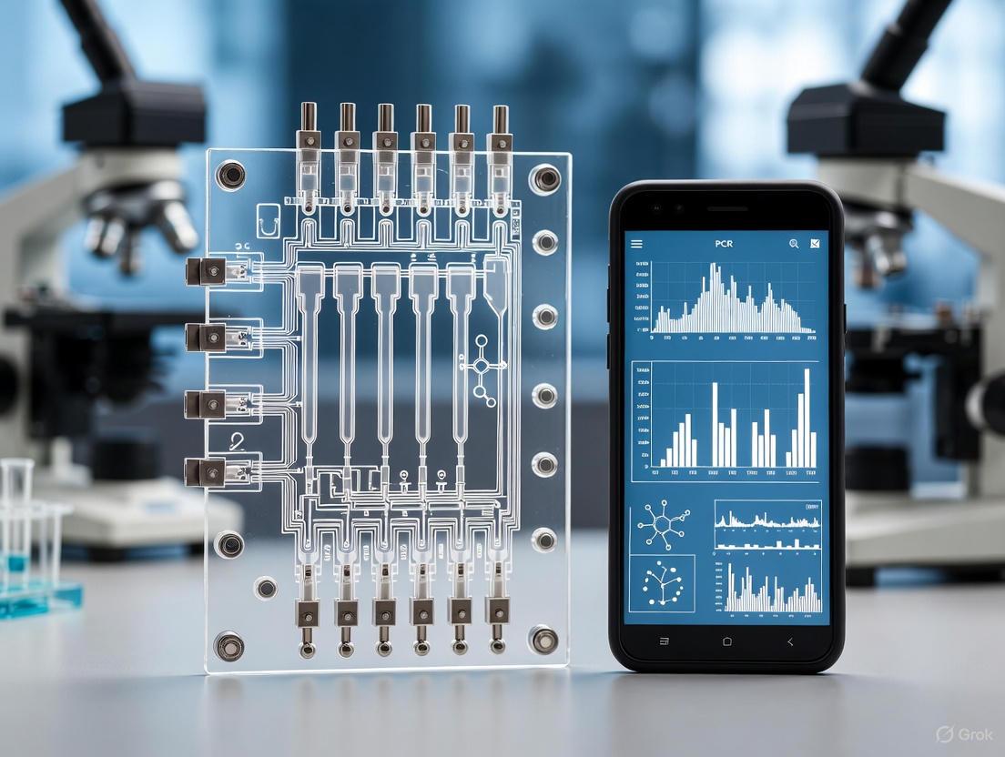
Designing a microfluidic chip for PCR-based detection of environmental pathogens requires the seamless integration of several functional units: sample preparation (e.g., filtration and concentration), nucleic acid amplification (PCR), and optical detection, all miniaturized and compatible with a smartphone readout.
Material Selection
The choice of material is critical and involves trade-offs between optical properties, manufacturability, chemical compatibility, and cost.
Table 2: Common Microfluidic Chip Materials
| Material | Properties | Advantages | Disadvantages |
|---|---|---|---|
| Polydimethylsiloxane (PDMS) [5] [3] | Elastomer; transparent; gas-permeable. | Excellent biocompatibility; easy and fast prototyping via soft lithography; high optical clarity for microscopy. | Can absorb small hydrophobic molecules; prone to swelling with organic solvents; not suitable for high-throughput mass production. |
| Polymethyl Methacrylate (PMMA) [8] [3] | Thermoplastic; rigid; transparent. | Good optical clarity; low cost; amenable to mass production (e.g., injection molding). | Lower chemical resistance than glass; can be brittle. |
| Glass [5] [3] | Inorganic solid; highly transparent; chemically inert. | Excellent optical properties and chemical stability; suitable for high temperatures (e.g., PCR) and high-pressure applications. | More expensive and fragile; microfabrication is complex and time-consuming. |
| Paper [2] | Cellulose network; porous and hydrophilic. | Very low cost; passive fluid transport via capillary action; easy disposal by incineration. | Low mechanical strength when wet; limited fluid control complexity. |
For PCR applications, the material must withstand repeated thermal cycling (typically 20–40 cycles between 50°C and 95°C). While silicon and glass were initially used for their thermal stability and chemical inertness, polymers like PDMS and PMMA are now widely adopted due to their lower cost and simpler fabrication [9] [5]. PDMS, in particular, is favored for research prototypes due to its ease of prototyping and optical transparency, which is crucial for subsequent fluorescence detection.
Chip Architecture and Integration
The architecture of a microfluidic PCR chip must facilitate the journey of the sample from introduction to result. A common approach is to create a monolithic chip with interconnected functional chambers for sample preparation, reagent mixing, PCR amplification, and detection [5]. For more complex assays, 3D microfluidic chips, constructed by stacking and bonding multiple layers of patterned PDMS or using a folding "origami" approach for paper-based devices, enable more complex fluidic routing and higher integration density in a small footprint [2].
Another powerful architecture is droplet-based microfluidics (DBM). This system generates thousands of picoliter-to-nanoliter sized, water-in-oil droplets, each acting as an isolated microreactor. In pathogen detection, this allows for the digital quantification of DNA targets, where a single DNA molecule can be amplified within a droplet. This digital PCR (dPCR) method provides absolute quantification without the need for a standard curve and can detect rare pathogens with high sensitivity by partitioning the sample [3]. Droplets are typically generated using passive flow-focusing geometry or T-junction designs within the chip [3].
Thermal Management for Microfluidic PCR
The core of any PCR chip is its thermal cycling system. The primary technical challenge is achieving rapid and precise temperature changes for the denaturation, annealing, and extension steps.
- Thin-Film Heaters: Microheaters made of thin films of platinum [9] or polysilicon can be fabricated directly onto the microfluidic chip, offering localized and fast heating with low thermal mass.
- Peltier Elements: These thermoelectric elements are commonly used in commercial systems and can be placed in direct contact with the chip for both heating and cooling. While reliable, their relatively high thermal mass can limit ramping rates [9].
- Flow-Through PCR: Instead of thermally cycling a stationary chamber, the sample solution is physically moved between three fixed temperature zones (e.g., 95°C, 60°C, 72°C) on the chip. This method enables very fast cycle times [9].
- Pulse Controlled Amplification (PCA): A novel method uses short, powerful electrical pulses to rapidly heat a very small volume of the sample directly. This approach, implemented in a handheld SARS-CoV-2 detection device, achieves PCR sensitivity with a limit of detection of 0.88 copies/μL in less than 40 minutes total analysis time [10].
Smartphone-Based Optical Detection Modalities
The smartphone serves as a potent all-in-one platform for image capture, data processing, and result reporting in POCT devices. Its CMOS camera, powerful processor, and connectivity make it ideal for reading optical signals from a microfluidic chip [8] [4].
Detection Methods
- Fluorescence Detection: This is the most common detection method for qPCR and is highly sensitive. The smartphone camera, often with an added external lens or filter, captures the fluorescence emission from intercalating dyes (e.g., SYBR Green) or specific probes (e.g., TaqMan) within the PCR chamber [9] [4] [6]. The resulting images are analyzed by an app to determine the cycle threshold (Ct) or perform end-point quantification.
- Colorimetric Detection: This method relies on a visible color change, often read by the naked eye or a smartphone camera. In paper-based chips (μPADs), assays for nutrients, heavy metals, or other contaminants can produce a color change. Smartphone apps can analyze the hue and intensity of the color for more quantitative results [2]. While simpler and lower cost, it is generally less sensitive than fluorescence.
- Surface-Enhanced Raman Scattering (SERS): SERS provides a highly specific "fingerprint" for molecules. Microfluidic chips can be functionalized with SERS-active nanoparticles (e.g., gold or silver) to capture and concentrate target pathogens. The smartphone platform can be integrated with a miniature Raman spectrometer to read the unique SERS signal, enabling highly specific identification [6].
Mobile Platform Integration
A complete mobile health (mHealth) platform requires more than just a phone and a chip. It typically includes:
- 3D-Printed Adapter: A custom-made holder that aligns the microfluidic chip with the smartphone camera and integrated optics [4].
- External Optics: Simple lenses can be added to achieve microscopic magnification for imaging cells or small features [4].
- Controlled Illumination: Integrated Light Emitting Diodes (LEDs) provide the necessary excitation light for fluorescence assays [4].
- On-Phone and Cloud-Based Software: Smartphone applications control the image acquisition, preprocess the data (e.g., color correction, background subtraction), and can run machine learning algorithms for classification (e.g., positive/negative). More complex data processing can be offloaded to cloud servers [4].
Experimental Protocols
Protocol: On-Chip Digital PCR for Pathogen Quantification
This protocol outlines the key steps for performing a digital PCR assay to absolutely quantify a bacterial pathogen in a water sample using a droplet-based microfluidic chip and smartphone detection.
Principle: The sample is partitioned into thousands of nanoliter droplets, following a Poisson distribution. After end-point PCR amplification, droplets containing the target sequence fluoresce. Counting the positive droplets allows for absolute quantification of the initial target concentration [9] [3].
Figure 2: Workflow for on-chip digital PCR detection of waterborne pathogens.
Materials and Reagents:
- Microfluidic Chip: Droplet generation chip (e.g., flow-focusing design) fabricated in PDMS/glass.
- Reagents: PCR master mix, forward and reverse primers specific to the target pathogen (e.g., E. coli uidA gene), fluorescent probe (e.g., FAM-labeled TaqMan probe), nuclease-free water.
- Oil Phase: Fluorinated oil with a biocompatible surfactant (e.g., 2% RAN Biotechnologies008-FluoroSurfactant).
- Sample: Environmental water sample, pre-filtered and concentrated if necessary, with extracted DNA.
- Equipment: Precision pressure pump or syringe pumps (e.g., OB1 MK3+), portable thermal cycler or custom chip heater, smartphone with a 3D-printed imaging adapter.
Procedure:
- Prepare PCR Mix: In a nuclease-free microtube, prepare the aqueous PCR phase. For a 50 μL reaction: 25 μL of 2x PCR master mix, 2.5 μL of forward primer (10 μM), 2.5 μL of reverse primer (10 μM), 1.0 μL of fluorescent probe (10 μM), 5.0 μL of DNA template, and 14.0 μL of nuclease-free water.
- Load Chip: Load the aqueous PCR mix into the sample inlet reservoir of the chip. Load the fluorinated oil into the oil inlet reservoir.
- Generate Droplets: Connect the chip to the pressure pump. Apply optimized pressures (e.g., 80 mbar for oil, 60 mbar for sample) to generate a stable stream of monodisperse water-in-oil droplets (~100 μm diameter) collected in a output tube or on-chip chamber.
- Seal and Amplify: If droplets are collected off-chip, seal the tube. Perform PCR amplification in a thermal cycler using standard cycling conditions for the target amplicon.
- Image and Analyze: After amplification, place the droplet emulsion in the imaging chamber of the chip or load it into a dedicated readout chip. Mount the chip onto the smartphone imaging adapter. Using the smartphone app, capture a fluorescence image of the droplet field. The app's algorithm will count the total number of droplets and the number of fluorescent (positive) droplets to calculate the original copy number concentration of the target DNA in the sample.
The Scientist's Toolkit: Key Research Reagent Solutions
Table 3: Essential Reagents for Microfluidic PCR Pathogen Detection
| Reagent/Material | Function | Example Specification/Note |
|---|---|---|
| PDMS (Sylgard 184) [5] [3] | Primary material for rapid prototyping of microfluidic chips. | Mixed in a 10:1 base-to-curing agent ratio; cured at 65°C for 2 hours. |
| SU-8 Photoresist [5] | Used to create high-resolution masters for PDMS soft lithography. | Enables creation of microchannels with features down to a few micrometers. |
| PCR Master Mix [9] | Contains Taq polymerase, dNTPs, and optimized buffer for amplification. | Should be selected for compatibility with on-chip thermocycling. |
| TaqMan Probes [9] | Sequence-specific fluorescent probes for real-time qPCR detection. | Provides higher specificity than intercalating dyes; requires a compatible qPCR master mix. |
| Fluorinated Oil & Surfactant [3] | Forms the continuous phase for stable water-in-oil droplet generation. | Prevents droplet coalescence during thermocycling (e.g., RAN Biotechnologies008-FluoroSurfactant). |
| Nucleic Acid Aptamers [8] [6] | Synthetic DNA/RNA molecules that bind specific pathogens; used for capture/detection. | Can be used as an alternative to antibodies in capture chambers or assays. |
| HEPES Buffer [8] | A buffering agent used to maintain stable pH during biochemical reactions on-chip. | Crucial for maintaining enzyme activity (e.g., reverse transcriptase, polymerase). |
The convergence of smartphone technology with microfluidic diagnostic platforms creates a powerful paradigm for decentralized environmental pathogen detection. Modern smartphones integrate sophisticated components—high-resolution CMOS cameras, multi-core processors, and ubiquitous connectivity—that can be repurposed to create portable, cost-effective analytical devices. These systems transform traditional laboratory-based molecular analyses, such as polymerase chain reaction (PCR), into field-deployable tools capable of rapid, on-site pathogen identification [11]. This application note details the essential smartphone components and provides structured protocols for implementing smartphone-based detection for environmental monitoring applications targeting pathogens.
The motivation for adopting smartphones as analytical platforms stems from their global ubiquity, integrated features, and advanced computing capabilities. Smartphones provide an unprecedented opportunity to deploy diagnostic technologies in resource-limited settings, enabling real-time environmental surveillance without requiring sophisticated laboratory infrastructure [12] [11]. By leveraging the existing smartphone ecosystem, researchers can develop detection systems that are both technologically advanced and economically viable for widespread implementation.
Smartphone Component Specifications for Analytical Applications
CMOS Camera Capabilities
The CMOS camera serves as the primary optical detector in smartphone-based diagnostic systems, capable of quantifying various signal types including fluorescence, colorimetry, and luminescence.
Table 1: CMOS Camera Specifications for Analytical Detection
| Smartphone Tier | Sensor Size (Notation) | Estimated Pixel Size | Useful Detection Modalities | Representative Application |
|---|---|---|---|---|
| Entry-Level | 1/3" | ~1.0 µm | Colorimetric LAMP, Visual ELISA | Educational tools, basic color change assays |
| Mid-Range | 1/2.8" | ~0.8 µm | Fluorescence detection, quantitative colorimetry | Pathogen detection via qLAMP [13] |
| High-End | 1/1.7" | ~0.7 µm | Low-light luminescence, high-resolution microscopy | Sensitive pathogen detection with low abundance targets |
CMOS sensors in smartphones are characterized by their high quantum efficiency across the visible spectrum, with peak sensitivity typically occurring at approximately 459 nm (blue), 520 nm (green), and 597 nm (red) [13]. This spectral sensitivity enables precise colorimetric quantification essential for molecular assays. The back-illuminated Exmor R CMOS sensor architecture, found in many modern smartphones, significantly enhances low-light performance critical for detecting faint fluorescent signals from pathogen amplification assays [13].
Processing Capabilities
Smartphone processors (SoCs - Systems on a Chip) provide the computational power required for real-time image analysis, data processing, and results quantification. Modern smartphone SoCs integrate multi-core CPUs, dedicated GPUs, and AI accelerators that enable sophisticated analytical functions:
- Real-time image processing: Automated analysis of color development in LAMP or PCR reactions
- Data quantification: Conversion of pixel intensity to target pathogen concentration
- Results interpretation: Machine learning algorithms for distinguishing positive from negative results
- User interface management: Touchscreen control of the analytical process
The processing capabilities allow implementation of advanced color models, including RGB (Red, Green, Blue) analysis and HSV (Hue, Saturation, Value) color space transformations, which provide more robust quantification compared to simple intensity measurements [13]. This processing power enables smartphones to perform functions that traditionally required desktop computers, making quantitative molecular analysis truly portable.
Connectivity Features
Smartphone connectivity options enable seamless data transfer, remote monitoring, and integration with broader surveillance networks:
- Cellular Networks (4G/5G): Enable real-time transmission of results from field testing sites to central laboratories or public health databases
- Wi-Fi and Bluetooth: Facilitate connection with peripheral devices and local network infrastructure
- GPS: Provides geographical tagging of environmental samples for spatial mapping of pathogen distribution
- Cloud Integration: Allows storage and analysis of large datasets, facilitating trend analysis and outbreak tracking
This connectivity framework supports the development of comprehensive environmental monitoring networks where multiple smartphone-based detectors can be deployed across a region, with all data streaming to a centralized analytical platform [14]. This creates an Internet of Things (IoT) for pathogen surveillance, potentially revolutionizing how environmental health threats are identified and managed.
Experimental Protocols for Pathogen Detection
Protocol: Colorimetric qLAMP with Smartphone Detection
This protocol describes quantitative Loop-Mediated Isothermal Amplification (qLAMP) for pathogen detection using smartphone-based colorimetric analysis [13].
Reagent Preparation
Table 2: Research Reagent Solutions for Smartphone-based Pathogen Detection
| Reagent/Material | Function | Specifications/Alternatives |
|---|---|---|
| LAMP Primer Mix | Target-specific amplification | Custom-designed for pathogen target; 6 primers per target |
| Isothermal Amplification Mix | DNA/RNA amplification | Contains Bst DNA polymerase, dNTPs, buffer |
| Colorimetric Indicator | Visual signal generation | Eriochrome Black T (EBT) or Hydroxy Naphthol Blue (HNB) |
| Sample Preparation Kit | Nucleic acid extraction | Silica-based columns or magnetic beads |
| Microfluidic Chip | Reaction chamber | Disposable chip with 7 reaction chambers [13] |
| Smartphone Enclosure | Light isolation | 3D-printed box with LED lighting [13] |
Prepare LAMP Master Mix:
- Combine 12.5 µL of isothermal amplification buffer
- Add 1.0 µL of each primer (F3, B3, FIP, BIP - total 6 primers)
- Include 1.0 µL of Bst DNA polymerase (8,000 U/mL)
- Add 1.5 µL of Eriochrome Black T indicator (200 µM stock)
- Add extracted template DNA (2-5 µL containing target sequence)
- Adjust total volume to 25 µL with nuclease-free water
Load Microfluidic Chip:
- Pipette 25 µL of reaction mixture into each chamber of the microfluidic chip
- Ensure no air bubbles are present in the reaction chambers
- Seal chambers with transparent adhesive tape if necessary
Amplification and Detection
Assemble Detection Device:
- Position smartphone in 3D-printed enclosure
- Align microfluidic chip with smartphone camera field of view
- Ensure uniform white LED illumination (6,000 K) of the chip [13]
- Maintain temperature at 65°C using integrated film heater
Execute Amplification and Monitoring:
- Initiate LAMP reaction at 65°C for 30-60 minutes
- Capture images of reaction chambers every 30 seconds using smartphone camera
- Process images through dedicated mobile application
- Extract RGB values from each reaction chamber using auto-select algorithm to exclude bubbles [13]
- Calculate hue values from RGB data for quantitative analysis
Data Analysis:
- Plot hue value versus time for each reaction
- Determine threshold time (Tt) for each sample
- Compare with standard curve of known concentrations
- Calculate initial template concentration in unknown samples
Figure 1: qLAMP Pathogen Detection Workflow
Protocol: PCR Microfluidic Chip with Smartphone Fluorescence Detection
This protocol adapts traditional PCR for smartphone detection using microfluidic chips and fluorescence detection [15].
Microfluidic Chip Design and Preparation
Chip Fabrication:
- Design microfluidic channels using AutoCAD or SolidWorks
- Fabricate chips from PDMS using soft lithography or PMMA via injection molding [12]
- Incorporate reaction chambers (10-20 µL volume) with transparent viewing windows
- Ensure chip compatibility with temperature cycling requirements
Surface Treatment:
- Treat PDMS surfaces with oxygen plasma to prevent biomolecule adsorption
- Coat channels with bovine serum albumin (BSA) to minimize non-specific binding
- Validate chip performance with control samples
Smartphone Fluorescence Detection Setup
Optical Configuration:
- Utilize smartphone LED flash as excitation source (may require filter modification)
- Add external lens system if needed for signal collection
- Implement emission filters compatible with fluorescent dyes (SYBR Green, EvaGreen, TaqMan probes)
- Ensure light-tight enclosure to minimize background signal
Temperature Cycling:
- Implement Joule heating, thermoelectric, or plasmonic heating systems [15]
- Achieve rapid thermal cycling: Denaturation (95°C), Annealing (55-65°C), Extension (72°C)
- Monitor temperature using integrated sensors with feedback control
PCR Execution and Data Analysis
Reaction Setup:
- Prepare PCR mix with fluorescence DNA binding dye (SYBR Green) or probe system (TaqMan)
- Load samples into microfluidic chip using capillary action or external pressure
- Seal chip to prevent evaporation during thermal cycling
Amplification and Detection:
- Execute 30-40 cycles of PCR with smartphone capturing fluorescence images at each cycle's extension step
- Use smartphone processor to plot fluorescence intensity versus cycle number
- Determine threshold cycle (Ct) for each sample
- Quantify initial template concentration using standard curve
Figure 2: Smartphone PCR Detection Workflow
Implementation Considerations and Troubleshooting
System Integration and Optimization
Successful implementation of smartphone-based pathogen detection requires careful integration of all system components:
- Optical Alignment: Precisely align excitation sources, filters, and camera field of view to maximize signal detection
- Temperature Uniformity: Ensure consistent temperature distribution across reaction chambers for reliable amplification
- Image Consistency: Maintain fixed distance and lighting conditions between smartphone camera and microfluidic chip
- Data Normalization: Implement reference standards in each assay to control for inter-experiment variability
Troubleshooting Common Issues
Table 3: Troubleshooting Guide for Smartphone-Based Detection Systems
| Problem | Potential Causes | Solutions |
|---|---|---|
| High Background Signal | Non-specific amplification, insufficient washing | Optimize primer specificity, increase stringency of wash steps |
| Low Signal Intensity | Inefficient amplification, suboptimal camera settings | Validate amplification efficiency, adjust smartphone exposure settings |
| Inconsistent Results | Temperature fluctuations, bubble formation | Improve temperature control, degas reagents before loading |
| Poor Standard Curve | Pipetting errors, degraded standards | Use fresh reference materials, implement automated liquid handling |
The integration of smartphone components with microfluidic PCR platforms creates a powerful tool for environmental pathogen detection. The CMOS camera provides sensitive optical detection, the processor enables real-time data analysis, and connectivity features facilitate data sharing and remote monitoring. The protocols outlined in this application note provide researchers with detailed methodologies for implementing these systems in both laboratory and field settings. As smartphone technology continues to advance, these systems will become increasingly sophisticated, offering new capabilities for environmental monitoring and public health protection.
The integration of microfluidic chips with polymerase chain reaction (PCR) and smartphone detection represents a transformative approach for the in-field monitoring of environmental pathogens. The performance, cost, and practicality of these diagnostic systems are profoundly influenced by the substrate material of the chip itself. Selecting an appropriate material is paramount, as it dictates the fabrication methodology, compatibility with biochemical reactions, and integration with optical detection systems. This application note provides a detailed comparison of three primary substrate categories—polymers, glass, and paper—for use in PCR microfluidic chips within environmental research. It further standardizes experimental protocols for chip evaluation to accelerate development in this critical field.
Material Comparison and Selection Guidelines
The choice of chip material involves balancing physical, chemical, and practical properties against the specific requirements of PCR amplification and smartphone detection. The table below summarizes the key characteristics of polymer, glass, and paper-based substrates.
Table 1: Comprehensive Comparison of Microfluidic Chip Substrate Materials
| Property | Polymers (e.g., PDMS, PMMA, COP) | Glass | Paper-Based Substrates |
|---|---|---|---|
| Typical Materials | Polydimethylsiloxane (PDMS), Polymethyl methacrylate (PMMA), Cyclic Olefin Copolymer (COP) [16] [17] | Borosilicate glass, Silica, Quartz [18] | Filter paper, Nitrocellulose membrane, Chromatography paper [19] [20] |
| Key Advantages | Low cost, ease of prototyping, good optical transparency, flexibility [16] [17] | Excellent optical clarity, high thermal stability, chemical inertness, reusable [21] [18] | Very low cost, biodegradable, passive fluid transport via capillarity, no external pumps needed [19] [20] |
| Primary Limitations | Can absorb small molecules, limited solvent resistance, autofluorescence in some types [17] | Higher cost, more complex and time-consuming fabrication, brittle [21] [18] | Limited structural integrity, not suitable for complex, multi-step liquid handling, low resolution [19] |
| Optical Clarity | Good to excellent (varies by polymer) [17] | Excellent (Superior for high-resolution detection) [18] | Opaque or semi-opaque (relies on surface detection) [20] |
| Thermal Conductivity | Low (e.g., PDMS: ~0.15 W/m•K) | High (~1 W/m•K) | Very Low |
| Biosensor Suitability | Good for integrated biosensors [17] | Excellent for electrochemical and optical sensors [18] | Ideal for disposable, single-use biosensors [19] [20] |
| Fabrication Methods | Soft lithography, hot embossing, injection molding, laser ablation [16] [17] | Photolithography, wet/dry etching, laser ablation [18] | Wax printing, inkjet printing, photolithography, cutting [19] [22] |
| Typical Applications | High-precision microreactors, organ-on-a-chip, dPCR chips [16] [17] | Capillary electrophoresis, high-temperature/reactivity reactions, Raman spectroscopy [18] | Lateral flow assays, rapid, low-cost diagnostic tests for pathogens [19] [20] |
Selection Guidelines for Pathogen Detection
- For High-Performance, Multi-step PCR: Glass or thermoplastic polymers (like COP or PMMA) are preferred when the protocol involves rigorous thermal cycling and requires superior optical detection for quantitative analysis. Their high thermal stability ensures consistent PCR efficiency [16] [18].
- For Rapid, Low-Cost, Point-of-Need Screening: Paper-based microfluidic analytical devices (μPADs) are ideal for detecting environmental pathogens in resource-limited settings. Their ability to wick fluids without pumps and extremely low cost make them unparalleled for disposable, on-site use [19] [20].
- For Prototyping and Complex Device Architectures: Polymers like PDMS are excellent for rapid prototyping and creating complex, layered structures (e.g., for valves and pumps) due to their flexibility and ease of fabrication via soft lithography [16] [17].
Experimental Protocols for Chip Evaluation
This section outlines standardized protocols for evaluating the performance of microfluidic chips fabricated from different materials, specifically for PCR amplification of environmental pathogens coupled with smartphone detection.
Protocol: Evaluating PCR Efficiency and Signal-to-Noise Ratio for Smartphone Detection
Objective: To quantify and compare the PCR amplification efficiency and the resulting optical signal-to-noise ratio achievable with polymer, glass, and paper-based chips when integrated with a smartphone detector.
Materials:
- Chip Fabrication: PDMS and glass chips with identical microchannel design (20 µm depth, 100 µm width); wax-patterned paper µPADs.
- Reagents: Prepared PCR mix containing target pathogen DNA (e.g., E. coli 16s rRNA gene), primers, dNTPs, Taq polymerase, and intercalating fluorescent dye (SYBR Green I).
- Equipment: Custom-built portable thermal cycler, smartphone in a darkbox with a holder, external lens, and a blue LED excitation source (~470 nm) with an emission filter (~520 nm).
Procedure:
- Chip Priming: Pre-treat the microchannels of polymer and glass chips with a 1% (w/v) solution of bovine serum albumin (BSA) in PBS for 30 minutes to prevent surface adsorption of enzymes. Paper chips require no priming.
- Sample Loading: Pipette 5 µL of the PCR mix into the reaction chamber of each chip type. For paper chips, pipette 2 µL directly onto the designated detection zone.
- Thermal Cycling: Place the loaded chips into the portable thermal cycler and run the following protocol:
- Initial Denaturation: 95°C for 120 s.
- 35 Cycles of:
- Denaturation: 95°C for 15 s.
- Annealing: 55°C for 30 s.
- Extension: 72°C for 30 s.
- Signal Acquisition: Upon completion, immediately transfer the chips to the smartphone darkbox. Capture an image of the fluorescent signal in the reaction chamber/detection zone using the smartphone camera with a fixed exposure time, ISO, and focus.
- Data Analysis:
- Use an image analysis software (e.g., ImageJ) to measure the mean fluorescence intensity of the reaction zone (Signal) and an adjacent empty zone (Background).
- Calculate the Signal-to-Noise Ratio (SNR) as: SNR = (Mean Signal Intensity - Mean Background Intensity) / Standard Deviation of Background Intensity.
- Plot the Ct (cycle threshold) values against known DNA concentrations to determine PCR amplification efficiency for each material.
Protocol: Assessing Chip-to-Chip Reproducibility
Objective: To determine the manufacturing reproducibility and operational consistency across multiple chips of the same material.
Materials: A batch of at least 10 chips per material type, standardized pathogen DNA sample.
Procedure:
- Batch Testing: Perform the PCR and detection protocol from section 3.1 simultaneously on all chips from the same batch.
- Data Collection: Record the final fluorescence intensity and Ct value (if applicable) for each chip.
- Statistical Analysis: Calculate the mean, standard deviation, and coefficient of variation (CV = Standard Deviation / Mean * 100%) for the fluorescence signals and Ct values. A CV of less than 10% is generally considered acceptable for analytical devices.
The Scientist's Toolkit: Essential Research Reagent Solutions
Table 2: Key Reagents and Materials for Microfluidic PCR Chip Development
| Item Name | Function/Application | Critical Notes |
|---|---|---|
| Polydimethylsiloxane (PDMS) | Elastomeric polymer for rapid prototyping of microfluidic devices via soft lithography [17]. | Prized for optical clarity and gas permeability; susceptible to absorbing small hydrophobic molecules. |
| Cyclic Olefin Copolymer (COP) | Rigid thermoplastic for high-integrity, mass-produced chips with low autofluorescence [21]. | Excellent for quantitative fluorescence detection; requires industrial fabrication like injection molding. |
| Nitrocellulose Membrane | Porous paper substrate for capillary-driven fluid transport and biomolecule immobilization [20]. | Backbone of lateral flow assays; pore size (e.g., 0.45 µm) dictates flow rate and binding capacity. |
| SYBR Green I Dye | Fluorescent intercalating dye for real-time quantification of amplified DNA in PCR [16]. | Compatible with standard FITC/GFP optical filters on smartphone detection setups. |
| BSA (Bovine Serum Albumin) | Used as a surface passivation agent to block non-specific adsorption in polymer and glass microchannels [16]. | Critical for maintaining PCR efficiency by preventing the loss of enzymes and DNA. |
| Taq DNA Polymerase | Thermostable enzyme for catalyzing DNA amplification in the PCR process [16]. | The workhorse enzyme for conventional PCR; performance must be validated in a miniaturized format. |
Workflow and Logical Relationship Diagram
The development and deployment of a material-optimized PCR microfluidic chip for environmental pathogen detection follow a structured workflow, from material selection to final result interpretation.
The accurate and rapid identification of environmental pathogens is a critical challenge for public health, clinical diagnostics, and epidemic prevention. Pathogens transmitted through air and water—such as Legionella, SARS-CoV-2, and Cryptosporidium—pose significant threats, causing illnesses ranging from gastrointestinal disorders to severe pneumonia and systemic infections [23] [24] [25]. The wide range of transmission routes and high risk of outbreaks necessitate ultrasensitive, specific, and rapid monitoring platforms.
Conventional detection methods, including culture-based techniques, immunoassays, and molecular diagnostics like polymerase chain reaction (PCR), are often hindered by complex workflows, prolonged analysis times (2–5 days for culture), and a reliance on sophisticated laboratory infrastructure and skilled personnel [23] [26]. These limitations render them unsuitable for rapid, on-site testing in resource-limited environments.
Microfluidic technology integrated with PCR (PCR-on-a-chip) and smartphone-based detection has emerged as a powerful solution, enabling automated, sample-to-answer analysis. These systems offer exceptional performance due to their miniaturization, low reagent consumption, high throughput, and portability [23] [26] [27]. When combined with smartphone analytics, they provide a potent platform for point-of-care testing (POCT), facilitating real-time, on-site pathogen detection [28]. This Application Note defines the primary airborne and waterborne pathogen targets and details protocols for their detection using an integrated PCR microfluidic chip and smartphone system.
Common Pathogen Targets
Effective environmental monitoring requires a clear understanding of the predominant pathogenic threats. The tables below catalog common waterborne and airborne pathogens, which are primary targets for microfluidic detection systems.
Table 1: Common Waterborne Pathogens and Associated Health Risks [24]
| Pathogen | Type | Primary Health Risks | Notable Characteristics |
|---|---|---|---|
| Legionella | Bacterium | Legionnaires' disease, a severe form of pneumonia | Grows in warm water systems (e.g., plumbing, cooling towers); inhaled via aerosolized droplets. |
| Pseudomonas aeruginosa | Bacterium | Pneumonia, urinary tract infections, sepsis | Opportunistic pathogen; found in soil, water, and moist environments; common in healthcare settings. |
| Acinetobacter | Bacterium | Pneumonia, skin infections, nosocomial infections | Opportunistic; often resistant to multiple antibiotics. |
| Nontuberculous Mycobacteria | Bacterium | Pulmonary infections, skin diseases | Hard outer shell makes it resistant to many antibiotics and disinfectants. |
| Burkholderia | Bacterium | Urinary tract infections, meningitis | Opportunistic pathogen; found in moist soil and water. |
| Stenotrophomonas | Bacterium | Pulmonary infections, urinary tract infections | Often resistant to many antibiotics; commonly associated with hospital equipment. |
| Cryptosporidium | Parasite | Gastroenteritis (diarrhea, cramps, dehydration) | Chlorine-resistant outer shell; low infectious dose. |
| Giardia | Parasite | Gastroenteritis ("beaver fever") | Chlorine-resistant cyst; spreads via contaminated water. |
Table 2: Common Airborne Pathogens and Representative Detection Targets [26] [25]
| Pathogen | Type | Primary Health Risks | Relevance to Detection |
|---|---|---|---|
| SARS-CoV-2 | Virus | COVID-19 (respiratory illness) | Representative target for airborne virus surveillance; detected in aerosols [25]. |
| Influenza Virus | Virus | Seasonal influenza | A major cause of airborne respiratory infections globally. |
| Mycobacterium tuberculosis | Bacterium | Tuberculosis | Airborne transmission; highlights need for sensitive nucleic acid detection. |
Integrated Pathogen Detection Platform
The proposed integrated platform combines a microfluidic chip for sample preparation and amplification with a smartphone for signal readout and analysis. The core technology leverages nucleic acid amplification tests (NAATs), such as PCR or isothermal methods, for high sensitivity and specificity.
Platform Workflow
The entire process, from sample introduction to result reporting, is automated within a single, compact device. The following diagram illustrates the integrated workflow of the pathogen detection platform.
Smartphone Detection Principle
Smartphones serve as a versatile analytical platform due to their powerful cameras, processors, and connectivity. In this setup, the optical biosensor in the microfluidic chip transduces the presence of a pathogen into a measurable optical signal.
Table 3: Optical Biosensing Modalities for Smartphone Detection [26]
| Sensing Modality | Principle | Smartphone Role |
|---|---|---|
| Colorimetric | Measures color change due to biochemical reaction or nanoparticle aggregation. | Camera captures image; software analyzes RGB values or hue. |
| Fluorescence | Detects light emission from fluorescent labels upon excitation. | Camera (often with a simple external filter) captures fluorescence intensity; app quantifies signal. |
| Surface-Enhanced Raman Scattering (SERS) | Enhances Raman signal of molecules adsorbed on nanostructured metals. | Camera captures unique spectral fingerprint; requires additional optics for spectroscopy. |
The following diagram outlines the functional principle of smartphone-based optical detection integrated with a microfluidic chip.
Detailed Experimental Protocols
Protocol 1: Detection of Airborne Viruses (e.g., SARS-CoV-2) via Integrated Aerosol Sampling and Microfluidic LAMP-CRISPR
This protocol describes a method for air-in-result-out detection of airborne viruses using a high-flow aerosol sampler coupled with a microfluidic chip for Loop-Mediated Isothermal Amplification (LAMP) and CRISPR-based detection [25].
4.1.1 Workflow
- Aerosol Sampling (45 minutes): Air is drawn at a high flow rate (e.g., >6000 L/min) into an electrostatic precipitator or similar high-efficiency sampler. Viral particles are captured into a liquid medium (aerosol-to-hydrosol) at the air-liquid interface.
- RNA Extraction & Purification (10 minutes): The collected sample is mixed with a lysis buffer containing magnetic beads. RNA binds to the beads, and a magnet is used to wash the beads and remove inhibitors. Pure RNA is eluted in a small volume.
- Microfluidic LAMP Amplification (30 minutes): The eluted RNA is injected into the LAMP chamber on the microfluidic chip. The chip temperature is maintained at a constant isothermal condition (e.g., 65°C). Reverse transcription and LAMP amplification occur simultaneously, generating large amounts of double-stranded DNA product.
- CRISPR Detection (10 minutes): The LAMP product is transferred within the chip to a CRISPR reaction chamber containing Cas12a enzyme and a fluorescent reporter probe. If the target viral sequence is present, the Cas12a complex is activated and cleaves the reporter probe, producing a fluorescent signal.
- Smartphone Readout: The smartphone's camera, equipped with a filter, captures the fluorescence intensity. A custom application processes the image and reports a positive/negative result.
4.1.2 The Scientist's Toolkit: Key Research Reagent Solutions
Table 4: Essential Reagents and Materials for Airborne Virus Detection Protocol
| Item | Function/Description |
|---|---|
| Lysis Buffer (Guanidine Thiocyanate) | Disrupts viral envelope and capsid, releasing RNA and protecting it from nucleases. |
| Silica-coated Magnetic Beads | Bind nucleic acids under high-salt conditions for purification and concentration; enable magnetic manipulation for washing. |
| LAMP Master Mix | Contains Bst DNA polymerase, dNTPs, and primers (FIP, BIP, F3, B3, LF, LB) specific to the target virus (e.g., SARS-CoV-2 N gene). |
| CRISPR/Cas12a Reagents | Includes the Cas12a enzyme and a guide RNA (crRNA) programmed to recognize the LAMP-amplified target sequence. |
| Fluorescent Reporter Probe | A single-stranded DNA oligonucleotide with a fluorophore (e.g., FAM) and a quencher (e.g., BHQ1); cleavage by activated Cas12a generates fluorescence. |
| Wash Buffers (Ethanol-based) | Remove salts, proteins, and other impurities from the nucleic acid-magnetic bead complex without eluting the RNA. |
| Elution Buffer (Nuclease-free Water) | A low-ionic-strength solution that releases purified RNA from the magnetic beads for downstream amplification. |
Protocol 2: Detection of Waterborne Bacteria (e.g., E. coli, Legionella) via Continuous-Flow PCR Microfluidic Chip
This protocol is optimized for the detection of bacterial pathogens in water samples using a continuous-flow PCR (CF-PCR) device fabricated from thermoplastics, suitable for global health applications [23] [29].
4.2.1 Workflow
- Sample Pre-concentration (30 minutes): A large volume of water (e.g., 1-10 L) is passed through a sterile filter membrane to capture bacterial cells. Cells are then back-flushed from the filter into a small volume (e.g., 1-5 mL).
- On-Chip Lysis and DNA Extraction (15 minutes): The concentrated sample is loaded into the microfluidic chip. Lysis is achieved chemically (e.g., with alkaline solution) or physically (e.g., electroporation). DNA is purified using immobilized silica membranes or magnetic beads within the chip.
- Continuous-Flow PCR Amplification (10-20 minutes): The purified DNA is injected into the CF-PCR chip. The chip contains a long, serpentine channel that passes through three fixed temperature zones on a hotplate or block: Denaturation (e.g., 95°C), Annealing (e.g., 60°C), and Extension (e.g., 72°C). The flow rate is controlled by a syringe pump to define the residence time in each zone, achieving 30-40 cycles in minutes.
- Endpoint Fluorescence Detection: Intercalating dyes (e.g., SYBR Green) in the PCR mix fluoresce upon binding to amplified DNA. The fluorescence is measured at the end of the channel.
- Smartphone Readout: The smartphone, placed in a custom holder with a blue LED for excitation, images the detection zone. The app quantifies the green fluorescence intensity to confirm amplification.
4.2.2 The Scientist's Toolkit: Key Research Reagent Solutions
Table 5: Essential Reagents and Materials for Waterborne Bacteria Detection Protocol
| Item | Function/Description |
|---|---|
| Sterile Filter Membranes (0.22µm or 0.45µm pore size) | Concentrate bacterial cells from large water volumes for analysis. |
| Bacterial Lysis Reagent (e.g., Lysozyme, Proteinase K) | Breaks down bacterial cell walls and proteins to release genomic DNA. |
| Hot-Embossed Thermoplastic CF-PCR Chip (e.g., Zeonex) | The core microfluidic device with a serpentine channel; fabricated for low-cost, high-throughput production [29]. |
| PCR Master Mix | Contains heat-stable DNA polymerase (e.g., Taq), dNTPs, MgCl₂, and primers specific to the target bacterium (e.g., E. coli uidA gene, Legionella mip gene). |
| SYBR Green I Dye | A double-stranded DNA intercalating dye that exhibits strong fluorescence enhancement when bound to PCR amplicons. |
| Thin-Film Heaters & Temperature Sensors | Create and maintain the three distinct temperature zones required for CF-PCR on the chip. |
| Programmable Syringe Pump | Precisely controls the flow rate of the PCR mixture through the microfluidic channel, determining cycle times. |
Performance Metrics and Validation
Robust validation is essential to demonstrate the reliability of the integrated platform for environmental monitoring. The following table summarizes typical performance targets based on current research.
Table 6: Representative Performance Metrics for Microfluidic Pathogen Detection [23] [27] [25]
| Assay Target | Technology Platform | Limit of Detection (LOD) | Total Assay Time | Key Performance Notes |
|---|---|---|---|---|
| SARS-CoV-2 | Microfluidic LAMP-CRISPR | 10 copies/reaction [25] | ~85 min (45 min sampling + 40 min detection) [25] | High specificity via CRISPR; integrated aerosol sampling. |
| SARS-CoV-2 | Integrated NAAT Chip (RT-LAMP) | <297 copies [27] | ~28 min [27] | Sample-to-answer cost ≈ $9.5; combines magnetic bead-based RNA extraction. |
| E. coli O157:H7 | Immunomagnetic Separation + ELISA | 3 × 10² CFU/mL [23] | ~3 hours [23] | Demonstrates utility of pre-concentration for sensitivity. |
| S. aureus, E. coli | Colorimetric Nanoarray | 10 CFU/mL [26] | <10 min [26] | Rapid colorimetric readout, suitable for smartphone camera analysis. |
Troubleshooting Guide
Table 7: Common Issues and Solutions in Microfluidic Pathogen Detection
| Problem | Potential Cause | Suggested Solution |
|---|---|---|
| Low or No Fluorescent Signal | Inhibitors from sample not fully removed. | Optimize wash steps during nucleic acid purification; include additional purification columns. |
| Low amplification efficiency. | Check primer design and concentration; optimize temperature zones and residence times in CF-PCR. | |
| Poor smartphone camera sensitivity. | Use an external lens filter to block excitation light; calibrate camera settings (ISO, exposure) via the app. | |
| High Background Signal | Non-specific amplification (especially in LAMP). | Integrate a CRISPR step for specific signal confirmation [25]; optimize primer specificity. |
| Probe degradation (in CRISPR assays). | Aliquot and store fluorescent reporter probes in the dark; avoid freeze-thaw cycles. | |
| Clogging of Microfluidic Channels | Particulate matter in water samples. | Pre-filter water samples through a coarse filter (e.g., 5µm) before on-chip processing. |
| Aggregation of magnetic beads. | Sonicate beads before use; ensure homogeneous suspension during loading. |
The Role of AI and Machine Learning in Automated Image and Data Analysis
The convergence of artificial intelligence (AI), microfluidic technology, and smartphone-based detection is creating a paradigm shift in environmental pathogen research. Traditional methods for analyzing pathogens are often time-consuming, require laboratory infrastructure, and lack real-time capabilities. The integration of miniaturized PCR microfluidic chips with the ubiquitous computing power of smartphones creates a powerful, portable diagnostic platform [30] [31]. However, these systems generate vast amounts of complex visual and data, necessitating advanced analytical tools. AI and machine learning (ML) have emerged as critical technologies for automating the analysis of images and data from these devices, transforming them from simple data collectors into intelligent, automated diagnostic systems [32] [33]. This integration enables rapid, accurate, and on-site detection of environmental pathogens, supporting applications from agricultural monitoring to public health.
AI and Machine Learning in Image Analysis for Microfluidics
Computer Vision for Microfluidic Image Analysis
Computer vision, a branch of AI, enables computers to interpret and understand visual data from the world. When applied to microfluidic PCR chips, it automates the extraction of meaningful information from images and videos captured via smartphone or integrated cameras [32]. The core tasks of computer vision in this context include:
- Classification: Categorizing droplets or cells within a chip as positive or negative for a target pathogen.
- Detection and Location: Identifying and pinpointing the precise location of specific targets, such as fluorescent amplicons.
- Segmentation: Distinguishing and outlining the boundaries of individual droplets, cells, or other regions of interest for further analysis [32].
These capabilities are crucial for handling the high-throughput, single-cell-level visual data that microfluidic chips produce, a task that is inefficient and prone to error when performed manually [32].
Deep Learning Architectures
Convolutional Neural Networks (CNNs) are the dominant deep learning architecture for image analysis tasks. Inspired by the human visual cortex, CNNs use layers of convolutional kernels to automatically and adaptively learn spatial hierarchies of features from images [32] [34]. In a CNN:
- Initial layers learn basic features like edges and corners.
- Deeper layers combine these into more complex, abstract features [32]. This makes CNNs exceptionally powerful for analyzing complex cellular or droplet images generated on microfluidic chips, enabling tasks such as label-free cell characterization and pathogen detection [32] [33].
AI-Enhanced Workflow for Pathogen Detection
The following diagram illustrates the integrated workflow of a smartphone-based PCR microfluidic chip system and the pivotal role of AI/ML in automating image and data analysis for environmental pathogen detection.
Diagram 1: Integrated AI and smartphone detection workflow for a PCR microfluidic chip system.
AI and Machine Learning in Data Interpretation and System Control
Beyond image analysis, AI and ML play a profound role in interpreting complex data patterns and optimizing the microfluidic system itself.
Predictive Modeling and Pathogen Identification
Machine learning models, particularly supervised learning algorithms, are trained on large datasets of known pathogen signatures. Once trained, these models can identify and classify pathogens from new, unseen data generated by the microfluidic chip [35] [34]. For instance, AI models can analyze multiplexed PCR results to detect multiple pathogens simultaneously from a single sample, a task that is highly complex for manual interpretation [36]. In genomics, AI tools like AI-MARRVEL have demonstrated a 98% precision rate in identifying disease-causing genetic variants, showcasing the potential for similar accuracy in identifying pathogen genetic markers [34].
AI for Chip Design and Droplet Control
AI is also revolutionizing the design and operation of microfluidic chips. The design of microchannels for optimal droplet generation is a complex, trial-and-error process. AI-powered design tools can now predict fluid dynamics and optimize chip layouts before fabrication, drastically reducing research and development time and costs [37] [33]. Furthermore, deep learning models like Artificial Neural Networks (ANNs) and Adaptive Neural-Fuzzy Inference Systems (ANFIS) can predict droplet characteristics (e.g., size, shape) based on input parameters such as flow rate and channel geometry, enabling precise control over the microfluidic environment [33].
Experimental Protocols
Protocol: AI-Assisted Analysis of Pathogen Detection in a Smartphone-Integrated PCR Microfluidic Chip
Objective: To detect and quantify a specific environmental pathogen (e.g., E. coli) from a water sample using a PCR microfluidic chip with smartphone imaging and AI-based data analysis.
I. Materials and Reagents
Table 1: Essential Research Reagent Solutions and Materials
| Item | Function in Protocol |
|---|---|
| Microfluidic PCR Chip (Disposable) | Miniaturized platform for nucleic acid amplification and reaction containment [30]. |
| Smartphone with High-Resolution Camera | Device for image capture, data processing, and user interface [28] [31]. |
| Lysis Buffer | Breaks down pathogen cells to release nucleic acids for amplification. |
| PCR Master Mix | Contains enzymes, dNTPs, and buffers necessary for DNA amplification. |
| Fluorescent DNA Intercalating Dye (e.g., SYBR Green) | Binds to double-stranded DNA and emits fluorescence upon excitation, enabling detection. |
| Positive Control (Target Pathogen DNA) | Validates the PCR reaction and AI model performance. |
| Negative Control (Nuclease-Free Water) | Checks for contamination or non-specific amplification. |
II. Procedure
Sample Preparation:
- Collect water sample from the environment (e.g., river, reservoir).
- Concentrate pathogens, if necessary, via filtration or centrifugation.
- Lyse the concentrated sample to release genomic DNA using the lysis buffer.
Chip Priming and Loading:
- Using a micropipette, load the prepared sample into the designated inlet port on the microfluidic chip.
- Similarly, load the positive and negative controls into their respective ports.
- The chip's capillary action or an applied pressure will draw the samples into the micro-reaction chambers.
On-Chip PCR Amplification:
- Place the loaded chip into the portable, smartphone-compatible thermal cycler.
- Initiate the pre-programmed thermal cycling protocol (e.g., 95°C for denaturation, 55-65°C for annealing, 72°C for extension) for 30-40 cycles.
Smartphone Image Acquisition:
- After amplification, place the chip into the smartphone imaging module, which includes a blue LED for excitation and an emission filter.
- Using the dedicated smartphone application, capture a high-resolution image of the entire chip, focusing on the micro-reaction chambers containing the amplified PCR products.
AI-Based Image and Data Analysis:
- The smartphone application automatically pre-processes the image (background subtraction, contrast enhancement).
- A pre-trained Convolutional Neural Network (CNN) model analyzes the image to:
- Segment and identify all reaction chambers.
- Classify each chamber as "positive" (fluorescent) or "negative" (non-fluorescent).
- Quantify the fluorescence intensity in positive chambers.
- The results are displayed on the smartphone screen, indicating the presence/absence and, if calibrated, the concentration of the target pathogen in the original sample.
III. Troubleshooting and Validation
- Low/No Fluorescence Signal: Verify PCR reagent activity, check thermal cycling temperatures, and ensure the integrity of the sample DNA.
- High Background Signal: Check for contamination of reagents and ensure the imaging module is clean and properly aligned.
- AI Model Misclassification: Retrain the CNN model with a larger and more diverse dataset of images that includes various environmental sample matrices.
Performance Metrics of AI in Diagnostic Analysis
The following table summarizes key quantitative performance data for AI models in related diagnostic and microfluidic applications, demonstrating their potential in environmental pathogen detection.
Table 2: Performance Metrics of AI/ML in Diagnostic and Microfluidic Analysis
| Application Domain | AI Model / System | Key Performance Metric | Result / Value | Citation |
|---|---|---|---|---|
| Rare Genetic Disease Diagnosis | AI-MARRVEL | Precision Rate | 98% | [34] |
| Dementia Differential Diagnosis | Deep Learning Classifier | Area Under the Curve (AUC) | 0.96 | [34] |
| Droplet Size Prediction | Adaptive Neural-Fuzzy Inference System (ANFIS) | Prediction Accuracy | 96% | [33] |
| Chest Radiograph Diagnosis | Commercial Deep Learning Model | Sensitivity | 99.1% | [34] |
| Colorectal Cancer Diagnosis | Interpretable Deep Learning System | Accuracy | 93.44% (Internal), 84.91% (External) | [34] |
The Scientist's Toolkit: Key Reagent Solutions
Table 3: Essential Research Reagent Solutions for PCR Microfluidic Systems
| Item | Function | Key Consideration |
|---|---|---|
| Lyophilized PCR Reagents | Stable, room-temperature storage for point-of-use applications [31]. | Enables long-term storage and portability of the diagnostic kit without cold chain. |
| Multiplex PCR Assay Kits | Simultaneous detection of multiple pathogen targets in a single reaction [30]. | Requires careful primer design and validation to avoid cross-reactivity. |
| Customized Microfluidic Chips (e.g., Polymer-based) | Flexible design for specific applications (e.g., specific channel geometry, surface chemistry) [37]. | Material must be compatible with biological samples and PCR reagents. |
| Fluorescent Probes & Dyes (e.g., TaqMan Probes, SYBR Green) | Specific and sensitive detection of amplified DNA [32]. | Choice depends on required specificity (probes) versus cost and simplicity (dyes). |
The synergy of PCR microfluidic chips, smartphone detection, and AI-powered analysis creates a transformative toolkit for environmental pathogen research. AI and machine learning are not merely incremental improvements but are foundational to managing the complexity and volume of data produced by these miniaturized systems. They enable a transition from manual, subjective interpretation to automated, high-throughput, and objective analysis directly in the field. As these intelligent systems evolve, they promise to deliver unprecedented capabilities in monitoring environmental health, tracking pathogen outbreaks, and safeguarding public and agricultural systems with speed and precision previously confined to the central laboratory.
From Sample to Answer: A Step-by-Step Guide to On-Site Pathogen Detection
Accurate sampling of airborne and waterborne pathogens is a critical prerequisite for effective environmental surveillance and outbreak investigation. The emergence of integrated diagnostic platforms, particularly those coupling PCR microfluidic chips with smartphone detection, places new emphasis on the initial sample collection and preparation steps. The performance of these advanced analytical systems is contingent on the quality and suitability of the input sample. This application note provides detailed protocols for the frontline collection of airborne and waterborne pathogens, with a specific focus on compatibility with downstream microfluidic concentration, nucleic acid amplification, and smartphone-based analysis.
Sampling Airborne Pathogens
The collection of airborne biological contaminants requires careful consideration of the sampling method, the preservation of pathogen viability, and the compatibility with subsequent molecular analysis.
Principles and Sampling Strategies
Airborne pathogens occur as bioaerosols—solid or liquid particles suspended in air—with particle sizes typically ranging from <1 μm to ≥50 μm. The size of these particles is critical, as particles ≤5 μm in diameter can reach the lungs, posing the greatest health risk [38]. When designing an air sampling strategy, several preliminary factors must be considered, including the characteristics of the aerosol, sampling time and duration, number of samples, and the method of microbiological assay [38].
Targeted microbiologic air sampling is indicated in several key situations [38]:
- Outbreak Investigation: When environmental reservoirs are implicated in disease transmission.
- Research: To provide new information on the spread of healthcare-associated diseases.
- Hazard Monitoring: To confirm the presence of a hazardous biological agent and validate its successful abatement.
- Quality Assurance: To evaluate the effects of a change in infection-control practice or to ensure equipment performance.
Electrostatic Sampling Protocol for Airborne Bacteria
Electrostatic samplers offer high physical collection efficiency and biological recovery for bacterial aerosols by using electrostatic attraction to concentrate particles into a liquid medium, making them ideal for subsequent molecular analysis [39].
- Application: Collection of gram-negative (e.g., Pseudomonas fluorescens) and gram-positive (e.g., Micrococcus luteus) bacterial aerosols for downstream molecular detection.
- Experimental Principle: Air is drawn through a corona charger where particles gain an electrical charge. These charged particles are then concentrated via an electric field into a small volume of liquid collection medium [39].
- Key Advantages: Low sampling velocity minimizes mechanical stress and damage to bacterial cells and DNA, enhancing biological recovery compared to impactors and impingers [39].
Materials and Equipment:
- Personal Electrostatic Particle Concentrator (EPC) or equivalent electrostatic sampler.
- Sterile plastic collection containers.
- Vacuum pump and airflow measuring device (flowmeter).
- Sampling media: Deionized (DI) water with 0.001–0.01% Sodium Dodecyl Sulfate (SDS).
- Vortex mixer.
- Auxiliary equipment: Optical Particle Counter (OPC) for physical efficiency measurement (optional).
Step-by-Step Protocol:
- Preparation: Aseptically add 1-2 mL of 0.001–0.01% SDS-DI water sampling medium into a sterile plastic container. Mount the container onto the collection electrode of the EPC. SDS acts as a surfactant to improve the recovery and stability of collected bacteria [39].
- Sampling: Turn on the corona charger and vacuum pump. Sample air at a low flow rate (e.g., 1-3 L/min) for a defined period (e.g., 15-60 minutes). The low flow rate preserves bacterial viability [39].
- Collection and Detachment: After sampling, carefully remove the container from the electrode. Securely cap the container and vortex it for 1-2 minutes at high speed to resuspend bacterial cells that may have deposited on the container walls via electrophoresis [39].
- Sample Storage and Transport: Immediately place the liquid sample on ice or refrigerate if it cannot be assayed promptly. The sample is now ready for nucleic acid extraction or direct analysis on an integrated microfluidic device.
- Note: Dry-phase electrostatic sampling followed by buffer addition and vortexing is not recommended for cultural analysis, as it leads to significant bacterial inactivation due to desiccation stress [39].
Data on Airborne Pathogen Sampling Methods
Table 1: Comparison of common bioaerosol sampling methods for pathogen detection.
| Method | Principle | Suitable for Measuring | Collection Media/Surface | Key Considerations for Downstream Analysis |
|---|---|---|---|---|
| Impingement in Liquids | Air drawn through a small jet and directed against a liquid surface [38]. | Viable organisms; concentration over time [38]. | Liquid (e.g., DI water, PBS, peptone water) [38]. | Provides liquid sample ideal for microfluidics; potential for mechanical damage at high velocities [39]. |
| Impaction on Solid Surfaces | Air drawn by vacuum and particles deposited on a solid surface via inertia [38]. | Viable particles; particle size distribution [38]. | Moist agar, gelatin membrane, coated glass slide [38]. | Requires elution step to create liquid sample; potential for cell damage and dehydration [39]. |
| Sedimentation | Particles settle onto surfaces by gravity [38]. | Qualitative or semi-quantitative viable particles [38]. | Agar plate (settle plate) [38]. | Simple but less quantitative; requires elution for molecular methods. |
| Filtration | Air drawn through a porous membrane that traps particles [38]. | Viable and non-viable organisms; concentration [38]. | Membrane filter (e.g., polycarbonate, gelatin) [38]. | High collection efficiency; desiccation stress can reduce viability; requires elution [39]. |
| Electrostatic Precipitation | Particles charged then collected on a liquid surface via an electric field [39]. | Viable organisms; concentration [39]. | Liquid medium (e.g., SDS-DI water) in a container [39]. | High physical collection and biological recovery; provides concentrated liquid sample ideal for on-site detection [39]. |
Diagram 1: Workflow for collecting airborne pathogens for microfluidic analysis.
Sampling Waterborne Pathogens
The detection of waterborne pathogens is crucial for public health, with traditional methods often being slow and laboratory-bound. Sampling for rapid, on-site platforms requires effective concentration and recovery of pathogens from large water volumes.
Principles and Sampling Strategies
Waterborne pathogen exposure occurs through multiple fecal-oral transmission pathways, including fluids (water), food, fingers, fields (soil), and fomites [40]. Exposure assessments can be categorized as:
- External Measures: Detecting indicators of fecal contamination or specific pathogens in environmental samples (water, soil). This identifies hazards and transmission pathways but must be combined with human interaction data to estimate ingested dose [40].
- Internal Measures: Using human biological specimens (e.g., serology, pathogen detection in feces) to infer past exposure events. This confirms ingestion but provides less information on the source [40].
Sampling for microfluidic detection primarily relies on external measures, aiming to provide a concentrated, purified sample of the target pathogen from a representative water volume.
Integrated Filtration and Elution Protocol for Water
This protocol describes a syringe-based filtration and elution method suitable for concentrating bacterial pathogens from water samples for on-site analysis.
- Application: Concentration of bacterial cells (e.g., Salmonella typhimurium, E. coli) from large-volume water samples (e.g., wastewater, surface water).
- Experimental Principle: A large volume of water is passed through a syringe filter, trapping microorganisms. The captured cells are then lysed on the filter, and their nucleic acids are purified using magnetic bead technology, all within a single device [41]. This method simplifies the traditionally labor-intensive process of nucleic acid extraction.
- Key Advantages: Rapid processing, multiple manual operations, and integration with downstream nucleic acid amplification detection methods [41].
Materials and Equipment:
- Syringe filter unit (e.g., equipped with a Flinders Technology Associates (FTA) membrane).
- Luer-lock syringe (50 mL).
- Phosphate Buffered Saline (PBS) or TE buffer for elution.
- Magnetic beads and magnet for separation.
- Lysis/binding buffer, wash buffers.
Step-by-Step Protocol:
- Sample Collection: Collect a representative water sample (e.g., 100 mL to 1 L) in a sterile container. If testing for chlorine-resistant pathogens, consider adding a neutralizer.
- Pre-filtration (Optional): For turbid samples, perform a coarse pre-filtration to remove large debris that could clog the filter.
- Concentration and Lysis: Attach the syringe to the filter unit. Pass the entire water sample through the filter manually or with a pump. Follow with an air push to clear residual fluid. Pass a lysis/binding buffer through the filter to lyse the captured cells on the membrane and bind nucleic acids to the integrated FTA matrix or introduced magnetic beads [41].
- Nucleic Acid Purification: If using magnetic beads, the bead-nucleic acid complex is held in place with a magnet while wash buffers are passed through to remove impurities. Finally, the purified nucleic acids are eluted in a small volume (e.g., 50-100 µL) of elution buffer [41].
- Sample Storage and Transport: The eluted nucleic acids are stable and can be stored at -20°C or immediately used as a template for on-chip RPA or LAMP amplification [41].
Data on Waterborne Pathogen Detection Methods
Table 2: Comparison of conventional and advanced methods for waterborne pathogen detection.
| Method | Principle | Time to Result | Sensitivity | Suitability for POC |
|---|---|---|---|---|
| Culture-Based Assays | Growth and isolation of pathogens on specific media [42]. | Days to weeks [42] | High (for cultivable organisms) [42] | Low (requires lab, skilled personnel) [42] |
| Immunomagnetic Separation (IMS) | Use of antibody-coated magnetic beads to isolate specific pathogens [42]. | Hours (when combined with PCR) [42] | Moderate to High [42] | Moderate (can be integrated into platforms) [42] |
| Enzyme-Linked Immunosorbent Assay (ELISA) | Detection via antigen-antibody interaction and enzyme-mediated color change [42]. | Several hours [42] | 10³–10⁵ CFU/mL [42] | Moderate (can be formatted into kits) [42] |
| Polymerase Chain Reaction (PCR) | Enzymatic amplification of specific nucleic acid sequences [42]. | 2-4 hours [42] | Very High (single copy detection) [42] | Low (requires thermal cycler) [42] |
| Isothermal Amplification (RPA/LAMP) | Amplification at a constant temperature [41]. | 20 minutes - 1 hour [41] | Very High (e.g., 100 GE/mL for SARS-CoV-2) [41] | High (simple heating source) [41] |
Diagram 2: Workflow for concentrating waterborne pathogens and purifying nucleic acids.
Integrated Protocol for Pathogen Detection via Microfluidic Chip with Smartphone Detection
This protocol integrates the sample preparation steps from above into a complete workflow from sample-to-answer using a 3D printed microfluidic chip and smartphone-based detection.
- Application: Multiplexed detection of pathogens (e.g., SARS-CoV-2, Salmonella typhimurium) in environmental samples.
- Experimental Principle: The platform integrates on-chip nucleic acid extraction, two-stage isothermal pre-amplification and amplification (e.g., RPA followed by synergetic enhanced colorimetric LAMP - SEC-LAMP), and smartphone-based colorimetric detection in a single, portable device [41]. The smartphone records the colorimetric signal in real-time, analyzes it, and can report results and locations via a custom website for spatiotemporal epidemiologic data collection [41].
- Key Advantages: Sensitivity (100 GE/mL for SARS-CoV-2), rapid results (<1 hour), portability, and connectivity for real-time reporting [41].
Research Reagent Solutions and Materials
Table 3: Key reagents and materials for integrated microfluidic pathogen detection.
| Item | Function/Description | Example/Reference |
|---|---|---|
| FTA Membrane | A sample preparation matrix for the rapid purification of nucleic acids from fresh or stored samples, integrated directly into the microfluidic chip [41]. | Whatman FTA Membrane [41]. |
| RPA Basic Kit | Provides enzymes and reagents for Recombinase Polymerase Amplification, a rapid isothermal (37-42°C) nucleic acid amplification method used for pre-amplification [41]. | TwistAmp Basic kit [41]. |
| Bst 2.0 DNA Polymerase | The strand-displacing DNA polymerase used in Loop-Mediated Isothermal Amplification (LAMP), which operates at a constant temperature (60-65°C) [41]. | New England BioLabs [41]. |
| Colorimetric Detection Reagent | A metal-ion indicator that changes color in response to the drop in pH (from proton release) or magnesium ion concentration (due to pyrophosphate complex formation) during nucleic acid amplification. Allows visual or smartphone-based detection [41]. | Eriochrome Black T (EBT) [41]. |
| 3D Printed Microfluidic Chip | A single device fabricated via 3D printing that integrates channels, chambers, and the FTA membrane for automated sample processing from extraction to detection [41]. | Clear resin (GPCL02) [41]. |
| Smartphone Detection Platform | A smartphone equipped with a custom app to record colorimetric changes in real-time, analyze the signal, and report results. Replaces the need for bulky, complicated equipment [41]. | Custom app and website framework [41]. |
Step-by-Step Integrated Protocol:
- Sample Input: The liquid sample from air sampling (Section 2.2) or the eluted nucleic acids from water sampling (Section 3.2) is injected into the sample inlet of the 3D printed microfluidic chip.
- On-Chip Nucleic Acid Preparation: The sample passes through the integrated FTA membrane, which captures and purifies nucleic acids from the complex environmental matrix. Purification reagents may be washed through the membrane on-chip [41].
- Two-Stage Isothermal Amplification:
- Pre-amplification (RPA): The purified nucleic acids are flushed into an RPA reaction chamber and incubated at ~40°C for 15 minutes. This step increases the target concentration for ultra-sensitive detection [41].
- Detection Amplification (SEC-LAMP): A portion of the RPA amplicon is transported into the SEC-LAMP chamber containing Bst polymerase, dNTPs, primers, and Eriochrome Black T. The chamber is heated to ~63°C for 30 minutes. As amplification proceeds, the color of the solution changes from purple to blue [41].
- Smartphone Detection and Analysis: The smartphone is mounted atop the chip to monitor the LAMP chamber in real-time. Its camera records the color change, and a dedicated application analyzes the kinetics of the color shift, providing a qualitative (positive/negative) or quantitative result [41].
- Data Reporting: Results, along with GPS location and time, can be automatically uploaded via the smartphone to a centralized database for epidemiological mapping and outbreak monitoring [41].
Diagram 3: Integrated microfluidic chip and smartphone detection workflow.
Integrated sample preparation, combining cell lysis, nucleic acid (NA) extraction, and purification on a single microfluidic chip, is a foundational technology for developing complete "sample-in-answer-out" systems. These systems are crucial for rapid, on-site detection of environmental pathogens, particularly when coupled with PCR and smartphone-based detection [43] [12]. The primary advantage of integration is the significant reduction in total analysis time and the elimination of cross-contamination risks associated with manual sample transfer between steps [27]. For environmental testing, where samples like water or soil can contain numerous PCR inhibitors, effective on-chip purification is essential for achieving high sensitivity and reliability [43].
This protocol details methods for realizing these integrated functions on-chip, specifically framed within a research context aiming to use a microfluidic PCR chip with smartphone detection for environmental pathogen analysis.
Key On-Chip Lysis Methods
Cell lysis is the first critical step, disrupting the cellular membrane to release nucleic acids for subsequent analysis. On-chip methods can be broadly categorized into chemical and physical mechanisms.
Chemical Lysis
Chemical lysis uses reagents to disrupt the cell membrane.
- Alkaline Lysis: This method uses a high-pH environment (pH 11.5-12.5) where hydroxide ions (OH⁻) break down ester bonds in the plasma membrane. Sodium dodecyl sulphate (SDS) is often added to dissolve proteins and membranes. While applicable to many cell types, its reaction rate can be slow for rapid detection. One study noted that E. coli (Gram-negative) lysed at pH 10 with best results at pH 13, while E. durans (Gram-positive) was barely lysed within 2 minutes using this method [43].
- Surfactant-Based Lysis: Non-ionic detergents are gentle on proteins and are effective for mammalian cells. For bacteria with robust outer layers, surfactants must be combined with lysozymes for effective lysis [43].
- Droplet-Based Mixing: To improve lysis efficiency, water-oil droplet systems can be employed. Secondary flows within the droplets enhance contact between the lysis buffer and the sample, improving speed and keeping the microchannel clean for reuse [43].
Physical Lysis
Physical methods mechanically disrupt the cell membrane.
- Bead Beating: Samples are ground with rigid beads (glass, ceramic, metal) at high speed. Efficiency depends on the bead size, shape, and composition [43].
- Acoustofluidic Lysis: This method uses acoustic streaming to generate high shear forces. One device with 180 pairs of sharp edges completed cell lysis efficiently. Another approach used acoustic streaming to induce collisions between silicon nanowires, achieving 97% lysis efficiency in just 10 seconds with less than 1 W of power [43].
- Micro-Patterned Silicon Structures: Silicon components fabricated with micro-patterns like pyramids, pillars, ridges, and needles have been tested to physically pierce and disrupt cells [43].
Table 1: Comparison of On-Chip Cell Lysis Methods
| Lysis Method | Mechanism | Typical Duration | Key Advantages | Key Challenges |
|---|---|---|---|---|
| Alkaline Lysis | Chemical membrane disruption | ~2 minutes [43] | Broad applicability | Slow for some Gram-positive bacteria |
| Surfactant Lysis | Chemical membrane dissolution | Varies | Gentle on proteins | Requires lysozyme for bacteria |
| Acoustofluidic | Shear force from acoustic streaming | ~10 seconds [43] | Rapid, high efficiency (>97%) | Potential for heat generation |
| Bead Beating | Mechanical grinding | Varies | Effective for tough cells | Channel clogging, bead removal |
On-Chip Nucleic Acid Extraction and Purification
Following lysis, nucleic acids must be separated from inhibitors like proteins and cell debris. Solid-phase extraction using magnetic beads is the most common method for on-chip integration.
Magnetic Bead-Based Extraction
This process involves the use of magnetic beads as a solid substrate for nucleic acids [43] [27].
- Adsorption: Under high ionic strength conditions (e.g., in lysis buffer containing chaotropic salts), the released nucleic acids bind to the surface of magnetic beads, forming an NA-MNP (nucleic acid-magnetic nanoparticle) complex [27].
- Magnetic Trapping and Washing: A permanent magnet mounted on a movable arm traps the NA-MNP complex. The complex is then transported through one or more wash chambers containing ethanol-based buffers or other solutions (e.g., mineral oil) to remove salts, proteins, and other inhibitors without eluting the nucleic acids [27].
- Elution: The washed NA-MNP complex is moved to a low-ionic-strength elution buffer (e.g., Tris-EDTA or nuclease-free water) or directly into the amplification chamber. The change in buffer condition causes the nucleic acids to detach from the beads into the solution, ready for amplification [27].
Integrated Workflow and Experimental Protocol
This protocol outlines the procedure for performing integrated lysis, extraction, and purification on a microfluidic chip, leading to PCR/LAMP amplification, suitable for a platform with smartphone detection.
Research Reagent Solutions and Materials
Table 2: Essential Materials and Reagents for Integrated On-Chip NA Preparation
| Item | Function/Description | Example/Note |
|---|---|---|
| Magnetic Beads | Solid-phase for NA binding and transport | Silica-coated or functionalized superparamagnetic particles |
| Lysis Buffer | Cell disruption and NA release | Contains chaotropic salts (e.g., guanidine thiocyanate) for binding; alkaline solution or surfactants [43] |
| Wash Buffer | Removal of impurities | Typically ethanol-based (70-80%) to remove contaminants while NAs remain bound [27] |
| Elution Buffer | NA release from beads | Low-ionic-strength buffer like TE or nuclease-free water [27] |
| Mineral Oil | Purification chamber fluid; prevents evaporation | Used in some designs to wash debris from the bead complex and seal ports [27] |
| PMMA Chip | Microfluidic substrate | Poly(methyl methacrylate), cost-effective for mass production [27] |
| Smartphone | Detection and data analysis | Integrated with optical attachment for fluorescence or colorimetric detection [12] |
Detailed Step-by-Step Protocol
Chip Design and Fabrication:
- Design: Use CAD software (e.g., SolidWorks, COMSOL) to design a chip with sequential chambers for lysis/mixing, washing, and elution/amplification. Incorporate a network of microchannels for fluid transport and magnetic bead movement [12] [27].
- Fabrication: For prototyping, a PMMA chip can be fabricated using computer numerical control (CNC) milling. The design is converted to a GDS II format and then to a format for CNC machining. Channels are milled into a ~3 mm PMMA sheet, which is then bonded to a ~1 mm flat PMMA sheet via chemically assisted thermal bonding [27].
Integrated Sample Preparation Workflow:
- Sample and Reagent Loading (~5 min):
- Mix the environmental sample (e.g., concentrated water sample) with the lysis buffer containing magnetic nanoparticles off-chip or in a dedicated on-chip chamber [27].
- Load the wash buffer (e.g., mineral oil or ethanol-based buffer) and elution/PCR master mix into their respective reservoirs on the chip.
On-Chip Lysis and Binding (~3 min):
- Pump the sample-lysis buffer mixture into the chip. Incubate to allow for complete cell lysis and binding of the released nucleic acids to the magnetic beads, forming the NA-MNP complex [27].
- For chemical lysis, ensure proper mixing in the chamber, potentially enhanced by droplet generation or channel geometry [43].
- For physical lysis, activate the mechanism (e.g., acoustic transducer) for the prescribed time [43].
Magnetic Purification (~3-5 min):
- Engage the external magnet to capture the NA-MNP complex.
- Move the magnet to transport the trapped complex through the wash chamber(s) to remove PCR inhibitors. In the protocol by [27], the complex was washed in a chamber containing mineral oil (M5904, Sigma-Aldrich).
- Finally, move the complex to the elution/amplification chamber.
Elution and Amplification (~20-60 min):
- In the amplification chamber, resuspend the washed NA-MNP complex in the elution buffer or directly in the PCR/LAMP master mix. The low-ionic-strength environment causes the nucleic acids to elute from the beads into the solution.
- Seal the chamber ports (e.g., with oil) to prevent evaporation.
- Initiate the thermal cycling protocol for PCR or isothermal amplification (e.g., RT-LAMP at 65°C). The entire assay from sample loading to result can be completed in as little as 28 minutes for some systems [27].
Smartphone Detection:
- For fluorescence detection, use a smartphone integrated with a blue LED (e.g., 465 nm) for excitation and the smartphone camera (with an emission filter) to capture fluorescence signals in real-time [12] [27].
- For colorimetric LAMP detection, use the smartphone camera to capture color changes (e.g., from neutral to green) in the reaction chamber [12].
Workflow Visualization
Integrated Workflow for Pathogen Analysis
Performance Data and Discussion
Integrated platforms have demonstrated high performance in rapid diagnostics. One reported system completed SARS-CoV-2 detection within 28 minutes (5 min sample load, 3 min RNA extraction, 20 min RT-LAMP) with a cost of approximately $9.5 per test and a potential limit of detection lower than 297 RNA copies [27]. While some RNA loss occurs during on-chip extraction, the efficiency remains sufficient for sensitive detection.
The choice between chemical and physical lysis depends on the target environmental pathogen. Gram-negative bacteria may be efficiently lysed with alkaline solutions, while Gram-positive bacteria or tougher spores may require mechanical methods like acoustofluidics. Magnetic bead-based purification is highly amenable to integration and automation, making it the dominant method. Isothermal amplification methods like LAMP are particularly advantageous for on-site use with smartphone detection due to their tolerance to some inhibitors and simpler heating requirements [43] [12]. Successful integration of these sample preparation steps with smartphone detection creates a powerful, portable tool for environmental pathogen research in resource-limited settings.
The detection of environmental pathogens is a critical public health imperative, necessitating technologies that are both rapid and deployable on-site. Within the context of a thesis focused on PCR microfluidic chips with smartphone detection, the selection of a nucleic acid amplification technique is a fundamental design decision that dictates the device's complexity, power requirements, and ultimate applicability in the field. Traditional Polymerase Chain Reaction (PCR) and isothermal amplification methods represent two divergent technological paths. PCR, the long-standing gold standard, offers robust performance but requires precise thermal cycling. Isothermal methods, which amplify nucleic acids at a single temperature, present a compelling alternative for point-of-care testing (POCT) due to their simplified instrumentation [44] [45]. This application note provides a detailed comparative analysis of these techniques, with a specific focus on their integration into microfluidic platforms coupled with smartphone-based detection for environmental pathogen research. We summarize key performance data in structured tables and provide detailed experimental protocols to guide researchers and scientists in the development of next-generation diagnostic tools.
Comparative Analysis of Amplification Techniques
The integration of an amplification technique into a microfluidic device requires careful consideration of its operational parameters and performance characteristics. The following table provides a direct comparison of traditional PCR and prominent isothermal methods.
Table 1: Comparison of Traditional PCR and Major Isothermal Amplification Methods
| Feature | Traditional PCR / qPCR | Loop-Mediated Isothermal Amplification (LAMP) | Recombinase Polymerase Amplification (RPA) |
|---|---|---|---|
| Reaction Temperature | Requires thermal cycling (typically 55–95°C) [46] | Isothermal (60–65°C) [44] [47] | Isothermal (37–42°C) [47] [48] |
| Reaction Time | 1.5 – 2 hours [49] | 30 – 45 minutes [49] [47] | 10 – 20 minutes [47] |
| Key Enzymes | Thermostable DNA polymerase (e.g., Taq) [46] | Bst DNA polymerase with strand-displacement activity [47] | Recombinase, single-stranded DNA-binding protein, strand-displacing polymerase [47] |
| Primer Design | Two primers [46] | Four to six primers, complex design [44] [47] | Two primers, similar to PCR [47] |
| Approximate Sensitivity | 30-50 RNA copies for SARS-CoV-2 RT-qPCR [49] | 120-500 RNA copies for SARS-CoV-2 RT-LAMP [49] | Comparable to PCR (varies by target) [47] |
| Microfluidic Integration Challenge | High (requires integrated heater and rapid thermal cycling) [44] | Medium (requires stable heating) [48] | Low (low-temperature operation) [48] |
| Suitability for Smartphone POCT | Lower due to power and control needs | High, with colorimetric or fluorescent detection [4] | Very high, due to low temperature and speed [45] |
Other isothermal techniques include Helicase-Dependent Amplification (HDA), which uses a helicase enzyme to unwind DNA [44] [47], Nucleic Acid Sequence-Based Amplification (NASBA), optimized for RNA targets [44] [47], and Rolling Circle Amplification (RCA) for circular DNA templates [44] [47]. LAMP and RPA are currently the most widely adopted for microfluidic platforms targeting foodborne and environmental pathogens [48].
Experimental Protocols for Microfluidic Integration
Protocol 1: One-Step RT-LAMP on a Microfluidic Chip
This protocol is adapted for the detection of RNA viruses (e.g., SARS-CoV-2) from environmental samples directly in a microfluidic chip, with results readable via a smartphone fluorescence detector [49] [45].
Research Reagent Solutions: Table 2: Key Reagents for One-Step RT-LAMP
| Reagent | Function | Final Concentration/Amount |
|---|---|---|
| Bst DNA Polymerase | Strand-displacing DNA polymerase for amplification | 4 U/μL [49] |
| Reverse Transcriptase (e.g., Reverase) | Reverse transcribes RNA target to cDNA | 0.4 U/μL [49] |
| LAMP Primers (F3, B3, FIP, BIP, LF, LB) | Specifically target six or eight regions of the genome for high specificity | Outer: 0.16 μM each; Inner: 1.6 μM each; Loop: 1.2 μM each [49] |
| dNTPs | Building blocks for new DNA strands | 0.5 mM each [49] |
| MgCl₂ | Cofactor for polymerase enzyme | 3.5 mM [49] |
| SYBR Green I | Fluorescent intercalating dye for real-time detection | 0.275x [49] |
| Reaction Buffer | Provides optimal pH and salt conditions | 1X [49] |
Methodology:
- Chip Priming: Pre-load the lyophilized or liquid LAMP reagent mix, excluding the template, into the reaction chambers of a polymer-based microfluidic chip (e.g., PDMS, PMMA). Seal the chambers to prevent evaporation [12] [45].
- Sample Introduction: Introduce the purified environmental RNA sample (5 - 5000 copies in 5 μL) into the chip's inlet. Use a manually actuated or capillary force-driven system to transport the sample to the reaction chambers [45].
- One-Step RT-LAMP Amplification: Place the entire microfluidic chip on a stable, portable heating block.
- Reverse Transcription: 50°C for 7 minutes.
- Isothermal Amplification: 66°C for 40 minutes [49].
- Smartphone Detection: After amplification, illuminate the reaction chamber with a blue LED. Use the smartphone's CMOS camera, coupled with an external lens and an emission filter, to capture the green fluorescence. A custom application can analyze the pixel intensity to provide a positive/negative result or estimate the viral load [4].
Protocol 2: Recombinase Polymerase Amplification (RPA) with Lateral Flow Readout
This protocol describes a low-energy RPA assay suitable for integration with a paper microfluidic lateral flow device and smartphone colorimetric detection [47] [48].
Research Reagent Solutions: Table 3: Key Reagents for RPA with Lateral Flow Detection
| Reagent | Function | Note |
|---|---|---|
| RPA Basic Kit | Contains recombinase, SSB, polymerase, and reaction buffer. | Commercially available (e.g., TwistAmp). |
| Forward and Reverse Primers | Bind target DNA sequence. | The reverse primer is typically biotin-labeled. |
| Probe | For lateral flow detection; contains FAM and a C3-spacer. | The probe is complementary to the target amplicon. |
| Magnesium Acetate | Essential cofactor to initiate the reaction. | Added last to start the reaction. |
| Lateral Flow Strip | Contains test (anti-FAM) and control lines. | Readable by smartphone camera. |
Methodology:
- Reaction Setup: In a microfluidic chamber or tube, prepare a 50 μL RPA reaction mix containing rehydration buffer, primers, the probe, and the DNA template from an environmental sample.
- Amplification Initiation: Add magnesium acetate to the required final concentration (as per kit instructions) to initiate the reaction. Incubate the chip or strip at 37–42°C for 15–20 minutes. This can be achieved using a simple hand-warmer or a miniaturized heater [47].
- Lateral Flow Detection: After amplification, apply the reaction product to the sample pad of a lateral flow strip. As the solution migrates, the amplicon (bound to both biotin and FAM) will be captured at the test line by anti-FAM antibodies, producing a visible band. Biotin is captured at the control line to validate the strip.
- Smartphone Analysis: Capture an image of the lateral flow strip using the smartphone camera. A dedicated app can perform color analysis to objectively interpret the result, reducing human error, and can even log the geotagged data for environmental monitoring [4].
Workflow Integration with Smartphone Detection
The integration of microfluidic amplification and smartphone detection creates a seamless mHealth platform. The following diagram illustrates the comparative workflows for PCR and isothermal amplification within this context.
Figure 1: Workflow comparison of PCR and isothermal methods integrated with smartphone detection. The isothermal path offers a simpler and faster route to a result.
Discussion and Concluding Remarks
The choice between traditional PCR and isothermal amplification in microfluidic design is governed by the application context. For a centralized laboratory, the high throughput and absolute quantification of qPCR or dPCR may be preferable [50]. However, for a thesis focused on field-deployable smartphone detection for environmental pathogens, isothermal methods like LAMP and RPA hold distinct advantages. Their minimal power requirements, operational simplicity, and rapid turnaround time align perfectly with the needs of point-of-care testing (POCT) in resource-limited settings [45].
The future of this field lies in the creation of fully integrated, sample-to-answer microfluidic devices. This will require overcoming challenges related to the automated preparation of complex biological samples (e.g., concentration and lysis of pathogens from large volumes of environmental water) on a chip [51] [45]. Furthermore, the combination of isothermal amplification with novel detection technologies, such as CRISPR-Cas, and the development of more robust smartphone-based optical systems and AI-driven analysis algorithms will further enhance sensitivity, specificity, and ease of use [4] [51]. For researchers, the decision matrix should prioritize the isothermal pathway when the design goal is maximum portability, speed, and autonomy, making PCR-on-a-chip the preferred option only when the ultimate in analytical sensitivity and multiplexing is required and can be supported by the necessary device infrastructure.
The integration of smartphones as optical analyzers represents a paradigm shift in point-of-care (POC) diagnostics and environmental pathogen research. By leveraging their advanced imaging sensors, computational power, and connectivity, smartphones transform into powerful, portable laboratories. This application note details the optical configurations and methodologies that enable smartphones to perform both fluorescence and bright-field imaging, with a specific focus on applications in polymerase chain reaction (PCR) microfluidic chip detection for environmental pathogens. The core advantage lies in deploying laboratory-grade sensitivity in field settings, enabling rapid, quantitative analysis critical for timely environmental monitoring and public health response [52] [53].
Optical Configurations for Smartphone-Based Imaging
The conversion of a smartphone into a functional microscope requires external optical components to guide light and create contrast. Two primary illumination modes are utilized: fluorescence for sensitive, specific detection of labeled targets, and bright-field for general sample visualization and positioning.
Fluorescence Imaging Configuration
Fluorescence imaging is the cornerstone of sensitive detection for applications like digital PCR (dPCR) and single-molecule assays. The configuration is designed to efficiently separate weak emission signals from intense excitation light.
- Excitation Source: Low-cost laser diodes or high-power Light Emitting Diodes (LEDs) are common. Lasers offer higher radiance and are preferred for high-sensitivity applications like single-molecule detection [52]. Typical wavelengths include 470 nm (blue) [54] [55] and 640 nm (red) [52], selected to match the absorption peaks of common fluorophores.
- Illumination Geometry: Total Internal Reflection (TIR) or Highly Inclined and Laminated Optical (HILO) sheet illumination is critical for achieving single-molecule sensitivity. These techniques confine the excitation light to a thin layer near the sample substrate, drastically reducing background signal from the bulk solution [52].
- Optical Path: Emitted fluorescence light is collected by a low numerical aperture (NA) air objective. An emission filter (EF), typically a long-pass or band-pass filter, is placed before the smartphone camera to block scattered laser light while transmitting the red-shifted fluorescence signal [52] [55]. The smartphone's built-in camera lens then acts as a tube lens to focus the image onto the CMOS sensor.
Bright-Field Imaging Configuration
Bright-field imaging provides a simple method for sample overview, focusing, and chip alignment. It is often integrated alongside fluorescence optics.
- Illumination Source: A simple white LED is sufficient, often incorporated directly into the sample stage [52].
- Optical Path: The sample is illuminated from below (trans-illumination), and light transmitted through the sample is collected by the objective and smartphone camera. This configuration does not typically require filters and provides a color image of the sample.
Table 1: Key Components of a Smartphone Fluorescence Microscope
| Component | Example Specifications | Function |
|---|---|---|
| Laser Diode | 640 nm, <50 mW [52] | Provides high-radiance excitation for fluorescence. |
| Objective Lens | Low NA air objective [52] | Collects light emitted from the sample. |
| Emission Filter | Long-pass filter with 500 nm cut-off [55] | Blocks excitation light; transmits only fluorescence. |
| External Lens | Focal length: 3.1 mm [55] | Works with smartphone camera to provide magnification. |
| Sample Stage | With x-y translation screws [52] | Holds and positions the microfluidic chip. |
Performance Metrics and Quantitative Data
Smartphone-based optical systems have achieved performance levels once restricted to expensive research-grade equipment.
- Sensitivity: A portable smartphone-based microscope has demonstrated direct single-molecule detection without signal amplification, achieving a signal-to-noise ratio of 3.3 on DNA origami structures [52].
- Resolution: When applied to Single-Molecule Localization Microscopy (SMLM), these systems can achieve a localization precision of 84 nm, enabling a 6.6-fold enhancement in resolution beyond the diffraction limit [52].
- Limit of Detection (LoD): For nucleic acid detection, integrated systems like the fully automated rotary microfluidic platform (FA-RMP) can detect 50 copies/μL of Mycoplasma pneumoniae DNA within 30 minutes using isothermal amplification [56].
Table 2: Comparison of Smartphone-Based Detection Systems
| System / Device | Detection Target | Assay Type | Key Performance Metric |
|---|---|---|---|
| Portable Smartphone Microscope [52] | Single DNA molecules, Ebola RNA | Single-molecule fluorescence, DNA-PAINT | Single-molecule SNR: 3.3; Localization precision: 84 nm |
| SPEED dPCR Device [54] [57] | SARS-CoV-2, Cancer genes | Digital PCR (dPCR) | Handheld (400g); 45 PCR cycles in ~49 min; 26,448 partitions |
| FA-RMP Platform [56] | Respiratory pathogens (MP, Flu A/B) | RT-LAMP (Isothermal) | LoD: 50 copies/μL; 4 samples in parallel in 30 min |
| Smartphone Sensing Platform [58] | Sulfite in food | Colorimetric/Fluorometric probe | Detection limit: 11.4 nM; Response time: <6 seconds |
Experimental Protocols
Protocol: Single-Molecule Fluorescence Detection on a DNA Origami Sample
This protocol outlines the procedure for detecting single fluorescent molecules using a smartphone-based microscope, as demonstrated in [52].
I. Research Reagent Solutions Table 3: Essential Reagents and Materials
| Item | Function / Specification |
|---|---|
| DNA Origami Fluorescence Standard | 2-layer sheet origami with centrally positioned ATTO 542 or ATTO 647N dye [52]. |
| Quartz Substrate | Low-fluorescence substrate for sample immobilization. |
| Immersion Oil | Matches refractive index between prism holder and sample substrate for TIR [52]. |
| Phosphate Buffered Saline (PBS) | Standard buffer for preparing and diluting biological samples. |
II. Workflow The following diagram illustrates the core workflow and optical path for single-molecule fluorescence detection.
III. Step-by-Step Procedure
- Sample Preparation: Immobilize biotinylated DNA origami structures on a quartz substrate at a low surface density suitable for isolating single molecules [52].
- Microscope Setup:
- Mount the smartphone onto the custom microscope casing, ensuring the camera is aligned with the objective lens and emission filter.
- Place the prepared sample on the stage and secure it with the top holder.
- Apply immersion oil between the prism holder and the sample substrate.
- Turn on the laser and use the alignment screws on the laser and objective stages to achieve TIR or HILO illumination. The illuminated area should be visible on the smartphone screen.
- Use the bright-field LED and the smartphone's live view to roughly focus on the sample surface.
- Data Acquisition:
- Switch off the bright-field LED and open the laser shutter.
- Using a dedicated application, record a time-lapse series of images or a video with an exposure time of 100 ms.
- Continue acquisition until the fluorescent molecules photobleach.
- Data Analysis:
- Extract fluorescence intensity traces over time from individual spots in the recorded images.
- Identify single-step photobleaching events, which confirm the detection of a single molecule [52].
Protocol: Endpoint Fluorescence Detection of a dPCR Chip
This protocol describes how to use a smartphone-based system, such as the SPEED device, to read the results of a digital PCR reaction [54] [57].
I. Workflow
II. Step-by-Step Procedure
- Chip Loading: Introduce the PCR master mix, containing sample DNA, primers, probes, and reagents, into the silicon-based dPCR chip. The chip's hydrophilic surface (contact angle 35.5°) facilitates loading into the microwells [54].
- Thermal Cycling: Place the dPCR chip onto the Peltier element within the SPEED device. Execute the pre-programmed PCR protocol (e.g., 45 cycles completed in approximately 49 minutes) [57].
- Fluorescence Imaging: After thermal cycling, the device's optical module, using LEDs for excitation and the smartphone camera for detection, automatically captures a fluorescence image of the entire dPCR chip [54].
- Image Analysis:
- A smartphone application runs an algorithm to correct for illumination non-uniformity and convert the image to a monochromatic format.
- The software identifies all partitions and classifies them as positive (bright) or negative (dark) based on fluorescence intensity.
- The original copy number concentration of the target DNA in the sample is absolutely quantified using Poisson statistics [54].
Troubleshooting and Optimization
- Low Signal-to-Noise Ratio: Ensure TIR/HILO illumination is properly aligned to minimize background. Increase laser power within the non-saturating range. Use emission filters with a narrow bandwidth to better block excitation light [52] [55].
- Poor Image Quality: Apply computational filters post-acquisition. A 3D Gaussian filter (kernel size 21x21x21, σ=5) has been shown to significantly enhance the signal-difference-to-noise ratio (SDNR) and contrast-to-noise ratio (CNR) in smartphone fluorescence images [55].
- Inconsistent dPCR Results: Verify that the dPCR chip surface is properly treated to be hydrophilic for uniform loading. Ensure the thermal cycler is calibrated for accurate temperature control [54].
The convergence of microfluidic technology, nucleic acid amplification, and smartphone-based detection is revolutionizing environmental monitoring. These integrated systems enable rapid, sensitive, and on-site identification of pathogenic threats, moving diagnostics from central laboratories directly into the field. This application note details deployable protocols and presents case studies for monitoring pathogens in agricultural, water, and air samples, providing a framework for researchers and scientists to implement these advanced biosensing platforms.
Case Study 1: Detection ofSalmonellain Agricultural Runoff Water
Application Note
The persistence of bacterial pathogens like Salmonella in agricultural water sources poses a significant risk of crop contamination and foodborne illness outbreaks. This protocol describes an integrated microfluidic system utilizing recombinase polymerase amplification (RPA) for the rapid, on-site detection of Salmonella in water samples, achieving results in under 90 minutes with a clinically relevant sensitivity of less than 100 CFU/mL [59].
Experimental Protocol
Workflow
Step-by-Step Procedure
Sample Collection and Pre-concentration
- Collect 100 mL of water from agricultural runoff or irrigation sources in a sterile container.
- Concentrate the sample to 500 µL using a portable, syringe-driven centrifugal concentrator [60].
- Transfer the concentrated sample to the inlet port of the microfluidic chip.
On-Chip Nucleic Acid Extraction
- Load lysis buffer and wash buffers into their respective reservoirs on the chip.
- Activate the chip's fluidic controls. In a vacuum-driven system, the pre-degassed PDMS material creates a vacuum to drive the sample and reagents through the purification chambers [60].
- Nucleic acids are captured and purified using magnetic bead technology, which is automated within the chip via electromagnetic actuation [61].
Recombinase Polymerase Amplification (RPA)
- The purified nucleic acids are transported to the isothermal amplification chamber.
- The chamber is pre-loaded with lyophilized RPA reagents, including primers specific to the Salmonella invA gene.
- Initiate amplification by heating the chamber to a constant 40°C for 20 minutes using the integrated heater [62] [60].
Smartphone-based Fluorescence Detection
- Following amplification, the reaction mixture is moved to the detection chamber.
- Activate the smartphone-based reader, which contains a blue LED for excitation and an emission filter.
- Use the smartphone camera to capture a fluorescence image of the detection chamber.
- A custom mobile application (e.g., built using OpenCV libraries) analyzes the image pixel intensity to provide a positive/negative result or a semi-quantitative estimate of pathogen load.
Performance Data
Table 1: Performance metrics for Salmonella detection in spiked water samples.
| Sample Type | Spiked Concentration (CFU/mL) | Assay Time (min) | Detection Limit | Specificity |
|---|---|---|---|---|
| Irrigation Water | 10^2 | 90 | <100 CFU/mL | 100% |
| Agricultural Runoff | 10^3 | 90 | <100 CFU/mL | 100% |
Case Study 2: Multiplex Detection ofE. coliandListeriain Food Samples
Application Note
This protocol leverages a multiplex microfluidic system based on loop-mediated isothermal amplification (LAMP) to simultaneously detect E. coli O157:H7 and L. monocytogenes from a single food sample homogenate. The system uses capillary action to split the sample into multiple reaction chambers, enabling high-throughput screening and strain differentiation with exceptional specificity [62] [60].
Experimental Protocol
Workflow
Step-by-Step Procedure
Sample Preparation and Target Enrichment
- Homogenize 25 g of food sample (e.g., leafy greens, ready-to-eat meat) in 225 mL of enrichment broth for 30 seconds.
- Incubate the homogenate for 6 hours at 37°C.
- Transfer 1 mL of enriched sample and mix with antibody-conjugated magnetic beads specific to E. coli O157:H7 and Listeria spp. for immunomagnetic separation [59].
On-Chip Lysis and Sample Loading
- After magnetic separation and washing, resuspend the bead-pathogen complex in lysis buffer.
- Load the lysate into the central inlet well of the multiplex LAMP (mμLAMP) chip.
- The sample is automatically distributed via capillary action into 10 identical reaction microchambers pre-loaded with specific LAMP primers [60].
Multiplex Loop-mediated Isothermal Amplification (mLAMP)
Colorimetric Smartphone Detection
- Post-amplification, a color change (e.g., from purple to blue) occurs in positive chambers due to pH shift or intercalating dyes.
- Place the entire chip into a smartphone reader accessory with uniform LED illumination.
- The smartphone app captures an image and uses color channel analysis (e.g., in the HSV color space) to identify positive chambers and assign the result to the specific pathogen.
Performance Data
Table 2: Analytical performance of the multiplex food pathogen detection system.
| Target Pathogen | Assay Sensitivity | Assay Specificity | Time-to-Result | Multiplexing Capacity |
|---|---|---|---|---|
| E. coli O157:H7 | 95% | 100% | < 90 min | 3 targets per sample |
| L. monocytogenes | 98% | 98% | < 90 min | 3 targets per sample |
Case Study 3: Airborne Influenza A Virus Monitoring in Indoor Air
Application Note
Monitoring airborne pathogens like Influenza A in high-traffic indoor environments (e.g., hospitals, schools) is critical for public health. This protocol outlines a method using a portable aerosol sampler coupled with a microfluidic immunoassay for virus detection. The system utilizes Virus Imprinted Polymer (VIP) technology for selective capture and enrichment, followed by smartphone-based fluorescence detection [61].
Experimental Protocol
Workflow
Step-by-Step Procedure
Aerosol Sampling and Elution
- Connect a portable bioaerosol sampler (e.g., a slit-to-liquid or cyclone sampler) to the microfluidic device inlet.
- Sample air at a flow rate of 3.5 L/min for 30 minutes in the environment of interest.
- The collected aerosols are directly introduced into a liquid collection medium (1 mL of PBS) within the chip [61].
On-Chip Enrichment and Detection
- The liquid sample is drawn over a microfluidic channel functionalized with a Virus Imprinted Polymer (VIP) specific to the H1N1 surface glycoproteins [61].
- The VIP layer selectively captures and concentrates the influenza virions from the complex sample matrix as the sample flows through.
- A solution of fluorescently labeled detection antibodies is then introduced, which bind to the captured virus, forming a "sandwich" complex.
Smartphone-based Fluoroimmunoassay
- After a wash step to remove unbound antibodies, the detection chamber is illuminated by the smartphone reader's laser diode.
- The resulting fluorescence signal, proportional to the captured viral load, is imaged by the smartphone camera through an emission filter.
- The accompanying application calculates the concentration based on a pre-loaded standard curve.
Data Reporting and Geotagging
- Results are automatically geotagged using the smartphone's GPS.
- Data can be securely transmitted to cloud-based dashboards for real-time epidemiological monitoring and alerting.
Performance Data
Table 3: Performance of the airborne influenza monitoring system.
| Sampling Parameter | Value | Detection Performance | Value |
|---|---|---|---|
| Air Volume Sampled | 100 L | Limit of Detection | 9 TCID₅₀/mL [61] |
| Sampling Duration | 30 min | Assay Time | 15 min (post-sampling) |
| Flow Rate | 3.5 L/min | Specificity | High (vs. other viruses) |
The Scientist's Toolkit: Research Reagent Solutions
Table 4: Essential reagents and materials for PCR microfluidic chips with smartphone detection.
| Item Name | Function & Application | Example Specification / Notes |
|---|---|---|
| Lyophilized RPA / LAMP Beads | Isothermal amplification master mix; enables rapid nucleic acid amplification at constant temperature. | Pre-aliquoted in microfluidic chambers to simplify workflow and enhance stability [62] [60]. |
| Pathogen-Specific Primers/Probes | Target recognition; ensures specific amplification of pathogen DNA (e.g., Salmonella invA gene). | Designed for high specificity and compatibility with isothermal methods; can be multiplexed [59]. |
| Immunomagnetic Beads (IMB) | Sample preparation; immunomagnetic separation and concentration of target bacteria from complex samples. | Coated with antibodies against E. coli O157:H7 or Listeria spp. for pre-concentration [59]. |
| Virus Imprinted Polymer (VIP) | Sample preparation; selective capture and enrichment of viral particles (e.g., Influenza A) from air samples. | Synthesized with 2-amino-1,3,4-thiadiazole for H1N1; integrated into microfluidic channels [61]. |
| Fluorescent DNA Intercalators/Dyes | Signal generation; binds to amplified DNA for fluorescence-based detection in nucleic acid assays. | e.g., SYBR Green or EvaGreen; compatible with smartphone camera detection [62]. |
| Fluorescently Labeled Antibodies | Signal generation; used in immunoassays for the detection of captured viral antigens. | Conjugated with dyes like FITC; excited by a blue LED in the smartphone reader [63] [61]. |
| PDMS-based Microfluidic Chip | Core platform; integrates sample prep, reaction, and detection; often gas-permeable and flexible. | Can be fabricated using soft lithography; may contain pre-degassed PDMS for vacuum-driven fluid control [63] [60]. |
Solving Real-World Challenges: A Troubleshooting Guide for Enhanced Performance
In the development of PCR microfluidic chips integrated with smartphone detection for environmental pathogen research, achieving consistent and robust amplification is a critical challenge. Failures in amplification, characterized by low signal or complete reaction failure, directly impact the reliability and detection limits of these portable diagnostic platforms. This protocol provides a systematic framework for researchers to troubleshoot and optimize the three core biochemical components—template quality, primer design, and enzyme selection—within the constraints of microfluidic environments and smartphone-based detection systems. The miniaturized nature of these lab-on-a-chip devices, while offering advantages in portability and cost, introduces unique challenges in fluid control, surface interactions, and reaction uniformity that must be addressed through targeted optimization strategies [12] [60].
Template Quality and Quantity Optimization
The integrity and concentration of nucleic acid template are foundational to successful amplification in microfluidic PCR. In environmental pathogen detection, samples often contain inhibitors or suffer from low target abundance, necessitating effective preparation and validation steps.
Template Preparation and Assessment
Environmental sample processing requires specialized approaches to concentrate targets and remove amplification inhibitors common in water, soil, or air samples. Digital plasma separation technologies, as demonstrated in self-powered integrated microfluidic platforms, can automatically separate and compartmentalize nucleic acids from complex samples into hundreds of microwells without manual intervention [64]. Microfluidic patterning techniques enable precise deposition of initiation reagents into defined reaction chambers, preserving template integrity and preventing adsorption to chip surfaces [64].
For inhibitor removal, incorporate integrated purification methods such as:
- Silica-based membrane columns in centrifugal microfluidic devices
- Magnetic bead-based extraction using actuated magnets alongside microchannels
- Electrokinetic separation leveraging surface charges to separate nucleic acids from contaminants
Template quality assessment should be performed prior to loading on chips using:
- UV spectrophotometry (A260/A280 ratios of 1.8-2.0 indicate pure DNA)
- Fluorometric quantification for low-concentration samples
- Gel electrophoresis to confirm high molecular weight and absence of degradation
Template Optimization Table
Table 1: Template-related causes and optimization strategies for PCR microfluidic chips.
| Issue | Possible Cause | Optimization Strategy | Expected Outcome |
|---|---|---|---|
| No amplification | Template degradation | Implement rapid on-chip lysis and stabilize with trehalose or BSA | >95% template integrity preservation |
| Low signal | Inhibitors from environmental samples | Integrate microfluidic membranes or IMB-based capture [59] | 10-fold improvement in detection limit |
| Inconsistent results | Low template concentration | Utilize digital amplification to partition single molecules [64] | Accurate quantification at <10 copies/μL |
| High baseline | Non-specific amplification | Optimize template input to 1-100 ng/reaction for conventional PCR | Signal-to-noise ratio improvement of 50% |
Primer Design and Formulation
Primer design for microfluidic PCR requires special consideration of reaction kinetics at small scales and compatibility with smartphone detection modalities.
Primer Design Specifications
Length and Melting Temperature (Tm):
- Optimal length: 18-25 bases
- Tm: 55-65°C with <2°C difference between forward and reverse primers
- Calculate Tm using the nearest-neighbor method for accurate prediction
Sequence Composition Guidelines:
- GC content: 40-60% for stable priming
- Avoid runs of identical nucleotides (especially G or C, with ≤3 consecutive bases)
- Ensure 3' end stability but avoid GC-rich 3' ends that promote mispriming
- Check for secondary structures using mfold or similar tools (ΔG > -4 kcal/mol acceptable)
- Verify specificity against environmental pathogen databases using BLAST
Microfluidic-Specific Considerations:
- Include modified bases (e.g., locked nucleic acids) for enhanced specificity in rapid cycling
- Incorporate fluorescent labels (FAM, HEX, Cy3) compatible with smartphone CMOS sensors
- Design for compatibility with isothermal methods (RPA, LAMP) as alternatives to PCR [60]
Primer Validation Protocol
- In silico validation: Use primer design tools (Primer-BLAST, OligoAnalyzer) to confirm specificity and absence of dimer formation
- Empirical testing:
- Perform gradient PCR (50-68°C) in conventional thermocycler
- Analyze products by gel electrophoresis for single amplicon of expected size
- Verify sensitivity with serial dilutions of target template (10^2-10^7 copies/μL)
- Test specificity against non-target environmental organisms
- On-chip validation:
- Load primers at 50-500 nM final concentration in microfluidic chambers
- Test with positive control template across 20 chip replicates
- Assess intra-chip and inter-chip variability
Table 2: Troubleshooting guide for primer-related amplification failures.
| Symptom | Root Cause | Solution | Validation Method |
|---|---|---|---|
| Primer-dimers | Complementary 3' ends | Redesign primers with different 3' bases | Gel electrophoresis showing clear target band |
| Non-specific amplification | Low annealing specificity | Increase annealing temperature by 2-5°C or add 1-3 mM MgCl₂ | Single band of expected size |
| Reduced efficiency in microfluidic format | Surface adsorption | Add carrier molecules (BSA, tRNA) or increase primer concentration | >90% efficiency compared to benchtop |
| Inconsistent fluorescence detection | Poor dye compatibility | Switch fluorophores or use quenchers (BHQ, TAMRA) | Signal-to-noise ratio >5:1 on smartphone camera |
Enzyme Selection and Reaction Chemistry
The choice of DNA polymerase and reaction formulation significantly impacts performance in microfluidic platforms, where surface-area-to-volume ratios can lead to enzyme inactivation.
Enzyme Selection Criteria
Thermostable Polymerases:
- Standard Taq polymerase: Cost-effective for targets <3 kb; add BSA (0.1 μg/μL) to prevent adsorption
- Hot-start enzymes: Critical for microfluidics to prevent primer-dimers; ideal for room temperature loading
- High-fidelity blends: Essential for sequencing applications; combine Taq with proofreading enzymes (Pfu)
Isothermal Alternatives: For resource-limited environmental monitoring, isothermal amplification methods offer advantages by eliminating thermal cycling requirements [60]:
- RPA (Recombinase Polymerase Amplification): Operates at 37-42°C, ideal for smartphone POCT platforms [64]
- LAMP (Loop-Mediated Isothermal Amplification): 60-65°C operation, highly sensitive but requires complex primer design
Enzyme Stabilization:
- Trehalose (0.4-0.6 M) as cryoprotectant for dried reagent storage [65]
- BSA (0.1-0.5 μg/μL) to prevent surface adhesion in PDMS chips
- Glycerol (5-15%) for enzyme storage at -20°C
Reaction Optimization Workflow
The following diagram illustrates the systematic approach to optimizing reaction chemistry in microfluidic PCR chips:
Systematic Optimization Steps:
- Enzyme Selection: Begin with standard Taq at 0.5-1 U/μL; if non-specific products occur, switch to hot-start variants
- Buffer Optimization:
- Titrate MgCl₂ from 1.0-4.0 mM in 0.5 mM increments
- Adjust pH to 8.0-9.0 (standard Tris-HCl buffer)
- Include stabilizers (BSA at 0.1 μg/μL, trehalose at 0.4 M)
- Add betaine (1-1.5 M) for GC-rich targets
- Thermal Cycling Parameters:
- Initial denaturation: 95°C for 2-5 min
- Cycling conditions: 25-40 cycles of:
- Denaturation: 95°C for 15-30 sec
- Annealing: Tm-5°C to Tm for 20-40 sec
- Extension: 68-72°C for 30-60 sec/kb
- Final extension: 68-72°C for 5-10 min
- On-Chip Validation: Test optimal conditions in at least 3 different chip lots to account for manufacturing variability
Integrated Workflow for Microfluidic PCR Optimization
Successful implementation of PCR in microfluidic chips with smartphone detection requires an integrated approach addressing all optimization parameters simultaneously. The following workflow provides a comprehensive troubleshooting framework:
The Scientist's Toolkit: Research Reagent Solutions
Table 3: Essential reagents and materials for microfluidic PCR optimization for environmental pathogen detection.
| Reagent/Material | Function | Example Applications | Optimization Tips |
|---|---|---|---|
| Hot-start DNA polymerases | Prevents non-specific amplification during reaction setup | Environmental samples with complex backgrounds | Use at 0.5-1.0 U/μL; activate at >90°C for 1-2 min |
| Plasmonic photothermal cycler | Rapid thermal cycling using gold nanofilms [66] | Portable pathogen detection systems | Enables 30 cycles in 13 min with 7.37°C/s heating rate |
| PDMS with mineral oil additive | Suppresses droplet evaporation during thermocycling [66] | Droplet-based digital PCR in microfluidics | Add 5-15% mineral oil to uncured PDMS before molding |
| Trehalose preservative | Stabilizes enzymes for dried reagent storage [65] | Point-of-care devices for resource-limited settings | Use at 0.4-0.6 M concentration during lyophilization |
| Magnetic beads with functionalized surfaces | Nucleic acid extraction and concentration [59] | Processing large-volume environmental samples | Silica coating enables binding under high chaotropic salt |
| RPA isothermal amplification kits | Amplification at constant temperatures (37-42°C) [64] | Smartphone-based field detection devices | Complete amplification in 10-20 minutes with good sensitivity |
| ABIL EM 90 surfactant | Prevents droplet coalescence in emulsion PCR [66] | Digital PCR in microfluidic platforms | Use at 3% (v/v) in carrier oil for stable droplet formation |
| UVO bonding treatment | Creates strong bonds between chip layers | Assembling multi-layer microfluidic devices | Enables robust fluidic connections without leakage |
Optimizing template, primer, and enzyme parameters is essential for developing reliable PCR microfluidic chips with smartphone detection for environmental pathogens. The miniaturized format introduces unique challenges that require specialized approaches, including surface passivation to prevent biomolecule adsorption, rapid thermal cycling enabled by innovative materials like plasmonic gold nanofilms, and stabilization strategies for long-term reagent storage. By systematically addressing each component through the protocols outlined here, researchers can achieve robust amplification with detection limits suitable for identifying low-abundance environmental pathogens. The integration of these optimized biochemical reactions with smartphone-based detection platforms promises to deliver powerful, field-deployable tools for environmental monitoring and public health protection.
In the development of PCR microfluidic chips integrated with smartphone detection for environmental pathogen research, the reliability of results is paramount. A key challenge in miniaturizing and automating polymerase chain reaction (PCR) within these chips is the formation of non-specific amplification products, such as primer-dimers and misprimed fragments. These artifacts compete for reaction reagents, reduce the yield of the desired amplicon, and can generate false-positive signals, severely compromising the accuracy of the readout, especially when using smartphone-based detection systems [16] [67].
Optimizing the reaction conditions is essential to suppress these non-specific events. This application note provides detailed protocols focused on two primary and interrelated strategies: the systematic optimization of annealing temperature and the precise adjustment of critical reaction components. By implementing these protocols, researchers can significantly enhance the specificity and sensitivity of their on-chip PCR assays, ensuring that the resulting smartphone-based detection is both robust and reliable for identifying environmental pathogens [16].
The Critical Role of Annealing Temperature
The annealing temperature is arguably the most critical parameter governing PCR specificity. It determines the stringency with which primers bind to the template DNA. If the temperature is too low, primers may bind to non-target sites with partial complementarity, leading to the amplification of non-specific products. Conversely, an excessively high temperature can prevent primer binding altogether, resulting in low or no yield of the desired product [16].
The optimal annealing temperature is primarily dependent on the melting temperature (Tm) of the primers. A general starting point is to set the annealing temperature 3–5°C below the calculated Tm of the primer with the lower melting temperature. However, for complex samples or multiplex assays targeting multiple environmental pathogens simultaneously, empirical determination through a gradient PCR is indispensable [68].
Protocol: Annealing Temperature Optimization via Gradient PCR
This protocol outlines the procedure for determining the optimal annealing temperature for a specific primer pair using a microfluidic PCR chip.
Materials:
- Prepared PCR mix (containing buffer, dNTPs, primers, polymerase, and template)
- Microfluidic PCR chip (compatible with gradient thermal cycling)
- Gradient-capable thermal cycler or a specialized microfluidic PCR system [16]
- Lab-on-a-chip analysis equipment (e.g., micro-electrophoresis unit or smartphone-based fluorescence detector)
Procedure:
- Chip Design and Loading: Utilize a microfluidic chip designed with multiple parallel reaction chambers or channels. Load an identical aliquot of the prepared PCR mix into each chamber.
- Gradient Setup: Program the thermal cycler to run a temperature gradient across the different chambers during the annealing step. A typical range is from 45°C to 65°C, spanning at least 8 chambers [68].
- Thermal Cycling: Execute the PCR protocol with the following steps for 30-40 cycles:
- Product Analysis: Analyze the amplification products from each chamber.
- Lab-on-a-Chip Electrophoresis: This is the preferred method for microfluidic systems, allowing for on-chip separation and quantification of DNA fragments [16].
- Smartphone Detection: If using a fluorescent dye (e.g., SYBR Green), use the integrated smartphone system to capture fluorescence data or melt curves. Specific amplification will yield a single, sharp peak in the melt curve [12] [69].
- Optimal Temperature Selection: The optimal annealing temperature is the highest temperature that produces a strong, specific band (as visualized by electrophoresis) or a single, specific melt peak (as detected by smartphone fluorescence), with no visible non-specific products or primer-dimers. Research indicates that 55°C is often an effective universal starting point for primers of approximately 20 nucleotides with 50% GC content [16] [68].
Table 1: Troubleshooting Annealing Temperature Issues
| Observation | Potential Cause | Solution |
|---|---|---|
| No amplification in any chamber | Annealing temperature too high; primer degradation | Lower the gradient range; prepare fresh primers |
| Non-specific bands/peaks at all temperatures | Annealing temperature too low; primer design issues | Raise the gradient range; re-design primers to avoid secondary structures |
| Specific product only in a narrow high-temperature range | Marginal primer specificity | Use the highest temperature that gives good yield; consider re-designing primers |
| Inconsistent results across chip chambers | Inefficient heat transfer in chip | Verify chip design and thermal contact; use CFD simulations for optimization [70] |
Adjustment of Reaction Components
Beyond annealing temperature, the composition of the PCR mix itself plays a vital role in enhancing specificity. The concentration of primers, magnesium ions, and the inclusion of specific additives can dramatically influence the reaction's fidelity.
Primer Concentration Optimization
Excessive primer concentrations promote off-target binding and the formation of primer-dimer complexes. The goal is to use the minimum primer concentration that supports robust amplification of the specific target.
Protocol: Primer Concentration Titration
Materials:
- Forward and Reverse Primers (100 µM stock)
- PCR master mix (lacking primers)
- Template DNA
- Microfluidic PCR chip
Procedure:
- Prepare Primer Mixes: Create a series of primer master mixes with combined forward and reverse primer concentrations of 4, 6, 8, 10, 12, and 14 µM [68].
- Load and Run: Load the PCR mixes, each with a different primer concentration, into separate chambers of the microfluidic chip. Perform amplification using the previously determined optimal annealing temperature.
- Analyze and Select: Analyze the products. The optimal concentration is the lowest one that yields a strong specific product with minimal non-specific amplification. Studies have shown that a final concentration of 10 µM for each primer is often optimal [68].
Magnesium Ion (Mg²⁺) Concentration
Mg²⁺ is a cofactor for DNA polymerase, and its concentration affects enzyme activity, primer annealing, and product specificity. A slight deviation from the optimum can increase non-specific amplification.
The Role of Additives and Nanoparticles
Incorporating certain additives into the PCR mix can further enhance specificity. Nanoparticles (NPs) have emerged as particularly effective PCR facilitators. Their unique properties, such as high thermal conductivity and surface charge, allow them to interact with PCR components to improve efficiency and specificity [67].
The proposed mechanisms include:
- Surface Interaction: NPs can adsorb the DNA polymerase, temporarily sequestering it and preventing non-specific extension during low-temperature phases like primer annealing. They are released during the high-temperature denaturation step [67].
- Improved Heat Transfer: The excellent thermal conductivity of NPs (e.g., gold, graphene) enables faster and more uniform heating within the microfluidic chamber, leading to more precise thermal cycling [67].
- Single-Stranded DNA Binding: Some functionalized NPs mimic single-stranded DNA binding proteins (SSBs), binding preferentially to single-stranded DNA and preventing mispriming [67].
Table 2: Common Reaction Component Additives for Enhancing Specificity
| Component | Function | Optimal Concentration Range | Considerations for Microfluidics |
|---|---|---|---|
| Primers | Provide specificity for target amplification | 0.1–0.5 µM each (e.g., 10 µM stock) [68] | Minimize consumption in costly chips; avoid dimerization. |
| MgCl₂ | Essential cofactor for DNA polymerase | 1.5–2.5 mM (titrate in 0.1–0.5 mM steps) | Can be pre-loaded in chip buffer reservoirs. |
| Gold Nanoparticles (Au NPs) | Enhances specificity & yield; improves thermal transfer [67] | 0.4–0.8 nM (e.g., 10–13 nm size) [67] | Biocompatible; easy to functionalize; suitable for photothermal PCR. |
| Graphene Oxide (GO) | Enhances specificity by binding ssDNA [67] | 20–50 ng/µL | High surface-to-volume ratio; can quench fluorescence if not immobilized. |
| DMSO | Reduces secondary structure in DNA/RNA | 2–10% (v/v) | Check compatibility with chip polymer materials (e.g., PDMS). |
| BSA | Stabilizes polymerase, neutralizes inhibitors | 0.1–0.5 µg/µL | Improves performance with complex environmental samples. |
Integrated Workflow for On-Chip Assay Development
The following diagram and workflow integrate the optimization of annealing temperature and reaction components into a coherent development process for a smartphone-based microfluidic PCR system.
Figure 1: A sequential workflow for optimizing specificity in microfluidic PCR chips, culminating in integration with a smartphone detection system.
The Scientist's Toolkit: Research Reagent Solutions
Successful implementation of these protocols requires high-quality materials. The following table lists key reagents and their functions for developing a specific and robust on-chip PCR assay.
Table 3: Essential Research Reagents for Microfluidic PCR Optimization
| Item | Function in Specificity Optimization | Example & Notes |
|---|---|---|
| Hot-Start DNA Polymerase | Reduces non-specific amplification and primer-dimer formation at low temperatures by requiring heat activation. | Essential for microfluidic workflows; many commercial blends available. |
| Nuclease-Free Water | Serves as the reaction solvent; ensures no enzymatic degradation of primers or template. | Use highest purity to avoid contaminants. |
| dNTP Mix | Building blocks for new DNA strands; unbalanced concentrations can promote misincorporation. | Use a balanced, high-quality mix. |
| 10x Reaction Buffer | Provides optimal pH and ionic strength for polymerase activity; often contains MgCl₂. | The provided MgCl₂ concentration is a starting point for titration. |
| SYBR Green I Dye | Fluorescent dye that intercalates with double-stranded DNA, enabling real-time detection and melt curve analysis. | Compatible with smartphone fluorescence detection [12]. |
| Gold Nanoparticles (10-15 nm) | PCR facilitator that improves specificity and yield through surface interactions and enhanced thermal conductivity [67]. | Optimal concentration ~0.7 nM; critical for photothermal PCR assays [67]. |
| Magnetic Beads (SiO₂-coated) | For solid-phase nucleic acid extraction and purification on-chip, removing PCR inhibitors from environmental samples. | Key for integrated "sample-in, answer-out" systems [69] [68]. |
| Chemically Modified Primers | 5'-end modifications (e.g., DIG, FAM) enable downstream detection on lateral flow dipsticks integrated with the chip [68]. | Crucial for creating portable, multiplexed detection systems. |
Achieving high-specificity amplification in PCR microfluidic chips is a foundational requirement for the accuracy of subsequent smartphone-based detection of environmental pathogens. By systematically optimizing the annealing temperature through gradient PCR and fine-tuning critical reaction components such as primer concentration and the use of novel additives like nanoparticles, researchers can effectively eliminate non-specific products. The integrated protocols and guidelines provided in this application note offer a clear pathway to developing robust, reliable, and field-deployable diagnostic platforms for environmental monitoring.
Overcoming Sample Matrix Inhibition from Complex Environmental Samples
The analysis of environmental pathogens using portable PCR microfluidic chips with smartphone detection represents a significant advancement in field-deployable diagnostics. However, the accuracy of these systems is critically compromised by matrix effects (ME), where co-extracted substances from complex environmental samples inhibit nucleic acid amplification and interfere with detection [71]. These interfering compounds can originate from soil, water, and biological debris, leading to false-negative results through polymerase inhibition or false-positive findings via non-specific amplification [72] [71]. Overcoming these challenges requires an integrated approach spanning sample preparation, chip design, and detection strategy. This application note provides detailed protocols and methodologies to mitigate matrix inhibition, enabling reliable pathogen detection in resource-limited settings using smartphone-based microfluidic platforms.
Understanding Matrix Effects in Environmental Samples
Matrix effects in environmental sampling occur through multiple mechanisms. Complex samples such as soil, wastewater, and surface water contain inhibitory substances including humic acids, polysaccharides, heavy metals, and organic pollutants that co-extract with target nucleic acids [71]. These compounds interfere with molecular analysis through:
- Polymerase inhibition: Binding to or denaturing DNA polymerase enzymes essential for amplification [29]
- Nucleic acid degradation: Acting as nucleases or preventing primer annealing through structural interference
- Detection interference: Quenching fluorescence or absorbing light in optical detection systems [72]
In LC-MS analysis, which shares analogous challenges with optical detection systems, matrix components cause ion suppression or enhancement by altering ionization efficiency when co-eluting with target analytes [73] [72]. Similarly, in smartphone-based fluorescent detection, matrix components can absorb excitation light or quench emission signals, reducing detection sensitivity [11].
Impact on Analytical Performance
The consequences of unaddressed matrix effects include:
- Reduced analytical sensitivity through suppression of amplification efficiency
- Impaired precision and accuracy from variable inhibition across samples
- Elevated limits of detection potentially missing clinically relevant pathogen concentrations
- Poor reproducibility between replicate samples and across different laboratories
Table 1: Common Matrix Inhibitors in Environmental Samples
| Sample Type | Primary Inhibitors | Impact on PCR | Impact on Detection |
|---|---|---|---|
| Soil | Humic acids, phenolic compounds, heavy metals | Polymerase inhibition, primer annealing interference | Fluorescence quenching, light absorption |
| Wastewater | Detergents, organic solvents, bile salts | Enzyme denaturation, nucleic acid degradation | Signal suppression, increased background noise |
| Surface Water | Algal polysaccharides, dissolved organic carbon | Moderate polymerase inhibition | Light scattering, reduced signal intensity |
| Marine Water | Salts, polysaccharides, colloidal particles | Primer dimer formation, Taq polymerase inhibition | Salt crystallization affecting optical clarity |
Strategies for Matrix Effect Mitigation
Sample Preparation and Cleanup
Effective sample preparation is the first defense against matrix effects. The goals are to concentrate target pathogens while removing inhibitory substances.
Protocol 3.1.1: Immunomagnetic Separation for Pathogen Concentration
Materials:
- Magnetic stand for microcentrifuge tubes
- Magnetic beads conjugated with appropriate antibodies
- Phosphate-buffered saline (PBS) with 0.1% Tween-20
- Microfluidic chip with integrated magnetic regions
- Sample collection buffer
Procedure:
- Mix 10μL of antibody-conjugated magnetic beads with 1mL of environmental sample
- Incubate with gentle agitation for 30 minutes at room temperature
- Place tube on magnetic stand for 5 minutes to capture bead-pathogen complexes
- Discard supernatant and wash twice with 500μL PBS-Tween buffer
- Resuspend beads in 50μL of elution buffer for microfluidic injection
- Transfer to microfluidic chip inlet reservoir for analysis
This method achieves 10-100x concentration of target pathogens while removing soluble inhibitors through washing steps [72].
Protocol 3.1.2: Silica-Based Nucleic Acid Purification in Microfluidic Format
Materials:
- Chaotropic binding buffer (e.g., guanidine thiocyanate)
- Wash buffer (ethanol or isopropanol based)
- Elution buffer (TE or nuclease-free water)
- Silica-coated microfluidic chambers
Procedure:
- Mix 200μL of processed sample with 400μL binding buffer in microfluidic chamber
- Allow nucleic acids to bind to silica surface for 5 minutes under continuous flow
- Wash with 500μL wash buffer using syringe pump at 10μL/min flow rate
- Dry chambers by flowing air for 2 minutes to remove residual ethanol
- Elute nucleic acids with 30μL elution buffer at elevated temperature (65°C)
- Transfer eluate directly to PCR chamber within the microfluidic device
Silica-based purification effectively removes >95% of humic substances and other common inhibitors from environmental samples [71].
Microfluidic Chip Design Considerations
Strategic chip design can mitigate matrix effects through integrated purification and optimized fluid dynamics.
Design Principle 1: Integrated Filtration Incorporate microfilters (0.5-5μm pore size) at sample inlets to remove particulate matter that may harbor inhibitors or interfere with optical detection. These can be implemented as:
- Weir-type filters fabricated during chip manufacturing
- Membrane sandwiches between chip layers
- Pillar arrays that trap particulates while allowing fluid passage
Design Principle 2: Chaotropic Capture Zones Design specific regions with silica-coated surfaces or functionalized polymers that selectively bind nucleic acids while allowing inhibitors to pass through during washing steps [12].
Design Principle 3: Dilution Factors Implement on-chip dilution using microfluidic networks to automatically dilute samples to concentrations below inhibition thresholds while maintaining detectable pathogen levels [72].
Table 2: Microfluidic Materials and Their Compatibility
| Material | Manufacturing Method | Chemical Resistance | Optical Properties | Suitability for Environmental Samples |
|---|---|---|---|---|
| Polydimethylsiloxane (PDMS) | Soft lithography | Moderate, absorbs small molecules | Excellent transparency | Good for prototyping, limited for complex matrices |
| Polymethylmethacrylate (PMMA) | Hot embossing, injection molding | Good chemical resistance | High transparency | Excellent for field-deployable devices |
| Polycarbonate | Injection molding | Good chemical resistance | High transparency | Good balance of properties and cost |
| Cyclic Olefin Copolymer (COC) | Hot embossing | Excellent chemical resistance | Low autofluorescence | Optimal for sensitive fluorescent detection |
| Paper | Wax printing, cutting | Limited to aqueous samples | Moderate, scattering | Simple, low-cost, single-use applications |
Analytical Condition Optimization
Protocol 3.3.1: PCR Additive Screening
Matrix effects can be counteracted by incorporating amplification enhancers into the reaction mixture.
Materials:
- Standard PCR reagents
- Potential additives: BSA (0.1-1μg/μL), betaine (0.5-2M), formamide (1-5%), T4 gene 32 protein (0.1-1μg/μL)
- Inhibitor-spiked positive controls
Procedure:
- Prepare master mix with standard concentrations of primers, polymerase, and nucleotides
- Aliquot into separate tubes and add different additives at varying concentrations
- Spike with known inhibitors (e.g., humic acid at 0.1-1μg/μL)
- Run amplification protocol with real-time monitoring
- Compare threshold cycles (Ct) and endpoint fluorescence with untreated controls
- Select additive providing greatest protection against inhibition without reducing specificity
Protocol 3.3.2: Digital PCR Partitioning
Using digital PCR principles to statistically overcome distributed inhibition.
Materials:
- Microfluidic chip with droplet generation or chamber partitioning capability
- Oil-surfactant mixture for droplet stabilization (if required)
- Reagents for digital PCR
Procedure:
- Load sample-primer-probe mixture into droplet generator or partitioning chip
- Create thousands of nanoliter or picoliter partitions
- Run amplification protocol
- Count positive and negative partitions using smartphone camera detection
- Calculate original template concentration using Poisson statistics
- Partitions without inhibitors will amplify normally, providing accurate quantification despite distributed inhibition [9]
Smartphone Detection and Data Analysis
Optical Considerations for Complex Matrices
Smartphone-based detection must overcome additional challenges from sample turbidity and color.
Protocol 4.1.1: Absorbance Compensation for Colored Samples
Materials:
- Smartphone with camera and flash
- 3D-printed attachment to align sample with optical path
- Microfluidic chip with transparent detection windows
- Reference standard of known concentration
Procedure:
- Capture image of detection chamber with sample before amplification
- Measure background intensity in all color channels (RGB)
- Perform amplification with intercalating dye (e.g., SYBR Green)
- Capture post-amplification image under blue excitation
- Calculate fluorescence intensity normalized to background absorption
- Use standard curve generated with similar background coloration
Protocol 4.1.2: Multi-Angle Detection to Reduce Scattering Effects
Materials:
- Smartphone attachment with multiple optical fibers at different angles
- Microfluidic chip with detection chamber
Procedure:
- Position optical fibers at 45°, 90°, and 135° relative to excitation source
- Capture simultaneous images from multiple angles
- Compare intensity ratios to calibrate for turbidity effects
- Use algorithmic correction based on angular distribution pattern
- Apply correction factors to fluorescence measurements [11]
Validation and Quality Control
Assessment of Matrix Effects
Protocol 5.1.1: Post-Extraction Spike Method for ME Quantification
Materials:
- Blank environmental matrix
- Target pathogen DNA at known concentration
- Extraction and purification reagents
Procedure:
- Prepare two sets of samples:
- Set A: Blank matrix spiked with pathogen before extraction
- Set B: Blank matrix extracted then spiked with pathogen post-extraction
- Process both sets through the complete analytical workflow
- Compare quantification results between sets
- Calculate matrix effect (ME) using formula: ME (%) = [(Signal Set B - Signal Set A) / Signal Set A] × 100
- ME values within ±25% are generally acceptable [72] [71]
Table 3: Matrix Effect Assessment and Interpretation
| ME Value Range | Inhibition/Enhancement Level | Recommended Action |
|---|---|---|
| ±10% | Negligible | No modification needed |
| ±10-25% | Mild | Monitor performance closely |
| ±25-50% | Moderate | Implement dilution or additive strategy |
| >±50% | Severe | Revise sample preparation protocol |
Internal Standards for Process Control
Incorporate internal controls to monitor extraction efficiency and amplification inhibition.
Protocol 5.2.1: External DNA Control
Materials:
- Non-target DNA sequence (e.g., from plant or synthetic source)
- Specific primers and probes for control sequence
Procedure:
- Spike consistent amount of control DNA (e.g., 10,000 copies) into each sample before processing
- Co-extract and co-amplify with target pathogens
- Monitor control amplification in separate fluorescence channel
- Reject samples with control Ct values exceeding mean by >2 cycles
- Normalize target signals based on control recovery efficiency [72]
Integrated Workflow
The following diagram illustrates the complete workflow for overcoming matrix inhibition in environmental pathogen detection:
The Scientist's Toolkit
Table 4: Essential Research Reagent Solutions for Matrix Effect Mitigation
| Reagent/Category | Function | Example Products | Working Concentration |
|---|---|---|---|
| Inhibitor-Removal Kits | Selective binding and removal of humic acids, polysaccharides, and other inhibitors | OneStep PCR Inhibitor Removal Kit, Zymo Research Inhibitor Removal Technology | Varies by kit; follow manufacturer protocols |
| Polymerase Enhancers | Stabilize polymerase activity in presence of inhibitors | BSA, T4 gp32 protein, betaine, formamide | BSA: 0.1-1μg/μL; Betaine: 0.5-2M |
| Digital PCR Reagents | Enable absolute quantification despite distributed inhibition | ddPCR Supermix, droplet generation oil | Follow manufacturer recommendations |
| Internal Standards | Monitor extraction efficiency and amplification inhibition | Synthetic DNA sequences, non-competitive analogs | 10^3-10^4 copies per reaction |
| Surface Modifiers | Functionalize microfluidic surfaces for specific capture | Silane-PEG compounds, silica coatings | Varies by application |
| Optical Reference Materials | Calibrate smartphone detection against turbidity and color | Fluorescent microspheres, absorbance standards | Manufacturer-specified concentrations |
Matrix effects present significant challenges for pathogen detection in complex environmental samples using smartphone-based microfluidic PCR systems. Through integrated approaches combining appropriate sample preparation, strategic chip design, analytical optimization, and detection compensation, these effects can be substantially mitigated. The protocols presented here provide a comprehensive framework for developing robust environmental monitoring platforms capable of reliable performance in field settings. As these technologies continue to evolve, the systematic addressing of matrix effects will be crucial for translating laboratory-developed assays into practical tools for environmental health protection.
In the development of a PCR microfluidic chip with smartphone detection for environmental pathogens, fluid control is a critical determinant of success. Effective management of fluid flow directly impacts the reliability, sensitivity, and accuracy of the entire analytical process. Clogging and inconsistent flow represent two of the most pervasive challenges in these miniaturized systems, particularly when processing complex environmental samples such as water, which may contain particulate matter, debris, and high biomass [23]. These issues can lead to assay failure, reduced detection sensitivity, and poor reproducibility, ultimately compromising the system's utility for on-site environmental monitoring. Within the specific context of a portable platform designed for pathogen detection, maintaining a consistent, clog-free flow is essential for the precise transport of the sample through DNA extraction, amplification, and detection zones. The miniaturized channels, while advantageous for reducing reagent consumption and increasing analysis speed, are exceptionally susceptible to blockage from particulates or air bubbles [74]. Furthermore, for quantitative PCR (qPCR) or digital PCR (dPCR) on-chip, flow consistency is non-negotiable for achieving accurate quantification of pathogen load [62]. This document outlines the primary fluid control challenges associated with this technology and provides detailed application notes and protocols to mitigate them, ensuring robust performance in the detection of environmental pathogens.
Analysis of Clogging Mechanisms and Flow Instability
Understanding the root causes of clogging and flow instability is the first step toward developing effective solutions. In microfluidic PCR chips designed for environmental samples, the challenges are multifaceted.
- Particulate Clogging: Environmental water samples are complex matrices containing silt, algae, and other suspended solids. When introduced into a microfluidic chip, these particles can physically obstruct channels, especially at junctions, valves, or in regions with sudden constrictions (e.g., near the flow-focusing unit for droplet generation) [75]. A common failure point is the PCR reaction chamber or the inlet channels, where accumulated debris can halt fluid movement entirely.
- Biological Fouling: The target pathogens themselves, along with other microorganisms in the sample, can adhere to channel walls. Furthermore, the biochemical reagents used in nucleic acid amplification, including proteins and enzymes, can non-specifically adsorb to surfaces, gradually reducing channel diameter and altering flow resistance [23].
- Bubble Formation and Occlusion: Temperature cycling is fundamental to PCR. Rapid heating and cooling can lead to the nucleation and growth of air bubbles from gases dissolved in the liquid reagents. These bubbles can cause flow instability and complete flow cessation, acting as a compressible gas piston within an incompressible liquid system. Bubbles are particularly detrimental in the detection zone, where they can interfere with optical measurements performed by the smartphone [74].
- Inconsistent Flow from Pressure Drivers: While pressure-driven flow control is often preferred for its rapid response and simplicity, the achieved flow rates are highly dependent on the hydrodynamic resistance of the entire system. Any partial clogging or change in fluid viscosity (e.g., due to temperature changes) will directly alter the flow rate, leading to inconsistent sample processing and amplification times [76].
Table 1: Common Clogging Mechanisms and Their Impact on PCR Chip Functionality
| Clogging Mechanism | Primary Cause | Impact on Chip Functionality |
|---|---|---|
| Particulate Clogging | Suspended solids in environmental samples | Physical blockage of channels and chambers; prevents sample/reagent delivery |
| Biological Fouling | Adsorption of cells, proteins, DNA to channel walls | Gradual increase in flow resistance; reduced assay efficiency and sensitivity |
| Bubble Formation | Outgassing from temperature cycling or imperfect priming | Flow instability; false negatives in detection; signal interference |
| Residual Reagents | Incomplete washing between steps in multi-step assays | Cross-contamination between samples; carryover affecting PCR efficiency |
Microfluidic Design Strategies for Clogging Prevention
Proactive chip design is the most effective method for mitigating fluid control issues. The following strategies, grounded in recent research, can significantly enhance operational robustness.
Strategic Channel Architecture
The geometry of microfluidic channels plays a pivotal role in preventing blockages.
- Low-Angle Bifurcations and Wide Curves: Avoid sharp, 90-degree turns. Instead, use gently curving channels or bifurcations with low angles to minimize regions where particles can lodge.
- Optimized Constrictions: In regions requiring narrow channels (e.g., for droplet generation or cell focusing), implement a gradual taper upstream and a gradual expansion downstream. This design reduces shear forces and the probability of particle capture. For instance, in flow-focusing geometries used for generating uniform droplets for digital PCR, a symmetrical, smooth constriction is critical for stable operation [75].
- Integrated Clog-Resistant Structures: Designs that incorporate self-cleaning mechanisms or structures that redirect flow around potential blockages can be highly effective. For example, creating redundant parallel channel networks can allow flow to bypass a clogged section.
Advanced Surface Treatments and Materials
The chemical nature of the microfluidic channel surface directly influences its propensity for fouling and bubble adhesion.
- Hydrophobic Coatings for Aqueous PCR: For chips performing PCR in aqueous droplets within an oil carrier phase (e.g., droplet digital PCR), treating the channel surfaces with a hydrophobic coating (e.g., Repel-Silane) is essential. This promotes the formation of stable droplets and prevents aqueous reagents from wetting and sticking to the walls [76]. This treatment also helps to reduce bubble adhesion.
- Passivation with Blocking Agents: Prior to running samples, passivating the channels with a solution of bovine serum albumin (BSA) or polyethylene glycol (PEG) can minimize the non-specific adsorption of enzymes (like polymerases) and DNA, thereby preserving reaction efficiency and preventing surface fouling that increases flow resistance [23].
- Material Selection: While polydimethylsiloxane (PDMS) is common for prototyping, its gas permeability can exacerbate bubble formation during thermal cycling. Alternative materials like thermoplastics (e.g., PMMA, COP) are less gas-permeable and can mitigate this issue [77].
Experimental Protocols for Clogging Mitigation and Flow Validation
Protocol 4.1: Pre-Filtration and Sample Preparation for Environmental Water
Objective: To remove particulate matter from environmental water samples prior to introduction into the microfluidic chip, thereby preventing physical clogging.
Materials:
- Syringe drive or peristaltic pump
- Syringe filters (e.g., 0.45 µm or 0.8 µm pore size, RC membrane) [78]
- Luer-lock to microfluidic chip interface
- Phosphate-buffered saline (PBS) or TE buffer
Method:
- Collect and Pre-sediment: Allow the environmental water sample (e.g., 10 mL) to settle for 15 minutes to let large particulates gravitate to the bottom.
- Primary Filtration: Draw the upper portion of the sample into a syringe and pass it through a 5.0 µm pre-filter to remove larger suspended solids.
- Final Sterile Filtration: Attach a 0.45 µm or 0.8 µm sterile syringe filter to the syringe. Gently push the pre-filtered sample through this final filter into a sterile collection vial. This step removes most bacteria-sized particles and silt while allowing the target pathogens (if smaller or later lysed) to be retained or their nucleic acids to pass.
- Chip Loading: The filtered sample is now ready to be loaded into the chip's sample reservoir or introduced via a pressure-driven system.
Protocol 4.2: Surface Passivation and Hydrophobic Treatment of Microfluidic Chips
Objective: To treat the internal surfaces of the microfluidic chip to reduce biofouling and, for droplet-based systems, to ensure stable droplet generation.
Part A: Surface Passivation for Adsorption Reduction
Materials:
- 1% (w/v) Bovine Serum Albumin (BSA) in DI water
- Pressure controller or syringe pump
- Washing buffer (e.g., 1x PBS)
Method:
- Fill the Chip: Using a pressure controller or syringe pump, flush the entire microfluidic network with 1% BSA solution. Ensure all channels and chambers are filled.
- Incubate: Allow the chip to incubate at room temperature for a minimum of 1 hour. For best results, incubate overnight at 4°C.
- Rinse: Gently flush the chip with 5-10 chip volumes of washing buffer to remove unbound BSA. The chip is now ready for use.
Part B: Hydrophobic Coating for Droplet PCR Chips
Materials:
- Repel-Silane ES (or equivalent dimethyldichlorosilane solution)
- Anhydrous ethanol
- Nitrogen gun
Method:
- Dry the Chip: Ensure the chip is completely dry by flushing with air or nitrogen.
- Introduce Coating: Flush the chip channels with a 2% (v/v) solution of Repel-Silane in octamethylcyclotetrasiloxane [76].
- Incubate and Rinse: Let the coating solution sit in the channels for 10 minutes. Flush thoroughly with ethanol to remove any residual silane solution.
- Cure: Dry the channels with a stream of nitrogen. The hydrophobic coating is now active.
Protocol 4.3: Priming and De-Bubbling Procedure
Objective: To remove all air bubbles from the microfluidic network prior to initiating an experiment.
Materials:
- Pressure-driven flow controller (e.g., Fluigent PX-1) or high-precision syringe pump [76]
- Priming solution (e.g., 0.1% Tween 20 in DI water)
- Reagents and sample solutions
Method:
- Wet with Priming Solution: Connect a reservoir containing the priming solution to the chip inlet. Apply a low, constant pressure (e.g., 50 mbar) to slowly fill the chip. The surfactant reduces surface tension, facilitating the wetting of all channel surfaces and displacing air.
- Inspect for Bubbles: Use a microscope or visual inspection to identify any trapped bubbles. Gently tapping the chip can help dislodge stubborn bubbles.
- High-Pressure Purge (if necessary): If bubbles persist, apply a short, controlled burst of higher pressure (e.g., 200-300 mbar for 1-2 seconds) to push them through the system. Caution is required to avoid damaging chip features.
- Equilibrate with Reagents: Once the chip is fully primed and bubble-free, flush it with several chip volumes of your running buffer or the first reagent in your protocol.
Table 2: Troubleshooting Guide for Common Fluid Control Issues
| Problem | Potential Cause | Corrective Action |
|---|---|---|
| Complete flow stoppage | Gross particulate clog | Reverse flush the chip if design allows. Otherwise, disassemble and clean. |
| Gradual flow rate decay | Progressive biofouling or small bubbles | Implement a more aggressive surface passivation protocol. Use degassed reagents. |
| Erratic flow/droplet generation | Unstable pressure source or small bubbles in lines | Check pressure controller calibration. Ensure all external tubing is securely connected and bubble-free. |
| PCR amplification failure | Enzyme inactivation from surface adsorption | Increase BSA concentration in passivation step; include BSA or PEG in the PCR master mix. |
The Scientist's Toolkit: Essential Research Reagent Solutions
Table 3: Key Reagents and Materials for Fluid Control in PCR Microfluidic Chips
| Item | Function/Application | Example & Notes |
|---|---|---|
| Syringe Filters | Pre-filtration of environmental samples | CHROMAFIL RC-45/25, 0.45 µm pore size [76]. Removes particulates to prevent physical clogging. |
| Surface Passivation Agent | Reduces non-specific adsorption of biomolecules | Bovine Serum Albumin (BSA), 1% solution. Coats channel walls to prevent loss of enzymes/DNA. |
| Hydrophobic Coating | Enables stable aqueous-in-oil droplet formation | Repel-Silane ES [76]. Creates a water-repellent surface critical for droplet-based dPCR. |
| Surfactant | Reduces surface tension; aids in priming and bubble prevention | Tween 20, 0.1% in priming solution. Helps wet channels and stabilizes droplets in the continuous oil phase. |
| Pressure Controller | Provides precise and stable fluid driving force | Fluigent PX-1 [76]. Enables fine control over pressure and flow rates for consistent operation. |
| Viscous Carrier Oil | Continuous phase for droplet generation | Fluorinated oil with 2% surfactant (e.g., dSurf from Fluigent) [76]. Prevents droplet coalescence. |
Diagrams and Workflows
The following diagram illustrates the interconnected nature of fluid control challenges and the corresponding design and procedural solutions within a PCR microfluidic chip.
Diagram 1: Fluid Control Challenge-Solution Map
This workflow details the sequential experimental procedure for operating a droplet PCR microfluidic chip, integrating the mitigation strategies from sample preparation to final detection.
Diagram 2: Droplet PCR Chip Experimental Workflow
The integration of smartphone-based detection with PCR microfluidic chips presents a transformative approach for monitoring environmental pathogens. This paradigm leverages the ubiquity and computational power of smartphones to create portable, cost-effective point-of-care testing (POCT) platforms. However, the optical limitations of smartphone cameras—including limited resolution, suboptimal lighting, and inherent noise—challenge the reliability of detecting low-abundance pathogens. This Application Note provides detailed protocols and data-driven strategies to overcome these hurdles, enabling researchers to achieve laboratory-grade imaging performance in field-deployable devices.
Key Challenges in Smartphone-Based Imaging for Pathogen Detection
Translating smartphone cameras into scientific detection tools requires addressing specific limitations that impact image quality and analytical sensitivity.
- Optical Limitations: Standard smartphone cameras have small pixel sizes and fixed-focus lenses not designed for microscopic imaging, resulting in resolution constraints when capturing microfluidic chip contents [4].
- Lighting Inconsistencies: Variations in illumination intensity and angle can cause flare, shadows, and uneven exposure, compromising quantification accuracy across different samples and imaging sessions [79].
- Signal-to-Noise Ratio (SNR): The small sensors in smartphones are prone to significant electronic noise, particularly under low-light conditions typical of fluorescence detection. This noise can obscure weak signals from low-concentration targets [79] [80].
Research Reagent Solutions for Smartphone Imaging
The table below catalogs essential materials and their functions for developing smartphone-based imaging platforms for pathogen detection.
Table 1: Key Research Reagents and Materials for Smartphone-Based Pathogen Detection
| Item | Function/Application | Example Specifications |
|---|---|---|
| Fluorescent Nanoparticles | Signal generation in diffusion-based assays; size change detection | 400 nm streptavidin-coated particles [81] |
| Biotinylated Primers | Incorporation into LAMP amplicons for nanoparticle binding | Targets specific pathogen genes (e.g., ctxA for V. cholerae) [81] |
| Fluorescent Beads | System calibration and image quality assessment | Sizes: 0.8 µm, 1 µm, 2 µm, 8.3 µm [80] |
| Long Pass Filter | Blocks excitation light in fluorescence microscopy; creates darkfield background | Cut-off wavelength: 500 nm [80] |
| Bandpass Filter | Ensures only desired wavelengths illuminate the sample | Example: 470 nm with ~40 nm bandwidth [80] |
| External Lens | Provides optical magnification for microscopic imaging | Focal length: 3.1 mm [80] |
| Blue Laser Diode | High-intensity excitation source for fluorescence | Wavelength: 450 nm [80] |
Quantitative Performance of Enhancement Strategies
Empirical data is critical for selecting the appropriate optimization technique. The following table summarizes the performance gains achieved by two advanced methods.
Table 2: Quantitative Comparison of Image Enhancement Techniques
| Method | Key Metric | Performance Before | Performance After | Application Context |
|---|---|---|---|---|
| HIST-DIP [79] | Peak Signal-to-Noise Ratio (PSNR) | 15.59 dB | 27.10 dB | Fluorescence microscopy image restoration |
| Structural Similarity Index (SSIM) | 0.035 | 0.82 | Fluorescence microscopy image restoration | |
| 3D Gaussian Filter [80] | Signal Difference-to-Noise Ratio (SDNR) | Varies by sample | Best results with σ=5, kernel size 21×21×21 | Enhancement of fluorescent beads and leukocyte images |
| Contrast-to-Noise Ratio (CNR) | Varies by sample | Best results with σ=5, kernel size 21×21×21 | Enhancement of fluorescent beads and leukocyte images |
Experimental Protocols
Protocol 1: HIST-DIP for Fluorescence Image Restoration
This unsupervised framework combines histogram thresholding with a Deep Image Prior to enhance image quality without pre-trained models [79].
Materials
- Smartphone fluorescence microscope (SFM)
- Sample slides (e.g., fluorescent beads, stained cells)
- Computer with Python and deep learning libraries (e.g., PyTorch)
Procedure
- Image Acquisition: Capture the fluorescence image
ILRusing the SFM. - Histogram Thresholding:
a. Compute the intensity histogram
H(k)of the grayscale imageI. b. Manually inspect the histogram and select a thresholdTin the tail region where background noise accumulates. c. Generate a binary maskM(i, j)where pixels with intensity aboveTare set to 0 (background), and others are set to 1 (signal). d. Create a masked target image:Itarget(i, j) = ILR(i, j) ⊙ M(i, j). - Deep Image Prior Setup:
a. Initialize a convolutional neural network (CNN)
fθwith random weights. b. Define the input as a random tensorz. c. Set the network output asx = fθ(z). - Network Optimization:
a. Minimize the loss function
L = ∥d(fθ(z)) - ILR∥², whered(·)is a downsampling operator. b. Use an early stopping strategy to prevent overfitting to noise. - Image Reconstruction: Use the optimized network to generate the final high-quality, denoised image.
Protocol 2: Computational Filtering for Image Enhancement
This protocol uses 3D linear filters to improve Signal Difference-to-Noise Ratio (SDNR) and Contrast-to-Noise Ratio (CNR) [80].
Materials
- SFM with oblique excitation modality
- Sample slides (e.g., fluorescent beads of 0.8-8.3 µm, fluorescently tagged leukocytes)
- Computer with image processing software (e.g., Python with SciKit-Image, MATLAB)
Procedure
- Image Acquisition: a. Capture multiple images or a z-stack of the sample using the SFM. b. For optimal results with sub-micron particles, use an oblique excitation angle of 15° [80].
- Filter Application: a. Averaging Filter: Apply a 3D averaging filter with a kernel size of 21x21x21. b. Gaussian Filter: Apply a 3D Gaussian filter with a standard deviation (σ) of 5 and a kernel size of 21x21x21.
- Quality Assessment: a. Use an automated algorithm (e.g., AQAFI) to calculate SDNR and CNR. b. Compare the values of the processed and original images to quantify improvement.
Protocol 3: Particle Diffusometry (PD) for Nucleic Acid Detection
This protocol leverages smartphone imaging to detect LAMP amplicons via nanoparticle Brownian motion, ideal for pathogen detection in microfluidic chips [81].
Materials
- Smartphone-based PD platform with 68x magnification
- Microfluidic chip or reaction chamber
- LAMP reagents with a biotinylated primer (e.g., LF primer)
- 400 nm streptavidin-coated fluorescent nanoparticles
Procedure
- LAMP Assay: a. Perform a LAMP reaction (30-35 minutes at 65°C) using a biotinylated primer specific to the target pathogen (e.g., V. cholerae ctxA gene). b. Include pond water or other environmental sample matrix in the reaction (50% v/v) [81].
- Nanoparticle Incubation: After amplification, add streptavidin-coated fluorescent nanoparticles to the LAMP products. Biotin-streptavidin binding will cause particle aggregation, increasing their effective size.
- Video Acquisition: a. Load the mixture into a microfluidic chamber on the smartphone PD platform. b. Record a 30-second video of the nanoparticles under fluorescence imaging.
- Data Analysis: a. Extract image sequences from the video. b. Perform auto- and cross-correlation analysis on the image sequences to calculate the diffusion coefficient of the nanoparticles. c. Interpret results: A lower diffusion coefficient indicates the presence of the target pathogen due to increased viscosity from amplicons and/or nanoparticle aggregation.
Workflow and System Integration Diagrams
The following diagrams illustrate the key experimental workflows and system components for optimizing smartphone-based pathogen detection.
Diagram 1: Particle Diffusometry Workflow for detecting pathogen-specific nucleic acids via LAMP and smartphone-based measurement of nanoparticle diffusion [81].
Diagram 2: HIST-DIP Image Restoration pathway, combining optical hardware components and computational processing to enhance raw images from a smartphone microscope [79] [80].
Benchmarking Performance: Validation, Comparison, and Future Directions
The integration of microfluidic chips with polymerase chain reaction (PCR) and smartphone-based detection represents a transformative advancement in the surveillance of environmental pathogens. This paradigm shift towards point-of-care testing (POCT) demands rigorous characterization of analytical performance—specifically, the limits of detection (LoD), sensitivity, and specificity—to ensure reliability in field settings. These parameters are critical for transforming complex laboratory procedures into simple, ubiquitous, integrated, and cost-effective (QUICK) diagnostic tools for researchers and public health professionals [15]. This document outlines standardized protocols and application notes for establishing these essential performance metrics within the context of a PCR microfluidic chip system coupled with smartphone detection.
Performance Metrics and Quantitative Benchmarks
The performance of a biosensing platform is quantitatively defined by its LoD, sensitivity, and specificity. The Limit of Detection (LoD) is the lowest concentration of an analyte that can be consistently distinguished from a blank sample. Analytical Sensitivity refers to the true positive rate, or the ability of the assay to correctly identify positive samples. Analytical Specificity is the true negative rate, indicating the assay's ability to exclusively detect the target pathogen without cross-reacting with non-target organisms [82] [83].
Table 1: Comparative Analytical Performance of Microfluidic PCR Platforms
| Pathogen/Target | Technology | LoD | Clinical Sensitivity | Clinical Specificity | Reference |
|---|---|---|---|---|---|
| Streptococcus pneumoniae | Multiplex ddPCR | 2.5 copies/μL | 100% | - | [82] |
| Mycoplasma pneumoniae | Multiplex ddPCR | 2.8 copies/μL | 100% | - | [82] |
| Haemophilus influenzae | Multiplex ddPCR | 2.0 copies/μL | 100% | - | [82] |
| EGFR Gene | Microdroplet PCR | 10¹ copies/μL (Linear Range: 10¹-10⁵) | - | - | [83] |
| Mycobacterium tuberculosis | Microfluidic Immunofluorescence | 100 CFU | - | - | [6] |
| Escherichia coli | Aptasensor with HCR | 250-400 CFU/mL | - | - | [6] |
The data in Table 1 demonstrates the enhanced sensitivity of microfluidic digital PCR (dPCR) formats. Droplet digital PCR (ddPCR) can achieve LoDs as low as 2.0 copies/μL and clinical sensitivity of 100% for respiratory pathogens, outperforming traditional qPCR due to its resistance to inhibitors and capability for absolute quantification without a standard curve [82]. The linear dynamic range of microfluidic PCR systems, often spanning from 10¹ to 10⁵ copies/μL, is also crucial for reliable quantification [83].
Detailed Protocol for Performance Establishment
This protocol provides a step-by-step methodology for determining the LoD, sensitivity, and specificity of a PCR microfluidic chip system designed for smartphone-based detection.
Reagent and Material Preparation
The Scientist's Toolkit: Essential Research Reagent Solutions
| Item | Function/Description | Application Note |
|---|---|---|
| Cyclic Olefin Copolymer (COC) | A thermoplastic polymer for chip fabrication; offers low autofluorescence, high chemical resistance, and thermal stability. | Ideal for high-performance PCR chambers due to excellent optical properties and biocompatibility [83] [84]. |
| Specific Primers & TaqMan Probes | Oligonucleotides designed for the specific amplification and detection of the target pathogen's DNA/RNA. | Fluorophore (e.g., FAM) and quencher labels are essential for real-time fluorescence detection via smartphone camera [82] [15]. |
| ddPCR Supermix | A PCR master mix optimized for the generation of stable, monodisperse droplets in oil. | Contains DNA polymerase, dNTPs, and buffers. Essential for droplet-based digital PCR assays [83]. |
| Droplet Generation Oil | An oil formulation used to encapsulate aqueous PCR reactions into nanoliter-sized droplets. | Provides a stable, compartmentalized environment for individual PCR reactions [83]. |
| Positive Control DNA/RNA | Purified nucleic acids from the target pathogen of known concentration. | Used for calibration, LoD determination, and as a positive control in each run. |
| Negative Control & Non-target Strains | Nucleic acids from closely related non-target pathogens and environmental samples. | Critical for establishing the analytical specificity of the assay and ruling out cross-reactivity [82]. |
Step-by-Step Experimental Workflow
- Chip Fabrication and Preparation: Fabricate microfluidic chips via injection molding of COC or hot embossing. The design should incorporate a droplet generation region (e.g., flow-focusing geometry), a PCR reaction chamber, and a detection zone. Prior to use, treat the chip's internal surfaces to enhance hydrophobicity and ensure droplet stability, for example, by baking at 105°C for 5 hours [83].
- Sample and Reaction Mix Preparation:
- Prepare a serial dilution of the positive control DNA, spanning from expected concentrations above the LoD to levels below it (e.g., from 10⁵ to 10⁰ copies/μL).
- For each concentration, prepare the PCR reaction mix containing ddPCR supermix, forward and reverse primers, TaqMan probes, and the template DNA in a total volume of 20 μL [82] [83].
- On-Chip Droplet Generation and PCR Amplification:
- Load the reaction mix and droplet generation oil into their respective inlets on the microfluidic chip.
- Use a constant pressure regulation device to generate ~50,000 water-in-oil droplets (~87 μm in diameter) from the 20 μL sample within 3 minutes. This method minimizes bubble formation, a common cause of PCR failure [83].
- Seal the chip and place it on a integrated thermal cycler. Run the optimized PCR protocol (e.g., 40 cycles of denaturation at 95°C, annealing/extension at 60°C) [82].
- Smartphone-Based Fluorescence Detection:
- After amplification, transfer the chip to the smartphone detection module. This module consists of a 3D-printed adapter, a laser or LED light source for excitation (e.g., blue LED for FAM), and an emission filter [4].
- Use the smartphone's CMOS camera to capture images of the fluorescence from the droplets in the detection zone.
- Image and Data Analysis with Artificial Intelligence:
- Process the captured images using a custom application on the smartphone. Apply a deep learning-based convolutional neural network (CNN) algorithm to classify each droplet as "positive" (bright) or "negative" (dark) with high accuracy [4].
- The concentration of the target nucleic acid in the original sample is calculated using Poisson statistics based on the ratio of positive to total droplets [83] [15].
Data Analysis and Metric Calculation
- Determining the Limit of Detection (LoD): Test the serial dilutions of the positive control in at least 20 replicates. The LoD is the lowest concentration at which ≥95% of the replicates test positive [82].
- Establishing Analytical Sensitivity and Specificity:
- Test a panel of known positive samples (n ≥ 50) and known negative samples (n ≥ 50), which include non-target but related strains.
- Sensitivity = [True Positives / (True Positives + False Negatives)] × 100%.
- Specificity = [True Negatives / (True Negatives + False Positives)] × 100%.
- As shown in Table 1, well-optimized multiplex ddPCR assays can achieve clinical sensitivities of 100% [82].
Figure 1: Experimental workflow for establishing analytical performance of a PCR microfluidic chip with smartphone detection.
Critical Factors Influencing Performance
Several technical factors are paramount to achieving optimal performance in an integrated system.
- Chip Material and Design: The choice of material (e.g., COC, PDMS, PMMA) affects optical clarity, thermal conductivity, and biochemical inertness. COC is often preferred for its low autofluorescence and excellent moldability [84]. The microfluidic design must ensure uniform droplet generation and prevent bubble formation during thermal cycling, which can be mitigated by constant pressure regulation [83].
- Smartphone Imaging Fidelity: The quality of the detection hardware accessory is critical. It must provide stable excitation light and precise optical alignment to capture high-contrast fluorescence images from the micro-droplets, ensuring accurate binary classification by the algorithm [4].
- Resistance to Inhibitors: A key advantage of ddPCR in microfluidics is its superior tolerance to inhibitors commonly found in complex environmental samples compared to qPCR, leading to more robust performance in real-world applications [82].
Figure 2: Key technical factors influencing the analytical performance of the integrated system.
The accurate and timely detection of environmental pathogens is a cornerstone of public health and environmental safety. For decades, the scientific community has relied on established laboratory techniques, primarily culture-based methods and polymerase chain reaction (PCR), which are considered gold standards for their sensitivity and specificity. However, the evolving demands for rapid, on-site monitoring in resource-limited settings have catalyzed the development of innovative alternatives. The convergence of microfluidic technology with the ubiquitous smartphone has given rise to a new class of portable, automated diagnostic platforms. This Application Note provides a detailed, evidence-based comparison of these emerging smartphone-microfluidic systems against traditional lab-based methods, framed within the context of environmental pathogen research. We present quantitative performance data, detailed experimental protocols for key assays, and a curated list of essential research tools to guide scientists in this rapidly advancing field.
Comparative Analysis: Performance and Operational Characteristics
A head-to-head comparison of smartphone-microfluidic platforms and traditional methods reveals a trade-off between operational convenience and absolute performance. The tables below summarize key quantitative metrics and operational characteristics.
Table 1: Quantitative Performance Comparison for Pathogen Detection
| Performance Metric | Smartphone-Microfluidic Platform | Traditional Culture | Traditional PCR |
|---|---|---|---|
| Agreement with Culture (Kappa, κ) | κ = 45.5% (Antigen Test) [85] | Gold Standard | κ = 10.0% [85] |
| Agreement with PCR (Kappa, κ) | κ = 87.1% (Antigen Test) [85] | N/A | Gold Standard |
| Time to Result | < 30 minutes - 1 hour [86] [11] | 2 - 5 days [23] | Several hours (includes processing) [23] |
| Sample Volume | Microliters (µL) [87] [88] | Milliliters (mL) | Milliliters (mL, often requires preconcentration) [23] |
| Detection Limit (Example) | ~10⁴ CFU/mL for E. coli (Colorimetric) [23] | Single organism (theoretically) | Very high (theoretically) |
Table 2: Operational and Logistical Characteristics
| Characteristic | Smartphone-Microfluidic Platform | Traditional Culture | Traditional PCR |
|---|---|---|---|
| Portability | High (Portable, field-deployable) [4] [86] | Low (Centralized lab) | Low (Centralized lab) |
| Assay Automation | High (Integrated on-chip) [23] [89] | Low (Extensive manual handling) | Medium (Instrument-based, manual prep) |
| Operator Skill Required | Low [86] [88] | High | High |
| Cost Per Test | Low (Minimal reagents) [89] | Medium | High (Reagents, specialized equipment) |
| Multiplexing Potential | High (Designed for multi-analyte detection) [4] [89] | Low | Medium |
The data shows that while traditional methods remain the benchmark for sensitivity, smartphone-microfluidic platforms offer compelling advantages in speed, portability, and operational simplicity, with performance that can, in some cases, surpass PCR in correlating with viable culture results [85].
Experimental Protocols
Below are detailed methodologies for implementing a smartphone-microfluidic detection assay and the traditional laboratory methods it aims to augment or replace.
Protocol: Smartphone-based RT-LAMP for Viral Pathogen Detection
This protocol outlines the steps for detecting viral RNA from environmental water samples using Reverse Transcription Loop-Mediated Isothermal Amplification (RT-LAMP) on a microfluidic chip coupled with smartphone detection [86] [11].
Primary Objective: To rapidly detect specific viral pathogens (e.g., SARS-CoV-2) in water samples with high specificity and sensitivity, outside a central laboratory.
Research Reagent Solutions & Materials:
- Microfluidic Chip: PDMS or PMMA chip with reaction chambers [4] [89].
- Smartphone with CMOS Camera: A standard smartphone, mounted on a custom 3D-printed holder.
- Portable Isothermal Heater: A small, programmable block heater or integrated Peltier element (60–65 °C).
- RT-LAMP Master Mix: Contains DNA polymerase with reverse transcriptase activity, dNTPs, and target-specific primers (ORF1ab, N, and E genes for SARS-CoV-2) [86].
- Fluorescent Intercalating Dye: e.g., SYBR Green or a proprietary fluorescent dye.
- Sample Preparation Kit: For viral concentration and RNA extraction/purification, potentially on-chip [23].
- LED Excitation Source: A blue LED for exciting the fluorescent dye.
Step-by-Step Workflow:
- Sample Pre-concentration: Concentrate viruses from a large volume of water (e.g., 1L) using membrane filtration or ultrafiltration [23].
- Nucleic Acid Extraction: Lyse the viral concentrate and extract RNA using a magnetic bead-based purification method, which can be automated on-chip [23].
- Chip Priming: Load the RT-LAMP master mix, including primers and fluorescent dye, into the reaction chamber of the microfluidic chip.
- Sample Loading: Introduce the purified RNA sample into the reaction chamber, mixing with the master mix.
- Sealing and Thermal Cycling: Seal the chamber and place the chip on the portable isothermal heater. Incubate at 60–65 °C for 20–30 minutes.
- Smartphone Detection: After incubation, illuminate the reaction chamber with the LED. Use the smartphone camera, housed in a dark enclosure, to capture the fluorescence image.
- Data Analysis: A dedicated smartphone application analyzes the fluorescence intensity from the image to provide a positive/negative result. Machine learning algorithms can be employed for image classification and to reduce false positives [4].
Protocol: Traditional Culture and PCR for Bacterial Pathogens
This protocol describes the standard laboratory method for detecting and confirming viable bacterial pathogens (e.g., E. coli) from water samples [23].
Primary Objective: To serve as a gold-standard method for culturing and genetically confirming the presence of viable bacterial pathogens.
Research Reagent Solutions & Materials:
- Culture Media: Selective and non-selective agars (e.g., m-Endo Agar for E. coli).
- Incubator: Maintained at appropriate temperatures (e.g., 35°C or 44.5°C for E. coli).
- Thermal Cycler: For PCR amplification.
- PCR Master Mix: Contains Taq DNA polymerase, dNTPs, MgCl₂, and primers specific to the target pathogen.
- Gel Electrophoresis System: For visualizing PCR amplicons.
- Centrifuge and Vortexer.
Step-by-Step Workflow:
- Sample Collection and Transport: Aseptically collect water samples and transport to the lab on ice.
- Culture-Based Enrichment:
- Filter a known volume of water through a membrane with a pore size of 0.45 µm.
- Place the membrane on selective agar and incubate for 18-24 hours at 35°C.
- Count characteristic colonies (e.g., E. coli colonies with a metallic sheen) to determine CFU/mL.
- PCR Confirmation:
- Pick suspect colonies and resuspend in sterile water.
- Lyse cells to release DNA (e.g., by boiling).
- Prepare PCR reactions with pathogen-specific primers.
- Run in a thermal cycler with standard denaturation, annealing, and extension cycles (typically 2-3 hours).
- Amplicon Detection: Separate PCR products by agarose gel electrophoresis and visualize under UV light to confirm the presence of the target amplicon.
The Scientist's Toolkit: Essential Research Reagent Solutions
The following table catalogs key materials and reagents critical for developing and implementing smartphone-microfluidic platforms for environmental pathogen detection.
Table 3: Key Research Reagent Solutions for Smartphone-Microfluidic Pathogen Detection
| Item Category | Specific Examples | Function & Application Note |
|---|---|---|
| Microfluidic Chip Substrates | Polydimethylsiloxane (PDMS), Polyethylene terephthalate (PET), Paper [89] | PDMS is popular for prototyping; paper is low-cost and biodegradable. Choice depends on required optical clarity, chemical resistance, and application. |
| Biological Recognition Elements | Antibodies [85], Aptamers [89], Molecularly Imprinted Polymers (MIPs) [87] [89] | Provide specificity. Aptamers and MIPs offer advantages in stability and cost over traditional antibodies for certain environmental applications. |
| Signal Amplification Nanomaterials | Gold Nanoparticles, Quantum Dots, Carbon Nanotubes, Graphene [88] [89] | Enhance detection sensitivity. Used as labels (e.g., in LFAs) or as sensing transducers (e.g., in electrochemical sensors). |
| Isothermal Amplification Reagents | RT-LAMP Master Mix, Primers [86] | Enable rapid nucleic acid amplification at constant temperature, eliminating the need for expensive thermal cyclers. |
| Portable Imaging Components | 3D-Printed Holder, LED Excitation Source, Additional Lenses [4] [11] | Create a compact, dark chamber to optimize smartphone camera performance for fluorescence or colorimetric detection. |
The increasing demand for rapid, on-site diagnostics in healthcare, food safety, and environmental monitoring has driven the development of versatile point-of-care (POC) testing platforms. Among these, lateral flow assays (LFAs) and modern biosensors represent two pivotal technologies. Lateral flow assays are well-established, low-cost, paper-based diagnostic tools that leverage capillary action to detect analytes, often providing a simple yes/no result [90] [91]. In contrast, the broader category of biosensors includes more advanced platforms such as electrochemical sensors, microfluidics, and paper-based biosensors that transduce a biological response into a quantifiable signal [92] [93]. Within the context of environmental pathogen research, particularly when integrated with PCR microfluidic chips and smartphone detection, understanding the capabilities, limitations, and optimal applications of each platform is crucial for method selection and development. This analysis compares these platforms based on performance parameters, operational complexity, and suitability for pathogen detection in resource-limited settings.
Lateral Flow Assays (LFAs)
LFAs are a mature POC technology characterized by their simplicity, low cost, and rapid results. A typical LFA strip consists of overlapping pads: a sample pad, conjugate pad, nitrocellulose membrane, and absorbent pad, all mounted on a backing card [91] [94]. The sample migrates via capillary action, and the result is typically visualized as a colored line within 5-15 minutes. LFAs are highly versatile and can be designed in various formats, primarily sandwich assays for larger analytes (where signal intensity increases with target concentration) and competitive assays for small molecules (where signal decreases with increasing analyte) [94].
Biosensor-Based Platforms
Biosensors are analytical devices that incorporate a biological recognition element (bioreceptor) coupled to a transducer. The term encompasses a diverse range of techniques, including:
- Electrochemical biosensors that measure electrical signals from biochemical reactions.
- Microfluidic biosensors that manipulate small fluid volumes in miniaturized channels for precise analysis.
- Optical biosensors that detect changes in light properties.
- Paper-based biosensors, which can include advanced lateral flow and dipstick assays [92] [93]. These platforms often integrate more complex instrumentation, such as smartphones for data capture and analysis, enabling quantitative measurements with high sensitivity [12].
Table 1: Fundamental Characteristics of Lateral Flow and Biosensor Platforms
| Characteristic | Lateral Flow Assays (LFAs) | Biosensor-Based Platforms |
|---|---|---|
| Principle | Capillary action, immunochromatography | Variable (optical, electrochemical, thermal, piezoelectric) |
| Format | Strip-based, paper/porous membranes | Chip-based, lab-on-a-chip, paper-based, microfluidic |
| Assay Types | Sandwich, competitive | Direct, indirect, sandwich, competitive |
| Key Components | Sample pad, conjugate pad, nitrocellulose membrane, absorbent pad | Biorecognition element, transducer, signal processor |
| Result Interpretation | Visual (qualitative/semi-quantitative), readers for quantitative | Often requires instrumentation, smartphones for POC |
| Typical Assay Time | 5–15 minutes | Minutes to hours (varies by type and complexity) |
Performance Comparison and Quantitative Data
The performance of LFA and biosensor platforms can be evaluated based on sensitivity, detection limits, dynamic range, and multiplexing capability. While both can be adapted for pathogen detection, their inherent design principles lead to significant differences.
Sensitivity and Detection Limits: Conventional LFAs using gold nanoparticles (AuNPs) typically have detection limits in the nanogram per milliliter (ng/mL) range, which may be insufficient for direct pathogen detection without pre-amplification. For example, a quantitative LFA for brain-derived neurotrophic factor (BDNF) achieved a limit of detection (LOD) of 14.12 pg/mL using a smartphone reader [91]. In contrast, biosensor platforms, particularly those employing signal amplification strategies or advanced transducers, can achieve significantly lower LODs. Fluorescence-based lateral flow systems using R-phycoerythrin (R-PE) demonstrated a wide dynamic range (0.4–4,000 ng/mL) with a 1,000-fold signal change, outperforming colloidal gold-based LFAs which showed a non-linear range of 16–4,000 ng/mL with only a 10-fold signal change [95].
Multiplexing Capability: Standard LFAs are typically limited to single-analyte detection, although multiplexing is possible by incorporating multiple test lines. Biosensors, particularly microfluidic platforms, excel at simultaneous multi-analyte detection due to their design flexibility, allowing integration of multiple reaction chambers or sensing elements [92] [12].
Table 2: Performance Metrics for Pathogen Detection
| Performance Metric | Lateral Flow Assays | Biosensor Platforms | Notes |
|---|---|---|---|
| Limit of Detection (LOD) | ~ng/mL for AuNPs; can reach pg/mL with readers/alternative labels [91] [95] | Can achieve fg/mL–pg/mL with advanced transducers and amplification [92] [12] | Pathogen detection often requires pre-amplification (e.g., PCR) for both, but biosensors generally offer higher inherent sensitivity. |
| Dynamic Range | ~2–3 orders of magnitude (AuNPs); wider with fluorescence [95] | 3–5 orders of magnitude or more [92] | Fluorescence detection in LFA can significantly improve dynamic range. |
| Multiplexing Capacity | Limited (typically 1–3 analytes) [92] | High (multiple analytes on a single chip) [12] | Microfluidic biosensors are particularly suited for multiplexed pathogen panels. |
| Quantification | Semi-quantitative with visual readout; quantitative with dedicated readers [92] [91] | Primarily quantitative, especially with smartphone integration [12] | Smartphone-based analysis is a bridge for both platforms. |
| Specificity | High (depends on antibody/aptamer affinity); non-specific binding can be an issue [96] | High (can use high-affinity bioreceptors and controlled assay conditions) [92] | Both platforms can suffer from matrix effects in complex samples (e.g., environmental samples). |
Experimental Protocols
Protocol 1: Developing a Competitive Lateral Flow Aptasensor for Mycotoxin Detection
This protocol outlines the development of a competitive LFA for a small molecule (e.g., Ochratoxin A) using an aptamer as the biorecognition element, suitable for food safety and environmental toxin monitoring [97].
Materials
- Nitrocellulose (NC) membrane, sample pad, conjugate pad, absorbent pad, backing card.
- Gold nanoparticles (AuNPs), 20–30 nm (e.g., synthesized by the Turkevich or Frens method).
- Biotinylated and thiol-modified DNA aptamers specific to the target.
- Streptavidin.
- Running buffer (e.g., phosphate buffer with surfactants and sucrose).
- Scanner or smartphone for image capture.
Procedure
- Aptamer-AuNP Conjugate Preparation:
- Incubate thiol-modified aptamer with AuNPs at an optimized volume ratio (e.g., 1:2 aptamer to AuNPs) in a suitable buffer for 16–24 hours.
- Block unreacted surfaces with a stabilizing agent (e.g., BSA or PEG-thiol).
- Purify the conjugate by centrifugation and resuspend in a storage buffer.
Conjugate Pad Preparation:
- Apply the aptamer-AuNP conjugate to the conjugate pad.
- Dry the pad thoroughly (e.g., 37°C for 1 hour or overnight at room temperature).
Membrane Preparation:
- Test Line: Dispense a biotinylated aptamer probe (e.g., 10 µM in PBS) onto the NC membrane using a dispenser. The biotinylated probe will be pre-incubated with streptavidin (e.g., 1 mg/mL) to form a streptavidin-biotin-probe complex.
- Control Line: Dispense a control probe (e.g., a complementary DNA sequence to the aptamer) onto the membrane.
- Dry the membrane.
Assembly:
- Assemble the strip by attaching the sample pad, conjugate pad, NC membrane, and absorbent pad to the backing card with ~2 mm overlaps.
- Cut the assembled card into individual strips of desired width (e.g., 3–4 mm).
Assay Execution:
- Apply the liquid sample (e.g., 70–100 µL) to the sample pad.
- Allow the sample to migrate by capillary flow for 15–20 minutes.
- For qualitative analysis, visually inspect the strip: the test line intensity decreases with increasing target concentration.
- For quantification, capture an image of the strip using a smartphone or scanner under consistent lighting. Analyze the color intensity of the test line using image analysis software (e.g., ImageJ) with the green color channel and applicable filters for optimal sensitivity [97].
Protocol 2: Fabrication of a Smartphone-Integrated Microfluidic Biosensor
This protocol describes the creation of a microfluidic biosensor with smartphone detection for quantitative analysis, aligning with the thesis context of environmental pathogen research [12].
Materials
- Microfluidic chip substrate: Polymethylmethacrylate (PMMA), polydimethylsiloxane (PDMS), or cyclic olefin copolymer (COC).
- Photolithography or micromachining equipment.
- Biorecognition elements: Antibodies, aptamers, or DNA probes.
- Fluorescent labels (e.g., R-phycoerythrin, quantum dots).
- Smartphone with a camera and customized imaging box.
- Optical components: LEDs, emission/excitation filters, lenses.
Procedure
- Chip Design and Fabrication:
- Design the microfluidic channel network using software (e.g., AutoCAD, COMSOL) to include features for sample introduction, mixing, and reaction chambers. For pathogen detection, integrate a chamber for PCR amplification if necessary.
- Select a substrate material based on application needs: PDMS for prototyping and optical clarity, PMMA for cost-effectiveness, or COC for low autofluorescence and high thermal stability for PCR [12].
- Fabricate the chip using appropriate techniques: soft lithography for PDMS, injection molding or laser cutting for PMMA/COC.
- Bond the fabricated layer to a cover (e.g., glass or another polymer layer) to enclose the channels.
Functionalization:
- Immobilize the capture bioreceptors (e.g., antibodies or DNA probes) in the detection zone of the microfluidic channel using suitable chemistry (e.g., adsorption, covalent bonding via EDC/NHS).
- Block the remaining surface with a blocking agent (e.g., BSA, casein) to minimize non-specific binding.
Reader Setup (Smartphone Integration):
- Construct a portable imaging box to house the smartphone and provide controlled illumination.
- Install an LED for excitation, aligned to illuminate the detection zone on the chip.
- Place an excitation filter between the LED and the chip to block unwanted wavelengths.
- Place an emission filter between the chip and the smartphone camera lens to ensure only the emission light from the fluorescent label is captured.
Assay Execution:
- Introduce the prepared sample (pre-amplified if for pathogen nucleic acid detection) into the microfluidic chip.
- Allow the assay to proceed (e.g., immuno-reaction or hybridization).
- Place the chip in the reader box and trigger the smartphone to capture an image of the detection zone.
- Use a dedicated smartphone application to analyze the fluorescence intensity, correlating it to the target concentration via a calibration curve.
Workflow and Signaling Pathways
The fundamental operational workflows for LFA and a microfluidic biosensor follow distinct pathways, from sample application to result interpretation. The diagrams below illustrate these logical relationships.
Workflow: Lateral Flow Assay (LFA)
Workflow: Microfluidic Biosensor with Smartphone Detection
The Scientist's Toolkit: Research Reagent Solutions
Selecting appropriate materials and reagents is fundamental to developing robust detection platforms. The following table details essential components for LFA and biosensor development.
Table 3: Essential Research Reagents and Materials
| Item | Function | Application Notes |
|---|---|---|
| Nitrocellulose Membrane | Porous matrix for capillary flow and immobilization of capture molecules. | Critical for LFA; choice depends on pore size, protein binding capacity, and wicking rate [92]. |
| Gold Nanoparticles (AuNPs) | Colorimetric label for visual detection. | Most common label in LFA; can be conjugated to antibodies or aptamers [92] [97]. |
| Fluorescent Dyes (e.g., R-PE) | Fluorescent label for enhanced sensitivity. | Used in both LFA and biosensors; provides wider dynamic range and lower LOD than AuNPs [95]. |
| Aptamers | Synthetic oligonucleotide biorecognition elements. | Alternative to antibodies; offer high stability, low cost, and wider target range [97] [91]. |
| Polydimethylsiloxane (PDMS) | Elastomeric polymer for microfluidic chip fabrication. | Popular for prototyping; optically clear, gas-permeable, but can absorb small molecules [12]. |
| Cyclic Olefin Copolymer (COC) | Polymer for microfluidic chip fabrication. | Ideal for commercial devices; low autofluorescence, high chemical resistance, suitable for PCR [12]. |
The integration of microfluidic chips with smartphone-based detection systems presents a transformative approach for monitoring environmental pathogens. This paradigm shift towards point-of-care (POC) testing demands rigorous assessment of key operational metrics: cost, portability, time-to-result, and user-friendliness. These parameters collectively determine the practical viability and deployment potential of these diagnostic platforms in real-world field settings, from water quality testing to airborne pathogen detection [12] [15]. This document provides a structured framework for evaluating these critical metrics, supported by experimental protocols and performance data relevant to environmental pathogen research.
Performance Metrics of Smartphone-Based PCR Microfluidic Systems
The operational performance of smartphone-integrated PCR microfluidic platforms can be evaluated against traditional laboratory-based methods. The data in Table 1 highlights the advantages of emerging systems.
Table 1: Comparative Operational Metrics for Pathogen Detection Systems
| System Type | Approx. Cost per Test | Time-to-Result | Portability (Instrument Size) | User-Friendliness (Steps) |
|---|---|---|---|---|
| Traditional Lab PCR | Moderate to High (reagents, facility costs) | 2 - 4 hours (after sample transport) | Non-portable (multiple large instruments) | Complex (requires trained technicians) [15] |
| iNAT System | Information Missing | ~30 minutes | Portable (compact, integrated device) | High (fully automated, sample-to-answer) [98] |
| dPCR System | Information Missing | ~2 hours (including partitioning) | Portable (battery-operated, compact system) | Moderate (requires sample loading) [99] |
| KASP Microfluidic Assay | Cost-effective [100] | ~2 hours | Portable (chip-based) | Moderate (requires DNA extraction) [100] |
Detailed Experimental Protocol for Chip-Based Pathogen Detection
This protocol outlines the procedure for using a smartphone-integrated digital PCR (dPCR) system for the absolute quantification of pathogen load in an environmental water sample, based on validated methodologies [99].
Materials and Equipment
- Smartphone-based dPCR Analyzer: A portable, battery-operated system incorporating a thermal cycler, fluorescence imaging module (e.g., CMOS sensor with LED excitation), and a smartphone for control and data analysis [99].
- Microfluidic dPCR Chip: A polycarbonate (PC) or cyclic olefin copolymer (COC) chip containing a microwell array for sample partitioning. Chips are treated with hydrophilic coatings (e.g., Polyvinyl Alcohol) to enhance sample loading [99].
- Reagents: PCR master mix, primers and TaqMan probes specific to the target pathogen (e.g., a waterborne bacterium), nuclease-free water.
- Environmental Sample: Water sample, filtered and concentrated if necessary.
- DNA Extraction Kit: For extracting genomic DNA from the concentrated water sample.
Procedure
Chip Preparation:
- Procure or fabricate a microwell-array dPCR chip via mass-production methods like microinjection molding to ensure reproducibility and low cost [99].
- Prior to use, treat the chip surface with oxygen plasma and coat with a hydrophilic agent to improve sample retention and reduce nonspecific binding [99].
Sample and Reaction Mixture Preparation:
- Concentrate pathogens from a defined volume of water sample (e.g., 1 L) using filtration.
- Extract genomic DNA from the concentrated sample using a commercial kit.
- Prepare the dPCR reaction mixture in a total volume of 100 µL, containing the DNA template, master mix, primers, and probes [99].
Chip Loading:
- Pipette the reaction mixture onto the chip's loading inlet. The solution will spontaneously fill the microwells via capillary action.
- Calculate the sampling efficiency by dividing the number of filled microwells by the total number of microwells, typically using a fluorescent dye for verification [99].
Sealing and Thermal Cycling:
- Seal the chip to prevent evaporation during thermal cycling.
- Place the loaded chip into the portable dPCR analyzer.
- Run the thermal cycling protocol (e.g., 95°C for denaturation, 60°C for annealing/extension) for 40-50 cycles.
Smartphone Detection and Data Analysis:
- After cycling, the integrated smartphone camera captures a fluorescence image of the entire microwell array.
- A custom application (App) on the smartphone analyzes the image to count the number of fluorescence-positive and negative microwells.
- The absolute concentration of the target pathogen (copies/µL) in the original sample is calculated using Poisson statistics [99].
Workflow for On-Site Pathogen Detection
The following diagram illustrates the integrated workflow of a smartphone-based microfluidic system for environmental pathogen detection, from sample collection to result reporting.
Researcher's Toolkit: Essential Reagents and Materials
Successful implementation of smartphone-microfluidic platforms relies on key reagents and materials. Table 2 lists critical components for assembling and operating these systems.
Table 2: Essential Research Reagent Solutions for Smartphone-Microfluidic Pathogen Detection
| Item | Function/Description | Key Considerations |
|---|---|---|
| Microfluidic Chip Material | Platform for housing the PCR reaction and guiding fluids. | PDMS: Ideal for prototyping; gas-permeable but can absorb small molecules. Thermoplastics (PMMA, COC, PC): Mass-producible via injection molding; high optical clarity and mechanical stability [101]. |
| Hydrophilic Coating (e.g., PVA) | Applied to the microfluidic chip surface to promote uniform well filling by reducing contact angle and nonspecific binding [99]. | Critical for achieving high sampling efficiency in microwell-based dPCR chips. |
| Lyophilized PCR Reagents | Pre-loaded, room-temperature-stable master mix, primers, and probes within the chip. | Enables cold-chain-free storage and transportation, enhancing portability and user-friendliness in resource-limited settings [98]. |
| TaqMan Probes | Sequence-specific hydrolysis probes for target detection in qPCR/dPCR. Provide high specificity through fluorescence quenching/release. | Essential for multiplexed detection in real-time microfluidic PCR systems; different fluorophores allow for multiple targets [102]. |
| Portable Power Supply | Battery pack powering the thermal cycler, LEDs, and smartphone. | Enables true field deployment for environmental monitoring outside of laboratory settings [99]. |
The operational metrics of cost, portability, speed, and ease-of-use are intrinsically linked and critical for the adoption of PCR microfluidic chips with smartphone detection. As evidenced by the data, integrated systems demonstrate significant advantages over traditional lab-bound methods, particularly for environmental applications requiring rapid, on-site results. Future development should focus on further reducing costs through scalable manufacturing, simplifying sample preparation, and validating these platforms across a wider range of environmental pathogens and sample matrices to fully realize their potential in public health and environmental surveillance.
The convergence of microfluidic PCR technology with smartphone-based detection creates powerful diagnostic tools for environmental pathogen detection. These systems, often configured as sample-in, answer-out platforms, offer significant advantages for field deployment, including portability, rapid results, and connectivity [103] [53]. However, their path to commercialization involves navigating complex regulatory pathways and solving substantial scalability challenges. This application note details the key considerations and protocols for transitioning these integrated systems from research prototypes to commercially viable products, with a specific focus on environmental pathogen detection applications.
The commercial potential of these systems is underpinned by their technical capabilities. Fully integrated microfluidic systems can implement various analytical operations without needing specialized laboratories or skilled personnel, making them ideal for resource-limited settings [103]. When combined with smartphone detection, these platforms leverage existing consumer hardware for image capture, data processing, and result transmission, potentially reducing costs and increasing accessibility [53] [4].
Regulatory Framework and Classification
Device Classification and Regulatory Pathways
Regulatory strategy must be established early in the development process, as it significantly influences design control, manufacturing, and clinical validation requirements. In the United States, the Food and Drug Administration (FDA) classifies diagnostic devices based on their intended use and risk profile. Similarly, in the European Union, the In Vitro Diagnostic Regulation (IVDR) establishes classification rules based on intended purpose and associated risks.
Table 1: Regulatory Classification Criteria for Diagnostic Devices
| Classification | Risk Level | Intended Use Examples | Regulatory Controls |
|---|---|---|---|
| Class I | Low | Environmental screening tests for research use only | General controls, establishment registration |
| Class II | Moderate | Detection of specific waterborne pathogens for public health monitoring | Special controls, performance standards, 510(k) clearance |
| Class III | High | Detection of pathogens directly linked to critical treatment decisions | Pre-market approval (PMA), rigorous clinical trials |
Most integrated microfluidic-smartphone platforms for environmental pathogen detection initially target Class II classification, though certain applications may fall into Class III if they detect pathogens with significant public health implications and require high-complexity testing [12] [51]. The recent focus on One Health approaches—recognizing the interconnection between human, animal, and environmental health—may influence regulatory thinking for environmental pathogen detection systems [87].
Quality Systems and Design Controls
Implementation of a Quality Management System (QMS) compliant with 21 CFR Part 820 (FDA) or ISO 13485 (international) is fundamental to commercialization. These frameworks require rigorous design controls throughout the product development lifecycle, including:
- Design and Development Planning: Establishing comprehensive plans for device development activities.
- Design Input: Converting user needs and intended uses into verifiable design requirements.
- Design Output: Documenting the complete design, including specifications, drawings, and materials.
- Design Verification: Confirming that design outputs meet design inputs through objective evidence.
- Design Validation: Ensuring devices meet user needs and intended uses under actual or simulated use conditions.
- Design Transfer: Facilitating the transition from development to production.
- Design Changes: Establishing procedures for review and documentation of design modifications.
For microfluidic-smartphone platforms, key design inputs typically include sensitivity, specificity, limit of detection (LoD), time-to-result, shelf life, environmental operating range, and usability by non-experts [103] [56]. These parameters must be thoroughly validated and documented for regulatory submissions.
Regulatory Pathway Diagram: This workflow outlines the key stages in the regulatory approval process for diagnostic devices.
Manufacturing and Scalability Considerations
Microfluidic Chip Fabrication at Scale
Transitioning from laboratory prototyping to mass production requires careful selection of materials and manufacturing processes. Different substrate materials present distinct advantage profiles necessitating comprehensive evaluation within the context of intended implementation scenarios [12].
Table 2: Material Selection for Scalable Microfluidic Device Fabrication
| Material | Advantages | Scalability Considerations | Suitable Manufacturing Methods |
|---|---|---|---|
| Polydimethylsiloxane (PDMS) | Excellent transparency, gas permeability, ease of fabrication | Limited scalability, material variability, prone to absorption | Injection molding with PDMS masters |
| Polymethylmethacrylate (PMMA) | Good optical clarity, chemical resistance, low cost | Limited chemical resistance to some solvents | Injection molding, hot embossing |
| Cyclic Olefin Copolymer (COC) | Low autofluorescence, high chemical resistance, biocompatibility | Higher material cost | Injection molding, hot embossing |
| Paper | Extremely low cost, capillary fluidics | Limited multiplexing capabilities, sample volume restrictions | Wax printing, cutting |
| Glass | Excellent optical properties, chemical resistance | Higher cost, fragile nature | Etching, milling |
For high-volume production, injection molding of thermoplastics like PMMA and COC offers favorable scalability and consistency [12]. Recent advances have significantly enhanced the creation of precise, miniaturized platforms that integrate complex sensing functions, offering improved performance for environmental monitoring applications with enhanced portability and cost-effectiveness [12].
System Integration and Assembly
Automated fluid handling, reagent integration, and final device assembly present significant scalability challenges. Successful commercial platforms often employ innovative approaches to simplify these processes:
- Lyophilized Reagents: Pre-loading lyophilized reaction mixtures in microfluidic chambers enhances stability and simplifies manufacturing [103] [56]. The FA-RMP platform, for instance, incorporates "LAMP lyophilization beads" pre-loaded into reaction chambers, which are rehydrated by the sample solution during testing [56].
- Modular Design: Implementing a modular approach where sample preparation, amplification, and detection occur in distinct but integrated modules can streamline manufacturing and quality control [87].
- Vertical Flow Design: Some newer platforms utilize vertical flow assay (VFA) configurations, which can simplify fluidic control and enhance multiplexing capabilities compared to traditional lateral flow formats [4].
Performance Validation and Benchmarking
Rigorous performance validation is essential for both regulatory approval and market acceptance. The validation process should demonstrate that the integrated system meets all claimed performance characteristics across multiple production lots and under anticipated use conditions.
Table 3: Performance Metrics for Integrated PCR Microfluidic-Smartphone Platforms
| Performance Parameter | Typical Validation Methods | Exemplary Performance Data from Literature |
|---|---|---|
| Analytical Sensitivity (LoD) | Probit analysis with serial dilutions | 10 copies/reaction (SP-PCR platform) [104], 50 copies/μL (FA-RMP platform) [56] |
| Analytical Specificity | Testing against cross-reactive organisms | No amplification with 8 non-target respiratory pathogens (FA-RMP) [56] |
| Precision/Reproducibility | Inter-run and intra-run CV assessment | Inter-batch CV: 0.08%-0.69%, Intra-batch CV: 0.9%-2.66% (Onestart system) [103] |
| Sample-to-Answer Time | Comparison to reference methods | 1.5 hours (Onestart system) [103], 30 minutes (FA-RMP platform) [56] |
| Multiplexing Capacity | Simultaneous detection of multiple targets | 21 pathogens (Onestart system) [103], 4 samples × 4 reactions (FA-RMP) [56] |
For environmental monitoring applications, additional validation should address performance in complex sample matrices such as water, soil, or air samples, which may contain inhibitors that affect PCR efficiency [12] [87]. The platform should demonstrate robustness across expected environmental conditions, including temperature and humidity variations encountered in field use.
Experimental Protocols
Protocol: Integrated Pathogen Detection Using Microfluidic PCR-Smartphone Platform
This protocol describes the complete workflow for detecting environmental pathogens using an integrated microfluidic PCR chip with smartphone detection, based on the operational principles of commercialized systems [103] [56].
Materials and Equipment
Table 4: Research Reagent Solutions and Essential Materials
| Item | Function | Example Specifications |
|---|---|---|
| Microfluidic Chip | Integrated nucleic acid extraction, purification, and amplification | Disposable cartridge with pre-loaded reagents [103] |
| Smartphone with Custom App | Image acquisition, data processing, and result reporting | iOS or Android device with minimum 12MP camera [4] [105] |
| Sample Lysis Buffer | Cell disruption and nucleic acid release | Contains guanidinium thiocyanate and surfactants [103] |
| Washing Buffer | Removal of inhibitors and impurities | Ethanol-based solution (70-80%) [103] |
| Elution Buffer | Nucleic acid elution from solid phase | Low-salt buffer (10 mM Tris-HCl, pH 8.0) [103] |
| Lyophilized PCR Reagents | Amplification of target sequences | Contains primers, probes, dNTPs, and polymerase [103] [56] |
| Positive Control | Verification of assay performance | Synthetic target sequence or inactivated pathogen |
Sample Preparation and Loading
- Environmental Sample Collection: Collect water or soil samples using appropriate sterile containers. For water samples, process 1-100mL through a 0.22μm filter to concentrate microorganisms. For soil samples, suspend 1g in 10mL of sterile phosphate-buffered saline and mix thoroughly.
- Sample Introduction: Transfer 200μL of processed sample to the sample reservoir of the microfluidic chip using a calibrated pipette.
- Reagent Release: Ensure all movable covers on reagent reservoirs are properly sealed. Apply firm, even pressure to the MB-lysis buffer reservoir cover to release the solution into the sample chamber.
On-Chip Nucleic Acid Extraction and Purification
- Automated Lysis and Binding: The system automatically mixes the sample with lysis buffer containing magnetic beads (MBs). Incubate for 5-10 minutes at room temperature to allow complete lysis and nucleic acid binding to MBs.
- Magnetic Separation: The microfluidic system transports the lysate through the SPE chamber where a magnetic field captures the MBs while liquid flows to waste.
- Washing Step: The washing buffer is released and passed through the SPE chamber to remove impurities. The system includes a stirring mechanism to enhance washing efficiency.
- Elution: The elution buffer is released into the SPE chamber, and the chamber is heated to 50°C for 5 minutes to elute purified nucleic acids from the MBs.
Microfluidic PCR Amplification
- Reagent Rehydration: The eluted nucleic acids are transported to the lyophilized PCR reagent storage tube and mixed to form a homogeneous reaction mixture.
- Reaction Mixture Distribution: The reaction mixture is distributed into the reaction chambers of the amplification module (32 chambers in the Onestart system, each receiving 3.5μL) [103].
- Thermal Cycling: The platform executes the optimized thermal cycling protocol:
- Initial Denaturation: 95°C for 2 minutes
- 45 Cycles of:
- Denaturation: 95°C for 15 seconds
- Annealing/Extension: 60°C for 60 seconds
- Real-Time Fluorescence Monitoring: The integrated detection system monitors fluorescence accumulation during each cycle.
Smartphone Detection and Data Analysis
- Image Acquisition: Position the smartphone in the integrated reader attachment. Using the custom application, capture images of the detection chambers under optimized LED excitation.
- Automated Image Analysis: The application automatically analyzes the images using predefined algorithms to quantify fluorescence signals.
- Result Interpretation: The application calculates the cycle threshold (Ct) values for positive reactions and compares them to established calibration curves.
- Data Reporting: Results are displayed on the smartphone screen and can be transmitted to cloud storage or healthcare providers via secure connections.
Experimental Workflow Diagram: This diagram visualizes the complete process from sample collection to result reporting for environmental pathogen detection.
Quality Control and Troubleshooting
- Positive and Negative Controls: Include positive control reactions with known target sequences and negative control reactions without template in each run to monitor assay performance.
- Sample Adequacy Control: Implement internal controls to verify sample quality and extraction efficiency, particularly important for environmental samples that may contain PCR inhibitors.
- Fluorescence Baseline Calibration: Perform regular calibration of the smartphone detection system using reference standards to maintain measurement accuracy.
- Common Issues and Solutions:
- Bubble Formation: Ensure proper degassing of liquids before loading and implement bubble traps in microfluidic design.
- Incomplete Filling: Verify adequate fluidic pressure and check for channel blockages.
- High Background Signal: Optimize washing steps and ensure proper sealing of reaction chambers.
Successful commercialization of PCR microfluidic chips with smartphone detection for environmental pathogens requires careful attention to regulatory strategy and scalability from the earliest development stages. By implementing robust design controls, selecting appropriate manufacturing approaches, and conducting thorough performance validation, developers can navigate the path from prototype to product effectively. The continuing evolution of microfluidic technologies, smartphone capabilities, and regulatory frameworks will further enhance the commercial viability of these promising diagnostic platforms in the coming years.
Emerging approaches such as modular design of devices allow adaptation to multiple analyte types or matrices, potentially streamlining regulatory approvals for platform technologies [87]. Similarly, advances in AI-driven analysis and image-based artificial intelligence on smartphone platforms are creating new opportunities for enhanced performance and usability [12] [4]. As these technologies mature, they hold significant potential to transform environmental monitoring and public health surveillance worldwide.
Conclusion
The integration of PCR microfluidic chips with smartphone detection represents a paradigm shift in environmental pathogen monitoring, moving powerful diagnostics from centralized laboratories directly to the field. This synthesis enables rapid, sensitive, and cost-effective detection, crucial for timely public health interventions. Key takeaways include the maturity of chip fabrication and smartphone imaging, the critical role of AI for data analysis, and the demonstrated success in real-world applications. Future advancements will hinge on developing more adaptive AI algorithms, creating robust and fully automated sample-to-answer systems, and establishing standardized validation frameworks. These intelligent, connected point-of-care tools are poised to become indispensable for proactive environmental surveillance and global health security.
References
- 1. Microfluidics: A general overview of microfluidics [https://elveflow.com/microfluidic-reviews/a-general-overview-of-microfluidics/]
- 2. Applications of microfluidic paper-based chips in ... - PMC - NIH [https://pmc.ncbi.nlm.nih.gov/articles/PMC9404056/]
- 3. Microfluidic chips: recent advances, critical strategies in ... [https://pmc.ncbi.nlm.nih.gov/articles/PMC8547131/]
- 4. Smartphone-based platforms implementing microfluidic ... [https://www.nature.com/articles/s41467-023-36017-x]
- 5. 微流控芯片技术:从基础原理到病原体检测的创新应用 [https://www.dxfluidics.com/technical-support/information/4885/%E5%BE%AE%E6%B5%81%E6%8E%A7%E8%8A%AF%E7%89%87%E6%8A%80%E6%9C%AF%EF%BC%9A%E4%BB%8E%E5%8E%9F%E7%90%86%E5%88%B0%E7%97%85%E5%8E%9F%E4%BD%93%E6%A3%80%E6%B5%8B%E7%9A%84%E5%88%9B%E6%96%B0%E5%BA%94%E7%94%A8/]
- 6. Recent advances in microfluidic-based spectroscopic ... [https://pmc.ncbi.nlm.nih.gov/articles/PMC11162289/]
- 7. 如何在微流控实验的微流控器件内实现流体动力学聚焦? [https://www.techusci.com/blog/ru-he-zai-wei-liu-kong-shi-yan-de-wei-liu-kong-qi/]
- 8. 基于微流控芯片和智能手机的便携式多目标同时检测系统 ... [https://patents.google.com/patent/CN114280041B/zh]
- 9. Microfluidic PCR & qPCR [https://elveflow.com/microfluidic-reviews/microfluidic-pcr-qpcr-rtpcr/]
- 10. Rapid detection of SARS-CoV-2 with a mobile device ... [https://www.sciencedirect.com/science/article/pii/S0956566324006328]
- 11. Smartphones as a platform for molecular analysis [https://pubs.rsc.org/en/content/articlehtml/2025/lc/d4lc00966e]
- 12. Microfluidic Sensors Integrated with Smartphones for ... [https://pmc.ncbi.nlm.nih.gov/articles/PMC12298308/]
- 13. Quantification of colorimetric isothermal amplification on the... [https://www.nature.com/articles/s41598-020-72095-3?error=cookies_not_supported&code=aa5104ee-3f7d-46fa-87bd-d65c8c634286]
- 14. Towards a bionic IoT: Environmental monitoring using ... [https://journals.plos.org/plosone/article?id=10.1371/journal.pone.0265856]
- 15. Towards practical point-of-care quick, ubiquitous ... [https://www.nature.com/articles/s41378-025-01057-4]
- 16. Present status of microfluidic PCR chip in nucleic acid detection and future perspective - ScienceDirect [https://www.sciencedirect.com/science/article/abs/pii/S0165993622002205]
- 17. Role of Polymers in Microfluidic Devices - PMC [https://pmc.ncbi.nlm.nih.gov/articles/PMC9738615/]
- 18. Development of glass-based microfluidic devices: A review ... [https://www.sciencedirect.com/science/article/pii/S0264127522011406]
- 19. Review of paper-based microfluidic analytical devices for ... [https://www.sciencedirect.com/science/article/pii/S0003267023008358]
- 20. Sustainable Sensing with Paper Microfluidics: Applications in ... [https://pmc.ncbi.nlm.nih.gov/articles/PMC11202065/]
- 21. Frontiers | Simple microfluidic devices for in situ detection of water contamination: a state-of-art review [https://www.frontiersin.org/journals/bioengineering-and-biotechnology/articles/10.3389/fbioe.2024.1355768/full]
- 22. Recent Advances in Microfluidic Paper-Based Analytical Devices toward High-Throughput Screening - PMC [https://pmc.ncbi.nlm.nih.gov/articles/PMC7412548/]
- 23. Recent Advances in Microfluidics-Based Monitoring of ... [https://pmc.ncbi.nlm.nih.gov/articles/PMC12029729/]
- 24. Most Common Waterborne Pathogens - LiquiTech [https://liquitech.com/waterborne-pathogens/]
- 25. A Microfluidic Chip-Based Integrated Device Combining ... [https://www.mdpi.com/2079-6374/15/8/475]
- 26. Research Progress on Multiplexed Pathogen Detection ... [https://pmc.ncbi.nlm.nih.gov/articles/PMC12191064/]
- 27. An integrated microfluidic platform for nucleic acid testing [https://www.nature.com/articles/s41378-024-00677-6]
- 28. Smartphone-enabled medical diagnostics and ... [https://www.sciencedirect.com/science/article/pii/S0039914025001894]
- 29. Plastic microfluidic chip for continuous-flow polymerase ... [https://pmc.ncbi.nlm.nih.gov/articles/PMC3049856/]
- 30. Global Microfluidic PCR Chip Trends: Region-Specific ... [https://www.datainsightsmarket.com/reports/microfluidic-pcr-chip-216163]
- 31. Portable solutions for plant pathogen diagnostics [https://www.frontiersin.org/journals/microbiology/articles/10.3389/fmicb.2025.1516723/full]
- 32. Computer vision meets microfluidics: a label-free method ... [https://www.nature.com/articles/s41378-023-00562-8]
- 33. Deep learning with microfluidics for on-chip droplet ... [https://pmc.ncbi.nlm.nih.gov/articles/PMC10282949/]
- 34. Artificial intelligence in medical image analysis and molecular ... [https://jmai.amegroups.org/article/view/10103/html]
- 35. A Perspective on Artificial Intelligence for Molecular ... [https://pmc.ncbi.nlm.nih.gov/articles/PMC12179507/]
- 36. Revenue Forecast for Microfluidic PCR Chip Market at a ... [https://www.linkedin.com/pulse/revenue-forecast-microfluidic-pcr-chip-market-projected-3eine]
- 37. Lab on Chip Market Size, Trends & Forecast, 2025-2032 [https://www.coherentmarketinsights.com/industry-reports/lab-on-chip-market]
- 38. F. Environmental Sampling | Infection Control [https://www.cdc.gov/infection-control/hcp/environmental-control/environmental-sampling.html]
- 39. Physical collection and viability of airborne bacteria ... [https://www.nature.com/articles/s41598-021-94033-7]
- 40. Measuring Environmental Exposure to Enteric Pathogens in ... [https://pmc.ncbi.nlm.nih.gov/articles/PMC7547864/]
- 41. Multiplexed colorimetric detection of SARS-CoV-2 and other... [https://pmc.ncbi.nlm.nih.gov/articles/PMC8183101/]
- 42. Point-of-Care Strategies for Detection of Waterborne ... [https://pmc.ncbi.nlm.nih.gov/articles/PMC6833035/]
- 43. Rapid on-site nucleic acid testing: On-chip sample ... [https://pmc.ncbi.nlm.nih.gov/articles/PMC9930993/]
- 44. Isothermal Amplification Methods for the Detection of ... [https://pmc.ncbi.nlm.nih.gov/articles/PMC4263587/]
- 45. Microfluidic chip and isothermal amplification technologies for ... [https://pmc.ncbi.nlm.nih.gov/articles/PMC9714395/]
- 46. Polymerase Chain Reaction: Basic Protocol Plus ... [https://pmc.ncbi.nlm.nih.gov/articles/PMC4846334/]
- 47. Isothermal amplification-based methods for assessment of ... [https://www.sciencedirect.com/science/article/abs/pii/S0956713520305958]
- 48. Isothermal amplification-based microfluidic devices for ... [https://pubs.rsc.org/en/content/articlelanding/2024/ay/d3ay02039h]
- 49. Combinations of PCR and Isothermal Amplification ... [https://pmc.ncbi.nlm.nih.gov/articles/PMC7672014/]
- 50. Quantitative or digital PCR? A comparative analysis for ... [https://www.sciencedirect.com/science/article/abs/pii/S0165993624001584]
- 51. Pathogen detection on microfluidic platforms [https://www.sciencedirect.com/science/article/pii/S2590137022000292]
- 52. Direct single-molecule detection and super-resolution ... [https://www.nature.com/articles/s41467-025-63993-z]
- 53. Interfacing Pathogen Detection with Smartphones for Point-of ... [https://pmc.ncbi.nlm.nih.gov/articles/PMC6867037/]
- 54. an integrated, smartphone-operated, handheld digital PCR ... [https://www.nature.com/articles/s41378-024-00689-2]
- 55. Enhancing Biomarker Detection and Imaging Performance ... [https://www.mdpi.com/2079-6374/15/7/403]
- 56. A fully automated rotary microfluidic platform for high ... [https://www.nature.com/articles/s41378-025-01044-9]
- 57. Smartphone integrated handheld (SPEED) digital ... [https://www.sciencedirect.com/science/article/abs/pii/S0956566323002610]
- 58. A portable smartphone sensing platform for simple, rapid ... [https://www.sciencedirect.com/science/article/abs/pii/S0956713525000349]
- 59. Advancements in microfluidic technology for rapid bacterial ... [https://pubs.rsc.org/en/content/articlehtml/2025/lc/d4lc00795f]
- 60. Microfluidic chip and isothermal amplification technologies for ... [https://jbioleng.biomedcentral.com/articles/10.1186/s13036-022-00312-w]
- 61. AI-Enabled Microfluidics for Respiratory Pathogen Detection [https://pmc.ncbi.nlm.nih.gov/articles/PMC12473228/]
- 62. Recent advances in microfluidic platforms utilizing nucleic ... [https://www.sciencedirect.com/science/article/abs/pii/S0924224425004224]
- 63. Advancing Microfluidic Immunity Testing Systems [https://www.mdpi.com/1420-3049/29/14/3322]
- 64. Self-powered integrated microfluidic point-of-care low -cost enabling... [https://pmc.ncbi.nlm.nih.gov/articles/PMC5362183/]
- 65. Construction of a microfluidic , using dried-down reagents, for... chip [https://pubs.rsc.org/en/content/articlelanding/2013/lc/c3lc51049b]
- 66. A plasmonic gold nanofilm-based microfluidic for rapid and... chip [https://www.nature.com/articles/s41598-021-02535-1?error=cookies_not_supported&code=18a7fe9d-bc97-4dd2-bbe6-9219eb16c949]
- 67. Nanomaterials in PCR: exploring light-to-heat conversion ... [https://pmc.ncbi.nlm.nih.gov/articles/PMC12177080/]
- 68. Construction of a portable multiplex detection system for four ... [https://bmcvetres.biomedcentral.com/articles/10.1186/s12917-025-04969-5]
- 69. AI-Enabled Microfluidics for Respiratory Pathogen Detection [https://www.mdpi.com/1424-8220/25/18/5791]
- 70. Multifunctional microfluidic chips in single-cell analysis [https://www.sciencedirect.com/science/article/abs/pii/S0165993625004224]
- 71. Challenges and strategies of matrix effects using ... [https://www.sciencedirect.com/science/article/abs/pii/S0165993620302971]
- 72. Compensate for or Minimize Matrix Effects? Strategies ... [https://pmc.ncbi.nlm.nih.gov/articles/PMC7412464/]
- 73. Strategies for the Detection and Elimination of Matrix ... [https://www.chromatographyonline.com/view/strategies-detection-and-elimination-matrix-effects-quantitative-lc-ms-analysis]
- 74. 10 Microfluidics Challenges in Diagnostics (and Solutions) [https://www.delve.com/insights/microfluidics-challenges-diagnostics]
- 75. Innovative Advances in Droplet Microfluidics - PMC [https://pmc.ncbi.nlm.nih.gov/articles/PMC12384975/]
- 76. Microfluidic Network Simulations Enable On-Demand ... [https://www.mdpi.com/2227-9717/9/8/1320]
- 77. Microfluidics in 2025: Applications, Trends & How It Work [https://eden-microfluidics.com/news-events/microfluidics-overview/]
- 78. Waterborne pathogens detection technologies - PubMed Central [https://pmc.ncbi.nlm.nih.gov/articles/PMC10708915/]
- 79. HIST-DIP: histogram thresholding and deep image priors assisted... [https://pubs.rsc.org/en/content/articlehtml/2025/an/d5an00487j]
- 80. Enhancing Biomarker Detection and Imaging Performance ... [https://pmc.ncbi.nlm.nih.gov/articles/PMC12293553/]
- 81. A Smartphone -Based Particle Diffusometry Platform for Sub-Attomolar... [https://pmc.ncbi.nlm.nih.gov/articles/PMC7532658/]
- 82. Multiplex droplet digital PCR for the detection and ... [https://pubmed.ncbi.nlm.nih.gov/40621164/]
- 83. Microdroplet PCR in Microfluidic Chip Based on Constant ... [https://pmc.ncbi.nlm.nih.gov/articles/PMC10305685/]
- 84. Microfluidics chips fabrication techniques comparison [https://www.nature.com/articles/s41598-024-80332-2]
- 85. Rapid Antigen and Antibody Microfluidic Immunofluorescence Assays... [https://pubmed.ncbi.nlm.nih.gov/37353225/]
- 86. The Potential Role of Smartphone-Based Microfluidic Systems ... [https://pmc.ncbi.nlm.nih.gov/articles/PMC7288261/]
- 87. Microfluidics and nanosensors for the detection of ... [https://www.sciencedirect.com/science/article/abs/pii/S0026265X25033120]
- 88. Microfluidics in environmental analysis - RSC Publishing [https://pubs.rsc.org/en/content/articlehtml/2024/lc/d3lc00871a]
- 89. Microfluidic Sensors for Micropollutant Detection in ... [https://www.mdpi.com/2079-6374/15/8/474]
- 90. Lateral Flow Assay: A Summary of Recent Progress for ... [https://pubmed.ncbi.nlm.nih.gov/37754072/]
- 91. Lateral Flow Assay: A Summary of Recent Progress for ... [https://www.mdpi.com/2079-6374/13/9/837]
- 92. Review Biosensors and lateral flow immunoassays [https://www.sciencedirect.com/science/article/abs/pii/S0009898125001512]
- 93. Biosensors and lateral flow immunoassays: Current state ... [https://pubmed.ncbi.nlm.nih.gov/40185383/]
- 94. A comprehensive review of competitive lateral flow assays ... [https://pubs.rsc.org/en/content/articlehtml/2025/lc/d4lc01075b]
- 95. A Low-Cost, High-Performance System for Fluorescence ... [https://www.mdpi.com/2079-6374/3/4/360]
- 96. Challenges of the nano-bio interface in lateral flow and ... [https://pmc.ncbi.nlm.nih.gov/articles/PMC5696013/]
- 97. An Aptamer-Based Lateral Flow Biosensor for Low-Cost ... [https://pmc.ncbi.nlm.nih.gov/articles/PMC10746035/]
- 98. System design and clinical validation of intelligent nucleic ... [https://www.sciencedirect.com/science/article/abs/pii/S0925400524005069]
- 99. Microinjection molded microwell array-based portable ... [https://pmc.ncbi.nlm.nih.gov/articles/PMC11928701/]
- 100. Using Microfluidic Chip and Allele-Specific PCR to Rapidly ... [https://pubmed.ncbi.nlm.nih.gov/37424666/]
- 101. Choosing the Right Microfluidic Chip: Materials, ... [https://conductscience.com/choosing-the-right-microfluidic-chip-materials-fabrication-and-cost-copy/?srsltid=AfmBOorxJ3iXToyuuACmEvQHB0dLjGBI-O7KgPyNa6M43FWd5dE3um8K]
- 102. Validation of a High-Throughput Microfluidic Real-Time ... [https://www.mdpi.com/2076-0817/14/5/491]
- 103. A fully automated microfluidic PCR-array system for rapid ... [https://pmc.ncbi.nlm.nih.gov/articles/PMC7829496/]
- 104. Point-of-Care Solid-Phase PCR in Vertical Microfluidic ... [https://www.medrxiv.org/content/10.1101/2025.05.24.25328289v1]
- 105. Development and Evaluation of a Smartphone App-Based ... [https://www.mdpi.com/2075-4418/15/22/2916]