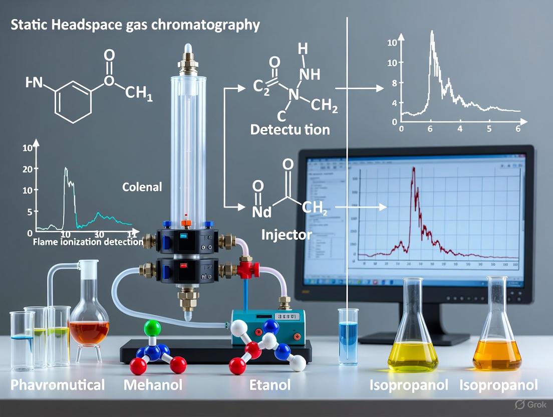
Static Headspace GC-FID for Pharmaceutical Residual Solvents: A Complete Guide from Method Setup to Validation
This article provides a comprehensive guide for researchers and drug development professionals on the application of Static Headspace Gas Chromatography with Flame Ionization Detection (HS-GC-FID) for analyzing residual solvents in...
Static Headspace GC-FID for Pharmaceutical Residual Solvents: A Complete Guide from Method Setup to Validation
Abstract
This article provides a comprehensive guide for researchers and drug development professionals on the application of Static Headspace Gas Chromatography with Flame Ionization Detection (HS-GC-FID) for analyzing residual solvents in pharmaceuticals. Covering foundational principles, method development, and practical application, it aligns with ICH and USP guidelines <467>. The content explores method optimization, common troubleshooting scenarios, and comparative analyses with techniques like HS-SPME and comprehensive two-dimensional GC. Finally, it outlines the critical steps for method validation and regulatory compliance, offering a complete resource for ensuring drug safety and quality control.
Understanding Static Headspace GC-FID: Principles and Regulatory Framework for Pharmaceutical Analysis
Static Headspace Gas Chromatography-Flame Ionization Detection (HS-GC-FID) is a premier technique for determining volatile and semi-volatile organic impurities in pharmaceutical products [1] [2]. The control of these residual solvents is critical, as their presence may pose toxic risks and negatively impact the stability and efficacy of drug substances and products [2]. The core of this technique lies in its reliance on the established principles of equilibrium and partitioning to provide a robust, accurate, and sensitive analysis, making it indispensable in drug development and quality control laboratories.
Core Theoretical Principles
The Equilibrium System
Static headspace sampling is an equilibrium-based technique [3]. A prepared sample is placed in a sealed vial and heated to a constant temperature, allowing the volatile analytes to distribute between the sample matrix (liquid or solid) and the vapor phase (headspace) above it [1]. The system is left until equilibrium is reached, a state where the rate of evaporation for each analyte from the liquid phase equals its rate of condensation from the gas phase [3]. Once equilibrium is established, a representative aliquot of the headspace vapor is injected into the GC system for separation and detection.
The Partition Coefficient
The distribution of an analyte between the sample matrix and the headspace at equilibrium is quantitatively described by its partition coefficient (K) [1] [3]. This is defined as:
[ K = \frac{CS}{CG} ]
Where:
- ( C_S ) is the concentration of the analyte in the sample phase.
- ( C_G ) is the concentration of the analyte in the gas phase (headspace) [1].
The value of K is a constant for a given analyte-matrix combination at a specific temperature and indicates the compound's volatility within that matrix [3].
- A high K value (e.g., ~500 for ethanol in water) indicates the analyte has a strong affinity for the sample matrix, resulting in a lower concentration in the headspace [1].
- A low K value (e.g., ~0.01 for hexane in water) indicates the analyte is highly volatile and will be predominantly found in the headspace, making it easier to detect [1].
The following diagram illustrates the workflow of a static headspace analysis, from sample preparation to data analysis.
Key Parameters Influencing Equilibrium and Partitioning
The sensitivity of a static headspace analysis is directly controlled by the concentration of the analyte in the headspace (( C_G )). This concentration is influenced by several experimental parameters that affect the partition coefficient and the phase ratio [1].
Table 1: Key Experimental Parameters in Static Headspace Analysis
| Parameter | Effect on Headspace Analysis | Practical Consideration |
|---|---|---|
| Sample Volume | Has a major effect for analytes with intermediate K values; minimal effect for analytes with very high or very low K [1]. | Using ~10 mL in a 20 mL vial (β = VG/VL = 1) simplifies calculations [1]. |
| Equilibration Temperature | Significantly increases headspace concentration for analytes with high K (matrix-bound) [1]. | Requires precise temperature control (±0.1°C for high K analytes). High pressure in aqueous samples can cause analyte loss upon needle insertion [1]. |
| Equilibration Time | Time required for the system to reach equilibrium [1]. | Must be determined empirically for each analyte/sample combination; not directly correlated with K value [1]. |
| Matrix Modification (Salting Out) | Adding salt (e.g., KCl) reduces the partition coefficient of polar analytes in polar matrices, driving them into the headspace [1] [3]. | An effective method to enhance sensitivity for specific analytes, particularly in aqueous samples [1]. |
Quantitative Analysis in Static Headspace
The quantitative nature of static headspace sampling is well-established, provided the system is calibrated appropriately [3]. The fundamental relationship used is:
[ CG = \frac{C0}{K + \beta} \quad \text{where} \quad \beta = \frac{VG}{VL} ]
Where:
- ( C_G ) is the analyte concentration in the gas phase.
- ( C_0 ) is the analyte concentration in the original sample.
- ( K ) is the partition coefficient.
- ( \beta ) is the phase ratio [1].
Because K is matrix-dependent, the most critical aspect of quantitative HS-GC is calibration with matrix-matched standards [1]. It is often challenging to obtain a completely analyte-free "blank" matrix. One technique to overcome this is to use exhaustive extraction (e.g., multiple headspace extractions) on a real sample to create a suitable blank for calibration [1].
Application Note: Protocol for Residual Solvents in an Active Pharmaceutical Ingredient (API)
Background
This protocol details the development and validation of an HS-GC-FID method for determining six residual solvents—Methanol, Ethyl Acetate, Isopropyl Alcohol (IPA), Triethylamine, Chloroform, and Toluene—in Losartan Potassium API, following ICH Q3C guidelines [2].
Materials and Reagents
Table 2: Research Reagent Solutions and Essential Materials
| Item | Function / Specification |
|---|---|
| Gas Chromatograph | Agilent 7890A, equipped with Flame Ionization Detector (FID) [2]. |
| Headspace Sampler | Agilent 7697A [2]. |
| Analytical Column | DB-624 capillary column (30 m × 0.53 mm × 3 µm film thickness) [2]. |
| Carrier Gas | Helium, constant flow of 4.718 mL/min. (Note: Hydrogen is a sustainable and efficient alternative carrier gas) [4]. |
| Sample Diluent | Dimethylsulfoxide (DMSO), GC grade. Chosen for its high boiling point and ability to dissolve the API effectively [2]. |
| Standard Solutions | Individual solvent standards in GC purity grade for calibration [2]. |
Detailed Methodology
Instrument Configuration
- GC Parameters: Inlet temperature: 190°C; Detector (FID) temperature: 260°C [2].
- Oven Program: Initial 40°C (hold 5 min), ramp to 160°C at 10°C/min, then to 240°C at 30°C/min (hold 8 min). Total run time: 28 min [2].
- Headspace Parameters: Equilibration: 30 min at 100°C; Syringe temperature: 105°C; Transfer line: 110°C; Split ratio: 1:5 [2].
Sample and Standard Preparation
- Standard Solution: Prepare in DMSO at concentrations based on ICH limits. Transfer 5.0 mL to a 20 mL headspace vial, cap, and crimp immediately [2].
- Sample Solution: Accurately weigh 200 mg of Losartan Potassium API into a 20 mL headspace vial. Add 5.0 mL of DMSO, cap, and crimp immediately [2].
- Vortex all vials for 1 minute to ensure mixing [2].
Method Validation
The developed method was validated per Brazilian guidelines (RDC 166/2017), proving to be:
- Selective: No interference from the API or diluent at the retention times of the target solvents [2].
- Linear: Correlation coefficient (r) ≥ 0.999 for all solvents over the specified range [2].
- Precise: Relative Standard Deviation (RSD) ≤ 10.0% for repeatability and intermediate precision [2].
- Accurate: Average recoveries ranged from 95.98% to 109.40% [2].
- Robust: Maintained performance under deliberate, small changes to chromatographic conditions [2].
Results and Discussion
The application of this validated method to a Losartan Potassium API batch detected only IPA and Triethylamine, demonstrating the effectiveness of the purification process in removing other synthesis solvents [2]. The use of DMSO as a diluent, coupled with optimized headspace conditions, was crucial for the precise and sensitive quantification of the diverse range of solvents, including the challenging analyte Triethylamine, which failed system suitability in a pharmacopeial method [2].
The following diagram summarizes the logical decision process involved in developing and optimizing a static headspace method.
The principles of equilibrium and partitioning form the scientific foundation of static headspace sampling. A deep understanding of how parameters like temperature, sample volume, and matrix composition affect the partition coefficient is essential for developing robust and sensitive HS-GC-FID methods. As demonstrated in the Losartan Potassium application, a meticulously optimized and validated static headspace method is a powerful tool for ensuring the safety and quality of pharmaceuticals by reliably monitoring harmful residual solvents. The technique's quantitative nature, when properly calibrated, makes it a cornerstone of modern pharmaceutical analysis.
The Role of Flame Ionization Detection (FID) in Universal Solvent Detection
Static headspace gas chromatography coupled with flame ionization detection (HS-GC-FID) is a cornerstone technique for analyzing residual solvents in active pharmaceutical ingredients (APIs) and drug products. The International Council for Harmonisation (ICH) Q3C(R8) guideline strictly controls these volatile organic impurities due to their potential toxicity, categorizing them into Classes 1-3 based on risk [5] [6]. The universal response of the FID to hydrocarbons, combined with its robustness and sensitivity, makes it exceptionally suitable for this regulated, high-throughput environment. This application note details the implementation of a generic HS-GC-FID method for the universal screening and quantification of residual solvents, providing a validated framework that accelerates pharmaceutical development while ensuring compliance.
Key Principles of FID and its Universality
The flame ionization detector operates on the principle of combusting organic analytes in a hydrogen/air flame. As compounds elute from the GC column, they are pyrolyzed. This process produces ions and electrons, which are collected by electrodes to generate a measurable electrical signal proportional to the mass of carbon in the flame [7].
- Broad Detection Capability: FID responds to virtually all organic compounds containing carbon-hydrogen bonds, which includes nearly all regulated residual solvents [7].
- Low Response to Key Exceptions: Crucially, FID has a negligible response to water and carbon dioxide, which are common sample matrix components, thereby minimizing potential interferences [7].
- Operational Benefits: The detector is valued for its simple design, stability, broad dynamic range, and low maintenance requirements, making it ideal for routine quality control (QC) laboratories [7].
Generic Method Development and Optimization
Chromatographic Conditions for Universal Separation
Developing a single GC method capable of resolving a wide array of solvents with varying polarities and volatilities is fundamental to universal detection. A mid-polarity stationary phase, such as a 6% cyanopropylphenyl / 94% dimethylpolysiloxane column (e.g., DB-624, RTx-624), has been widely established as the industry standard for this application [8] [9] [6]. This phase provides an optimal balance for separating diverse solvent mixtures.
Table 1: Exemplary Generic GC-FID Conditions for Residual Solvent Analysis
| Parameter | Specification |
|---|---|
| Column | DB-624, 30 m × 0.32 mm, 1.8 µm (or equivalent) |
| Carrier Gas | Hydrogen or Helium, constant flow (~1.5 mL/min) |
| Oven Program | 40°C (hold 20 min), ramp to 200°C at 15°C/min (hold 5 min) |
| Injector Temp | 200°C |
| Detection | FID @ 250°C |
The method successfully resolves common solvents, including critical pairs like 2-methylpentane and dichloromethane [8]. Using hydrogen as a carrier gas is a superior and more sustainable alternative to helium, offering better chromatographic efficiency and faster analysis times due to its lower viscosity and more favorable van Deemter characteristics [4].
Headspace Sampling Parameters
Headspace sampling is the preferred technique for residual solvent analysis as it introduces volatile analytes into the GC system while excluding non-volatile sample components, thereby protecting the column and detector [9] [6].
Optimized Headspace Parameters:
- Diluent: N,N-Dimethylacetamide (DMA), N,N-Dimethylformamide (DMF), or 1,3-Dimethyl-2-imidazolidinone (DMI). These high-boiling point solvents effectively dissolve many APIs and create a favorable partition coefficient for a wide range of volatile solvents [9] [6].
- Sample Concentration: Typically 50-100 mg of API per 1 mL of diluent [9].
- Vial Equilibration Temperature: 80-120°C, balanced to ensure sensitivity for high-boiling solvents without degrading the sample [9] [6].
- Equilibration Time: 20-60 minutes with vigorous shaking to ensure complete partitioning equilibrium is reached [9].
Experimental Protocol: A Generic HS-GC-FID Method
Reagents and Materials
The Scientist's Toolkit: Essential Research Reagents and Materials
| Item | Function & Specification |
|---|---|
| GC-FID System | Agilent, PerkinElmer, or equivalent, equipped with a headspace autosampler. |
| DB-624 Column | A mid-polarity 6% cyanopropylphenyl/94% dimethylpolysiloxane column for separation. |
| High-Purity Diluent | DMA, DMF, or DMI (HS-GC grade) to dissolve samples with minimal background. |
| Residual Solvent Standards | Certified reference materials for target solvents (e.g., methanol, acetone, THF, toluene). |
| Positive Displacement Pipettes | Essential for accurate and reproducible transfer of volatile solvent standards [6]. |
| Headspace Vials & Seals | 10-20 mL vials with PTFE/silicone septa and crimp caps to maintain a sealed system. |
Step-by-Step Procedure
Standard Solution Preparation:
- Prepare a stock standard solution in DMA/DMI by accurately pipetting known volumes of neat solvent standards using positive displacement pipettes [6].
- Dilute the stock solution to create a working standard containing all target solvents at concentrations relative to their ICH limits (see Table 2).
Sample Solution Preparation:
- Accurately weigh approximately 100 mg of the API into a headspace vial.
- Add 1.0 mL of diluent (e.g., DMA) via pipette, immediately crimp seal the vial, and vortex to dissolve or suspend the sample [9].
Instrumental Analysis:
- Load vials into the HS autosampler tray in the recommended sequence: system blank, sensitivity check standard, followed by six replicates of the working standard for system suitability, and finally the prepared samples [9].
- Initiate the analytical sequence using the parameters defined in Table 1 and Section 3.2.
System Suitability Test:
- Before quantifying samples, verify system performance. Critical pairs (e.g., methyl ethyl ketone/ethyl acetate) must have a resolution (Rs) ≥ 0.9 [9]. The %RSD for six replicate injections of the working standard should be ≤ 15.0%, and the signal-to-noise (S/N) ratio for solvents at the limit of quantitation (LOQ) must be ≥ 10 [9].
The following workflow diagram illustrates the generic HS-GC-FID analytical procedure:
Method Validation and Performance Data
A generic HS-GC-FID method must be validated per ICH Q2(R1) guidelines to prove its reliability for regulatory testing. The following table summarizes typical validation outcomes for a well-designed generic method.
Table 2: Representative Method Validation Data for a Generic HS-GC-FID Method
| Validation Parameter | Performance Criteria | Exemplary Results from Literature |
|---|---|---|
| Linearity & Range | Correlation coefficient (r²) across ICH limit range (e.g., LOQ-150%). | > 0.999 for ethanol, acetone, acetonitrile, THF [10]; > 0.9998 for benzyl chloride [11]. |
| Precision (Repeatability) | Relative Standard Deviation (%RSD) of replicate analyses. | < 6.5% for portable GC-PID [5]; < 5% for benzyl chloride [11]; 0.4-4.4% for PET radiopharmaceuticals [10]. |
| Accuracy (Recovery) | Mean recovery of spiked solvents at various levels. | 99.3% - 103.8% for 8 solvents in radiopharmaceuticals [10]; Within 95-105% for benzyl chloride [11]. |
| Limit of Quantitation (LOQ) | The lowest concentration that can be quantified with acceptable precision and accuracy. | Ethanol: 0.48 mg/L; Acetone: 0.42 mg/L; DMSO: 0.50 mg/L [10]; Benzyl chloride: 0.1 μg/g [11]. |
| Specificity | Resolution of all analyte peaks from each other and from the diluent blank. | Resolution (Rs) ≥ 1.5 for all solvents in mixed standard [6]. No interference from diluent or API [11] [6]. |
Case Studies and Applications
The generic HS-GC-FID approach has been successfully implemented across a wide spectrum of pharmaceutical compounds.
- Favipiravir API: A validated HS-GC-FID method was developed for methanol, dichloromethane, acetonitrile, and toluene, demonstrating linearity, precision (%RSD < 10), and accuracy suitable for routine QC [12].
- Arterolane Maleate: A method for ten residual solvents (including pentane, ethanol, dichloromethane, and n-heptane) was validated, showing specificity, sensitivity, and was successfully applied to a bulk drug sample [8].
- PET Radiopharmaceuticals: A novel GC method quantified ethanol, acetone, acetonitrile, and others in radioactive drugs like [¹⁸F]FDG with high precision (RSD 0.4-4.4%) and excellent accuracy (99.3-103.8% recovery) [10].
Flame Ionization Detection remains an indispensable tool for universal solvent detection within the pharmaceutical industry. Its combination of universal response to hydrocarbons, robustness, and sensitivity perfectly aligns with the requirements for monitoring diverse residual solvents as per ICH Q3C(R8). The establishment of validated generic HS-GC-FID methods, as detailed in this application note, provides a powerful strategy for laboratories to enhance efficiency, reduce method development timelines, and ensure consistent compliance in the quality control of pharmaceuticals.
Navigating ICH Q3C and USP <467> Guidelines for Residual Solvents
Residual solvents are organic volatile chemicals that may remain in active pharmaceutical ingredients (APIs), excipients, or finished drug products after manufacturing. These solvents, while useful in the synthesis and processing of pharmaceuticals, offer no therapeutic benefit and may pose significant toxic risks to patients if not properly controlled. The International Council for Harmonisation (ICH) Q3C guideline and the United States Pharmacopeia (USP) General Chapter <467> provide the primary frameworks for controlling these impurities in pharmaceutical products. Both guidelines aim to protect patient safety by establishing stringent limits for residual solvents based on their toxicity profiles, ensuring that drug manufacturers worldwide adhere to consistent safety standards [13] [14].
ICH Q3C serves as an internationally recognized guideline, while USP <467> represents an enforceable compendial standard in the United States. A crucial distinction lies in their scope of application: ICH Q3C primarily addresses new drug products, whereas USP <467> applies to all compendial drug substances, excipients, and products, whether new or existing. This broader application makes USP <467> particularly significant for manufacturers of generic drugs and existing pharmaceutical products [14]. Understanding the nuances, harmonization, and differences between these two documents is essential for pharmaceutical scientists developing robust analytical methods for residual solvent analysis.
Solvent Classification and Regulatory Limits
Categorization of Residual Solvents
Both ICH Q3C and USP <467> classify residual solvents into three categories based on their toxicity and potential risk to human health. This classification system enables manufacturers to prioritize control strategies based on the inherent risk associated with each solvent.
Class 1 solvents are considered the most hazardous and are to be avoided in the manufacture of drug substances, excipients, and drug products. These solvents include known human carcinogens, strongly suspected carcinogens, and environmental hazards. Examples include benzene (a known carcinogen) and carbon tetrachloride [15] [14].
Class 2 solvents are considered less toxic than Class 1 but still present significant potential health risks, thus requiring limitation in pharmaceutical products. These solvents are associated with irreversible toxicity, such as neurotoxicity or reproductive toxicity, or other significant but reversible toxicities. Common examples include methanol, acetonitrile, toluene, and chloroform [2] [15].
Class 3 solvents are considered the least toxic, with low toxic potential to humans. While they are subject to control, they present lower risk at levels typically found in pharmaceuticals. Examples include acetone, ethanol, and ethyl acetate [2] [15].
Table 1: Classification and Limits of Common Residual Solvents
| Solvent | Class | PDE (mg/day) | Concentration Limit (ppm) | Toxicological Concern |
|---|---|---|---|---|
| Benzene | 1 | 0.02 | 2 | Carcinogen |
| Carbon Tetrachloride | 1 | 0.04 | 4 | Hepatotoxin, Carcinogen |
| Methanol | 2 | 30.0 | 3000 | Neurotoxin |
| Acetonitrile | 2 | 4.1 | 410 | Cyanide precursor |
| Toluene | 2 | 8.9 | 890 | Developmental toxin |
| Chloroform | 2 | 0.6 | 60 | Carcinogen |
| Ethanol | 3 | 50.0 | 5000 | Low toxicity |
| Acetone | 3 | 50.0 | 5000 | Low toxicity |
| Isopropyl Alcohol | 3 | 50.0 | 5000 | Low toxicity |
Options for Establishing Compliance
USP <467> and ICH Q3C provide two primary options for demonstrating compliance with Class 2 solvent limits, offering flexibility in quality control approaches:
Option 1 involves testing individual components against fixed concentration limits. If all drug substances and excipients in a formulation meet the limits specified in the Option 1 table, these components may be used in any proportion without further calculation, provided the daily dose does not exceed 10 g. This approach simplifies compliance but may be unnecessarily restrictive for components used in small proportions [14].
Option 2 allows for a more nuanced approach by calculating the cumulative solvent load based on the formulation composition. This option acknowledges that a drug substance or excipient comprising only a small fraction of the final drug product may contain higher levels of residual solvents without exceeding the overall permitted daily exposure (PDE) in the finished product. The concentration limits for each component are calculated using the formula: Concentration (ppm) = (1000 × PDE) / dose. The sum of the calculated PDE values for all components must not exceed the established limit [14].
Analytical Method Development for Static Headspace GC-FID
Instrumentation and Research Reagent Solutions
The development of a robust static headspace gas chromatography with flame ionization detection (HS-GC-FID) method requires careful selection of both instrumentation and reagents. The following table outlines essential materials and their functions in residual solvents analysis:
Table 2: Key Research Reagent Solutions for HS-GC-FID Analysis of Residual Solvents
| Item | Function/Application | Example Specifications |
|---|---|---|
| DB-624 Capillary Column | Separation of volatile solvents | 30 m × 0.53 mm × 3 µm film thickness; 6% cyanopropyl-phenyl, 94% dimethyl polysiloxane stationary phase [2] |
| Dimethylsulfoxide (DMSO) | Sample diluent | High purity GC grade; high boiling point (189°C) to minimize interference [2] |
| Helium Gas | Carrier gas | High purity (99.999%); constant flow rate (e.g., 4.718 mL/min) [2] |
| HS Vials | Sample containment | 20 mL volume; sealed with crimp caps with PTFE/silicone septa [2] |
| Residual Solvents Standards | Method calibration and validation | Individual and mixed standards in GC purity grade [2] |
Critical Method Development Parameters
Several parameters require careful optimization during HS-GC-FID method development to ensure accurate and reproducible results:
Sample Diluent Selection: The choice of diluent significantly impacts method sensitivity and accuracy. While water is sometimes used, dimethylsulfoxide (DMSO) often provides superior performance due to its higher boiling point (189°C), which reduces diluent interference, and its ability to dissolve a wide range of pharmaceutical compounds. Studies have demonstrated that DMSO typically yields higher precision and sensitivity with improved recoveries compared to aqueous diluents [2].
Headspace Conditions Optimization: Incubation time and temperature must be optimized to ensure efficient transfer of volatile solvents into the headspace without degrading the sample. Typical incubation conditions range from 30 minutes at 100°C, though these parameters should be adjusted based on the specific solvents of interest and the sample matrix. Proper optimization ensures equilibrium between the sample solution and headspace, critical for quantitative analysis [2].
Chromatographic Conditions: Column selection, temperature programming, and carrier gas flow rates significantly impact separation efficiency. The DB-624 column has proven effective for separating diverse solvent mixtures. Temperature programs often begin with an isothermal hold (e.g., 40°C for 5 minutes) followed by controlled ramping (e.g., 10°C/min to 160°C, then 30°C/min to 240°C) to resolve solvents with varying volatilities. Split ratios (e.g., 1:5) should be optimized to balance sensitivity and resolution [2].
Case Study: Method Development for Losartan Potassium
A recent study demonstrated the development and validation of an HS-GC-FID method for determining six residual solvents (methanol, ethyl acetate, isopropyl alcohol, triethylamine, chloroform, and toluene) in losartan potassium raw material. Initial screening using the USP procedure A revealed inadequate performance for triethylamine, necessitating method development. DMSO was selected as the diluent with incubation at 100°C for 30 minutes. Chromatographic separation was achieved using a DB-624 column with a temperature program from 40°C (held for 5 minutes) to 160°C at 10°C/min, then to 240°C at 30°C/min with an 8-minute hold. The total run time was 28 minutes with a split ratio of 1:5 [2].
The method was validated according to Brazilian guidelines (RDC 166/2017), demonstrating selectivity for all target solvents, with limits of quantification below 10% of ICH specification limits. Precision was excellent (RSD ≤ 10.0%), with strong linearity (r ≥ 0.999) and accurate recoveries ranging from 95.98% to 109.40%. The method proved robust under deliberate variations of chromatographic conditions. Application to an actual losartan potassium batch detected only isopropyl alcohol and triethylamine, indicating effective purification during manufacturing [2].
Experimental Protocol: Residual Solvents Analysis by HS-GC-FID
Sample Preparation Protocol
Standard Solution Preparation: Prepare stock solutions of each target solvent in DMSO GC grade, based on ICH limits. For a typical mixed standard, combine appropriate volumes to achieve the following concentrations: methanol (600 µg/mL), isopropyl alcohol (1000 µg/mL), ethyl acetate (1000 µg/mL), chloroform (12 µg/mL), triethylamine (1000 µg/mL), and toluene (178 µg/mL). Transfer 5.0 mL of this solution to a 20 mL headspace vial and cap immediately with crimp caps [2].
Sample Solution Preparation: Accurately weigh approximately 200 mg of the drug substance into a 20 mL headspace vial. Add 5.0 mL of DMSO GC grade, cap the vial immediately, and mix on a vortex shaker for 1 minute to ensure complete dissolution [2].
Quality Control Samples: Prepare system suitability samples and quality control samples at appropriate concentrations to verify method performance during analysis.
Instrumental Analysis Protocol
Headspace Conditions:
- Incubation temperature: 100°C
- Incubation time: 30 minutes
- Syringe temperature: 105°C
- Transfer line temperature: 110°C
- Pressurization time: 1 minute [2]
GC-FID Conditions:
- Column: DB-624 capillary column (30 m × 0.53 mm × 3 µm)
- Carrier gas: Helium, constant flow rate of 4.718 mL/min
- Oven temperature program:
- Initial: 40°C held for 5 minutes
- Ramp 1: 10°C/min to 160°C
- Ramp 2: 30°C/min to 240°C, held for 8 minutes
- Inlet temperature: 190°C
- Detector temperature: 260°C
- Split ratio: 1:5
- Total run time: 28 minutes [2]
Method Validation Protocol
Validation should be performed according to ICH Q2(R2) guidelines, assessing the following parameters [16]:
Selectivity: Demonstrate the ability to unequivocally identify and quantify all target residual solvents in the presence of potentially interfering components. Analyze the diluent (DMSO), standard solutions of individual solvents, mixture of solvents, API, and API spiked with the solvent mixture [2].
Linearity: Prepare three independent calibration curves with at least six concentration levels ranging from the limit of quantitation (LOQ) to 120% of the specification limit for each solvent. Determine correlation coefficients (r ≥ 0.999 is typically acceptable) and linear regression equations [2].
Limit of Quantification (LOQ): Prepare decreasing concentrations of individual solvent solutions and determine the lowest concentration that can be quantified with acceptable precision and accuracy (typically signal-to-noise ratio ≥ 10:1). LOQ values should be below 10% of the specification limits [2].
Precision:
- Repeatability: Analyze six individual samples at 100% concentration level for each solvent.
- Intermediate precision: Have a second analyst perform the analysis on a different day using different equipment if possible.
- Calculate relative standard deviations (RSD ≤ 10.0% typically acceptable) [2].
Accuracy: Perform recovery studies by spiking known quantities of individual residual solvents into API samples at three levels (low, middle, and high) in triplicate. Average recoveries should typically fall between 80-115% [2].
Robustness: Evaluate the method's resilience to deliberate, small variations in chromatographic conditions such as oven initial temperature (±5°C), gas linear velocity (e.g., 29 or 39 cm/s), and different column batches. System suitability criteria should be maintained under all conditions [2].
Regulatory Compliance and Current Updates
The regulatory landscape for residual solvents continues to evolve, with recent updates to both ICH Q3C and USP <467>. The ICH Q3C(R9) version, published in April 2024, includes revisions to section 3.4 on Analytical Procedures. The updated text states: "Residual solvents are typically determined using chromatographic techniques such as gas chromatography. Any harmonised procedures for determining levels of residual solvents as described in the pharmacopoeias should be used, if feasible. Otherwise, manufacturers would be free to select the most appropriate validated analytical procedure for a particular application" [16].
This revision reinforces the principle that while pharmacopeial methods are preferred, alternative validated methods are acceptable when justified. This flexibility is crucial for addressing complex analytical challenges where compendial methods may be insufficient, such as in the analysis of losartan potassium where the USP procedure A proved inadequate for quantifying triethylamine [2].
ICH Q3C remains a guideline, while USP <467> constitutes an enforceable standard for products with USP monographs. However, regulatory agencies such as the FDA apply ICH Q3C principles to all drug applications, including those without USP monographs. For global submissions, compliance with both documents is typically necessary, with manufacturers often applying the more stringent requirement where discrepancies exist [15] [14].
Successful navigation of ICH Q3C and USP <467> guidelines requires a comprehensive understanding of both regulatory expectations and analytical methodology. The static headspace GC-FID technique has proven to be a robust, sensitive, and versatile approach for residual solvents analysis across diverse pharmaceutical matrices. By following systematic method development and validation protocols, pharmaceutical scientists can establish reliable analytical procedures that ensure product safety and regulatory compliance.
The case study of losartan potassium demonstrates that even when compendial methods prove inadequate, well-designed and thoroughly validated alternative methods can successfully address analytical challenges. As regulatory frameworks continue to evolve, the fundamental principles of patient safety, scientific rigor, and quality by design remain paramount in residual solvents control.
In the pharmaceutical industry, residual solvents are defined as organic volatile chemicals that remain in active pharmaceutical ingredients (APIs), excipients, or final drug products after manufacturing [17]. These solvents are used or produced during synthesis, purification, or formulation processes but provide no therapeutic benefit [18]. The International Council for Harmonisation (ICH) guideline Q3C establishes a unified framework for classifying these solvents and setting safety-based limits to protect patient health [19] [6]. The United States Pharmacopeia (USP) general chapter <467> provides the official compendial procedures for testing residual solvents, making compliance with these standards mandatory for pharmaceutical products in the U.S. market [20] [17].
The classification system categorizes solvents based on their toxicity profiles and environmental hazards, with stringent testing required to ensure that residual levels do not exceed established safety limits [20]. This application note details the categorization, allowable limits, and analytical protocols for residual solvent analysis, with specific focus on implementation via static headspace gas chromatography with flame ionization detection (HS-GC-FID) within pharmaceutical research and development.
Solvent Classification and Allowable Limits
Classification System and Risk Assessment
The ICH Q3C guideline categorizes residual solvents into three classes based on their toxicity and environmental impact [20] [18]. This risk-based classification enables manufacturers to prioritize solvent control strategies and implement appropriate testing protocols.
Class 1 solvents (Solvents to Be Avoided) consist of known or strongly suspected human carcinogens, as well as substances presenting significant environmental hazards [20] [17]. The use of these solvents in pharmaceutical manufacturing should be avoided unless their application is unavoidable in producing a drug product with significant therapeutic benefit [18].
Class 2 solvents (Solvents to Be Limited) include substances with inherent but reversible toxicities, such as nongenotoxic animal carcinogens or solvents suspected of causing other irreversible toxicity like neurotoxicity or teratogenicity [17] [6]. These solvents are commonly employed in pharmaceutical synthesis and require strict limitation with concentration monitoring [20].
Class 3 solvents (Solvents with Low Toxic Potential) encompass chemicals with low toxic potential to humans, where no health-based exposure limit is typically needed [17] [6]. While these solvents are generally regarded as less hazardous, they must still be controlled under good manufacturing practice principles and general quality management systems [18].
Allowable Limits for Residual Solvents
The acceptable concentration limits for residual solvents are established based on toxicological data and calculated as Permitted Daily Exposure (PDE) values, typically assuming a maximum daily drug product intake of 10 grams [6] [18]. The following tables summarize the classification and limits for representative solvents in each category.
Table 1: Class 1 Solvents - Solvents to Be Avoided
| Solvent | PDE (mg/day) | Concentration Limit (ppm) | Risk Basis |
|---|---|---|---|
| Benzene | 0.02 | 2 | Known human carcinogen [20] |
| Carbon tetrachloride | 0.04 | 4 | Environmental hazard, toxic [20] |
| 1,2-Dichloroethane | 0.05 | 5 | Strongly suspected human carcinogen [20] |
Table 2: Class 2 Solvents - Solvents to Be Limited
| Solvent | PDE (mg/day) | Concentration Limit (ppm) | Toxicological Basis |
|---|---|---|---|
| Methanol | 30.0 | 3000 | Systemic toxin [20] |
| Acetonitrile | 4.1 | 410 | Toxic [20] |
| Toluene | 8.9 | 890 | Developmental toxicity [20] |
| Chloroform | 0.6 | 60 | Carcinogenic potential [17] |
| Hexane | 2.9 | 290 | Neurotoxicity [17] |
| Ethylene Glycol | 6.2 | 620 | Corrected PDE value per ICH Q3C(R9) [19] |
Table 3: Class 3 Solvents - Solvents with Low Toxic Potential
| Solvent | PDE (mg/day) | Concentration Limit (ppm) | Note |
|---|---|---|---|
| Ethanol | 50.0 | 5000 | Low toxic potential [20] |
| Acetone | 50.0 | 5000 | Low toxic potential [20] |
| Acetic Acid | 50.0 | 5000 | Low toxic potential [17] |
| Isopropanol | 50.0 | 5000 | Low toxic potential [17] |
For Class 3 solvents, concentration limits of 0.5% (5000 ppm) are generally acceptable without justification, though higher amounts may be acceptable with proper scientific rationale [18]. The limits specified in the tables assume a maximum daily dose of 10 grams of drug product; adjustments are required for products with higher daily intake [6].
Analytical Methodology: Static Headspace GC-FID
Technical Principle and Rationale
Static headspace gas chromatography with flame ionization detection (HS-GC-FID) represents the gold standard technique for residual solvent analysis in pharmaceuticals [20]. This methodology separates volatile analytes through partitioning between the sample matrix and the gas phase in a sealed vial, followed by chromatographic separation and detection.
The HS-GC-FID approach offers significant advantages for pharmaceutical analysis:
- Prevention of Instrument Contamination: The headspace sampling technique avoids direct introduction of API solutions into the GC injection port, preventing non-volatile residue accumulation and system contamination [6].
- Enhanced Sensitivity for Volatiles: Headspace sampling provides improved response characteristics for volatile solvents through favorable gas phase partitioning, enabling detection at low ppm levels [6].
- Regulatory Compliance: USP <467> designates HS-GC-FID as a compendial method for residual solvent analysis, ensuring regulatory acceptance [20] [17].
The flame ionization detector provides excellent sensitivity for organic compounds, with linear response across the concentration ranges required for pharmaceutical testing [20].
Experimental Workflow for Residual Solvent Analysis
The following diagram illustrates the complete analytical workflow for residual solvent determination using static HS-GC-FID:
Diagram 1: HS-GC-FID Analytical Workflow
Detailed Experimental Protocols
Method Conditions for Generic Residual Solvent Analysis
A robust generic GC method can efficiently quantify multiple residual solvents across different API projects, significantly reducing method development time [6]. The following protocol details established conditions for comprehensive residual solvent analysis:
Table 4: Generic GC Method Conditions for Residual Solvent Analysis
| Parameter | Specification | Rationale |
|---|---|---|
| GC Column | 60 m × 0.32 mm, 1.80 µm DB-624 | Mid-polarity (6% cyanopropyl-phenyl) for broad solvent polarity range [6] |
| Carrier Gas | Hydrogen | Optimal chromatographic efficiency with appropriate safety precautions |
| Headspace Sampler Temperature | Optimized between 80-100°C | Balances sensitivity for high-boiling solvents with minimal API degradation [6] |
| Equilibration Time | 30-45 minutes | Ensures complete partitioning equilibrium between phases |
| Diluent | 1,3-Dimethyl-2-imidazolidinone (DMI) | High boiling point (225°C), minimal interference, sharp solvent peak [6] |
| Sample Concentration | 50 mg/mL | Provides appropriate sensitivity for ICH limit testing [6] |
| Injection Volume | 1.0 mL headspace gas | Standard headspace injection volume |
Sample and Standard Preparation Protocol
Materials Required:
- Positive displacement pipettes (essential for accurate transfer of volatile and non-aqueous liquids) [6]
- Certified reference standards of target solvents
- High-purity DMI diluent
- 10 mL headspace vials with crimp caps and PTFE/silicone septa
Sample Preparation Procedure:
- Accurately weigh approximately 500 mg of API into a headspace vial [20].
- Add 10 mL of DMI diluent using a positive displacement pipette [6].
- Immediately seal the vial with a crimp cap to prevent solvent loss.
- Gently mix or vortex until the API is completely dissolved.
Mixed Stock Standard Preparation:
- Prepare a mixed stock standard containing all target solvents at concentrations calculated according to Equation 1 [6]: Where PDE is the permitted daily exposure in mg/day, and the factor 400 accounts for standard and sample dilution parameters.
- For the working standard, dilute 4.0 mL of mixed stock standard to 100 mL with DMI diluent [6].
- Transfer 1.0 mL of working standard to headspace vials for analysis.
System Suitability and Method Validation
System suitability must be established before sample analysis to ensure method integrity [18]. Critical validation parameters for residual solvent methods include:
- Specificity: Resolution (R) ≥ 1.5 between all target solvent peaks [6]
- Linearity: Demonstrated across 10-120% of ICH limit concentrations with insignificant intercepts [6]
- Sensitivity: Limit of quantitation (LOQ) established at 10% of specification limit [6]
- Accuracy: Verified through spike recovery studies (typically 80-120% recovery) using API material dissolved in mixed standard preparation [6]
The Scientist's Toolkit: Essential Materials and Reagents
Table 5: Key Research Reagent Solutions for HS-GC-FID Analysis
| Item | Function/Purpose | Technical Specifications |
|---|---|---|
| DMI Diluent | Sample and standard dissolution | High boiling point (225°C), minimal interference, suitable for broad solvent polarity range [6] |
| DB-624 GC Column | Chromatographic separation | 60 m × 0.32 mm, 1.80 µm; 6% cyanopropyl-phenyl mid-polarity stationary phase [6] |
| Positive Displacement Pipettes | Accurate liquid transfer | Essential for non-aqueous and volatile standard preparation [6] |
| Certified Solvent Standards | Quantitation reference | Certified reference materials for all target Class 1, 2, and 3 solvents |
| Headspace Vials/Closures | Sample containment | 10-20 mL vials with PTFE/silicone septa for volatile retention |
Case Study: USP <467> Compliance for API Manufacturer
A mid-sized U.S. API manufacturer faced a compliance challenge when in-house testing detected methanol levels exceeding the Class 2 limit of 3000 ppm in a new API [20]. The laboratory implemented the following resolution:
- Analytical Approach: Applied HS-GC-FID with validation for methanol detection down to 50 ppm sensitivity [20]
- Sample Analysis: Tested 250 mg sample aliquots in triplicate for statistical confidence [20]
- Results: Initial batch contained 3400 ppm methanol; post-purification batch demonstrated 240 ppm, well within acceptable limits [20]
- Impact: Successful product release with full documentation aligned with FDA audit standards, achieved within 48-hour turnaround [20]
This case demonstrates the critical importance of robust analytical methods with appropriate sensitivity to ensure compliance with regulatory limits for Class 2 solvents.
The classification of solvents into Categories 1, 2, and 3 based on toxicity, with corresponding allowable limits, provides a scientifically sound framework for managing residual solvents in pharmaceutical products. Static headspace GC-FID methodology offers a robust, compliant approach for quantifying these solvents across all categories. The experimental protocols detailed in this application note enable pharmaceutical scientists to implement reliable testing strategies that ensure product safety and regulatory compliance while optimizing laboratory efficiency through generic method approaches.
Advantages of Static Headspace for Complex Pharmaceutical Matrices
Static headspace gas chromatography (HS-GC) coupled with flame ionization detection (FID) represents a premier analytical technique for determining volatile organic impurities in pharmaceutical substances. Its superiority lies in exceptional sensitivity, minimal sample preparation, and robust performance across diverse drug matrices. This application note delineates the fundamental advantages of static HS-GC-FID for pharmaceutical residual solvents analysis, provides a detailed protocol compliant with international pharmacopeial standards, and presents experimental data validating its efficacy for complex drug substances.
In the synthesis of active pharmaceutical ingredients (APIs) and excipients, organic solvents are employed which must be subsequently removed to safe levels as defined by regulatory bodies. Static headspace gas chromatography (HS-GC) has emerged as the dominant technique for monitoring these residual solvents due to its ability to analyze volatiles in complex matrices with minimal interference from non-volatile components [21] [22]. The technique involves heating a sealed vial containing the sample to achieve equilibrium partitioning of volatile analytes between the condensed phase and the vapor phase (headspace), followed by instrumental analysis of the vapor phase [23].
This application note frames the significant advantages of static HS-GC within the broader context of pharmaceutical solvents research, particularly highlighting its alignment with Quality by Design (QbD) principles through robust, transferable methods that enhance laboratory efficiency and data reliability.
Core Advantages for Pharmaceutical Matrices
Simplified Sample Preparation and Enhanced Matrix Tolerance
Static headspace requires only that the sample is dissolved or suspended in a suitable diluent within a sealed vial, dramatically reducing preparation time and associated errors [22]. By analyzing only the vapor phase, it effectively excludes non-volatile matrix components (e.g., API polymers, excipients) that would otherwise contaminate the GC inlet and column, leading to longer column life and reduced system maintenance [21] [22]. This is particularly advantageous for complex pharmaceutical formulations like liposomes and nanoformulations, where the matrix would severely interfere with direct injection techniques [24].
Superior Analytical Performance and Reproducibility
The closed-system nature of static headspace sampling provides excellent analytical precision. By avoiding manual injection variability and minimizing matrix effects, it delivers high reproducibility essential for regulatory compliance [22] [9]. The technique demonstrates robust sensitivity, routinely achieving detection from parts-per-billion (ppb) to percentage levels, sufficient to meet the strict limits stipulated by ICH Q3C guidelines for all classes of residual solvents [23] [25].
Direct Regulatory Compliance and Method Flexibility
Static headspace is explicitly referenced in compendial methods from the United States Pharmacopeia (USP) <467> and the European Pharmacopoeia (Ph. Eur.) [26] [25] [9]. This facilitates direct implementation in quality control laboratories. Furthermore, the technique offers significant flexibility through parameter optimization (temperature, equilibration time, phase ratio) to enhance sensitivity for specific analytes or to overcome challenging sample solubilities, for instance by using water-DMF mixtures [23] [25].
Comparative Analysis: Static vs. Dynamic Headspace
The following table summarizes key distinctions between static and dynamic headspace (purge-and-trap) techniques, underscoring the operational advantages of static headspace for routine pharmaceutical analysis [21].
| Feature | Static Headspace GC | Dynamic Headspace GC |
|---|---|---|
| Principle | Equilibrium-based sampling | Continuous purging with inert gas |
| Sample Prep | Minimal preparation required | Requires setup for gas flow and trapping |
| Sensitivity | Good for many volatiles (ppm-ppb) | Higher sensitivity for trace-level analysis |
| Analysis Time | Longer equilibration time | Generally faster analysis |
| Complexity | Simpler setup and operation | More complex setup; requires trapping |
| Matrix Tolerance | High (minimal contamination risk) | Potential for foaming and trap clogging |
| Ideal Use Case | Routine analysis of residual solvents | Trace analysis of very low-concentration volatiles |
Detailed Experimental Protocol
This protocol outlines a generic static HS-GC-FID method for determining residual solvents in a typical active pharmaceutical ingredient (API), adaptable for various drug substances and products [9].
Materials and Reagents
- GC System: Gas chromatograph equipped with Flame Ionization Detector (FID) and an automated headspace autosampler (e.g., PerkinElmer HS system, Agilent G1888) [24] [9].
- GC Column: Fused-silica capillary column, 30 m × 0.32 mm ID, 1.8 µm film thickness, with a 6% cyanopropylphenyl/94% dimethylpolysiloxane stationary phase (e.g., Agilent DB-624, or equivalent USP G43 phase) [26] [9].
- Chemicals: High-purity N,N-Dimethylacetamide (DMA) or Dimethyl sulfoxide (DMSO) as sample diluent [9]. Use GC-grade or "Headspace Grade" solvents to minimize background interference [26].
- Reference Standards: USP-class residual solvent reference standards or certified reference materials of ≥98.5% purity.
- Vials: 10-mL or 20-mL headspace vials with PTFE/silicone septa and aluminum crimp caps.
Instrumentation and Conditions
Gas Chromatography Conditions [9]:
- Injector: Split mode (split ratio 5:1 to 10:1), temperature: 150-200°C
- Carrier Gas: Helium or Nitrogen, constant flow mode: 1.5 - 2.0 mL/min
- Oven Program: 40°C (hold 5-10 min), ramp at 10-15°C/min to 200-240°C (hold 0-5 min)
- FID Temperature: 250-280°C
Static Headspace Sampler Conditions [9]:
- Oven Temperature: 80-120°C (Optimize based on sample and analytes)
- Transfer Line Temperature: 10-20°C above oven temperature
- Vial Equilibration Time: 15-30 minutes (with vigorous shaking if available)
- Pressurization Time: 0.5 - 2.0 minutes
- Injection Volume: 0.5 - 1.0 mL of headspace vapor
Sample and Standard Preparation
- Standard Solution: Accurately weigh reference solvent standards and dilute to appropriate concentrations in DMA. A working standard solution should contain all target solvents at concentrations near their ICH limits (e.g., 100% of specification) [9].
- Sample Solution: Accurately weigh approximately 100 mg of the API or drug product into a headspace vial. For insoluble samples, create a fine suspension. Pipette 1.0 mL of DMA into the vial and immediately crimp seal. Vortex to mix or dissolve. Note: Sonication is not recommended as it may promote degradation [9].
System Suitability and Quantitation
- System Suitability Test: Prior to sample analysis, inject the working standard solution six times. The method is considered valid if the relative standard deviation (RSD) of the peak areas for each solvent is ≤15.0%, and resolution (Rs) between critical peak pairs (e.g., methyl ethyl ketone and ethyl acetate) is ≥0.9 [9].
- Quantitation: Use external standard calibration. The concentration of a residual solvent in the sample is calculated by comparing its peak area to that of the standard, corrected for sample weight [9].
Experimental Data and Validation
Representative Performance Data
A validated generic method for 28 common solvents demonstrates the capability of static HS-GC-FID to meet rigorous acceptance criteria [9]. The table below summarizes key validation parameters for a selection of Class 2 and 3 solvents.
Table: Method Performance Data for Selected Residual Solvents [9]
| Solvent | ICH Limit (ppm) | Relative Standard Deviation (RSD, %) | Approx. Limit of Quantitation (LOQ, ppm) |
|---|---|---|---|
| Acetonitrile | 410 | < 6.0 | ~100 |
| Chloroform | 60 | < 8.0 | ~10 |
| Dichloromethane | 600 | < 5.0 | ~100 |
| Ethanol | 5000 | < 4.0 | ~500 |
| n-Heptane | 5000 | < 7.0 | ~500 |
| Methanol | 3000 | < 5.0 | ~300 |
| Tetrahydrofuran | 720 | < 5.0 | ~100 |
| Toluene | 890 | < 4.0 | ~100 |
Case Study: Overcoming Sample Solubility Challenges
A study on a poorly water-soluble drug substance highlights the adaptability of static HS-GC. The API was insoluble in water and only soluble in DMF at elevated temperatures, posing a challenge for the Ph. Eur. method. The solution employed a water-DMF (3:2) mixture as the diluent, which successfully solubilized the sample at headspace oven temperature. This adaptation provided good recoveries for ethanol, tetrahydrofuran, and toluene, and enabled detection of all Class 1 and 2 solvents at ICH limits, validating the method for quantitative analysis [25].
The Scientist's Toolkit: Essential Research Reagent Solutions
Table: Key Reagents and Materials for Static HS-GC Pharmaceutical Analysis
| Item | Function & Importance | Exemplary Products / Grades |
|---|---|---|
| Headspace-Grade Solvents | High-purity diluents (DMA, DMF, DMSO) with low background to prevent artifactual peaks and ensure accurate quantitation. | "GC-HS Grade" solvents microfiltered and packed under inert gas [26]. |
| Pharmacopeial Reference Standards | Certified mixtures of Class 1, 2, and 3 solvents for system suitability, identification, and quantitation as per regulatory methods. | USP Residual Solvents Mixtures (Class 1, Class 2 A/B) [26] [27]. |
| GC Columns (USP G43 Phase) | Capillary columns specifically tested for resolving the 61 ICH-listed solvents, ensuring compliance with USP <467> and Ph. Eur. methods. | Agilent DB-624, Supelco OVI-G43, or equivalent [26] [9]. |
| Deactivated Guard Column | A short (~5 m) pre-column to protect the analytical column from non-volatile residues, extending its lifetime. | Deactivated fused silica guard column [26]. |
Static headspace gas chromatography with FID detection stands as an indispensable analytical technique in pharmaceutical development and quality control. Its core advantages—minimal sample preparation, exceptional protection of the GC system, robust performance, and direct regulatory compliance—make it uniquely suited for the analysis of volatile impurities in complex drug matrices. The detailed protocols and supporting data provided herein offer a reliable foundation for scientists to implement and leverage this powerful technique, thereby ensuring the safety and quality of pharmaceutical products.
Implementing HS-GC-FID: A Step-by-Step Guide to Method Development and Application
In the analysis of residual solvents in pharmaceutical drug substances using static headspace gas chromatography coupled with flame ionization detection (HS-GC-FID), the control of method parameters is not merely a procedural step but a fundamental determinant of analytical success. The optimization of vial temperature and equilibration time directly influences the partitioning of volatile analytes between the sample matrix and the headspace gas, thereby dictating the method's sensitivity, precision, and accuracy [25] [28]. This application note provides a detailed examination of these critical parameters, framed within the context of pharmaceutical quality control and compliance with International Conference on Harmonisation (ICH) Q3C guidelines and European Pharmacopoeia (Eur. Ph.) requirements [25]. It presents optimized protocols and datasets to guide researchers and drug development professionals in establishing robust and reliable HS-GC-FID methods.
The Impact of Temperature and Equilibration Time
The foundational principle of static headspace analysis is based on the equilibrium distribution of an analyte between the sample (liquid or solid) phase and the gas phase in a sealed vial. The concentration of an analyte in the gas phase ((CG)) is related to its original concentration in the sample ((CO)) by the partition coefficient ((K)) and the phase ratio ((\beta = VG/VL)), as described by the equation: (CG = CO / (K + \beta)) [28]. Both vial temperature and equilibration time are pivotal in controlling this equilibrium.
- Vial Temperature: Increasing the vial temperature enhances the vapor pressure of analytes, shifting the equilibrium towards the headspace gas and improving sensitivity. This effect is most pronounced for analytes with high solubility in the sample matrix (high (K) values) [28]. However, temperature must be precisely controlled; for analytes with a high partition coefficient, a variation of just ±0.1 °C may be required to achieve a precision of 5% [28]. Excessive temperatures can also increase pressure within the vial, potentially leading to analyte loss upon needle insertion [28].
- Equilibration Time: This parameter defines the duration allowed for the system to reach equilibrium at the set temperature. The required time is dependent on a complex interplay of analyte vapor pressure, sample concentration, phase ratio, and temperature [28]. It is crucial to determine the minimum time required for equilibrium for each analyte-sample combination to ensure reproducibility without unnecessarily lengthening the analytical cycle.
Optimized Experimental Protocol for Residual Solvents
The following protocol is adapted from pharmacopeial methods and recent research for the determination of Class 1, 2, and 3 residual solvents in a typical pharmaceutical drug substance [25].
Materials and Reagents
- Drug Substance: The analyte of interest, often an Active Pharmaceutical Ingredient (API).
- Reference Standards: Certified reference materials for all target residual solvents (e.g., ethanol, toluene, tetrahydrofuran) and an internal standard (e.g., n-propanol) [25].
- Diluent: A suitable solvent or solvent mixture. Water is often preferred for sensitivity, but for insoluble drug substances, a water-N,N-Dimethylformamide (DMF) mixture (e.g., 3:2 v/v) may be necessary to achieve solubilization and good recovery [25].
- Headspace Vials: 20 mL vials sealed with crimp caps fitted with polytetrafluoroethylene (PTFE)/silicone septa [25] [29].
Equipment
- Gas Chromatograph equipped with a Flame Ionization Detector (FID).
- Static Headspace Autosampler.
- Capillary GC Column, such as a 6%-cyanopropyl-phenyl-94%-dimethylpolysiloxane stationary phase, common in pharmacopeial methods [25].
Procedure
- Sample Preparation: Accurately weigh an appropriate amount of the drug substance into a headspace vial. A typical sample size is 1 g for solids or 1 mL for liquids [29]. Add a suitable diluent (e.g., 5 mL of water-DMF mixture) and 30 µL of internal standard solution (0.1% n-propanol). Seal the vial immediately [25] [29].
- Standard Preparation: Prepare calibration standards by spiking the diluent with known concentrations of the target residual solvents, covering the range from below to above the ICH limit concentrations [25].
- Headspace Analysis:
- Load the prepared vials into the headspace autosampler.
- Set the vial equilibration temperature and time according to the optimized conditions, for instance, 100 °C for 15 minutes [29] [30].
- Set the sample loop and transfer line temperatures at least 20 °C above the vial oven temperature to prevent condensation [28].
- Inject a defined volume of headspace gas (e.g., 0.5-1 mL) into the GC system in split or splitless mode, as required for sensitivity.
- GC-FID Analysis:
- Use a temperature program optimized for the separation of all target solvents. A common starting point is an oven temperature hold at 40 °C for 20 minutes, then ramped to 240 °C [25].
- Maintain the FID temperature at ~250 °C.
Data Presentation and Optimization Findings
Table 1: Quantitative Data from a Validated HS-GC-FID Method for a Drug Substance
This table summarizes validation data for three common residual solvents, demonstrating the method's performance at ICH-specified limits [25].
| Solvent | ICH Classification | ICH Limit (ppm) | Optimized Vial Temperature | Optimized Equilibration Time | Precision (%RSD) |
|---|---|---|---|---|---|
| Toluene | Class 2 | 890 | 100 °C | 15 min | < 5% |
| Tetrahydrofuran (THF) | Class 2 | 720 | 100 °C | 15 min | < 5% |
| Ethanol | Class 3 | 5000 (0.5%) | 100 °C | 15 min | < 5% |
Table 2: Effect of Method Parameters on Analyte Response (Partition Coefficient, K)
This table generalizes the impact of changing key parameters based on an analyte's partition coefficient (K) [28].
| Analyte Solubility (K value) | Effect of Increasing Sample Volume | Effect of Increasing Temperature | Effect of "Salting Out" |
|---|---|---|---|
| High K (Soluble, e.g., Ethanol in water) | Negligible increase in headspace concentration | Significant increase in headspace concentration | Significant reduction of K, increasing headspace concentration |
| Low K (Poorly soluble, e.g., Hexane in water) | Large increase in headspace concentration | Lesser effect, possible reduction | Minimal effect |
Workflow and Parameter Relationships
The following diagram illustrates the decision-making workflow for optimizing vial temperature and equilibration time in HS-GC-FID method development.
The Scientist's Toolkit: Essential Research Reagent Solutions
Table 3: Key Materials and Reagents for HS-GC-FID Analysis of Residual Solvents
| Item | Function / Rationale | Example / Specification |
|---|---|---|
| Water (HPLC Grade) | Primary diluent for water-soluble APIs; preferred for its low volatility and cost. | Organic-free water [27]. |
| N,N-Dimethylformamide (DMF) | Co-solvent used to dissolve poorly water-soluble drug substances [25]. | High purity (e.g., 99.9%) for spectroscopy [27]. |
| Potassium Chloride (KCl) | "Salting-out" agent. High concentrations reduce the partition coefficient (K) of polar analytes in aqueous matrices, boosting headspace concentration [28]. | Saturated salt solution. |
| Certified Reference Standards | Used for accurate identification and quantitation of target residual solvents. Essential for calibration. | USP/Ph. Eur. Class 1, 2, and 3 solvent mixtures [25] [27]. |
| Internal Standard (e.g., n-propanol) | Added in a known concentration to all samples and standards to correct for injection volume variability and matrix effects [25] [29]. | High purity (>99%) for trace analysis [25]. |
| Headspace Vials & Seals | Provide an inert, sealed environment for equilibrium. Septa must be non-reactive and maintain a tight seal at high temperatures. | 20 mL vials with PTFE/silicone septa [29]. |
The rigorous optimization of vial temperature and equilibration time is a critical success factor in developing a robust, sensitive, and compliant HS-GC-FID method for pharmaceutical residual solvents. As demonstrated, a temperature of 100 °C and an equilibration time of 15 minutes can serve as an effective starting point for many methods, but fine-tuning based on the specific drug substance matrix and target analytes is imperative. By adhering to the detailed protocols and leveraging the optimization strategies outlined in this application note, scientists can ensure their analytical methods meet the stringent requirements of modern pharmaceutical development and quality control.
In the pharmaceutical industry, the accurate analysis of residual solvents is a critical component of drug quality control and safety assurance. These volatile organic compounds, used or produced during the manufacturing of active pharmaceutical ingredients (APIs) and drug products, provide no therapeutic benefit and must be controlled to safe levels [27]. Static headspace gas chromatography coupled with flame ionization detection (HS-GC-FID) has emerged as a premier technique for this analysis, offering the significant advantage of introducing only volatile compounds into the chromatographic system, thereby minimizing interference from non-volatile sample components and reducing instrument maintenance [31].
The selection of an appropriate diluent represents one of the most critical methodological choices in HS-GC-FID, directly influencing key performance parameters including sensitivity, accuracy, and precision. This application note provides a detailed examination of two primary diluents—dimethyl sulfoxide (DMSO) and water—contextualized within pharmaceutical solvents research. We present structured experimental protocols, quantitative data comparisons, and practical guidance to enable researchers to make scientifically sound diluent selections that balance the competing demands of sample solubility and analyte volatility.
Scientific Principles and Diluent Comparison
The fundamental principle of static headspace analysis involves heating a sample in a sealed vial to allow volatile components to partition into the gas phase above the sample [31]. The partition coefficient (K), defined as the ratio of an analyte's concentration in the sample phase to its concentration in the gas phase at equilibrium, is a central parameter governing method sensitivity [31]. A lower K value favors higher analyte concentration in the headspace, thereby enhancing detection. Diluent selection directly impacts this coefficient by influencing both the solubility of the analyte and its volatility.
Table 1: Key Properties of DMSO and Water as HS-GC Diluents
| Property | Dimethyl Sulfoxide (DMSO) | Water |
|---|---|---|
| Polarity | Highly polar, aprotic | Highly polar, protic |
| Boiling Point | 189°C | 100°C |
| Key Advantage | Excellent solubility for a wide range of APIs and organic compounds | High volatility for many common residual solvents, low background |
| Primary Limitation | Low volatility for many analytes, requiring higher transfer temperatures | Poor solubility for many non-polar APIs and compounds |
| Ideal For | Samples with poor water solubility, analytes requiring high-temperature partitioning | Water-soluble samples, analytes with high volatility in aqueous matrices |
Water is often the diluent of choice for water-soluble samples due to its high volatility for many common residual solvents, leading to strong headspace concentration. However, its application is limited by the poor solubility of many modern, highly non-polar pharmaceutical compounds [27].
DMSO serves as a powerful alternative, capable of dissolving a vast spectrum of organic molecules, including those with poor aqueous solubility. This makes it invaluable in early drug discovery and for analyzing complex natural products [32]. A study analyzing botanicals like Coffeeberry extract and pomegranate powder demonstrated the utility of DMSO, though it noted that recovery rates for certain residual solvents could be highly variable, ranging from 77% to 151% depending on the matrix and detection method [33]. The primary challenge with DMSO is its low volatility, which can suppress the headspace concentration of some analytes unless method parameters like oven temperature are optimized to facilitate transfer [32].
Table 2: Quantitative Recovery Comparison for Residual Solvents in Different Matrices and Diluents
| Matrix | Spike Level (μg/g) | Average Recovery (GC-MSD) | Average Recovery (GC-FID) | Primary Diluent |
|---|---|---|---|---|
| Coffeeberry Extract | 10 | 91% - 121% | 77% - 110% | Not Specified (DMSO used in validation) |
| Coffeeberry Extract | 100 | 105% - 123% | 87% - 112% | Not Specified (DMSO used in validation) |
| Pomegranate Powder | 10 | 95% - 124% | 72% - 151% | Not Specified (DMSO used in validation) |
| Pomegranate Powder | 100 | 109% - 135% | 97% - 127% | Not Specified (DMSO used in validation) |
| Vitreous Humor | 0.2 - 2.5 mg/mL | N/A | Validated for forensic ethanol testing [34] | Water |
Experimental Design and Workflow
The following section outlines a standardized workflow for method development, starting with sample preparation and culminating in data analysis.
Diagram 1: Experimental workflow for diluent selection and HS-GC-FID analysis. This flowchart outlines the decision-making process for selecting between aqueous and DMSO-based protocols based on sample solubility, followed by critical optimization steps.
The Scientist's Toolkit: Essential Research Reagents and Materials
Table 3: Key Research Reagent Solutions for HS-GC-FID Analysis of Residual Solvents
| Item | Function/Application | Example/Specification |
|---|---|---|
| GC System with HS Sampler | Core instrumental setup for separation and analysis | System equipped with HS autosampler (e.g., Agilent 7694) [27] |
| Capillary GC Column | Stationary phase for analyte separation | Mid-polarity column (e.g., 30m x 0.53mm ID Zebra BAC1 [34] or 30m x 0.25mm DB-624 [27]) |
| Flame Ionization Detector (FID) | Detection and quantitation of organic compounds | Standard FID, gases: H₂ (40 mL/min), air (400 mL/min), N₂ carrier (30 mL/min) [34] [35] |
| Diluent: Water | For water-soluble APIs and excipients | Organic-free, HPLC/LC-MS grade water [27] |
| Diluent: DMSO | For poorly water-soluble compounds | High-purity, spectroscopy grade (e.g., 99.9%) [27] |
| Reference Standards | Identification and quantitation | USP Class 1, Class 2A, and Class 2B Residual Solvent Mixtures [27] |
| Internal Standard | Correction for injection variability | n-Propanol (for forensic ethanol [34]) or 13C-labeled compounds (e.g., 13C7-toluene [27]) |
Detailed Protocols
Protocol 1: HS-GC-FID Analysis Using an Aqueous Diluent
This protocol is suitable for water-soluble drug substances, excipients, and finished dosage forms [27].
- Sample Preparation: Weigh an appropriate amount of sample into a headspace vial. The sample size should be representative and calculated to target the specific concentration limits of the solvents of interest. Dilute with organic-free water to a known volume (e.g., 1-5 mL). Seal the vial immediately with a septum and crimp cap [27].
- Standard Preparation: Prepare a reference standard solution containing the target residual solvents at their specified concentration limits (e.g., the "100% limit concentration"). Use the same aqueous diluent and similar matrix if possible [27].
- Headspace Conditions:
- Oven Temperature: 80-85°C [27]
- Equilibration Time: 30-60 minutes
- Needle/Temperature: 90-100°C
- Transfer Line Temperature: 100-110°C
- GC-FID Conditions:
- Column: Mid-polarity stationary phase (e.g., 6% cyanopropylphenyl / 94% dimethyl polysiloxane)
- Carrier Gas: Nitrogen or Helium, constant flow (~3-5 mL/min)
- Oven Program: Initial temperature 40°C (hold), ramp to 240°C
- Injector: Split mode (split ratio 1:1 to 5:1), temperature 140-150°C [27]
- FID Temperature: 250-260°C [34]
- System Suitability: Prior to analysis, inject the standard solution. The chromatogram must meet predefined criteria for resolution and peak response (signal-to-noise ratio) to ensure the system is performing adequately [27].
Protocol 2: HS-GC-FID Analysis Using a DMSO Diluent
This protocol is designed for APIs and natural products with limited water solubility [33] [32].
- Sample Preparation: Weigh the sample into a headspace vial. Add a suitable volume of high-purity DMSO. For quantitative work, ensure complete dissolution of the sample matrix, which may require vortex mixing or brief sonication. Seal the vial securely [33].
- Standard Preparation: Spike the target residual solvents into DMSO at the required concentrations. Due to the hygroscopic nature of DMSO, ensure minimal water absorption.
- Headspace Conditions:
- Oven Temperature: May require higher temperatures (e.g., 100-120°C) to compensate for the lower volatility of analytes in DMSO.
- Equilibration Time: 45-60 minutes to ensure equilibrium is reached.
- Needle/Transfer Line: Set ~10°C above the oven temperature to prevent condensation.
- GC-FID Conditions:
- The GC-FID conditions can be similar to Protocol 1. However, a higher final oven temperature may be necessary to ensure all high-boiling components are eluted from the column, preventing carryover.
- Validation: As demonstrated in studies of botanical extracts, method validation for accuracy (recovery), precision, and linearity is crucial when using DMSO, as recovery can vary significantly with the sample matrix [33].
Discussion and Concluding Recommendations
The choice between DMSO and water is not merely a procedural detail but a fundamental determinant of analytical success. The decision flowchart (Diagram 1) provides a clear pathway for this selection. For water-soluble samples, an aqueous diluent is generally preferred due to its clean chromatographic background and efficient partitioning of volatile analytes, as evidenced by its use in validated forensic methods for ethanol in vitreous humor [34].
However, the growing complexity of pharmaceutical compounds, particularly in early discovery stages, often necessitates the use of DMSO as a "universal solvent." Its ability to dissolve a wide range of chemical structures is invaluable, though analysts must be aware of its potential to suppress headspace yield for some solvents. The quantitative data in Table 2 underscores the importance of rigorous method validation, as recovery rates can be matrix-dependent. Machine learning models are now being employed to predict DMSO solubility, which can aid in pre-screening compounds and optimizing analytical workflows [32].
In conclusion, balancing solubility and volatility requires a strategic approach:
- Prioritize Sample Solubility: The diluent must fully dissolve the sample matrix. This is the primary constraint.
- Optimize for Volatility: Once a suitable diluent is identified, fine-tune headspace parameters (temperature, equilibration time) to maximize the transfer of target analytes into the gas phase.
- Validate Comprehensively: Especially when using a non-ideal diluent like DMSO for volatility, a thorough validation—including recovery studies at multiple concentration levels—is non-negotiable for generating reliable, high-quality data compliant with regulatory standards [27].
Column Selection and Temperature Programming for Optimal Separation
Within the framework of pharmaceutical quality control, the precise identification and quantification of residual solvents in active pharmaceutical ingredients (APIs) and finished drug products is a non-negotiable safety requirement. Static headspace gas chromatography coupled with flame ionization detection (HS-GC-FID) has emerged as the premier technique for this analysis, offering superior robustness by introducing only volatile compounds into the GC system, thereby protecting the column and instrumentation from non-volatile sample components [36]. The core challenge for analysts lies in selecting an appropriate GC column and developing an efficient temperature program to achieve optimal separation of the diverse solvent mixtures encountered in pharmaceutical synthesis. This application note details a systematic approach to column selection and method optimization, providing validated protocols to ensure reliable, reproducible, and compliant analysis of residual solvents.
The Scientist's Toolkit: Essential Materials for HS-GC-FID Analysis
The following table catalogs the fundamental reagents and materials required for developing and implementing HS-GC-FID methods for residual solvent analysis.
Table 1: Key Research Reagent Solutions and Essential Materials
| Item | Function & Importance | Common Examples |
|---|---|---|
| GC Diluent | Dissolves the sample matrix; its polarity and boiling point critically influence solvent partitioning into the headspace. | Water, Dimethylsulfoxide (DMSO) [2], N,N-Dimethylacetamide (DMA) [9], 1,3-Dimethyl-2-imidazolidinone (DMI) [36] |
| Capillary GC Column | The core separation component; its stationary phase determines the selectivity and resolution of the target solvents. | DB-624 (6% Cyanopropylphenyl/94% Dimethyl polysiloxane) [2] [9] [37], DB-1/DB-5 [38], DB-Wax [38] |
| Reference Standards | Used for instrument calibration, method validation (accuracy, linearity), and system suitability tests. | GC/HPLC-grade solvents (e.g., Methanol, Acetone, Toluene, Dichloromethane) [2] [39] |
| Internal Standard (Optional) | Corrects for analytical variability; improves the precision and accuracy of quantitative results. | Isopropyl acetate (IPAC) [39] or other solvents not present in the sample |
A Strategic Guide to GC Column Selection
Choosing the correct capillary column is the most critical determinant for a successful separation. The stationary phase's chemical composition directly governs its interactions with analytes, thereby controlling selectivity—the ability to distinguish between different solvents [38].
Stationary Phase Polarity and Selectivity
The polarity of the stationary phase should be matched to the analytes of interest. A general rule is that analytes will be more strongly retained on a stationary phase of similar polarity due to stronger intermolecular forces [38]. Selectivity, which refers to the column's ability to separate analytes based on differences in their chemical interactions (e.g., dipole-dipole, hydrogen bonding), is even more important than general polarity. For instance, a trifluoropropylmethyl polysiloxane phase (e.g., Rtx-200) is highly selective for analytes containing lone pair electrons, such as halogenated or carbonyl-containing solvents [38].
The DB-624: A Benchmark for Residual Solvent Analysis
The DB-624 column (and equivalents from other manufacturers) with a 6% cyanopropylphenyl / 94% dimethyl polysiloxane stationary phase (USP G43) is widely regarded as a benchmark for residual solvent testing [9] [37]. Its intermediate polarity offers a balanced selectivity capable of resolving a wide range of common Class 2 and Class 3 solvents, from low-boiling alcohols to higher-boiling aromatics. Its application in the analysis of solvents in losartan potassium and numerous other APIs is well-documented [2] [9] [39]. The following diagram illustrates the logical workflow for selecting a GC column for residual solvent analysis.
Column Dimensions and Film Thickness
Beyond the stationary phase, column physical dimensions are crucial for optimization:
- Length: Longer columns (e.g., 30-60 m) provide higher theoretical plates and better resolution for complex mixtures but increase analysis time. A 30 m column is often a practical starting point [9] [39].
- Internal Diameter (ID): Narrower ID columns (e.g., 0.25-0.32 mm) offer higher efficiency, while wider ID columns (0.53 mm) provide higher sample capacity and are more robust for dirty samples [2].
- Film Thickness: Thicker films (e.g., 1.8 - 3.0 µm) retain analytes longer, improving the separation of very volatile solvents. Thinner films (e.g., 0.25-1.0 µm) are suited for high-boiling compounds as they elute faster with less peak broadening [38]. For a generic method targeting a wide boiling range, a 1.8 µm film is an excellent compromise [39].
Table 2: Common Stationary Phases for Residual Solvent Analysis
| Stationary Phase (USP Nomenclature) | Polarity | Common Brand Names | Ideal Application |
|---|---|---|---|
| 6% Cyanopropylphenyl / 94% Dimethyl polysiloxane (G43) | Intermediate | DB-624, VF-624ms, ZB-624 | Broad-range residual solvents; balances retention of volatiles and high-boiling compounds [2] [9]. |
| 20% Diphenyl / 80% Dimethyl polysiloxane (G28) | Low-Intermediate | Rtx-20 | General-purpose analysis where slightly different selectivity from G43 is needed. |
| Polyethylene Glycol (Wax) (G14) | High | DB-WAX, HP-INNOWax | Excellent for polar solvents (alcohols, ketones); often has a lower temperature limit [38]. |
| 100% Dimethyl polysiloxane (G1) | Non-Polar | DB-1, Rtx-1 | Separates primarily by boiling point; good for screening or non-polar solvents. |
| Trifluoropropylmethyl polysiloxane (G6) | Mid-Polar | Rtx-200 | Highly selective for lone-pair electrons; ideal for halogenated solvents [38]. |
Temperature Programming for Peak Resolution and Speed
A well-designed temperature program is essential to separate a complex mixture of solvents with varying volatilities within a reasonable runtime. The goal is to find a balance between sufficient resolution of all critical peak pairs and a short analysis cycle to maximize throughput.
Foundational Principles
- Initial Temperature: Set low enough to adequately retain and resolve the most volatile solvents (e.g., dichloromethane, acetone). A typical starting temperature is 40°C, often held for a few minutes to ensure consistent elution of early peaks [2] [39].
- Ramp Rate: The rate of temperature increase controls the compromise between resolution and speed. A moderate ramp of 10°C/min is a common and effective choice for separating a wide range of solvents [2]. Faster ramps (e.g., 20°C/min) can be used later in the program to quickly elute high-boiling compounds [39].
- Final Temperature and Hold Time: The upper temperature should be high enough to elute the highest-boiling solvent of interest (e.g., toluene, butyl acetate) and be held long enough to ensure its elution and to bake out the column in preparation for the next run. A final temperature of 200-240°C is typical [2] [39].
Exemplary Temperature Programs from Literature
The following table summarizes temperature programs from validated methods for different APIs, demonstrating the application of these principles.
Table 3: Temperature Program Examples for Different Pharmaceutical Applications
| API Analyzed | Column | Temperature Program | Total Runtime | Key Separations Achieved |
|---|---|---|---|---|
| Losartan Potassium [2] | DB-624 (30 m x 0.53 mm, 3.0 µm) | 1. 40°C for 5 min2. →160°C at 10°C/min3. →240°C at 30°C/min4. Hold for 8 min | 28 min | Methanol, Ethyl acetate, IPA, Chloroform, Triethylamine, Toluene |
| Avibactam Sodium [39] | DB-624UI (30 m x 0.32 mm, 1.8 µm) | 1. 40°C for 5 min2. →120°C at 20°C/min3. Hold for 2 min4. →200°C at 20°C/min5. Hold for 5 min | 20 min | 12 solvents including Methanol, DCM, THF, Toluene, Butyl acetate |
| Generic Method [9] | DB-624 (30 m x 0.32 mm, 1.8 µm) | Optimized to resolve 28 solvents | ~25 min | A comprehensive mixture of common Class 2 and Class 3 solvents |
Comprehensive Experimental Protocol
This section provides a detailed, step-by-step protocol for determining residual solvents in an API using HS-GC-FID, based on established methodologies [2] [9] [39].
Materials and Equipment
- Gas Chromatograph: Agilent 7890A or equivalent, equipped with Flame Ionization Detector (FID).
- Headspace Autosampler: Agilent 7697A or equivalent.
- Data System: OpenLAB, ChemStation, or equivalent.
- GC Column: DB-624 capillary column, 30 m length × 0.32 mm ID × 1.8 µm film thickness.
- Chemicals: GC-grade or HPLC-grade DMSO (or DMA/DMI), reference standards of target solvents, high-purity Helium or Nitrogen carrier gas.
Preparation of Solutions
- Standard Solution: Accurately weigh and dilute reference standard solvents in the chosen diluent (e.g., DMSO) to prepare a stock solution. Serially dilute this stock to create a working standard solution containing all target solvents at concentrations near their specification limits, typically between 50-100% of the ICH limit [9] [39]. An internal standard (e.g., Isopropyl Acetate) may be added at this stage.
- Sample Solution: Accurately weigh approximately 100-200 mg of the API into a 20 mL headspace vial. Add 1-2 mL of diluent via pipette, immediately cap the vial, and vortex to dissolve or suspend the sample [9] [39].
Instrumental Conditions
- Headspace Conditions:
- GC Conditions:
- Inlet: Split mode (split ratio 1:5 to 20:1) [2] [39], Temperature: 190-200°C [2].
- Carrier Gas: Helium, constant flow mode at 1.5 - 2.0 mL/min.
- Oven Temperature Program: Refer to Table 3 for specific examples. A generic starting program is: 40°C (hold 5 min) → →120°C at 10°C/min → →240°C at 30°C/min (hold 5 min).
- Detector (FID): Temperature: 250-260°C [2] [39].
System Suitability and Sequence
Before sample analysis, ensure system performance meets acceptance criteria. A typical injection sequence is: blank (diluent), system suitability/standard solution (6 replicates), followed by samples [9]. Key system suitability criteria include:
- Precision: Relative Standard Deviation (RSD) of peak areas for 6 replicate injections of the standard should be ≤ 15.0% [9].
- Resolution (Rs): Resolution between critical peak pairs (e.g., Methyl ethyl ketone–Ethyl acetate) should be ≥ 0.9 [9].
- Signal-to-Noise (S/N): S/N for each peak in a sensitivity solution should be ≥ 10 [9].
The combination of a DB-624 capillary column and a carefully optimized temperature program provides a robust, reliable foundation for the analysis of residual solvents in pharmaceuticals. The protocols and data presented herein offer a clear pathway for method development and validation, ensuring that analyses meet the stringent requirements for selectivity, sensitivity, and speed demanded in modern drug development and quality control. By adhering to this structured approach, scientists can generate data that guarantees patient safety and upholds the highest standards of pharmaceutical quality.
Within pharmaceutical quality control, the analysis of residual solvents is a critical safety requirement, mandated by pharmacopeial standards such as USP General Chapter <467> [27]. Static headspace gas chromatography coupled with flame ionization detection (HS-GC-FID) is a widely adopted technique for this analysis, prized for its ability to introduce only volatile compounds into the GC system, thereby minimizing non-volatile matrix interference and extending column life [40] [9]. A central pillar of this methodology is demonstrating system suitability, which verifies that the analytical system performs adequately for its intended purpose. A classic challenge in this domain is the chromatographic separation of critical pairs, particularly acetonitrile and methylene chloride (dichloromethane), which can co-elute under suboptimal conditions [9]. This application note details a robust, generic HS-GC-FID method and provides a structured protocol to achieve baseline resolution of this critical pair, ensuring compliance and data integrity within a pharmaceutical research context.
The United States Pharmacopeia (USP) general chapter <467> provides a structured, multi-step process for identifying and quantifying Class 1 and Class 2 residual solvents [27]. The described protocol offers a generic HS-GC-FID method that aligns with this framework while providing specific adaptations to resolve challenging separations. The core principle of static headspace analysis involves heating a sample in a sealed vial to allow volatile analytes to partition between the sample matrix (liquid or solid phase) and the gas phase (headspace) above it [40]. Once equilibrium is established, an aliquot of the headspace is injected into the GC system for separation and detection.
The quantitative relationship in static headspace is derived from the following equation [9]:
A = k * C₀ / (K + β)
Where:
Ais the peak area of the analyte.kis a proportionality constant.C₀is the original concentration of the solvent in the sample solution.Kis the partition coefficient (CS/CG), representing the distribution of the analyte between the sample (CS) and gas (CG) phases.βis the phase ratio (VG/VS), the ratio of gas phase volume to sample phase volume in the vial.
Accurate quantification relies on maintaining consistent K and β between standard and sample solutions, which underscores the importance of precise sample preparation and strict control of headspace equilibrium conditions [9].
Experimental Protocol
Research Reagent Solutions and Essential Materials
The following table catalogs the key reagents, standards, and materials required to perform this analysis.
Table 1: Essential Research Reagents and Materials for HS-GC-FID Analysis of Residual Solvents
| Item | Function/Description | Example/Note |
|---|---|---|
| GC System with FID | Separation and detection of volatile compounds. | Agilent 6890N or 7890 GC system [41] [9]. |
| Headspace Autosampler | Automated and reproducible sampling of vial headspace. | Agilent G1888 autosampler [41] [9]. |
| Capillary GC Column | Medium-polarity column for separating diverse residual solvents. | DB-624 (30 m × 0.32 mm, 1.8 µm film) or equivalent USP G43 phase [41] [9]. |
| High-Purity Diluent | Dissolves sample and provides matrix for equilibration. | N, N-Dimethylacetamide (DMA) is recommended for its broad solubility and performance [9]. DMSO or NMP are alternatives [41] [9]. |
| Residual Solvent Standards | For identification, calibration, and system suitability testing. | USP Class 1 and Class 2 Mixture Reference Standards; neat solvents for preparing custom mixes [27] [9]. |
| Headspace Vials and Seals | Secure container for sample equilibration. | 10-mL vials with PTFE/silicone septa and aluminum crimp caps [41] [9]. |
| Carrier and Detector Gases | Mobile phase for GC (Carrier) and fuel for FID. | Helium, Nitrogen, or Hydrogen (Carrier); Hydrogen and Zero Air (for FID) [40]. |
Detailed Stepwise Procedure
Step 1: Standard and Sample Preparation
- Stock Standard Solution: Using Class A glass pipettes, aliquot appropriate volumes of neat solvent standards, including acetonitrile and methylene chloride, into a 250-mL volumetric flask containing approximately 100 mL of DMA. Bring to volume with DMA and mix thoroughly. Critical: Pipette directly into the diluent to prevent evaporation losses of highly volatile solvents [9].
- Working Standard Solution: Dilute a 5-mL aliquot of the stock solution into a 200-mL volumetric flask with DMA. The final concentration should be at or near the 100% limit concentration (the allowed ppm limit for each solvent) [27] [9].
- Sample Solution: Accurately weigh about 100 mg of the Active Pharmaceutical Ingredient (API) into a 10-mL headspace vial. Pipette 1.0 mL of DMA into the vial and immediately seal with a crimp cap. Vortex or swirl until the API is completely dissolved or finely suspended [9].
Step 2: Instrumental Parameter Configuration
Adhere to the following instrument parameters, which are critical for achieving the necessary separation.
Table 2: Generic HS-GC-FID Method Parameters for Residual Solvent Analysis [41] [9]
| Parameter | Setting |
|---|---|
| Headspace Sampler | |
| Equilibration Temperature | 110 °C |
| Equilibration Time | 11-15 minutes (with shaking) |
| Loop Temperature | 120 °C |
| Transfer Line Temperature | 130 °C |
| Injection Volume | 1 mL (from sample loop) |
| Gas Chromatograph | |
| Column | DB-624, 30 m x 0.32 mm, 1.8 µm |
| Carrier Gas & Flow Rate | Helium, 1.5 mL/min (constant flow) |
| Inlet Temperature | 160 °C |
| Split Ratio | 1:40 |
| Oven Temperature Program | Initial: 35 °C for 15 min → Ramp 1: 10 °C/min to 90 °C → Ramp 2: 45 °C/min to 200 °C, hold 5 min |
| Detector (FID) | |
| Temperature | 250 °C |
| Hydrogen Flow | 40 mL/min |
| Air Flow | 400 mL/min |
| Make-up Gas (He/N₂) | 30 mL/min |
Step 3: Injection Sequence and System Suitability
Run samples in the following sequence: 1) Diluent blank, 2) Sensitivity check standard, 3) Six replicates of the working standard solution [9]. Before quantifying samples, verify system suitability based on the working standard injections [9]:
- Resolution (Rs): The resolution between the critical pair, acetonitrile and methylene chloride, must be ≥ 0.9. Baseline resolution (Rs ≥ 1.5) is the ideal target for robust methods.
- Precision: The relative standard deviation (RSD) of the peak areas for six replicates of each solvent must be ≤ 15.0%.
- Signal-to-Noise (S/N): The S/N for each solvent in the sensitivity check standard must be ≥ 10.
The following workflow diagram illustrates the complete analytical process from sample preparation to system suitability assessment.
Results and Discussion
Resolving the Critical Pair
The primary challenge of separating acetonitrile and methylene chloride arises from their similar boiling points (82°C and 40°C, respectively) and chromatographic behavior on many common stationary phases. The method described herein, utilizing a mid-polarity DB-624 column (6% cyanopropylphenyl / 94% dimethyl polysiloxane) combined with a low initial oven temperature (35°C) and a shallow ramp, is specifically designed to exploit subtle differences in their polarity and interaction with the stationary phase [9]. Holding the initial temperature for an extended period (15 minutes) is critical for achieving the necessary separation before eluting higher-boiling point solvents.
Mass spectrometry (MS) provides an orthogonal identification tool. If co-elution persists, GC-MS can spectrally identify and confirm individual solvents in a peak, as acetonitrile and methylene chloride have unique mass spectra [27]. This is a key advantage when chromatographic resolution is difficult to achieve.
Troubleshooting and Method Robustness
Should system suitability fail, a systematic investigation is required.
- Poor Resolution: Verify the GC column's integrity and confirm it is a USP G43-type phase. Slightly adjusting the initial oven temperature hold time or the temperature ramp rate between 35°C and 90°C can fine-tune the separation. Ensure the carrier gas flow rate is stable and correctly set.
- Poor Precision (High RSD): Check the headspace autosampler for leaks or inconsistent operation. Ensure the sample vials are sealed properly and that the sample equilibration time and temperature are sufficient and uniform across all vials. Review sample preparation techniques for consistency [40] [9].
- Low Sensitivity: Consider the matrix effect. The presence of a high concentration of API can alter the activity coefficient of the analyte, potentially suppressing or enhancing the headspace concentration [41]. A standard addition experiment can diagnose this. To enhance sensitivity for trace-level solvents, one can optimize headspace temperature or utilize matrix modifiers that reduce the partition coefficient (K), thereby increasing the analyte concentration in the headspace [41].
Achieving and demonstrating system suitability is a non-negotiable aspect of validating any chromatographic method for pharmaceutical analysis. The critical pair of acetonitrile and methylene chloride presents a known challenge that can be reliably overcome using the described generic HS-GC-FID protocol. The key to success lies in the careful selection of the chromatographic column (DB-624 or equivalent) and the temperature program, particularly the extended isothermal hold at a low initial temperature. By adhering to this detailed protocol and systematically addressing any suitability failures, researchers and quality control scientists can ensure the generation of reliable, defensible data that complies with regulatory standards and safeguards patient safety.
The control of residual solvents in pharmaceutical products is a critical requirement mandated by the International Council for Harmonisation (ICH Q3C guideline) [24]. Gas Chromatography with Flame Ionization Detection (GC-FID) has become the premier technique for this analysis due to its compatibility with volatile organic solvents, excellent capillary column efficiency, and the universality and sensitivity of FID detection [4]. The drive for greater efficiency in drug development laboratories demands analytical methods that are not only robust and compliant but also high-throughput. This case study, set within the broader context of static headspace gas chromatography-flame ionization detection (HS-GC-FID) for pharmaceutical solvents research, details the application of fast GC techniques to simultaneously analyze over 20 common residual solvents, significantly reducing analysis times while maintaining regulatory compliance.
Experimental Protocols and Methodologies
Generic Fast GC-FID Method for Pharmaceutical Solvents
A validated, generic HS-GC-FID method capable of resolving over 30 solvents in a single, rapid chromatographic run has been reported [4]. The key to this method's speed and efficiency is the use of hydrogen (H₂) as the carrier gas. Hydrogen's lower viscosity and high diffusivity compared to helium allow for operation at higher optimal linear velocities, facilitating faster analysis without a loss of resolution [4].
2.1.1 Detailed Protocol
- Instrumentation: Gas chromatograph equipped with a flame ionization detector (FID) and a static headspace autosampler.
- Column: A mid-polarity 6% cyanopropylphenyl / 94% dimethylpolysiloxane capillary column (e.g., 30 m x 0.25 mm ID, 1.4 µm df) [24] [42].
- Carrier Gas: Hydrogen (H₂), generated on-demand.
- Flow Rate: Constant flow mode at 4.0 mL/min [42].
- Injector Temperature: 150°C
- Split Ratio: 2.5:1
- Oven Temperature Program:
- Initial Temperature: 40°C
- Initial Hold: 1.5 minutes
- Ramp Rate: 30°C/min [42]
- Final Temperature: 240°C
- Final Hold: 1.22 minutes
- Total Run Time: < 10 minutes
- FID Temperature: 250°C
2.1.2 Sample Preparation Samples are prepared in appropriate diluents, typically water or dimethyl sulfoxide (DMSO), within headspace vials. The vials are sealed and incubated in the autosampler to achieve equilibrium between the liquid sample and the headspace gas before injection [4].
Confirmatory Analysis Using GC-VUV
An alternative confirmatory technique utilizes Gas Chromatography with Vacuum Ultraviolet detection (GC-VUV). The VUV detector provides characteristic absorbance spectra for each analyte, allowing for high-confidence identification without a secondary, lengthy confirmatory run on a different stationary phase [42].
2.2.1 Detailed Protocol
- Instrumentation: Gas chromatograph coupled to a VUV detector.
- Column: Rxi-624Sil MS (6% cyanopropylphenyl / 94% dimethylpolysiloxane), 30 m x 0.25 mm ID, 1.40 µm df.
- Carrier Gas: Helium (He).
- Flow Rate: Constant flow at 4.0 mL/min.
- Oven Temperature Program:
- Initial Temperature: 35°C
- Initial Hold: 2 minutes
- Ramp Rate: 30°C/min
- Final Temperature: 220°C
- Final Hold: 0 minutes
- Total Run Time: < 8 minutes
- VUV Detection: Wavelength range 120 - 240 nm.
Data Presentation and Comparison
The following tables summarize the quantitative performance of the fast GC techniques and list the solvents analyzed.
Table 1: Method Validation Parameters for the Generic GC-FID Method [4]
| Validation Parameter | Result and Performance |
|---|---|
| Specificity | No interference from sample matrices. Baseline resolution for over 30 solvents. |
| Linearity | Demonstrated for all solvents with correlation coefficients (R²) > 0.995. |
| Accuracy | Recoveries within acceptable ranges (e.g., 80-120%) for all solvents. |
| Precision | Repeatability and intermediate precision meet ICH guidelines (%RSD < 10-15%). |
| Sensitivity | Limits of detection and quantitation suitable for ICH Q3C concentration limits. |
Table 2: Comparison of Fast GC Techniques for Residual Solvent Analysis
| Parameter | Static Headspace GC-FID (H₂ Carrier) [4] | Static Headspace GC-VUV [42] |
|---|---|---|
| Target Analytes | >30 common pharmaceutical solvents | Class 1, 2, and 3 solvents |
| Typical Run Time | < 10 minutes | < 8 minutes |
| Carrier Gas | Hydrogen (H₂) | Helium (He) |
| Detection Method | Flame Ionization (FID) | Vacuum Ultraviolet (VUV) |
| Key Advantage | Fast, universal, green (H₂), ICH-validated | Co-elution deconvolution, high-confidence ID |
| Regulatory Use | Suitable for quality control and release testing | Ideal for confirmatory analysis and R&D |
Table 3: Select Solvents Analyzed by Fast GC Techniques [24] [4]
| Solvent | ICH Class | Solvent | ICH Class |
|---|---|---|---|
| Methanol | 2 | Ethyl Acetate | 3 |
| Ethanol | 3 | Tetrahydrofuran | 2 |
| Acetone | 3 | Dichloromethane | 2 |
| Diethyl Ether | 3 | Chloroform | 2 |
| 2-Propanol | 3 | 1-Butanol | 3 |
| Acetonitrile | 2 | Toluene | 2 |
| 1-Propanol | 3 | Pyridine | 2 |
The Scientist's Toolkit: Essential Research Reagents and Materials
Table 4: Key Research Reagent Solutions and Materials
| Item | Function / Application |
|---|---|
| Elite-624 / Rxi-624Sil MS Column | A 6% cyanopropylphenyl / 94% dimethylpolysiloxane stationary phase; the workhorse column for separating a wide range of volatile solvents [24] [42]. |
| Hydrogen (H₂) Generator | Provides a sustainable, on-demand supply of carrier gas, offering superior chromatographic efficiency for fast GC methods compared to helium [4]. |
| High-Purity Helium (He) | The traditional carrier gas for GC, still widely used and specified in many pharmacopeial methods [24]. |
| Static Headspace Autosampler | Automates the injection of the vapor phase above a sample, minimizing instrument contamination and improving reproducibility for volatile analytes [4]. |
| Certified Reference Standards | Pure, certified solvents used for accurate identification (retention time) and quantitation of residual solvents in samples [24]. |
| Dimethyl Sulfoxide (DMSO) | A common, high-boiling solvent used for preparing standard and sample solutions for solvents with low water solubility [4]. |
Workflow and Signaling Diagrams
High-Throughput Solvent Analysis Workflow
The following diagram illustrates the logical workflow for the high-throughput analysis of residual solvents in pharmaceuticals, from sample preparation to data interpretation and confirmation.
Static Headspace Gas Chromatography coupled with Flame Ionization Detection (HS-GC-FID) has become a cornerstone technique for analyzing volatile organic compounds in pharmaceutical products. The control of residual solvents in drug substances and intermediates represents a critical aspect of pharmaceutical development and manufacturing, directly impacting product safety, efficacy, and regulatory compliance. These volatile organic chemicals, used or produced during the synthesis of drug substances or excipients, offer no therapeutic benefit and may pose substantial toxic risks to patients if not properly controlled [2]. The International Council for Harmonisation (ICH) Q3C guideline establishes permitted daily exposure limits for these solvents, classifying them into three categories based on their risk toxicity [25]. This application note details specific, validated methodologies for analyzing residual solvents in pharmaceutical compounds using HS-GC-FID, providing researchers with robust protocols for ensuring drug product quality and safety.
Practical Applications in Pharmaceutical Analysis
Analysis of Residual Solvents in Losartan Potassium
Losartan potassium, a widely prescribed antihypertensive agent, often contains residual solvents from its synthetic pathway. A recent 2025 study developed and validated a specific HS-GC-FID method for quantifying six residual solvents in losartan potassium raw material [2]. The method successfully addresses challenges such as the analysis of triethylamine, which failed system suitability in the United States Pharmacopeia (USP) general method due to excessive peak tailing [2].
Key Method Parameters and Results:
- Sample Diluent: Dimethylsulfoxide (DMSO) was selected over water, resulting in improved precision, sensitivity, and higher recoveries [2].
- Headspace Conditions: Incubation temperature of 100°C for 30 minutes [2].
- Chromatography: Separation was achieved on a DB-624 column (30 m × 0.53 mm × 3.0 µm) with a programmed temperature ramp [2].
- Validation: The method demonstrated suitable sensitivity, precision (RSD ≤ 10.0%), linearity (r ≥ 0.999), and accuracy (recoveries of 95.98–109.40%) [2].
- Real-World Finding: Application to a commercial losartan potassium batch detected only isopropyl alcohol and triethylamine, indicating effective purification during manufacturing [2].
A Green Chemistry Approach Using Ionic Liquids
The pursuit of greener analytical methods has led to the investigation of alternative sample diluents. One study employed the ionic liquid (IL) 1-ethyl-3-methylimidazolium ethyl sulfate ([EMIM][EtSO₄]) as a diluent for analyzing isopropyl alcohol (IPA) and dichloromethane (DCM) in hydrochlorothiazide and losartan potassium tablets [43].
The IL's low vapour pressure, thermal stability, and negligible volatility offered distinct advantages over conventional solvents, including reduced environmental hazards, improved peak resolution, and minimized risk of headspace vial leakage during heating [43]. The method was validated per ICH Q2(R1) guidelines, showing a linear range of 24.96–374.43 µg mL⁻¹ for IPA and 3.53–52.92 µg mL⁻¹ for DCM [43].
Determination of Formaldehyde in Excipients
Trace levels of reactive impurities like formaldehyde can compromise drug stability by forming adducts with Active Pharmaceutical Ingredients (APIs). A robust HS-GC-FID method was developed for formaldehyde in excipients like polyvinylpyrrolidone (PVP) and polyethylene glycol (PEG) after derivatization with acidified ethanol to form diethoxymethane [44]. This approach allows the use of cost-effective FID detection instead of mass spectrometry, making it accessible for routine quality control. The method showed a limit of detection of 2.44 µg/g [44].
Detailed Experimental Protocols
Protocol: Residual Solvent Analysis in Losartan Potassium API
This protocol is adapted from a validated method for the simultaneous determination of six Class 2 and 3 solvents [2].
Materials and Instrumentation
- GC System: Agilent 7890A Gas Chromatograph with FID
- Headspace Sampler: Agilent 7697A
- Column: DB-624 capillary column (30 m × 0.53 mm × 3.0 µm film thickness)
- Chemicals: GC-grade DMSO, methanol, isopropyl alcohol, ethyl acetate, chloroform, triethylamine, toluene
- Samples: Losartan potassium API
Preparation of Standard and Sample Solutions
- Standard Solution: Prepare a stock solution in DMSO containing methanol (600 µg/mL), isopropyl alcohol (1000 µg/mL), ethyl acetate (1000 µg/mL), chloroform (12 µg/mL), triethylamine (1000 µg/mL), and toluene (178 µg/mL). Transfer 5.0 mL to a 20 mL headspace vial and seal immediately [2].
- Sample Solution: Accurately weigh 200 mg of losartan potassium API into a 20 mL headspace vial. Add 5.0 mL of DMSO, seal the vial, and mix on a vortex shaker for 1 minute until dissolved [2].
Instrumental Conditions
- Headspace Parameters:
- Incubation Temperature: 100°C
- Equilibration Time: 30 minutes
- Syringe Temperature: 105°C
- Transfer Line Temperature: 110°C
- Injection Volume: 1 mL (gas phase) [2]
- GC Parameters:
- Carrier Gas: Helium, constant flow of 4.7 mL/min
- Inlet Temperature: 190°C, Split Ratio 1:5
- Oven Temperature Program: 40°C (hold 5 min), ramp to 160°C at 10°C/min, then to 240°C at 30°C/min (hold 8 min)
- FID Temperature: 260°C
- Total Run Time: 28 minutes [2]
Quantification
Quantify the residual solvents in the sample using the external standard method [45]. Construct a calibration curve for each solvent by analyzing the standard solution at multiple concentrations.
Protocol: Managing Diluent and Matrix Effects
The choice of sample diluent significantly impacts method sensitivity. Understanding diluent effects is crucial for robust method development [46].
- Polarity Principle: Analyte solvents with polarities higher than the diluent will show increased peak responses in a less polar diluent, and vice versa [46]. For example, replacing DMSO with the less-polar DMA increases the response for methanol (a polar solvent) by 47.1% but decreases the response for n-hexane (a non-polar solvent) by 49.1% [46].
- Strategy: If analyzing polar solvents (e.g., alcohols), a less polar diluent like DMA or DMF can enhance sensitivity. For non-polar solvents (e.g., hexane, toluene), a more polar diluent like DMSO or water/DMF mixtures will improve their response [25] [46].
The following workflow outlines the key stages of HS-GC-FID analysis for residual solvents, from sample preparation to data interpretation:
Summarized Quantitative Data
Table 1: Summary of Validated Analytical Methods for Residual Solvent Analysis
| Drug Substance / Analyte | Linear Range (µg mL⁻¹) | Correlation Coefficient (r) | Precision (RSD) | Accuracy (% Recovery) | Key Method Parameters | Citation |
|---|---|---|---|---|---|---|
| Losartan Potassium (6 solvents) | LQ to 120% of spec. limit | ≥ 0.999 | ≤ 10.0% | 95.98 – 109.40% | Diluent: DMSO; Column: DB-624 | [2] |
| Hydrochlorothiazide/Losartan (IPA) | 24.96 – 374.43 | Not specified | Not specified | Not specified | Diluent: Ionic Liquid [EMIM][EtSO₄]; Column: DB-1 | [43] |
| Hydrochlorothiazide/Losartan (DCM) | 3.53 – 52.92 | Not specified | Not specified | Not specified | Diluent: Ionic Liquid [EMIM][EtSO₄]; Column: DB-1 | [43] |
| Formaldehyde in Excipients | Not specified | Not specified | Not specified | Not specified | Derivatization to diethoxymethane; LOD: 2.44 µg/g | [44] |
Table 2: Effect of Sample Diluent on HS-GC Peak Response (Relative to DMSO) [46]
| Analyte Solvent | Polarity Index | % Change in Peak Area in DMA | % Change in Peak Area in DMF |
|---|---|---|---|
| Methanol | 5.1 | +47.1% | Similar to DMA |
| Ethanol | 4.3 | +25.5% | Similar to DMA |
| Isopropyl Alcohol (IPA) | 4.3 | +20.5% | Similar to DMA |
| Acetonitrile | 4.3 | +14.8% | Similar to DMA |
| Dichloromethane (DCM) | 3.4 | -13.7% | Similar to DMA |
| Toluene | 2.4 | -24.8% | Similar to DMA |
| n-Hexane | 0.0 | -49.1% | Similar to DMA |
The Scientist's Toolkit
Table 3: Essential Reagents and Materials for HS-GC-FID Analysis of Residual Solvents
| Item | Function / Purpose | Example Specifics |
|---|---|---|
| DB-624 Capillary Column | Chromatographic separation of volatile mixtures. Standard for residual solvent analysis. | 6% cyanopropyl phenyl / 94% dimethyl polysiloxane stationary phase. Common dimensions: 30 m x 0.53 mm x 3.0 µm [2] [46]. |
| High-Purity Dimethylsulfoxide (DMSO) | Sample diluent for dissolving APIs and preparing standards. | Aprotic, polar solvent with high boiling point (189°C) to minimize interference [2]. |
| Alternative Diluents (DMA, DMF) | Sample diluents for optimizing sensitivity based on analyte polarity. | Less polar than DMSO; can enhance response for polar solvents like alcohols [46]. |
| Ionic Liquid Diluent ([EMIM][EtSO₄]) | Green solvent alternative. Improves peak shape and reduces vial leakage risk. | Low vapour pressure, thermally stable, negligible volatility [43]. |
| p-Toluenesulfonic Acid | Acid catalyst for derivatization reactions (e.g., of formaldehyde). | Used in acidified ethanol to convert formaldehyde to volatile diethoxymethane [44]. |
| Diethoxymethane Standard | Reference standard for quantifying formaldehyde via its derivative. | High-purity (≥99.0%) compound for accurate calibration [44]. |
| Headspace Vials and Seals | Containment for samples during incubation and vapor sampling. | 20 mL amber vials with magnetic screw caps and PTFE/silicone septa to maintain integrity [44] [2]. |
Troubleshooting HS-GC-FID: Solving Common Problems and Enhancing Method Performance
In the analysis of residual solvents for pharmaceutical development using static headspace gas chromatography with flame ionization detection (HS-GC-FID), a stable baseline is a fundamental prerequisite for generating accurate, reliable, and reproducible data. Baseline instability, drift, and excessive noise directly compromise data integrity by affecting the precision of peak integration, the accuracy of quantification, and the ability to detect trace-level impurities, which is critical for compliance with ICH Q3C guidelines [47] [6] [5]. Among the most prevalent and challenging sources of these baseline anomalies are column bleed and system contamination. This application note provides a detailed examination of these issues, offering scientists structured diagnostic workflows, targeted experimental protocols, and practical solutions to maintain optimal system performance.
In HS-GC-FID, the baseline is the detector signal recorded in the absence of eluting analytes. An ideal baseline is flat and quiet, but in practice, it can exhibit several types of anomalies, each with distinct characteristics and origins. Baseline drift refers to a continuous, gradual rise or fall in the baseline signal, often correlated with the GC oven temperature program. Baseline instability or noise presents as rapid, erratic signal fluctuations that are not reproducible across runs [47]. A specific and common form of drift is column bleed, which is the continuous, temperature-dependent release of degradation products from the stationary phase of the GC column.
Primary Causes: Column Bleed and Contamination
The following table summarizes the primary causes and characteristics of baseline issues related to column bleed and contamination.
Table 1: Common Sources of Baseline Anomalies in HS-GC-FID
| Source Category | Specific Source | Manifestation | Primary Cause |
|---|---|---|---|
| Column-Related | Normal Column Bleed | Gradual rise with temperature; reproducible pattern. | Thermal degradation of the stationary phase at high temperatures. |
| High Column Bleed | Abnormally elevated baseline; may not stabilize. | Column degradation from oxygen exposure, moisture, or acid/base injections [48]. | |
| Column Contamination | Rising baseline, ghost peaks, noisy signal. | Accumulation of non-volatile residues from samples or septa [47]. | |
| Inlet & Gas System | Contaminated Inlet | Baseline instability, ghost peaks, noise. | Dirty liner, septum particles, or degraded gold seal [47]. |
| Contaminated/ Poor Quality Gases | Noisy, unstable, or drifting baseline. | Impurities in carrier, fuel, or make-up gas (e.g., oxygen, hydrocarbons, water) [48]. | |
| Gas Leaks | Baseline instability and noise. | Introduction of oxygen, which accelerates column degradation [47] [48]. | |
| Detector | Dirty/Unstable Detector | High noise, drifting baseline. | Contamination of the FID jet or ion collector. |
Diagnostic and Troubleshooting Workflow
A systematic approach is essential for efficiently identifying and resolving the root cause of baseline problems. The following diagnostic protocol guides the user from initial observation to targeted resolution.
Visual Diagnostic Guide
The diagram below outlines a logical decision-making process for diagnosing common baseline issues.
Experimental Protocols for Diagnosis and Resolution
Protocol 1: Condensation Test for Isolating Contamination Source
This test isolates the source of contamination to either the sample introduction system (gas lines and inlet) or the column/detector [47].
- Preparation: Ensure the GC system is at normal operating conditions. Do not inject any sample.
- Cool the Oven: Set the GC oven temperature to 40°C and hold for 5-10 minutes.
- Create Condensation: Rapidly increase the oven temperature to the method's maximum temperature (do not exceed the column's temperature limit) and hold.
- Observation: Monitor the baseline during the temperature ramp and hold.
- Interpretation: A large peak or significant baseline disturbance during the ramp indicates contaminants were condensed in the inlet or front of the column and have now revolatilized. This confirms a contamination source in the sample introduction pathway [47].
- Resolution: If the test is positive, proceed to clean the inlet (see Protocol 3).
Protocol 2: Column Bleed Assessment and Bake-Out
This protocol assesses the column's condition and can sometimes restore a usable baseline.
- Establish Baseline Bleed Profile: With a well-maintained system, create a reference chromatogram by running a temperature program from a low temperature (e.g., 40°C) to the method's maximum temperature without injecting any sample. This is your "normal bleed profile."
- Compare to Current Performance: Run the same program with the column in question. A significantly elevated baseline, particularly at higher temperatures, indicates excessive column bleed [48].
- Column Bake-Out Procedure:
- Disconnect the column from the detector.
- Set the carrier gas flow to 1-2 mL/min.
- Program the oven to hold at the maximum isothermal temperature limit of the column (or 20°C above the method's highest temperature, but never exceed the column's stated limit).
- Hold for 1-2 hours, or until the baseline stabilizes at an acceptable level [47].
- Post-Bake-Out Check: Reconnect the column to the detector, re-ignite the FID, and re-run the temperature program. If the baseline remains high, the column may be permanently damaged and require replacement.
Protocol 3: Inlet Maintenance and Cleaning
A contaminated inlet is a common source of ghost peaks and baseline instability [47].
- Materials: New inlet liner, new septum, new gold seal (if applicable), lint-free gloves, appropriate cleaning tools.
- Procedure:
- Follow manufacturer instructions to safely access the inlet.
- Remove and inspect the liner. Replace it with a new, deactivated liner.
- Replace the septum.
- Inspect the inlet ferrule (gold seal) and replace it if it shows signs of charring or damage.
- Reassemble the inlet and perform a leak check.
- Leak Check: Use an electronic leak detector to check all inlet and column connections. Even a minor leak can introduce oxygen, causing baseline drift and column damage [48].
The Scientist's Toolkit: Essential Research Reagents and Materials
The following table details key materials and reagents essential for maintaining a stable HS-GC-FID system for pharmaceutical residual solvent analysis.
Table 2: Essential Research Reagent Solutions for HS-GC-FID Maintenance
| Item | Function/Justification | Application Notes |
|---|---|---|
| High-Purity Carrier Gas | He, H₂, or N₂, with built-in hydrocarbon, oxygen, and moisture traps. | Prevents stationary phase degradation and baseline noise. Purity should be ≥99.999% [48]. |
| Deactivated Inlet Liners | Provides a clean, inert surface for vaporization; minimizes analyte adsorption and decomposition. | Select a liner design appropriate for the headspace transfer volume and method. Replace regularly. |
| High-Temperature Septa | Seals the inlet; low-bleed septa prevent introduction of contaminants. | Use septa rated for the inlet temperature. Automatic plungers can extend septum life. |
| Mid-Polarity GC Column | e.g., (6% Cyanopropylphenyl)-dimethylpolysiloxane (e.g., DB-624, VF-624). | Industry standard for broad-range residual solvent separation per ICH Q3C [6]. |
| High-Boiling Point Diluent | 1,3-Dimethyl-2-imidazolidinone (DMI), N,N-Dimethylformamide (DMF). | High boiling point (e.g., DMI @ 225°C) ensures sharp solvent peak and minimal interference with volatile analytes [6]. |
| Certified Residual Solvent Standards | For system qualification and calibration. | Prepared in a suitable solvent like DMI at concentrations per ICH Q3C classes [6] [34]. |
| Positive Displacement Pipettes | For accurate and precise transfer of volatile and non-aqueous standards. | Essential for preparing calibration standards with high reproducibility [6]. |
Maintaining a stable baseline in HS-GC-FID is critical for the integrity of pharmaceutical residual solvent data. By understanding the common culprits of column bleed and contamination, and by implementing the systematic diagnostic workflows and detailed maintenance protocols outlined in this note, scientists can proactively manage their instrument's health. Regular preventive maintenance, the use of high-quality consumables, and a structured troubleshooting approach are the most effective strategies for minimizing downtime, ensuring regulatory compliance, and generating data of the highest quality.
Within the framework of research utilizing static headspace gas chromatography coupled with flame ionization detection (HS-GC-FID) for pharmaceutical residual solvents analysis, peak shape and resolution are paramount for accurate identification and quantification. Peak tailing, fronting, and poor resolution directly compromise data integrity, leading to inaccurate integration, misidentification, and failure to meet regulatory standards such as those outlined in USP <467> [27]. These deformations frequently originate from two primary sources: active sites on the chromatographic system and mass/volume overloading of the analytical column. This application note provides detailed protocols and data to diagnose and correct these critical issues, ensuring robust and reliable analytical methods for drug development professionals.
Theoretical Background and Common Causes
Active Sites and Secondary Interactions
Active sites refer to locations within the GC system that undesirably retain analytes through non-ideal interactions, most notably with underivatized silanol groups (-Si-OH) on the silica surface of the column [49]. Trace metal impurities within the silica can exacerbate this issue, particularly for analytes capable of chelation [49]. In the context of pharmaceutical residual solvent analysis, while the volatile analytes are generally less prone to these interactions than heavier, polar compounds, the high sensitivity required means even minor secondary interactions can cause significant peak tailing. This is especially critical when analyzing solvents with heteroatoms (such as oxygen or nitrogen) that can interact with these active sites.
Column Overloading
Column overloading occurs when the amount of analyte introduced onto the column exceeds its capacity, leading to a non-linear distribution isotherm and distorted peak shapes. In HS-GC-FID, this can manifest as two types of overloading:
- Mass Overload: The total mass of a specific analyte is too high, typically causing peak fronting.
- Concentration/Volume Overload: The injection volume of the headspace gas is too large, leading to band broadening and loss of resolution [50].
The FID's wide dynamic range makes it susceptible to such overloads if method parameters are not carefully optimized [51].
Material and Methods
Research Reagent Solutions
The following table details essential materials and their functions for establishing a robust HS-GC-FID method for residual solvents.
Table 1: Key Research Reagents and Materials for HS-GC-FID Analysis of Residual Solvents
| Item | Function and Importance |
|---|---|
| High-Purity Silica GC Column (e.g., DB-624, OVI-G43) | Specially prepared columns with low metal content and designed for residual solvent separation per USP <467>, minimizing active sites and secondary interactions [49] [52]. |
| Headspace-Grade Solvents (DMSO, DMF, Water) | High-purity solvents microfiltered and packed under inert gas to minimize background volatile impurities that can cause ghost peaks or elevated baseline noise [52]. |
| USP/Ph.Eur. Residual Solvent Reference Standards | Certified mixtures and individual standards for system suitability, identification via retention time, and quantification, ensuring regulatory compliance [27] [52]. |
| Deactivated Guard Column | A short, deactivated pre-column installed before the analytical column to trap non-volatile residues and protect the main column from active site generation, extending column life [52]. |
| Hydrogen and Zero-Air Gases | High-purity hydrogen (fuel) and air (oxidant) are critical for stable, sensitive FID operation. Impurities can cause flame instability and excessive baseline noise [51]. |
| Helium, Nitrogen, or Hydrogen Carrier Gas | Ultra-high-purity carrier gas with built-in purification traps to remove oxygen, moisture, and hydrocarbons, preventing column degradation and detector noise [51] [53]. |
Instrumentation and Initial Conditions
The experimental protocols were developed using an Agilent 6890N GC system equipped with a 7694 Headspace Sampler and a Flame Ionization Detector. Data was processed using MSD ChemStation software. A DB-624 capillary column (30 m × 0.25 mm, 1.4 µm film thickness) was used [27].
Table 2: Typical Initial HS-GC-FID Instrumental Parameters
| Parameter | Setting |
|---|---|
| Headspace Sampler | |
| Oven Temperature | 80-110 °C (optimized for sample) |
| Vial Equilibration Time | 15-45 minutes |
| Transfer Line Temperature | 110-130 °C |
| Gas Chromatograph | |
| Injector Temperature | 150-250 °C |
| Split Ratio | 5:1 to 20:1 (depending on concentration) |
| Carrier Gas & Flow | Helium or Nitrogen, 1.0 - 2.0 mL/min |
| Oven Program | 40 °C for 5 min, then ramp at 10-20 °C/min to 220-240 °C |
| Flame Ionization Detector (FID) | |
| Temperature | 250-300 °C |
| Hydrogen Flow | 30-45 mL/min |
| Air Flow | 300-450 mL/min |
| Makeup Gas (Nitrogen) | 25-30 mL/min |
Experimental Protocols for Diagnosis and Correction
Protocol 1: Diagnosing and Mitigating Active Sites
Objective: To identify and correct peak tailing caused by secondary interactions with active sites.
Procedure:
- Symptom Identification: Inject a test mixture containing a low-level (e.g., at the specification limit) of a known problematic solvent (e.g., a base like pyridine or a chelating compound). Observe the chromatogram for significant tailing, calculated using the Tailing Factor (TF) = W₀.₀₅/2f, where W₀.₀₅ is the peak width at 5% height and f is the distance from the peak front to the peak apex. A TF > 2.0 is typically considered problematic [49].
- Benchmarking: Run a benchmarking method on a known good column and system. If the peak shape is acceptable on the benchmark system but poor on the system under test, the issue is likely column-specific or due to system contamination [49].
- Column Conditioning: If the column is new or has been exposed to contaminants, perform a high-temperature conditioning without the detector connected, following the manufacturer's maximum temperature guidelines.
- Systematic Troubleshooting:
- Check Inlet Liner and Seals: Replace the inlet liner, cut a few centimeters from the column inlet, and reinstall with new ferrules to remove potentially contaminated or active sections.
- Evaluate Mobile Phase pH: While less common in GC than HPLC, the sample solution's pH can affect the volatility of ionizable impurities. Ensure the sample matrix is compatible with the analysis.
- Confirm Detector Temperature: Ensure the FID temperature is maintained at least at 150 °C, and 20-50 °C above the maximum column temperature to prevent water condensation, which can cause noise and drift [51].
- Solution Implementation: If tailing persists, the primary solution is to use a high-quality column with low metal content and well-endcapped silanols [49]. For severe cases, a column designed specifically for active compounds may be required.
Protocol 2: Optimizing Conditions to Prevent Overloading
Objective: To distinguish and correct peak distortions caused by mass or volume overloading.
Procedure:
- Symptom Identification:
- Peak Fronting: This is a classic indicator of mass overload. The adsorption isotherm becomes non-linear because the stationary phase is saturated with analyte.
- Peak Broadening & Loss of Resolution: This can indicate volume overload (injection volume too large) or a carrier gas flow rate that is too high or too low [51] [54].
- Makeup Gas Optimization: For capillary columns with low carrier gas flows (<10 mL/min), adding a makeup gas (e.g., nitrogen) at 25-30 mL/min at the detector is crucial. It maintains optimal flow through the FID jet, ensuring high sensitivity and a stable flame, and sweeps out dead volume to prevent peak broadening [51].
- Split Ratio and Injection Volume Optimization:
- Prepare standards at concentrations spanning 25% to 200% of the target limit concentration [27].
- Inject these standards at different split ratios (e.g., 5:1 and 50:1). A logarithmic amplifier in the FID electrometer can be beneficial for viewing this wide dynamic range [51].
- Observe the peak shape. If fronting decreases at a higher split ratio (e.g., 50:1), mass overload was the cause. If broadening decreases with a smaller headspace injection volume (e.g., 0.5 mL vs. 1 mL), volume overload was a factor.
- Flow Rate and Temperature Verification:
- Ensure hydrogen and air flows are set to manufacturer recommendations (typically a 10:1 ratio, e.g., 30-45 mL/min H₂ and 300-450 mL/min air). Deviations significantly reduce sensitivity and linear dynamic range [51].
- Verify the GC oven temperature program. A faster ramp rate may be necessary to sharpen later-eluting peaks and improve overall resolution.
The following workflow diagram summarizes the systematic approach to diagnosing and resolving these issues.
Diagram: Troubleshooting workflow for common GC peak shape issues.
Results and Discussion
Quantitative Data from Parameter Optimization
Systematic optimization of FID and headspace parameters is critical for eliminating peak shape issues. The data below summarizes the impact of key variables.
Table 3: Effect of FID Hydrogen Flow Rate on Relative Response [51]
| Hydrogen Flow Rate (mL/min) | Relative FID Sensitivity |
|---|---|
| 20 | ~60% |
| 30 | ~95% |
| 40 | 100% (Optimum) |
| 50 | ~85% |
| 60 | ~70% |
Table 4: Impact of Analytical Parameters on Peak Shape and Resolution
| Parameter | Effect of Sub-Optimal Setting | Corrective Action |
|---|---|---|
| Split Ratio Too Low | Peak fronting due to mass overload; may saturate detector. | Increase split ratio incrementally until symmetric peak is achieved. |
| Active Inlet/Column | Peak tailing for specific analytes due to secondary interactions. | Use high-quality, deactivated columns and liners; trim column inlet. |
| Incorrect H₂/Air Flow | Reduced sensitivity, increased noise, poor peak shape. | Adjust to manufacturer specs (e.g., H₂: 40 mL/min, Air: 400 mL/min). |
| No/Low Makeup Gas | Peak broadening and tailing due to detector dead volume. | Add makeup gas (N₂) at 25-30 mL/min for capillary columns. |
| Excessive Headspace Volume | Band broadening and co-elution due to volume overload. | Reduce headspace injection volume (e.g., from 1 mL to 0.5 mL). |
Achieving optimal peak shape and resolution in HS-GC-FID for pharmaceutical solvents is a systematic process of eliminating active sites and preventing column overloading. This application note has provided detailed protocols demonstrating that the primary path to success involves: 1) selecting appropriate, high-quality materials and columns with low active sites; 2) meticulously optimizing FID gas flows and split ratios to prevent mass overload; and 3) utilizing makeup gas and controlled injection volumes to mitigate volume overload effects. By adhering to this structured troubleshooting framework, scientists can ensure their methods generate reliable, high-fidelity data that meets the stringent requirements of modern pharmaceutical development and regulatory compliance.
In the analysis of pharmaceutical residual solvents using static headspace gas chromatography with flame ionization detection (HS-GC-FID), the integrity of results is paramount. The presence of ghost peaks and carryover can compromise data, leading to false positives and inaccurate quantitation. These extraneous peaks are frequently traced to contamination within the syringe and injection port components. This application note provides detailed protocols and data-driven strategies to identify, troubleshoot, and eliminate these contamination sources, ensuring the reliability of analytical methods for drug development.
Ghost peaks are unexplained, symmetrical, or asymmetrical peaks that are not expected to come from the current sample, while carryover peaks are specific peaks from a previous injection that reappear in a subsequent run [55]. In the context of static headspace analysis, the syringe and injection port are critical points where contamination can be introduced.
A primary diagnostic tool is the no-injection instrument blank. By running the instrument without making an injection, analysts can determine if the system itself is the source of the contamination. The observation of broad peaks in this blank run strongly indicates carryover from a previous analysis, whereas sharp, well-defined peaks suggest a different source of contamination, such as a contaminated syringe or injection port liner [56].
A common chemical source of ghost peaks is siloxanes. These compounds can originate from vial cap septa, inlet septa, or even column bleed. Mass spectral analysis can help pinpoint the source: septum bleed often shows a base peak of m/z 73, while column bleed from a standard polydimethylsiloxane (PDMS) column typically has base peaks of m/z 207 and m/z 281 [57]. Silicone-based lubricants in system valves can also be a source, introducing contamination that is eliminated by using appropriate gas purifiers [57].
Table 1: Common Contamination Sources and Characteristics
| Contamination Source | Manifestation | Key Identifying Features |
|---|---|---|
| Syringe Contamination | Carryover peaks | Peaks from a previous sample; resolved by replacing the syringe [55]. |
| Inlet Septum Degradation | Ghost peaks (often siloxanes) | A series of late-eluting, evenly spaced peaks; base mass spectral peak m/z 73 [57]. |
| Contaminated Inlet Liner | Ghost and carryover peaks | Presence of active sites can cause adsorption and degradation, leading to extra peaks [55]. |
| Vial Cap Septa | Ghost peaks (often siloxanes) | Sharp, repetitive peaks even after multiple blank runs; can also cause analyte degradation [57]. |
Experimental Protocols for Diagnosis and Resolution
Protocol: Systematic Diagnostic Procedure
Objective: To isolate the source of ghost or carryover peaks within the syringe and injection port assembly.
Materials:
- Contamination-free solvent (as verified by a blank run)
- A known-clean syringe
- New inlet septum
- New, deactivated inlet liner
- New gold seal
Procedure:
- Execute a No-Injection Blank: Program the GC system to perform a full analytical cycle without an injection. Observe the resulting chromatogram.
- Observation: Presence of broad peaks.
- Interpretation: Suggests system carryover. Proceed to increase the run time and/or final oven temperature to elute all compounds from the column [56].
- Observation: Presence of sharp, well-defined peaks.
- Interpretation: Suggests contamination introduced at the front end (e.g., during injection). Proceed to step 2 [56].
- Replace the Syringe: Install a known-clean syringe. Perform an injection of a clean solvent.
- Observation: Ghost/carryover peaks disappear.
- Interpretation: The original syringe was contaminated. The issue is resolved.
- Observation: Ghost/carryover peaks persist.
- Interpretation: The contamination lies elsewhere in the system. Proceed to step 3.
- Replace the Inlet Septum: Install a new, high-quality septum appropriate for the inlet temperature. Perform an injection of a clean solvent.
- Observation: Ghost/carryover peaks disappear.
- Interpretation: The old septum was bleeding or contaminated. The issue is resolved.
- Observation: Ghost/carryover peaks persist.
- Interpretation: Proceed to step 4 [55].
- Replace the Inlet Liner and Gold Seal: Install a new, deactivated inlet liner appropriate for the injection volume and mode. Replace the gold seal. Perform an injection of a clean solvent.
- Observation: Ghost/carryover peaks disappear.
- Interpretation: The old liner and/or seal was contaminated. The issue is resolved.
- Observation: Ghost/carryover peaks persist.
- Interpretation: The contamination may be further downstream (e.g., the analytical column or detector), or a more thorough inlet bake-out is required [55].
The following workflow diagram summarizes the diagnostic pathway:
Protocol: Inlet Bake-Out Procedure
Objective: To remove volatile and semi-volatile contaminants that have accumulated inside the GC inlet.
Caution: This procedure should be performed with the column disconnected from the detector to prevent contamination of the detector. Follow manufacturer-specific guidelines for your instrument.
Procedure:
- Cool the GC oven to 40°C.
- Disconnect the column from the detector. Cap the detector port if possible.
- Set the inlet temperature to the maximum allowable temperature for the inlet components (typically 300°C or higher, but verify manufacturer limits).
- Set the inlet purge flow to a high rate (e.g., 50-100 mL/min).
- Initiate the temperature program and hold for 30-60 minutes.
- After the bake-out, cool the inlet, reconnect the column, and re-check system performance with a blank injection [55].
The Scientist's Toolkit: Essential Research Reagents and Materials
Preventing contamination requires the use of high-quality, dedicated consumables. The following table details essential items for reliable HS-GC-FID analysis of pharmaceutical solvents.
Table 2: Essential Research Reagents and Consumables
| Item | Function & Importance | Selection & Handling Guidance |
|---|---|---|
| High-Purity Syringes | Introduces the headspace vapor into the GC inlet. Contamination here is a direct source of carryover. | Use a syringe with a tapered cone-style needle (e.g., 23s-26g) to minimize septum coring. Have dedicated, clean spares for troubleshooting [57]. |
| Advanced Grade Inlet Septa | Seals the injection port. Low-quality or overheated septa bleed siloxanes, causing ghost peaks. | Select high-temperature, low-bleed septa. Change regularly according to injection volume and temperature; daily change is recommended during continuous use [55] [57]. |
| Deactivated Inlet Liners | Provides the vaporization chamber for the sample. A dirty or active liner causes adsorption, degradation, and ghost peaks. | Use a liner with the appropriate volume and design (e.g., gooseneck, wool) for your method. Replace regularly or after dirty samples [55]. |
| High-Purity Vial Cap Septa | Seals the headspace vial. Siloxanes can be leached by solvents, especially with multiple penetrations. | Use septa with a PTFE (polytetrafluoroethylene) silicone interior layer to prevent solvent contact. Do not re-use vial caps for critical low-level analysis [57]. |
| Gas Purification Filters | Purifies carrier and detector gases. Contaminated gas lines are a known source of siloxanes and other contaminants. | Install triple filters (trapping hydrocarbons, moisture, and oxygen) on gas lines and replace as indicated [57]. |
Data Presentation and Quantitative Considerations
While the primary focus is on identification, documenting the magnitude of contamination is crucial for assessing the success of remediation efforts. Tracking the area counts of specific ghost peaks before and after maintenance actions provides quantitative proof of resolution.
Table 3: Example Data from a Contamination Resolution Study
| Intervention Step | Peak Area of Target Ghost Peak (m/z 73) | % Reduction from Baseline | Observation Notes |
|---|---|---|---|
| Baseline (Contaminated System) | 45,800 | -- | Large, sharp ghost peak interfering with analyte of interest. |
| Post-Syringe Replacement | 44,500 | 2.8% | Minimal change, ruling out syringe as primary source. |
| Post-Inlet Septum Replacement | 12,200 | 73.4% | Significant reduction, confirming septum bleed contribution. |
| Post-Inlet Liner & Seal Replacement | 950 | 97.9% | Peak reduced to acceptable baseline noise level. |
Eliminating ghost peaks and carryover originating from the syringe and injection port is a systematic process achievable through rigorous diagnostic protocols and disciplined maintenance. The consistent use of high-quality consumables, as outlined in the Scientist's Toolkit, is a foundational practice for prevention. By adhering to the detailed procedures described in this application note, scientists and drug development professionals can ensure the generation of reliable, high-fidelity data essential for the accurate monitoring of residual solvents in pharmaceutical products.
Strategies for Improving Sensitivity and Detection Limits
In the quality control of pharmaceutical ingredients, the precise determination of residual solvents is a regulatory requirement to ensure patient safety. Static Headspace Gas Chromatography coupled with Flame Ionization Detection (HS-GC-FID) has emerged as a premier technique for this analysis, prized for its ability to minimize sample preparation and reduce instrument contamination [58]. The sensitivity of an HS-GC-FID method and its detection limits are pivotal performance characteristics, directly influencing the ability to reliably quantify trace-level volatile impurities as stipulated by ICH guidelines [59] [60]. This application note delineates targeted strategies to enhance these critical parameters, providing pharmaceutical scientists with validated protocols to optimize their analytical methods.
Critical Factors Influencing Sensitivity
The sensitivity in HS-GC-FID is governed by the interplay of three core areas: the efficiency of volatile compound transfer from the sample to the headspace (headspace conditions), the chromatographic separation itself (GC conditions), and the sample preparation fundamentals. A systematic optimization of these factors is essential for achieving maximal detection capability.
The logical relationship between these optimization strategies and their impact on the final analytical signal is summarized in the workflow below.
Figure 1: A comprehensive workflow for optimizing HS-GC-FID sensitivity, covering headspace conditions, GC parameters, and sample preparation strategies.
Headspace Condition Optimization
The headspace sampling step is a equilibrium process, and its conditions directly control the amount of analyte available for injection.
Equilibration Temperature and Time: Increasing the vial temperature enhances the partition coefficient (K), driving more volatile analytes from the sample matrix into the headspace gas phase [60]. For diluents with high boiling points like Dimethylsulfoxide (DMSO, b.p. 189°C), temperatures up to 100°C can be used safely to significantly improve solvent recovery and method sensitivity compared to aqueous diluents [59] [2] [60]. Sufficient equilibration time (typically 15-30 minutes) is critical for the system to reach a stable equilibrium, ensuring reproducible results [2] [44].
Matrix Modification - The Salting-Out Effect: The addition of inorganic salts such as sodium chloride (NaCl) or magnesium sulfate (MgSO₄) to the aqueous sample matrix decreases the solubility of organic analytes in the liquid phase. This "salting-out" effect favors their partitioning into the headspace, thereby increasing the analytical signal. This technique has been successfully applied in the analysis of dissolved methane in wastewater, where adding 25% NaCl significantly improved method reproducibility and accuracy [61].
GC-FID Parameter Optimization
Post-headspace injection, the chromatographic conditions determine the separation efficiency and the quality of the signal reaching the detector.
Carrier Gas Selection: Hydrogen (H₂) is increasingly recognized as a superior carrier gas to helium for GC methods. Its flatter van Deemter curve allows for operation at higher optimal linear velocities without a significant loss of efficiency, enabling faster analysis times and potentially better sensitivity [4]. Hydrogen can be generated on-demand, making it a greener and more sustainable option [4].
Injection Split Ratio and Column Selection: A lower split ratio or splittless injection mode directs a larger portion of the headspace sample onto the column, directly enhancing sensitivity at the cost of potential solvent front focusing requirements. The choice of capillary column is also critical; mid-polarity columns like the DB-624 (6% cyanopropylphenyl / 94% dimethyl polysiloxane) are widely used for residual solvent analysis due to their strong separation performance for a broad range of volatiles [59] [2] [4].
Sample Preparation Strategies
Foundational choices in sample preparation can have a profound impact on the overall method performance.
Sample Diluent: The selection of an appropriate diluent is paramount. DMSO is frequently the diluent of choice for analyzing residual solvents in pharmaceutical compounds due to its high boiling point, excellent solvent power for many drug substances, and thermal stability. These properties allow for high incubation temperatures, which improves the transfer of analytes to the headspace and consequently boosts sensitivity [59] [2] [60]. One study demonstrated that using DMSO instead of water resulted in more precise and sensitive analyses with higher recoveries for residual solvents in losartan potassium [2].
Internal Standard Calibration: The use of an Internal Standard (IS) is a powerful strategy to minimize variability from the sample preparation and injection steps. An ideal IS is a volatile compound not present in the sample, which elutes near the analytes of interest but is well-resolved. It is added in a constant amount to all samples, standards, and blanks. By monitoring the ratio of the analyte response to the IS response, the method's precision and accuracy are significantly improved [59] [34]. For example, n-propanol is commonly used as an IS in the determination of ethanol in biological matrices [34].
Table 1: Summary of Key Optimization Strategies and Their Impacts on Sensitivity
| Factor | Optimization Strategy | Impact on Sensitivity & Detection Limits | Exemplary Data |
|---|---|---|---|
| Headspace Temperature | Increase within safe limits of vial/diluent | Enhances volatility, increasing analyte concentration in headspace | Incubation at 100°C in DMSO improved recovery vs. 80°C in water [2] [60] |
| Equilibration Time | Allow sufficient time for equilibrium (e.g., 30 min) | Ensures result reproducibility and maximum analyte transfer | 30 min sonication prior to analysis improved accuracy for dissolved methane [61] |
| Sample Diluent | Use high-bo-point solvents (e.g., DMSO) | Enables higher incubation temperatures and better analyte solubility | DMSO provided higher precision and sensitivity vs. water for losartan analysis [2] |
| Matrix Modification | Add salts (e.g., 25% NaCl) | "Salting-out" effect reduces analyte solubility, favoring gas phase | Significantly improved reproducibility for dissolved methane analysis [61] |
| Carrier Gas | Use hydrogen over helium or nitrogen | Superior efficiency allows for faster analysis and potential sensitivity gains | Generic method for ~30 solvents developed with H₂ as carrier gas [4] |
| Injection Mode | Minimize split ratio or use splittless | More sample enters the column, directly boosting signal | A split ratio of 1:5 was used for sensitive determination of residual solvents [2] |
| Calibration | Implement internal standard (IS) | Corrects for volumetric and injection variances, improves precision | n-Propanol used as IS for ethanol determination in vitreous humor [34] |
Experimental Protocol: A Practical Guide
This section provides a detailed, step-by-step protocol for developing and validating a sensitive HS-GC-FID method for the determination of multiple residual solvents in an active pharmaceutical ingredient (API), based on optimized parameters.
Materials and Instrumentation
The Scientist's Toolkit
Table 2: Essential Reagents and Materials for HS-GC-FID Analysis of Residual Solvents
| Item | Function / Role | Recommendation / Example |
|---|---|---|
| GC System | Core instrument for separation and detection | Agilent 7890A/6890 GC system with FID [2] [60] |
| Headspace Sampler | Automated, thermostatically controlled sample introduction | Agilent 7697A or equivalent [2] [44] |
| Capillary GC Column | Stationary phase for analyte separation | Agilent DB-624 (30 m × 0.53 mm, 3.0 µm) or ZB-WAX [59] [2] [44] |
| Dimethylsulfoxide (DMSO) | High-boiling point sample diluent | GC grade, ≥99.9% purity [59] [2] |
| Internal Standard (IS) | Correction for analytical variability | n-Propanol for neutral analytes [34] |
| Salting-Out Agent | Improves partitioning into headspace | Sodium Chloride (NaCl), analytical grade [61] |
| Headspace Vials | Container for sample equilibration | 20 mL amber glass vials with PTFE/silicone septa [44] |
Step-by-Step Procedure
1. Sample Preparation:
- Weigh accurately 200 mg of the API into a 20 mL headspace vial [2] [60].
- Add 5 mL of GC-grade DMSO as the diluent. If analytes are poorly partitioned, consider adding 0.5 - 1 g of NaCl to leverage the salting-out effect [61].
- If using an Internal Standard, add a known, precise volume of the IS stock solution at this stage [59] [34].
- Immediately seal the vial with a magnetic crimp cap equipped with a PTFE/silicone septum to ensure no volatile loss.
2. Headspace Instrument Conditions:
- Equilibration Temperature: 100 °C [2]
- Equilibration Time: 30 minutes [61] [2]
- Loop/Syringe Temperature: 105 °C [2]
- Transfer Line Temperature: 110 °C [2]
- Injection Volume: 1 mL from the headspace [2]
- Agitation: Enable (e.g., 500 rpm) to facilitate equilibrium [44].
3. GC-FID Instrument Conditions:
- Injector: Temperature 190 °C, Split Ratio 1:5 [2]
- Carrier Gas: Hydrogen, constant flow mode at 1.9 mL/min [59] [4]
- Oven Temperature Program:
- Initial: 40 °C held for 5 minutes
- Ramp 1: 10 °C/min to 160 °C
- Ramp 2: 30 °C/min to 240 °C, held for 8 minutes [2]
- FID Detector: Temperature 260 °C [2]
- Hydrogen flow: 40 mL/min
- Air flow: 400 mL/min
- Make-up gas (Nitrogen): 30 mL/min
4. Calibration and Quantification:
- Prepare a series of standard solutions containing the target residual solvents in DMSO across the concentration range of interest, encompassing the ICH specification limits [2].
- Analyze the standards following the same HS-GC-FID conditions as the samples.
- Construct a calibration curve by plotting the peak area ratio (analyte/IS) against the concentration of each analyte. The method should demonstrate linearity with a correlation coefficient (r) of ≥ 0.999 [2].
Validation and Application
Once the method is optimized, its performance must be rigorously validated. A method developed for losartan potassium, for instance, was proven to be selective, precise (RSD ≤ 10.0%), accurate (average recoveries from 95.98% to 109.40%), and linear (r ≥ 0.999) for six residual solvents [2]. The practical application of such an optimized method is evident in its ability to detect and quantify trace levels of impurities, such as a method achieving a Limit of Quantification (LOQ) for chloroform as low as 12 µg/mL, well below the ICH-specified limit [2]. Furthermore, the flexibility of HS-GC-FID allows for the analysis of challenging compounds like formaldehyde via derivatization, demonstrating the technique's wide applicability in ensuring pharmaceutical safety [44].
Optimizing the sensitivity and detection limits of static HS-GC-FID methods is a multi-faceted endeavor. A holistic approach that integrates optimized headspace conditions (temperature, time, matrix), judicious GC parameter selection (carrier gas, split ratio), and strategic sample preparation (diluent choice, salting-out, internal standardization) is fundamental to success. The experimental protocol detailed herein provides a robust framework for pharmaceutical scientists to develop highly sensitive, reliable, and validated methods compliant with regulatory standards, thereby effectively supporting the critical objective of ensuring drug product safety and quality.
In the field of pharmaceutical analysis, static headspace gas chromatography coupled with flame ionization detection (HS-GC-FID) is a benchmark technique for determining residual solvents in active pharmaceutical ingredients (APIs) and drug products. The choice of carrier gas is a critical parameter, directly influencing method separation efficiency, analysis time, cost, and environmental impact. Historically, helium has been the preferred choice, but global supply shortages and rising costs have necessitated a reevaluation of alternatives, primarily hydrogen and nitrogen. This application note provides a detailed, evidence-based comparison of these three carrier gases—helium, hydrogen, and nitrogen—within the context of pharmaceutical residual solvents analysis. It offers structured experimental protocols and practical guidance to enable scientists to make an informed, optimal selection for their specific applications.
Theoretical Foundations of Carrier Gas Selection
The efficiency of a chromatographic separation is profoundly affected by the physical properties of the carrier gas, which are best understood through the van Deemter equation. This equation describes the relationship between the height equivalent to a theoretical plate (HETP, a measure of efficiency) and the linear velocity of the carrier gas.
- Hydrogen exhibits a low viscosity and high diffusion coefficient. Its van Deemter curve is relatively flat, meaning high efficiency can be maintained over a wide range of linear velocities [62]. This allows for faster analyses without significant loss of resolution, as the optimal linear velocity for hydrogen is approximately 38–45 cm/s [63].
- Helium has a van Deemter curve with a slightly broader minimum compared to hydrogen, offering good efficiency at linear velocities of 25–33 cm/s [63]. It is chemically inert, making it a safe and reliable standard.
- Nitrogen provides the highest theoretical efficiency at its optimum linear velocity. However, this optimum occurs at a much lower velocity (8–14 cm/s), and the van Deemter curve is steep, meaning efficiency drops off rapidly if the velocity deviates from this narrow optimum [63] [64]. This typically results in longer analysis times to achieve maximum resolution.
The following diagram illustrates the decision-making workflow for selecting and optimizing a carrier gas for HS-GC-FID methods.
Comparative Analysis of Carrier Gases
A comprehensive evaluation of carrier gases requires balancing analytical performance, cost, safety, and supply stability. The tables below summarize key quantitative and qualitative data for direct comparison.
Table 1: Performance and Physical Properties of Carrier Gases
| Property | Hydrogen | Helium | Nitrogen |
|---|---|---|---|
| Optimal Linear Velocity (cm/s) | 38 - 45 [63] | 25 - 33 [63] | 8 - 14 [63] |
| Relative Analysis Speed | Fastest | Intermediate | Slowest |
| Chromatographic Efficiency | High over a wide velocity range | High | Highest, but over a narrow velocity range [64] |
| Viscosity | Lowest | Low | High |
| Diffusivity | High | Intermediate | Low |
| Chemical Reactivity | Potentially reactive (hydrogenation risk) [65] | Inert | Inert |
| Safety Concerns | Flammable; requires safety measures | None | Asphyxiant |
Table 2: Economic and Practical Considerations
| Consideration | Hydrogen | Helium | Nitrogen |
|---|---|---|---|
| Relative Gas Cost | 2-5x less expensive than He [64] | High and volatile | 10x less expensive than He [64] |
| Supply Stability | Abundant, sustainable | Finite, supply shortages frequent [65] | Abundant, renewable |
| Recommended Supply Mode | On-site generator [66] [62] | Cylinders | On-site generator or cylinders |
| Compatibility with GC-MS | Requires hardware/software consideration [62] [65] | Standard | Requires specific ion sources (e.g., APGC) [64] |
| Regulatory Status | Permitted in some newer monographs [65] | Traditional pharmacopeia standard | Permitted in some methods |
Key Insights from Comparative Data
- Hydrogen for Speed and Efficiency: For most pharmaceutical methods, including residual solvents analysis, hydrogen provides the best balance of speed and efficiency [62]. Its low viscosity allows for faster flow rates, significantly reducing run times. For instance, one study demonstrated that a residual solvents method could be optimized with hydrogen to achieve reduced run times and improved peak shape compared to helium [62].
- Nitrogen for Maximum Resolution: If analysis time is not a constraint and the goal is to achieve the absolute maximum resolution for a critical pair of peaks, nitrogen can be the best choice due to its high plate count at optimal velocity [64].
- Helium's Diminishing Role: While a proven and inert standard, helium's high cost and unreliable supply make it increasingly difficult to justify for routine use [65] [64]. Its continued use is often tied to legacy methods or specific regulatory requirements.
Experimental Protocols for Method Conversion and Optimization
This section provides a detailed protocol for converting an existing helium-based HS-GC-FID method for residual solvents to use hydrogen or nitrogen.
Reagent and Instrument Configuration
Research Reagent Solutions & Essential Materials
| Item | Function/Description | Example |
|---|---|---|
| Carrier Gas | Mobile phase for chromatographic separation. | High-purity Hydrogen, Helium, or Nitrogen. |
| Sample Diluent | Dissolves the API; high boiling point is critical. | Dimethylsulfoxide (DMSO, b.p. 189°C) is preferred for its stability and high sample load capacity [2] [60]. |
| Residual Solvent Standards | For system calibration and qualification. | Certified reference materials for ICH Q3C Class 1, 2, and 3 solvents (e.g., Methanol, Chloroform, Toluene, IPA) [2]. |
| GC Capillary Column | Stationary phase for analyte separation. | Mid-polarity column such as Agilent DB-624 (30 m x 0.53 mm x 3 µm) [2]. |
| Headspace Vials/Caps | Contain the sample and maintain pressure integrity. | 20 mL headspace vials with PTFE/silicone septa and crimp caps [67]. |
Instrumental Configuration:
- GC System: Agilent 7890A GC or equivalent.
- Detector: Flame Ionization Detector (FID). Note: When using hydrogen as a carrier gas, the total hydrogen flow to the FID (carrier + fuel) must be adjusted to maintain a stable, stoichiometric flame [62].
- Headspace Sampler: Agilent 7697A or equivalent. Standard conditions: incubation temperature of 100°C, incubation time of 30 min, syringe/transfer line temperatures of 105-110°C [2].
- Data System: OpenLAB or equivalent.
Step-by-Step Conversion Protocol
Initial System Check
- Install the appropriate GC column (e.g., DB-624).
- Ensure the carrier gas supply is connected and leak-free. If using hydrogen, confirm that all safety protocols and generator checks are operational [62].
Establish Baseline with Helium
- If possible, run the existing helium-based method to establish a baseline chromatogram. Note the retention times, resolution of critical peak pairs, and run time. A standard mixture of residual solvents, such as methanol, isopropyl alcohol, ethyl acetate, triethylamine, chloroform, and toluene, should be used [2].
Direct Carrier Gas Substitution
- Change the carrier gas in the GC method from helium to hydrogen.
- Critical Step: To maintain a similar elution pattern, set the average linear velocity for hydrogen to be approximately the same as that used for helium [63]. This provides a starting point for optimization.
- Inject the standard mixture and analyze the chromatogram. You will likely observe faster elution of all peaks with hydrogen at the same linear velocity.
Method Optimization for Hydrogen
- Increase Linear Velocity: To leverage hydrogen's flat van Deemter curve, systematically increase the linear velocity (or flow rate). This will further reduce analysis time. A generic starting point for linear velocity with hydrogen is 40 cm/s, but this should be optimized for your specific column and analyte set [63].
- Adjust Oven Program: With faster elution, the oven temperature program may need to be modified. This could involve a steeper ramp rate or a higher final temperature to ensure all high-boiling solvents are eluted efficiently. The method for losartan potassium, for example, used a ramp to 160°C at 10°C/min and then to 240°C at 30°C/min [2].
- Re-evaluate Resolution: After these changes, carefully check the resolution between all critical peak pairs to ensure it remains acceptable (typically >1.5).
Alternative Protocol: Conversion with Column Scaling (Advanced)
- For a more fundamental method transfer that maintains identical retention times and resolution, the column dimensions can be scaled when switching gases. When converting from helium to nitrogen, this is often necessary to avoid prohibitively long run times [64].
- Strategy: When switching to nitrogen, use a shorter, narrower-bore column to compensate for its lower optimal linear velocity. For example, a method using a 30m x 0.25mm column with helium can be transferred to a 20m x 0.15mm column with the same stationary phase and film thickness using nitrogen [64].
Discussion and Recommendations
Safety and Operational Considerations
- Hydrogen Safety: The primary concern with hydrogen is its flammability. The safest approach is to use an on-site hydrogen generator rather than high-pressure cylinders. Modern generators produce gas on-demand at low pressure and are equipped with multiple safety features, including leak detection, automatic shut-off, and pressure monitoring, effectively mitigating explosion risks [66] [62].
- Regulatory Compliance: For pharmacopeial methods, the carrier gas type is often specified and is not an "allowable adjustment." Therefore, any change from a compendial method (e.g., USP) requires full validation to demonstrate equivalence [65]. However, for in-house methods, hydrogen is an excellent choice, and regulatory agencies will accept validated methods using hydrogen carrier gas.
Final Recommendations
- For High-Throughput Laboratories: Hydrogen is the strongly recommended carrier gas for most new methods and conversions. It provides the best combination of speed, efficiency, and cost-effectiveness. Its compatibility with FID is excellent, making the transition relatively straightforward [62] [63].
- For Maximum Resolution (Time-Insensitive Analyses): Nitrogen can be considered if the highest possible resolution is the sole priority and long run times are acceptable. For GC-MS with atmospheric pressure ionization (API) sources, nitrogen is an excellent and native carrier gas [64].
- For Legacy Methods and Specific Regulations: Helium should be reserved for methods where conversion is not feasible or where compendial methods explicitly mandate its use until such monographs are updated.
The optimization of carrier gas selection is a crucial step in modernizing and improving HS-GC-FID methods for pharmaceutical residual solvents. While helium has a long history as a standard, the compelling advantages of hydrogen in terms of analysis speed, cost savings, and supply stability make it the superior choice for most contemporary laboratories. By following the structured experimental protocols outlined in this application note, scientists can confidently transition their methods to hydrogen, ensuring robust, efficient, and sustainable analytical operations in drug development and quality control.
Method Validation and Comparative Analysis: Ensuring Regulatory Compliance and Choosing the Right Technique
Within pharmaceutical development, the control of residual solvents in active pharmaceutical ingredients (APIs) is a critical safety requirement, governed by stringent regulatory guidelines such as ICH Q3C [68] [2]. Static Headspace Gas Chromatography coupled with Flame Ionization Detection (HS-GC-FID) has emerged as the gold-standard technique for this analysis, effectively separating volatile impurities from non-volatile sample matrices [2] [69]. The reliability of these analytical methods hinges on rigorous validation, a cornerstone of the analytical procedure lifecycle as outlined in modern regulatory frameworks like ICH Q14 [68]. This application note delineates the core validation parameters—Linearity, Precision, Specificity, and Sensitivity—providing detailed protocols and data interpretation frameworks to ensure methods are fit-for-purpose in compliance-driven environments.
Core Validation Parameters: Protocols and Data Analysis
Specificity
Definition and Purpose: Specificity is the ability of the method to unequivocally assess the analyte in the presence of components that may be expected to be present, such as impurities, degradants, or the sample matrix itself [2]. For HS-GC-FID, this translates to the chromatographic resolution between all target solvent peaks and any interfering peaks from the diluent or the API.
Experimental Protocol:
- Preparation: Separately prepare and analyze the following solutions in triplicate:
- Blank Solution: The chosen diluent (e.g., NMP, DMSO, or water).
- Standard Solution: A mixture of all target residual solvents at their specification limits, prepared in the diluent.
- Sample Solution: The API (e.g., Losartan Potassium, Imatinib Mesylate) dissolved in the diluent, without spiking with solvents.
- Spiked Sample Solution: The API sample solution spiked with the standard mixture of residual solvents at the specification level [2] [69].
- Analysis: Analyze all solutions using the optimized HS-GC-FID method.
- Data Interpretation: Overlay the resulting chromatograms. The method is considered specific if:
- The blank chromatogram shows no interfering peaks at the retention times of the target solvents.
- All solvents in the standard and spiked sample are resolved from each other and from any API-derived peaks. Resolution (Rs) between any two adjacent peaks should be greater than 1.5 [39].
- There is no significant shift in retention times between the standard and the spiked sample.
Case Study - Losartan Potassium: During method development for Losartan Potassium, water and DMSO were evaluated as diluents. The use of DMSO resulted in superior precision, sensitivity, and higher recoveries, demonstrating how diluent selection is a critical factor in achieving method specificity [2].
Sensitivity: Limit of Detection (LOD) and Quantitation (LOQ)
Definition and Purpose: Sensitivity defines the lowest levels of detection and quantitation that a method can reliably achieve. The LOD is the lowest concentration that can be detected, but not necessarily quantified. The LOQ is the lowest concentration that can be quantified with acceptable precision and accuracy, typically defined by a signal-to-noise ratio (S/N) of 3:1 for LOD and 10:1 for LOQ [2].
Experimental Protocol:
- Preparation: Prepare a series of diluted standard solutions at progressively lower concentrations.
- Analysis: Inject each solution and measure the signal-to-noise ratio for each solvent.
- Data Interpretation: Identify the concentrations where the S/N criteria for LOD and LOQ are met for each solvent. An alternative approach involves establishing the LOD and LOQ based on the standard deviation of the response and the slope of the calibration curve.
- Acceptance Criteria: The LOQ for each residual solvent should be sufficiently low, ideally below 10% of its specification limit as per ICH guidelines, to ensure the method is capable of controlling solvents at the required level [2].
Exemplar Data: The developed method for Losartan Potassium demonstrated suitable sensitivity, with the LOQ for all six solvents found to be below 10% of their respective ICH specification limits [2].
Linearity
Definition and Purpose: Linearity is the ability of the method to elicit test results that are directly proportional to the concentration of the analyte within a given range. The range should encompass the specification limit, typically from LOQ to 120% or 150% of the limit [2] [39].
Experimental Protocol:
- Preparation: Prepare a minimum of five concentration levels for each solvent across the specified range (e.g., LOQ, 40%, 80%, 100%, 120%, 200%). For the avibactam sodium method, six concentration levels from LOQ to 200% were used [39].
- Analysis: Analyze each level in triplicate.
- Data Interpretation: Plot the mean peak area (or peak area ratio to an internal standard, if used) against the corresponding concentration for each solvent. Perform linear regression analysis to calculate the correlation coefficient (r), coefficient of determination (R²), y-intercept, and slope.
- Acceptance Criteria: The method is considered linear if the correlation coefficient (r) is ≥ 0.999 and the y-intercept is not significantly different from zero [2] [39].
Table 1: Linearity Data from Validated Methods
| API / Study | Target Solvents | Concentration Range | Correlation Coefficient (r) | Coefficient of Determination (R²) |
|---|---|---|---|---|
| Losartan Potassium [2] | Methanol, Ethyl Acetate, IPA, etc. | LOQ to 120% of specification | ≥ 0.999 | Not Specified |
| Avibactam Sodium [39] | Methanol, Ethanol, Acetone, etc. (12 solvents) | LOQ to 200% of working concentration | Not Specified | ≥ 0.99 |
| Platform Procedure [68] | Multiple Class 1, 2, and 3 solvents | Established via MODR | Meets ATP criteria | Meets ATP criteria |
Precision
Precision, the closeness of agreement between a series of measurements, is evaluated at two levels: repeatability and intermediate precision.
Experimental Protocol:
- Repeatability (Method Precision): Analyze six independent preparations of the standard solution spiked at the 100% specification level (e.g., prepared from the same stock) by the same analyst, using the same instrument, on the same day [2] [69].
- Intermediate Precision: A second analyst repeats the repeatability study on a different day, often using a different instrument or column batch [2].
Data Interpretation: For both studies, calculate the Relative Standard Deviation (RSD%) of the peak areas (or calculated concentrations) for each solvent.
Acceptance Criteria: The method is considered precise if the RSD for the calculated concentrations from the six preparations is ≤ 10.0% for each solvent [2]. The results from the repeatability and intermediate precision studies should show no significant statistical difference.
Table 2: Precision and Accuracy Data from a Validated Method for Losartan Potassium
| Residual Solvent | Repeatability (RSD%) | Intermediate Precision (RSD%) | Average Recovery (%) |
|---|---|---|---|
| Methanol | ≤ 10.0% | ≤ 10.0% | 95.98 - 109.40 |
| Ethyl Acetate | ≤ 10.0% | ≤ 10.0% | 95.98 - 109.40 |
| Isopropyl Alcohol | ≤ 10.0% | ≤ 10.0% | 95.98 - 109.40 |
| Triethylamine | ≤ 10.0% | ≤ 10.0% | 95.98 - 109.40 |
| Chloroform | ≤ 10.0% | ≤ 10.0% | 95.98 - 109.40 |
| Toluene | ≤ 10.0% | ≤ 10.0% | 95.98 - 109.40 |
The Scientist's Toolkit: Essential Research Reagents and Materials
Table 3: Key Reagents and Materials for HS-GC-FID Method Development and Validation
| Item | Function / Rationale | Common Examples |
|---|---|---|
| GC Column | The stationary phase for chromatographic separation of volatiles. | DB-624 (6% cyanopropylphenyl/94% dimethylpolysiloxane), a mid-polar column standard for residual solvents [2] [69]. |
| Sample Diluent | To dissolve the API; high boiling point and aprotic solvents minimize interference. | Dimethyl sulfoxide (DMSO), N-Methyl-2-pyrrolidone (NMP) [2] [39] [69]. |
| Carrier Gas | The mobile phase that carries volatilized analytes through the column. | Helium (traditional) or Nitrogen (economical) [2] [69]. |
| Reference Standards | To identify and quantify target solvents by matching retention times and calibrating response. | USP Class 1, 2A, 2B Mixtures; individual solvents of high purity (GC/HPLC grade) [2] [27]. |
| Internal Standard | To correct for vial-to-vial variability in headspace sampling and injection (optional). | Isopropyl Acetate (IPAC) was used in the avibactam sodium method [39]. |
Integrated Workflow for Method Validation
The following diagram illustrates the logical sequence and key decision points in the validation workflow for an HS-GC-FID method, integrating the four core parameters discussed.
Figure 1: HS-GC-FID Method Validation Workflow.
The rigorous validation of linearity, precision, specificity, and sensitivity forms the foundation of any reliable HS-GC-FID method for residual solvent analysis. As demonstrated through the cited case studies, adherence to structured protocols and predefined acceptance criteria—such as a correlation coefficient ≥ 0.999 for linearity and RSD ≤ 10.0% for precision—ensures that methods are not only scientifically sound but also compliant with global regulatory standards. The adoption of a systematic, risk-based approach, as championed by ICH Q14,, including the establishment of an Analytical Target Profile (ATP) and a Method Operable Design Region (MODR), significantly enhances method robustness and flexibility throughout its lifecycle [68]. By applying these detailed protocols and validation principles, scientists can confidently develop and implement analytical procedures that safeguard patient safety and guarantee the quality of pharmaceutical products.
Comparing Static Headspace with Dynamic Headspace and HS-SPME
This application note provides a systematic comparison of three principal headspace sampling techniques—Static Headspace (SHS), Dynamic Headspace (DHS), and Headspace Solid-Phase Microextraction (HS-SPME)—for the analysis of residual solvents in pharmaceuticals using Gas Chromatography-Flame Ionization Detection (GC-FID). The determination of residual solvents is a critical requirement in pharmaceutical development and manufacturing, as mandated by ICH Q3C guidelines. Each technique offers distinct advantages and limitations in terms of sensitivity, reproducibility, complexity, and applicability to different sample matrices. Herein, we detail the fundamental principles, provide optimized experimental protocols, and present a comparative performance evaluation to guide scientists and drug development professionals in selecting the most appropriate methodology for their specific analytical needs.
The control of residual solvents in active pharmaceutical ingredients (APIs) and drug products is essential for patient safety and is a regulatory requirement. Static Headspace (SHS) is a widely recognized technique for this application, involving the equilibration of volatile analytes between the sample matrix and the gas phase in a sealed vial, with subsequent injection of the vapor into the GC system. Its simplicity and robustness make it a cornerstone in quality control laboratories. In contrast, Dynamic Headspace (DHS), also known as Purge and Trap, continuously strips volatiles from the sample using an inert gas, concentrating them onto a sorbent trap before thermal desorption into the GC. This technique offers superior sensitivity. Headspace Solid-Phase Microextraction (HS-SPME) represents an intermediate approach, where a fiber coated with a stationary phase is exposed to the headspace to adsorb and concentrate analytes, combining extraction and enrichment into a single, solvent-free step.
The choice of sampling technique directly impacts the method's detection limits, precision, and overall efficiency, making a clear understanding of their comparative profiles indispensable for method development within the pharmaceutical industry.
Fundamental Principles
- Static Headspace (SHS): This technique relies on establishing equilibrium in a closed system between the non-volatile sample matrix and the gas phase (headspace). An aliquot of this headspace is then introduced into the GC. Its performance is governed by the partition coefficient (K) of the analyte, which is influenced by temperature and the sample matrix's nature [70] [71]. It is considered relatively simple and rapid [72].
- Dynamic Headspace (DHS): DHS is a non-equilibrium exhaustive extraction technique. An inert gas purges the sample, continuously removing volatile compounds. These analytes are transferred to and concentrated on a sorbent trap (e.g., Tenax TA). After the sampling period, the trap is heated to rapidly desorb the trapped volatiles into the GC column [72]. This process results in a higher enrichment factor compared to static techniques.
- Headspace SPME (HS-SPME): HS-SPME is an equilibrium technique that involves exposing a fused-silica fiber coated with a stationary phase to the sample headspace. Analytes partition from the headspace into the coating. The fiber is then retracted and introduced directly into the GC injector for thermal desorption. It integrates sampling, extraction, and concentration into a single step without solvents [70] [73]. The selectivity of the extraction can be tailored by choosing fibers with different coating chemistries.
Comparative Technique Profiles
Table 1: High-level comparison of the primary headspace sampling techniques.
| Feature | Static Headspace (SHS) | Dynamic Headspace (DHS) | Headspace SPME (HS-SPME) |
|---|---|---|---|
| Principle | Equilibrium-based single aliquot | Exhaustive extraction & trapping | Equilibrium-based sorption |
| Sensitivity | Lower (suitable for µg/L - mg/L) | Excellent (pg/L - ng/L range) [74] | Good (ng/L - µg/L range) [72] [73] |
| Enrichment Factor | Low | Very High | Moderate to High |
| Relative Complexity | Low | High | Moderate |
| Analysis Time | Fast | Slow (longer purge/trap times) | Moderate (requires extraction time) |
| Automation | Highly automated | Automated systems available | Highly automated |
| Carry-over Risk | Low (with proper purging) [71] | Moderate (requires trap baking) | Moderate (requires fiber reconditioning) |
| Key Advantage | Simplicity, robustness for high conc. | Ultimate sensitivity for traces | Solvent-free, good sensitivity & versatility |
Experimental Protocols
Protocol 1: Static Headspace for Residual Solvents per USP <467>
This protocol is optimized for the analysis of Class 1 and 2 solvents in pharmaceutical matrices using an automated static headspace sampler [4] [71].
Research Reagent Solutions & Materials:
- Standard Solutions: Certified reference standards of target solvents (e.g., methanol, acetone, dichloromethane, toluene, DMF) in appropriate diluents.
- Diluent: N,N-Dimethylformamide (DMF) or water, suitable for GC analysis. The choice depends on the solubility of the drug substance [4] [71].
- Internal Standard (Optional): 1,2-Dichloroethane-d4 or other deuterated solvents, if specified by the method.
- Sample Vials: Certified clear glass headspace vials (10-20 mL) with PTFE/silicone septa and aluminum crimp caps.
- Control Samples: Blank (diluent only) and quality control samples spiked with known concentrations of solvents.
Procedure:
- Sample Preparation: Accurately weigh approximately 100 mg of the drug substance into a headspace vial. Add 5.0 mL of the chosen diluent (DMF for water-insoluble APIs) and seal the vial immediately [71].
- Standard Preparation: Prepare a series of standard solutions in the same diluent and matrix as the samples to create a calibration curve.
- Instrument Conditions:
- GC System: Equipped with a FID detector and a mid-polarity stationary phase column (e.g., 6% cyanopropylphenyl / 94% dimethylpolysiloxane, 30 m x 0.32 mm ID, 1.8 µm film thickness) [4].
- Carrier Gas: Hydrogen or Helium. Hydrogen offers superior efficiency and is a more sustainable option [4].
- Headspace Sampler:
- Oven Temperature: 80-95°C [based on typical pharmacopeial methods]
- Needle Temperature: 100°C [based on typical pharmacopeial methods]
- Transfer Line Temperature: 110°C [based on typical pharmacopeial methods]
- Vial Equilibration Time: 15-30 minutes
- Pressurization Time: 1-2 minutes
- Injection Volume: 1 mL of headspace gas
- GC-FID Parameters:
- Injector Temperature: 200°C
- Split Ratio: 5:1
- Oven Program: Initial temperature 40°C (hold 5 min), ramp to 240°C at 15-20°C/min.
- FID Temperature: 250°C
- Data Analysis: Identify solvents based on retention times and quantify using peak areas from the external standard calibration curve.
Protocol 2: Dynamic Headspace (Purge & Trap) for Trace-Level Volatiles
This protocol is designed for detecting ultra-trace volatile organic compounds (VOCs) in high-purity water or dissolved API samples [74].
Procedure:
- Sample Preparation: Transfer 5-10 mL of aqueous sample to a purging vessel. Add an internal standard if required.
- Purge and Trap:
- Purge the sample with high-purity helium or nitrogen at a flow rate of 20-40 mL/min for 10-20 minutes at ambient temperature.
- The volatiles are transferred and focused onto a sorbent trap (e.g., Tenax TA, activated charcoal, or a multi-bed trap).
- Desorption and Injection:
- After purging, the trap is heated rapidly (180-250°C) while the carrier gas flow is reversed to backflush the trapped analytes into the GC column.
- The injection is typically performed in a splitless mode to maximize sensitivity.
- Baking: The trap is baked at an elevated temperature after desorption to eliminate any potential carry-over.
- GC-FID Parameters: Similar to Protocol 1, but the initial oven temperature is often held lower (e.g., 35°C) to refocus the analytes at the column head.
Protocol 3: HS-SPME for Broad-Range Volatile Profiling
This protocol is suitable for a wide range of volatile and semi-volatile compounds and can be applied to both liquid and solid pharmaceutical samples [70] [73].
Research Reagent Solutions & Materials:
- SPME Fibers: Available with various coatings. A Carboxen/Polydimethylsiloxane (CAR/PDMS) fiber is highly recommended for a broad range of volatile compounds, including halogenated solvents [73]. Alternatives include Divinylbenzene/PDMS (DVB/PDMS) and pure PDMS.
- Salting-Out Agents: Sodium chloride (NaCl), analytical grade, to increase ionic strength and improve the extraction efficiency of polar volatiles [73].
Procedure:
- Sample Preparation: Place the sample (e.g., 2-5 mL of an aqueous solution or a weighed solid) into a 10-20 mL headspace vial. For aqueous samples, adding ~1 g of NaCl can enhance sensitivity. Seal the vial.
- Equilibration: Incubate the vial in the headspace sampler or a heating block at a controlled temperature (40-60°C) with agitation for 5-15 minutes.
- Extraction: Expose the preconditioned SPME fiber to the sample headspace for a defined extraction time (15-30 minutes) at the same temperature, with continuous agitation [73].
- Desorption: Retract the fiber and immediately insert it into the GC injector for thermal desorption (2-5 minutes at 220-250°C) in splitless mode.
- Fiber Reconditioning: After desorption, recondition the fiber in a dedicated port or the GC injector according to the manufacturer's instructions to prevent carry-over.
- GC-FID Parameters: Similar to Protocol 1. Ensure the injector liner is designed for SPME (narrow id) to ensure efficient desorption and sharp peak shapes.
Results and Discussion
Quantitative Performance Comparison
A systematic comparison of the three techniques reveals clear performance trade-offs, as summarized in Table 2.
Table 2: Quantitative performance metrics for different headspace techniques.
| Technique | Typical Extraction Yield (%) | Typical Method Detection Limit (MDL) | Precision (%RSD) | Number of VOCs Identified in a Model Study* |
|---|---|---|---|---|
| Static Headspace (SHS) | ~10-20% [74] | ~100 ng L⁻¹ [74] | < 5% (for modern autosamplers) [71] | Lower than enrichment techniques [72] |
| Dynamic Headspace (DHS) | Up to ~80% (exhaustive) [74] | Low pg L⁻¹ range [74] | < 10% [74] | Highest [72] |
| HS-SPME | Varies with Kow & time | Low ng L⁻¹ range [73] | 5.08% - 8.07% [73] | Higher than SHS, lower than DHS [72] |
Note: The "Number of VOCs Identified" is highly dependent on the sample and analytical conditions. The data here is inferred from comparative studies, such as one on kimchi where HS-HIT (a dynamic enrichment technique) identified 59 VCs, outperforming HS-SPME and SHS [72].
Application-Specific Selection Guide
The choice of technique is dictated by the analytical question.
- SHS is the undisputed choice for routine residual solvent testing in pharmaceuticals, where solvents are often present at ppm levels and regulatory methods like USP <467> are established. It offers unparalleled robustness, ease of use, and precision for this specific application [4] [71].
- DHS should be reserved for applications requiring the ultimate sensitivity, such as identifying and quantifying trace-level impurities, degradation products, or leachables/extractables that are beyond the scope of SHS.
- HS-SPME excels in research and development contexts, especially for volatile profiling. Its solvent-free nature, flexibility in fiber chemistry, and good sensitivity make it ideal for method scouting and for samples where exhaustive extraction is not necessary or practical [70].
Visual Workflow Comparison
The following diagram illustrates the fundamental operational differences between SHS, DHS, and HS-SPME workflows.
Within the pharmaceutical laboratory, Static Headspace GC-FID remains the gold standard for compliance testing of residual solvents due to its direct alignment with pharmacopeial methods, exceptional precision, and operational robustness. However, a comprehensive analytical toolkit is vital for modern drug development. Dynamic Headspace provides an essential capability for ultra-trace analysis, while Headspace SPME offers a versatile and solvent-free approach for research-oriented volatile profiling. The selection of the optimal technique must be a strategic decision, balancing the requirements for sensitivity, throughput, regulatory adherence, and the specific nature of the analytical problem at hand.
Within pharmaceutical development, the precise and accurate determination of residual solvents is a non-negotiable aspect of quality control and product safety, mandated by stringent international guidelines [59]. Static Headspace (HS) and Headspace Solid-Phase Microextraction (HS-SPME) have emerged as two preeminent, solvent-free sample preparation techniques for gas chromatography (GC). This application note delves into a direct comparison of these two methods, critically evaluating the core trade-off between analytical precision and operational speed to guide scientists in selecting the optimal technique for pharmaceutical solvent analysis. Framed within a broader research context utilizing static headspace gas chromatography with flame ionization detection (HS-GC-FID), this document provides structured quantitative data, detailed protocols, and practical insights for the drug development professional.
Fundamental Principles
Static Headspace (HS) is a well-established sampling technique where a sample is placed in a sealed vial and thermostated until the volatile compounds equilibrate between the sample matrix and the gas phase (headspace). An aliquot of this gas phase is then introduced into the GC column for separation and detection [75]. Its robustness and ease of automation have led to its adoption in numerous regulatory protocols [75].
In contrast, Headspace Solid-Phase Microextraction (HS-SPME) is a more recent technique that combines extraction, concentration, and introduction into a single step. It involves exposing a fused-silica fiber coated with a polymeric stationary phase to the headspace above the sample. Volatile analytes are adsorbed onto the fiber and are subsequently thermally desorbed directly into the GC injector [70] [76]. Its non-invasive nature makes it particularly suitable for complex or dirty samples, as the fiber does not contact the sample matrix directly [75].
Quantitative Performance Comparison
A direct comparative study of the two techniques for determining volatile organochlorine compounds provides clear, quantitative metrics on their performance characteristics [77] [75]. The table below summarizes the key findings.
Table 1: Quantitative Comparison of HS-SPME and Static HS for Volatile Organochlorine Compound Analysis [77] [75]
| Analytical Parameter | Static Headspace (HS) | Headspace SPME (HS-SPME) |
|---|---|---|
| Extraction/Equilibration Time | 15 minutes | 2 minutes |
| Analytical Precision (RSD) | 1 - 3% | Higher than HS (exact range not specified) |
| Detection Limits | Sub-ppb range (50–100 pg mL⁻¹) | Sub-ppb range (50–100 pg mL⁻¹) |
| Sample Volume | 5 mL in 10 mL vials | Typically smaller than HS |
| Key Advantage | Superior analytical precision | Faster analytical response; no dilution needed |
The data reveals a clear dichotomy: Static HS offers better analytical precision, making it a robust choice for validated quantitative analysis where reproducibility is paramount. Conversely, HS-SPME provides a significantly faster analytical response, reducing the sample preparation time from 15 minutes to just 2 minutes in the cited study [77] [75]. This speed advantage, coupled with its inherent preconcentration capability, makes HS-SPME highly attractive for high-throughput screening or method development stages.
Experimental Protocols
Detailed Static Headspace Protocol for Residual Solvents
This protocol is adapted from methodologies used for the determination of residual solvents in pharmaceuticals like Linezolid and is optimized using a systematic experimental design [78] [59].
Sample Preparation:
- Weigh an appropriate amount of the drug substance (e.g., 100-500 mg) into a headspace vial.
- Add an internal standard (IS) such as acetonitrile or dimethylacetamide to correct for potential injection variability [59].
- Dissolve the sample in a high-boiling, aprotic solvent like Dimethyl Sulfoxide (DMSO). DMSO is preferred for its ability to dissolve a wide range of pharmaceutical compounds and allows for higher incubation temperatures without excessive pressure buildup [59].
- Seal the vial immediately with a crimp-top cap with a PTFE/silicone septum.
Instrumental Parameters (HS-GC-FID):
- HS Sampler:
- Equilibration Temperature: 80-120 °C (optimized based on solvents)
- Equilibration Time: 15-45 minutes
- Loop/Transfer Line Temperature: 10-20 °C above equilibration temperature
- Carrier Gas: Nitrogen or Helium
- Vial Pressurization: ~16 psi
- GC-FID:
- Column: Mid-polarity stationary phase (e.g., DB-624, 30 m x 0.53 mm i.d., 3.0 µm)
- Oven Program: Initial temperature 30-40 °C (hold), ramp to 220-240 °C
- Injector: Split mode (e.g., 5:1 ratio), temperature 90-150 °C
- FID Temperature: 280 °C
- HS Sampler:
Optimization Strategy:
- Employ a Central Composite Design (CCD) to optimize critical factors like initial oven temperature, final temperature, and carrier gas flow rate to achieve optimal resolution of all analytes in the shortest possible time [59].
Detailed HS-SPME Protocol for Volatile Analysis
This protocol is informed by applications in pharmaceutical and environmental analysis, highlighting the critical role of fiber selection [75] [79].
Sample Preparation:
- Prepare the sample solution in a suitable solvent (often water or a buffered solution) in a headspace vial. For complex matrices, salt (e.g., NaCl) can be added to adjust ionic strength and improve volatilization.
- Acidification may be necessary to convert analytes like short-chain fatty acids to their undissociated, more volatile form [80].
- Seal the vial.
SPME Procedure:
- Fiber Selection: Choose a fiber coating based on the analytes' chemical nature. A triphasic DVB/CAR/PDMS (Divinylbenzene/Carboxen/Polydimethylsiloxane) fiber is highly recommended for a wide range of volatile and semi-volatile compounds with varying polarities [79].
- Extraction:
- Desorption:
- Retract the fiber and immediately introduce it into the GC injector.
- Desorb the analytes thermally for 1-5 minutes in splitless mode to ensure complete transfer.
Instrumental Parameters (GC-FID):
- Similar to the static HS setup, but the injector must be configured for a narrow-bore SPME liner and operated in splitless mode during desorption.
The following workflow diagram illustrates the core steps and decision points for both techniques.
The Scientist's Toolkit: Essential Research Reagents and Materials
Successful implementation of these techniques relies on the use of specific, high-quality materials. The following table catalogues the key consumables and reagents.
Table 2: Key Research Reagent Solutions for Headspace Analysis
| Item Name | Function / Description | Application Note |
|---|---|---|
| High-Purity DMSO | A high-boiling point, aprotic solvent for dissolving drug substances. Prevents excessive vial pressure and allows high incubation temperatures. | Critical for Static HS to dissolve APIs and modulate partitioning [59]. |
| Internal Standards (e.g., Acetonitrile) | A compound added in a constant amount to correct for analytical variability during sample preparation and injection. | Improves the precision and accuracy of the Static HS method [59]. |
| DVB/CAR/PDMS SPME Fiber | A triphasic fiber coating combining the extraction properties of divinylbenzene (DVB), Carboxen (CAR), and polydimethylsiloxane (PDMS). | Ideal for a broad range of volatile and semi-volatile analytes with different polarities [79] [81]. |
| DB-624 (or equivalent) GC Column | A mid-polarity 6% cyanopropylphenyl / 94% polydimethylsiloxane capillary column. | The standard workhorse column for separating volatile organic compounds, including residual solvents [59]. |
Application in Pharmaceutical Research
The choice between HS and HS-SPME is application-dependent. Static HS-GC-FID, with its superior precision and robust validation profiles, is often the default choice for quality control (QC) release testing of final drug substances and products, where regulatory compliance is critical [78] [59]. Its well-defined protocols align perfectly with the requirements of a pharmaceutical stability study or batch release.
HS-SPME-GC-MS, with its enhanced sensitivity due to the preconcentration step, is exceptionally valuable in early-stage development and troubleshooting. It is ideal for identifying and quantifying low-level volatile impurities or degradation products that may fall near or below the detection limit of static HS [76]. Furthermore, its speed makes it suitable for high-throughput screening of synthetic reaction mixtures or for profiling volatile components in complex natural product extracts used as starting materials [33].
Both Static Headspace and HS-SPME are powerful, complementary techniques within the pharmaceutical analyst's arsenal. Static HS-GC-FID remains the gold standard for validated, high-precision quantitative analysis of residual solvents, directly supporting the core thesis of its irreplaceable role in pharmaceutical quality control. HS-SPME, however, presents a compelling alternative when analytical speed and enhanced sensitivity are the primary drivers, such as in method development and impurity profiling. The decision between them should be guided by a clear understanding of the project's requirements, prioritizing either the unwavering precision of Static HS or the rapid, sensitive capabilities of HS-SPME.
The Role of GC×GC and GC-MS for Confirmatory Analysis and Complex Mixtures
Within pharmaceutical development, controlling residual solvents is a critical safety and quality requirement, as these organic volatile impurities offer no therapeutic benefit and can pose significant toxic risks. Static Headspace Gas Chromatography with Flame Ionization Detection (HS-GC-FID) has become a cornerstone technique for this analysis, prized for its robustness and reproducibility in quality control environments [2] [9]. However, the analysis of complex mixtures or the need for unambiguous identification of unknown volatile impurities often demands more advanced analytical tools.
This is where the complementary techniques of Comprehensive Two-Dimensional Gas Chromatography (GC×GC) and Gas Chromatography-Mass Spectrometry (GC-MS) demonstrate their critical value. While HS-GC-FID excels at quantitative routine testing, GC-MS provides definitive confirmatory analysis through spectral identification, and GC×GC offers superior separation power for the most complex samples [82] [27]. This application note details their specific roles within a framework built upon validated HS-GC-FID methodologies, providing researchers with detailed protocols for deploying these powerful confirmatory tools.
The Confirmatory Power of GC-MS
GC-MS couples the separation power of gas chromatography with the identification capability of mass spectrometry. This combination allows for the simultaneous identification and quantitation of residual solvents. The mass spectrometer acts as a highly specific detector that can identify compounds based on their unique mass spectral fragmentation patterns, even in the presence of co-eluting interferences that would challenge a conventional FID [27].
This orthogonal identification capability means that chromatographic resolution, while still desirable, is not always mandatory for positive identification. For instance, co-eluted compounds with different mass spectra can be identified and quantified using selected ions, significantly reducing method development and analysis time compared to procedures that require baseline separation [27].
The Separation Power of GC×GC
GC×GC provides a monumental leap in separation capacity over traditional one-dimensional GC. In GC×GC, two separate chromatographic columns with orthogonal separation mechanisms (e.g., a non-polar first dimension and a polar second dimension) are connected via a modulator. The modulator continuously traps, re-focuses, and re-injects effluent from the first column onto the second column, resulting in a two-dimensional chromatogram where analytes are spread across a plane rather than along a single line [68].
The key advantages of this approach include:
- Enhanced Peak Capacity: Dramatically increased ability to separate individual components in complex mixtures.
- Improved Sensitivity: The focusing effect of the modulation process lowers detection limits.
- Structured Chromatograms: Patterns emerge based on compound class, aiding in the identification of unknown impurities.
Although the provided search results do not contain specific experimental data for GC×GC, its theoretical and practical superiority for complex samples is well-established in the field and it represents the cutting edge for analyzing the most challenging formulations.
Application in Pharmaceutical Analysis
Analysis of Complex Plant-Based Substances
The analysis of novel, multi-component, plant-based active substances presents a significant challenge due to the complexity of the matrix and the diverse nature of volatile components. A recent study developed and validated a specific GC-MS method for a substance containing Melaleuca alternifolia leaf oil, 1,8-cineole, and (-)-α-bisabolol [82].
The method successfully identified fifteen chemical phytoconstituents and was validated for the quantification of the key markers: (-)-α-bisabolol (27.67%), 1,8-cineole (25.63%), and terpinen-4-ol (16.98%). The validation confirmed the method was specific, linear (R² > 0.999), accurate (recoveries of 98.3–101.60%), and precise (RSD ≤ 2.56%) [82]. In this application, GC-MS was indispensable for both qualitative profiling and quantitative control, a task beyond the capability of FID alone when dealing with unknown or complex matrices.
Confirmatory Analysis and Regulatory Flexibility
The current USP <467> methodology for residual solvents relies on multiple GC-FID procedures with different columns to achieve the necessary identifications, a process that can be time-consuming. Research has demonstrated that a single GC-MS method can streamline this process, combining identification and quantitation into one analytical sequence [27].
This approach uses unique quantifying and qualifying ions for each solvent, providing orthogonal identification parameters that reduce reliance on absolute chromatographic retention time matching. For example, co-eluting compounds like 1,2-dichloroethane and carbon tetrachloride can be distinguished by their distinct mass spectra [27]. This not only shortens the total analysis time but also enhances the confirmatory power of the method, a crucial factor for regulatory compliance and investigating out-of-specification results.
Table 1: Key Performance Data for GC-MS and GC-FID Methods in Residual Solvent Analysis
| Analytical Technique | Application Context | Key Performance Metrics | Reference |
|---|---|---|---|
| GC-MS | Plant-based antimicrobial substance | Linear R² > 0.999; Accuracy: 98.3-101.6%; Precision RSD: ≤ 2.56% | [82] |
| GC-MS | General residual solvents (Class 1 & 2) | Provides spectral confirmation; enables identification of co-eluting peaks | [27] |
| HS-GC-FID | Losartan potassium API | Linear R² ≥ 0.999; Accuracy: 96-109.4%; Precision RSD: ≤ 10.0% | [2] |
| HS-GC-FID | Nanoformulations (13 solvents) | Validated as specific, linear, accurate, precise, and sensitive | [24] |
Experimental Protocols
Protocol 1: GC-MS Analysis of a Plant-Based Substance
This protocol is adapted from the method developed for the analysis of a novel plant-based substance with antimicrobial activity [82].
1. Instrumentation and Reagents:
- Gas Chromatograph: System capable of temperature programming.
- Mass Spectrometer Detector (MSD): Equipped with electron ionization (EI) source.
- Column: A mid-polarity fused-silica capillary column (e.g., 5% phenyl polysilphenylene-siloxane equivalent to the one used in the study).
- Carrier Gas: Helium, constant flow mode.
- Reagents: High-purity standards of (-)-α-bisabolol, 1,8-cineole (eucalyptol), and terpinen-4-ol.
- Diluent: Suitable solvent for the substance, such as methanol or dimethyl sulfoxide (DMSO).
2. Sample Preparation:
- Accurately weigh an appropriate amount (e.g., ~100 mg) of the plant-based substance.
- Transfer to a volumetric flask and dissolve/dilute with the diluent to a known concentration.
- For the standard solution, prepare a mixture of the target analytes (-)-α-bisabolol, 1,8-cineole, and terpinen-4-ol in the diluent at concentrations spanning the expected range in the samples.
3. GC-MS Conditions:
- Injector Temperature: 250°C
- Injection Volume: 1 µL, split mode (e.g., 10:1 split ratio)
- Oven Temperature Program: Optimized to elute all analytes of interest; initial temperature 60°C (hold 1 min), ramp to 300°C at 10°C/min (hold 5 min).
- Transfer Line Temperature: 280°C
- Ion Source Temperature: 230°C
- Quadrupole Temperature: 150°C
- Mass Scan Range: 40-500 m/z
4. System Suitability:
- Prior to analysis, inject a standard mixture to verify chromatographic performance. The method should demonstrate significant peak separation, symmetry, and resolution between the key phytochemicals [82].
5. Identification and Quantitation:
- Identify compounds by comparing their retention times and mass spectra with those of authentic standards.
- For quantitation, use a calibration curve built from the standard solutions. The selected quantifying and qualifying ions for each analyte should be monitored to ensure specificity.
Protocol 2: A Generic HS-GC-MS Method for Residual Solvents
This protocol outlines a generic approach for using HS-GC-MS as a confirmatory test for residual solvents in active pharmaceutical ingredients (APIs), adapting principles from the literature [9] [27].
1. Instrumentation and Reagents:
- Gas Chromatograph with a Mass Spectrometer Detector.
- Static Headspace Autosampler.
- Column: DB-624 capillary column (30 m × 0.32 mm, 1.8 µm) or equivalent.
- Diluent: High-purity N,N-Dimethylacetamide (DMA) or Dimethyl sulfoxide (DMSO).
- Standards: USP Class 1 and Class 2 Residual Solvent Reference Standards.
2. Headspace Conditions:
- Oven Temperature: 80-100°C (optimize for sample matrix)
- Needle Temperature: 105-110°C
- Transfer Line Temperature: 110-120°C
- Vial Equilibration Time: 30-60 minutes
- Vial Pressurization Time: 1-2 minutes
- Injection Volume: 1 mL (from headspace vial)
3. GC-MS Conditions:
- Carrier Gas: Helium, constant flow of 1.5 mL/min.
- Injector Temperature: 190°C
- Split Ratio: 5:1 to 50:1 (adjust based on sensitivity requirements for different concentration levels) [27].
- Oven Temperature Program: 40°C for 5 min, increased to 160°C at 10°C/min, then to 240°C at 30°C/min, hold for 8 min. Total run time: ~28 min [2].
- MS Solvent Delay: Set appropriately for the diluent peak.
- MS Scan Range: 35-300 m/z.
4. System Suitability:
- The method should be able to identify all target residual solvents in a standard mixture based on their retention times and mass spectra.
- A check standard at the limit concentration should yield a signal-to-noise ratio (S/N) of ≥10 for the quantifying ion of each solvent.
5. Analysis:
- Prepare samples by dissolving the API in the selected diluent (e.g., 100 mg/mL in DMA) in a headspace vial [9].
- Identify solvents in the sample by matching both retention time and mass spectrum to the standards.
- For quantitation, use an external standard method, measuring the peak area of a primary quantifying ion for each solvent.
Table 2: Essential Research Reagent Solutions for HS-GC and GC-MS Analysis of Residual Solvents
| Reagent / Material | Function / Application | Specific Examples & Notes |
|---|---|---|
| DB-624 Capillary Column | Primary separation column for volatile solvents; USP G43 phase. | 6% cyanopropylphenyl / 94% dimethylpolysiloxane stationary phase [24] [9]. |
| High-Purity Diluents | To dissolve sample and facilitate vapor-liquid partitioning in headspace. | DMSO, DMA, DMF, NMP; high boiling point minimizes interference [2] [9]. |
| Residual Solvent Reference Standards | For instrument calibration, identification, and quantitation. | USP Class 1, 2A, 2B Mixtures; used to prepare working standard solutions at ICH limit concentrations [27]. |
| Helium Carrier Gas | Mobile phase for chromatographic separation. | Requires ultra-high-purity grade; constant flow mode (e.g., 1.5 mL/min) is typical [24] [9]. |
Workflow and Decision Pathway
The following diagram illustrates the integrated analytical strategy for residual solvents analysis, positioning GC×GC and GC-MS within a workflow grounded by HS-GC-FID.
While HS-GC-FID remains the workhorse for high-throughput, quantitative analysis of residual solvents in pharmaceutical quality control, its limitations in identifying co-eluting peaks or unknown volatile impurities are evident. GC-MS and GC×GC serve as powerful orthogonal techniques that address these limitations directly.
GC-MS provides definitive confirmatory analysis by adding spectral identification to chromatographic separation, streamlining regulatory procedures and ensuring the identity of volatile impurities. For the most complex samples, such as those derived from natural products or involving intricate synthetic pathways, GC×GC provides the ultimate separation power, unveiling components that would otherwise remain hidden in a one-dimensional chromatogram. By integrating these techniques into a single, coherent strategy—initially screening with a robust HS-GC-FID method and then deploying GC-MS or GC×GC for targeted investigations—scientists can ensure the highest standards of pharmaceutical safety and quality.
When is Revalidation Required? Assessing Changes like Carrier Gas Switching
In the quality control of pharmaceutical products, the analysis of residual solvents is a critical safety requirement, as these organic volatile impurities can possess significant toxicity. Static headspace gas chromatography coupled with flame ionization detection (HS-GC-FID) has emerged as a benchmark technique for this analysis, leveraging its ability to introduce volatile analytes into the GC system while excluding non-volatile matrix components that could degrade instrument performance [27] [83]. The International Council for Harmonisation (ICH) Q3C guideline provides a foundational framework for classifying residual solvents and establishing permissible concentration limits, thereby making robust analytical methods essential for regulatory compliance [39].
A validated HS-GC-FID method represents a controlled system where any modification has the potential to affect its performance. Among such changes, switching the carrier gas is a significant event. This application note, framed within a broader thesis on pharmaceutical solvent analysis, delineates a structured protocol to assess whether a carrier gas change necessitates a full, partial, or no revalidation of the HS-GC-FID method. The decision is based on objective data derived from a targeted experimental assessment, ensuring continued method integrity and the reliability of patient-safety data.
The Revalidation Framework: A Scientific and Regulatory Imperative
Revalidation is the process of demonstrating that an analytical method remains fit for its intended purpose after a change to the established procedure. According to regulatory guidelines like ICH Q2(R1) and regional directives such as Brazil's RDC 166/2017, the extent of revalidation depends on the nature of the change [2]. A carrier gas switch is not merely an operational tweak; it can directly influence fundamental aspects of the chromatographic process, including linear velocity, viscosity, and column efficiency, thereby potentially altering retention times, peak resolution, and detector response [83].
Consequently, a systematic evaluation is not just a regulatory formality but a critical scientific exercise to safeguard the method's accuracy, precision, and specificity. The following protocol provides a step-by-step guide for this assessment, focusing on the key parameters that must be verified.
Experimental Protocol: Assessing a Carrier Gas Switch
This protocol outlines the methodology for evaluating the impact of changing the carrier gas from Helium (He) to Hydrogen (H₂), a common switch in many laboratories due to cost and efficiency considerations. The experiment is designed to be performed using a standard mixture of residual solvents, ensuring a practical and relevant assessment.
Research Reagent Solutions and Materials
Table 1: Essential Materials and Reagents for the Revalidation Study
| Item | Function/Description | Example from Literature |
|---|---|---|
| GC System with FID | Core instrument for separation and detection of volatile solvents. | Agilent 7890A [2] |
| Headspace Autosampler | Automated system for sampling the vapor phase, improving precision and reproducibility. | Agilent 7697A [2] |
| Capillary GC Column | Stationary phase for chromatographic separation; a mid-polarity column is standard. | DB-624 (6% cyanopropyl-phenyl) [2] [37] |
| Carrier Gases | Mobile phase; the subjects of the switch (e.g., Helium vs. Hydrogen). | Helium, ultra-high-purity grade [27] |
| Residual Solvent Standards | Target analytes for method performance assessment. | Methanol, IPA, Ethyl Acetate, Toluene, etc. [2] [39] |
| High-Boiling Point Diluent | Solvent for dissolving samples; minimizes interference from solvent peak. | Dimethylsulfoxide (DMSO) [2] [37] |
| Internal Standard (Optional) | Compound used to normalize analytical responses and correct for variability. | Isopropyl acetate (IPAC) [39] |
Detailed Experimental Workflow
The logical flow of the experimental assessment and subsequent decision-making is summarized in the diagram below.
Diagram 1: Experimental workflow for assessing the impact of a carrier gas switch on method performance. The path taken after the decision node determines the required level of revalidation.
Step 1: Preparation of Standard and Sample Solutions
- Standard Solution: Prepare a mixture of residual solvents in DMSO at concentrations that reflect the specification limits for the target analytes. For example, a mixture could include Methanol (600 µg/mL), Isopropyl Alcohol (1000 µg/mL), and Toluene (178 µg/mL), following the approach used in losartan potassium analysis [2]. Using a high-boiling-point diluent like DMSO minimizes its interference in the chromatogram [37].
- Sample Solution: If available, prepare a sample of the active pharmaceutical ingredient (API) spiked with the target solvents at the 100% level to evaluate performance in a true matrix. Dissolve an appropriate amount of API (e.g., 200 mg) in 5.0 mL of DMSO in a 20 mL headspace vial [2].
Step 2: Instrumental Parameters and Data Acquisition
- Chromatographic Conditions: Maintain consistent HS and GC conditions. The column should be a DB-624 or equivalent. The oven temperature program should be optimized for the separation of all solvents of interest, as seen in methods developed for losartan potassium and avibactam sodium [2] [39].
- Headspace Conditions: Use established parameters, for example: equilibration time of 30 minutes at 80-100°C [2] [39]. Note that studies have shown equilibration times longer than 10-15 minutes may not significantly increase signal for some analytes, allowing for potential method optimization in the future [83].
- Data Collection: Analyze the standard and sample solutions in a minimum of six replicates using the original carrier gas (He) and then the new carrier gas (H₂). The carrier gas flow rate for H₂ should be adjusted to achieve the same linear velocity as He to ensure a comparable chromatographic environment.
Quantitative Assessment and Acceptance Criteria
The quantitative data from the experiment must be compiled and evaluated against pre-defined acceptance criteria. These criteria are derived from standard method validation practices, such as those outlined for losartan potassium [2] and avibactam sodium [39].
Table 2: Key Performance Parameters and Acceptance Criteria for Revalidation
| Parameter | Experimental Action | Acceptance Criteria | Rationale |
|---|---|---|---|
| System Precision | Calculate %RSD of peak areas/ratios for 6 replicates. | RSD ≤ 10.0% [2] | Ensures the analysis is sufficiently reproducible with the new gas. |
| Retention Time Stability | Compare absolute retention times (RT) or relative RT. | RSD of RT ≤ 2.0% (for stable identification) | Confirms that solute migration is consistent and predictable. |
| Peak Resolution (Rs) | Measure resolution between the closest eluting peak pair. | Rs ≥ 1.5 [39] | Verifies that the separation power of the method is maintained. |
| Tailing Factor (Tf) | Calculate the tailing factor for each peak. | Tf ≤ 2.0 [2] | Indicates that active sites in the system are minimized, ensuring accurate integration. |
| Signal-to-Noise (S/N) Ratio | Measure S/N for the solvent at the limit of quantitation. | S/N ≥ 10 [2] | Confirms that method sensitivity is not adversely affected. |
Decision Logic and Revalidation Pathways
The data collected in Section 3.3 must be rigorously compared against the acceptance criteria to determine the necessary level of revalidation.
No Revalidation Required: If all parameters listed in Table 2 for both the standard and sample solutions meet the acceptance criteria when using H₂, the method is considered robust against the carrier gas change. The change can be implemented with documentation of the successful assessment.
Partial Revalidation Required: If minor deviations are observed that do not impact the core integrity of the method (e.g., a slight but consistent shift in retention times that does not affect resolution or identification), a partial revalidation is warranted. This typically involves re-demonstrating:
- Specificity/Selectivity: To ensure peak identification and resolution remain unambiguous.
- Precision: To confirm the reproducibility of the method with the new baseline conditions.
- An example from literature shows that a method may need development to adequately quantify a specific solvent like triethylamine, which initially failed system suitability for tailing factor [2].
Full Revalidation Required: If critical parameters fail—such as resolution falling below 1.5, a significant loss of precision (RSD > 10%), or a failure in accuracy—the method is deemed to have been substantially altered. In this case, a full revalidation as per ICH Q2(R1) guidelines must be conducted, re-establishing all validation parameters including linearity, accuracy, precision, specificity, and robustness [43] [2] [39].
Switching the carrier gas in an HS-GC-FID method for residual solvent analysis is a modification that demands a structured, data-driven assessment. The protocol outlined herein provides pharmaceutical scientists and quality control professionals with a clear framework for this evaluation. By systematically testing key chromatographic performance parameters against predefined acceptance criteria, a scientifically justified decision can be made regarding the extent of revalidation required. This approach not only ensures ongoing regulatory compliance but, more importantly, upholds the fundamental commitment to drug safety and quality by guaranteeing that analytical data remains accurate and reliable.
In the pharmaceutical industry, controlling residual solvents is a critical requirement for ensuring drug safety and quality, as outlined in ICH Q3C guidelines. Static headspace gas chromatography coupled with flame ionization detection (HS-GC-FID) is a well-established, compendial method for this analysis. This application note demonstrates the method equivalency and complementary roles of GC-FID and the more advanced GC-MS for residual solvent analysis. We provide a detailed protocol for a generic static HS-GC-FID method and data comparing its performance with GC-MS, highlighting how these techniques can be used in tandem to enhance analytical capabilities in pharmaceutical development.
Residual solvents in pharmaceutical substances are organic volatile impurities classified by ICH Q3C into three classes based on their toxicity [60]. Class 1 solvents (known human carcinogens) should be avoided, Class 2 solvents (with reversible toxicity) must be limited, and Class 3 solvents (low toxic potential) have higher permitted limits. Monitoring these impurities is crucial for patient safety and product quality, requiring robust, sensitive, and specific analytical methods [60] [84].
Static headspace gas chromatography (HS-GC) is particularly suited for this analysis as it introduces volatile analytes into the GC system while minimizing non-volatile matrix components that could contaminate the instrument [60] [9]. While HS-GC-FID is the benchmark for quantitative analysis due to its robust performance, HS-GC-MS offers superior capabilities for identifying unknown volatile impurities [85]. This document provides experimental data and protocols demonstrating how these techniques can be used equivalently for quantitative determination and complementarily for comprehensive volatile impurity profiling.
Theoretical Background and Technique Comparisons
Fundamental Principles of Static Headspace Gas Chromatography
Static headspace analysis is based on partitioning volatile analytes between the sample matrix (liquid or solid) and the gas phase in a sealed vial at a controlled temperature. After equilibrium is reached, a portion of the gas phase is injected into the GC system.
The fundamental relationship is described by the equation [9]:
Cg = C0 / (K + β)
Where:
Cg= Concentration of solvent in the gas phaseC0= Original concentration of solvent in the sample solutionK= Partition coefficient (Cs/Cg)β= Phase ratio (Vg/Vs)
Key parameters affecting sensitivity include sample diluent, equilibration temperature and time, vial size, and sample volume [60] [9].
GC-FID and GC-MS: Complementary Detection Technologies
The following dot language code defines the logical relationship and workflow between GC-FID and GC-MS applications:
Figure 1: Logical workflow diagram illustrating the complementary roles of GC-FID and GC-MS in pharmaceutical residual solvent analysis.
Flame Ionization Detection (FID) measures the concentration of organic compounds by combusting them in a hydrogen-air flame, which generates ions proportional to the number of carbon atoms in the analyte [85]. FID offers excellent linearity, wide dynamic range, and robust performance for quantifying hydrocarbons and most organic compounds, but provides no structural information [85].
Gas Chromatography-Mass Spectrometry (GC-MS) separates compounds and then ionizes them, typically by electron impact ionization, followed by separation based on mass-to-charge ratio (m/z) [85]. This provides both quantitative data and molecular structure information for compound identification [85].
Comparative Performance Characteristics
Table 1: Comparison of GC-FID and GC-MS for Residual Solvent Analysis
| Parameter | GC-FID | GC-MS |
|---|---|---|
| Detection Principle | Combustion in hydrogen flame producing ions | Ionization and mass separation |
| Primary Application | Target compound quantification | Identification and quantification |
| Sensitivity | Parts-per-million (ppm) range [85] | Parts-per-billion (ppb) to parts-per-trillion (ppt) range [85] |
| Selectivity | Limited - responds to most organic compounds | High - specific mass spectra for compound identification |
| Linear Dynamic Range | ~10⁷ [85] | ~10⁵ |
| Structural Information | No | Yes - provides mass spectra |
| Cost (Acquisition & Maintenance) | Lower [85] | Higher [85] |
| Operational Complexity | Lower - simpler operation and maintenance [85] | Higher - requires specialized training [85] |
| Ideal Use Case | Routine quality control of known solvents | Identification of unknowns, method development, confirmation |
Experimental Protocol: Generic Static HS-GC-FID Method for Residual Solvents
Materials and Reagents
Table 2: Essential Research Reagent Solutions
| Reagent/Solution | Function/Application | Key Specifications |
|---|---|---|
| DMSO (Headspace Grade) | Sample diluent for insoluble APIs [60] [86] | High boiling point (189°C), high purity, microfiltered (0.2 μm), low volatile impurities |
| DMAC or DMF (Headspace Grade) | Alternative diluents for specific applications [60] [86] | High solvent capacity, high boiling point, low residual solvents |
| Water (Headspace Grade) | Preferred diluent for water-soluble samples [86] | Microfiltered, low volatile organic content |
| Residual Solvent Standards | Calibration and quantification | USP/Ph.Eur. compliant mixtures, certified concentrations |
| DB-624 Column (or equivalent) | GC separation of volatile compounds | 6% cyanopropylphenyl/94% dimethylpolysiloxane, USP phase G43 [86] [9] |
| Helium or Nitrogen | GC carrier gas | High purity (≥99.999%) |
Sample Preparation
Standard Solution Preparation: Prepare stock standard solution by accurately pipetting appropriate volumes of each solvent of interest into a volumetric flask containing approximately 100 mL of DMSO. Bring to volume with DMSO and mix well [9].
Working Standard Solution: Dilute stock solution appropriately with DMSO to achieve concentrations near the target specification limits [9].
Sample Solution: Accurately weigh approximately 100-200 mg of drug substance into a headspace vial. Add 1-2 mL of DMSO, seal immediately with a crimp cap, and vortex to ensure complete dissolution or uniform suspension [60] [9].
Instrumental Conditions
Table 3: Generic HS-GC-FID Conditions for Residual Solvent Analysis
| Parameter | Setting |
|---|---|
| Headspace Sampler | |
| Equilibration Temperature | 100-120°C [60] |
| Equilibration Time | 30-45 minutes [60] [2] |
| Transfer Line Temperature | 105-110°C [2] |
| Carrier Gas | Helium |
| Vial Pressurization | 15-20 psi |
| Injection Volume | 1 mL |
| Gas Chromatograph | |
| Column | DB-624, 30 m × 0.32 mm, 1.8 μm (or equivalent) [9] |
| Carrier Gas Flow | 1.5-2.0 mL/min constant flow [9] |
| Inlet Temperature | 190-200°C [2] |
| Split Ratio | 1:5 to 1:10 [60] [2] |
| Oven Temperature Program | 40°C (hold 5 min), ramp to 160°C at 10°C/min, then to 240°C at 30°C/min (hold 8 min) [2] |
| Flame Ionization Detector | |
| Detector Temperature | 260°C [2] |
| Hydrogen Flow | 30-40 mL/min |
| Air Flow | 300-400 mL/min |
| Make-up Gas (Nitrogen) | 25-30 mL/min |
System Suitability and Quality Control
- System Suitability Test: Perform six replicate injections of working standard solution. The relative standard deviation (RSD) for peak areas should be ≤15.0% for all target solvents [9].
- Resolution: Critical peak pairs (e.g., methyl ethyl ketone–ethyl acetate) should have resolution ≥0.9 [9].
- Signal-to-Noise: ≥10 for each peak at the limit of quantitation [9].
Demonstrating Method Equivalency
Comparative Performance Data
Recent studies have directly compared GC-FID with alternative techniques for residual solvent analysis. The following data demonstrates comparable performance between GC-FID and selected ion flow tube mass spectrometry (SIFT-MS), a direct-MS technique:
Table 4: Comparative Performance of GC-FID and SIFT-MS for Residual Solvent Analysis [84]
| Performance Parameter | GC-FID | SIFT-MS | Acceptance Criteria |
|---|---|---|---|
| Linearity (R²) | >0.94 | >0.97 | - |
| Repeatability (%RSD) | <17% | <10% | - |
| Accuracy/Recovery | Within 20% for most compounds | Within 20% with fewer failures | Within 20% |
| Sample Throughput | ~36 samples in 36 hours | ~36 samples in 3 hours | - |
While GC-FID demonstrates slightly lower precision in this comparison, it still meets acceptance criteria for pharmaceutical analysis. The significantly faster analysis time of MS-based techniques must be balanced against their higher operational costs and complexity [85].
Method Validation Parameters
For equivalency demonstration, both GC-FID and GC-MS methods should be validated according to ICH guidelines, addressing the following parameters:
- Specificity: No interference from diluent, sample matrix, or other solvents.
- Linearity: Correlation coefficient (r) ≥0.999 for all solvents over the specified range [2].
- Accuracy: Average recoveries between 90-110% for all target solvents [2].
- Precision: RSD ≤10.0% for repeatability and intermediate precision [2].
- Limit of Quantitation (LOQ): Signal-to-noise ratio ≥10, with precision and accuracy at this level [2].
Complementary Applications in Pharmaceutical Development
Workflow for Comprehensive Residual Solvent Analysis
The following dot language code defines the experimental workflow for complementary use of GC-FID and GC-MS:
Figure 2: Experimental workflow for the complementary use of GC-FID and GC-MS in comprehensive residual solvent analysis.
Implementation Strategies
Method Development and Troubleshooting: Use GC-MS during method development to identify potential interferences and confirm analyte identities before transferring to GC-FID for routine analysis [85].
Unknown Peak Identification: When unknown peaks appear in routine GC-FID analysis, GC-MS provides the capability to identify these impurities without requiring authentic standards [85] [87].
Method Verification: Periodically use GC-MS to verify the specificity of GC-FID methods, particularly when method changes are implemented or new synthetic routes are developed.
GC-FID remains the gold standard for routine quantification of residual solvents in pharmaceutical materials due to its robustness, cost-effectiveness, and reliability [60] [9]. GC-MS provides complementary capabilities for identifying unknown impurities and troubleshooting analytical methods [85] [87]. The experimental protocols and comparative data presented herein demonstrate that these techniques can be used equivalently for quantitative analysis while offering complementary strengths that, when used strategically, provide a comprehensive solution for residual solvent analysis throughout the drug development lifecycle. Pharmaceutical laboratories can leverage this equivalency to optimize their analytical workflows, using GC-FID for high-throughput quality control and GC-MS for method development and investigation.
Conclusion
Static Headspace GC-FID remains a robust, reliable, and regulatory-accepted platform for the monitoring of residual solvents in pharmaceuticals. Its simplicity, effectiveness with complex matrices, and direct alignment with pharmacopeial methods make it a cornerstone of quality control labs. Future directions point toward the adoption of faster GC techniques to enhance throughput, the integration of advanced detection methods like VUV spectroscopy for deconvoluting co-elutions, and a continued focus on method sustainability, such as the validated transition to alternative carrier gases. Mastery of this technique, from foundational principles through rigorous validation, is essential for ensuring the safety, efficacy, and quality of modern drug products, directly impacting positive outcomes in biomedical and clinical research.
References
- 1. Static Headspace Sampling [https://www.chromatographyonline.com/view/static-headspace-sampling]
- 2. Analytical Method by Headspace‐Gas Chromatography for ... [https://pmc.ncbi.nlm.nih.gov/articles/PMC12614197/]
- 3. Is Headspace Sampling Quantitative? [https://www.chromatographyonline.com/view/headspace-sampling-quantitative-0]
- 4. Generic gas chromatography flame ionization detection ... [https://www.sciencedirect.com/science/article/abs/pii/S0731708518316649]
- 5. Monitoring residual solvents in pharmaceutical products ... [https://www.sciencedirect.com/science/article/abs/pii/S0731708524003674]
- 6. Generic Method Approach for the Determination of ... [https://www.chromatographyonline.com/view/generic-method-approach-for-the-determination-of-residual-solvents-in-active-pharmaceutical-ingredients-by-gas-chromatography]
- 7. GC with Flame Ionization Detector (FID) [https://www.phenomenex.com/knowledge-center/gc-knowledge-center/gc-with-flame-ionization-detector?srsltid=AfmBOoo_Gl6eWU3cpsjPLwz0FW7qUvbHWMzTV0spXWyX4H94HhonEMyf]
- 8. Development and validation of a headspace gas ... [https://pmc.ncbi.nlm.nih.gov/articles/PMC3146089/]
- 9. A Generic Headspace GC Method for Residual Solvents in ... [https://www.chromatographyonline.com/view/generic-headspace-gc-method-residual-solvents-pharmaceutical-benefits-rationale-and-adaptations-new]
- 10. A generic gas chromatography method for determination of ... [https://pubmed.ncbi.nlm.nih.gov/34656936/]
- 11. Development and Validation of a Solvent-Free Headspace ... [https://www.mdpi.com/2227-9040/11/9/487]
- 12. HS-GC-FID Method Development and Validation for ... [https://www.pnrjournal.com/index.php/home/article/view/9459]
- 13. FAQs: <467> Residual Solvents [https://www.usp.org/frequently-asked-questions/residual-solvents]
- 14. Clearing the Air on Residual Solvents [https://www.pharmtech.com/view/clearing-air-residual-solvents]
- 15. ICH Q3C Residual Solvents Case Study: Generic Drug ... [https://resolvemass.ca/ich-q3c-residual-solvents-case-study-generic-drug-project/]
- 16. ICH Q3C: New Version of the Guideline for Residual ... [https://www.gmp-compliance.org/gmp-news/ich-q3c-new-version-of-the-guideline-for-residual-solvents-published]
- 17. Residual Solvents In Therapeutic Products [https://cptclabs.com/residual-solvents-in-therapeutic-products/]
- 18. Residual Solvent Analysis in Pharmaceuticals [https://www.pharmtech.com/view/residual-solvent-analysis-pharmaceuticals-0]
- 19. ICH Q3C (R9) Residual solvents - Scientific guideline [https://www.ema.europa.eu/en/ich-q3c-r9-residual-solvents-scientific-guideline]
- 20. Residual Solvent Analysis in US Pharma: Class 1,2,3 Guide [https://resolvemass.ca/residual-solvent-analysis-in-us-pharma-class-123-guide/]
- 21. Static vs. Dynamic Headspace GC: Key Differences Explained [https://www.hplcvials.com/knowledge/static-vs-dynamic-headspace-gc-key-differences-explained.html]
- 22. Headspace Gas Chromatography: Types and Uses [https://www.phenomenex.com/knowledge-center/gc-knowledge-center/headspace-gc-principles-applications?srsltid=AfmBOor4O33nX_wjHNGPgYXJwB8v705x29gjGfYulA4RcBbFUcTQ_doX]
- 23. Novel techniques for enhancing sensitivity in static ... [https://www.sciencedirect.com/science/article/abs/pii/S0021967310000166]
- 24. A Static Headspace Gas Chromatography Method ... - PubMed [https://pubmed.ncbi.nlm.nih.gov/38506993/]
- 25. Static headspace gas chromatographic method for ... [https://www.sciencedirect.com/science/article/abs/pii/S0021967304016012]
- 26. Residual Analysis for Static Headspace GC Applications [https://www.sigmaaldrich.com/US/en/technical-documents/protocol/analytical-chemistry/gas-chromatography/residual-analysis-optimized-static-headspace-gc-applications?srsltid=AfmBOopCcPjmBbiEQgdVkn8cvTh6Y1THt5LD2quVHLcqCIO7gaNzOmZF]
- 27. Static Headspace GC–MS Detection of Residual Solvents ... [https://www.chromatographyonline.com/view/static-headspace-gc-ms-detection-residual-solvents-possible-simultaneous-identification-and-quantita]
- 28. Optimizing Sample Introduction for Headspace GC [https://www.chromatographyonline.com/view/optimizing-sample-introduction-headspace-gc]
- 29. Optimization of a static headspace GC-MS method and its ... [https://www.frontiersin.org/journals/plant-science/articles/10.3389/fpls.2022.1050289/full]
- 30. Optimization of a static headspace GC-MS method and its ... [https://pmc.ncbi.nlm.nih.gov/articles/PMC9772436/]
- 31. Headspace Gas Chromatography: Types and Uses [https://www.phenomenex.com/knowledge-center/gc-knowledge-center/headspace-gc-principles-applications?srsltid=AfmBOoq71oje1RQ_-ZLi_Izu8aHZrgjo4WSksZdyo_yoQGCWjfszf66a]
- 32. Development of Dimethyl Sulfoxide Solubility Models Using ... [https://pmc.ncbi.nlm.nih.gov/articles/PMC3760295/]
- 33. Determination and Quantitation of Residual Solvents in ... [https://www.sciencedirect.com/science/article/pii/S0362028X22087531]
- 34. Development and validation of headspace gas ... [https://pmc.ncbi.nlm.nih.gov/articles/PMC11737362/]
- 35. GC-FID | Gas Chromatography Flame Ionization Detector [https://scioninstruments.com/blog/gc-fid-gas-chromatography-flame-ionization-detector/]
- 36. Static headspace gas chromatographic method for the ... [https://www.sciencedirect.com/science/article/abs/pii/S0731708510004632]
- 37. A general static-headspace gas chromatographic method ... [https://www.sciencedirect.com/science/article/abs/pii/S0731708510005182]
- 38. Guide to GC Column Selection and Optimizing Separations [https://www.restek.com/articles/guide-to-gc-column-selection-and-optimizing-separations?srsltid=AfmBOopd4V8itDU6LrdhWiKhKnOH2b1_CbbpoqEgmX0pJDMfPEoTm4TQ]
- 39. Development and Validation of a Headspace Gas ... [https://www.researchsquare.com/article/rs-7203829/latest]
- 40. Headspace Gas Chromatography: Types and Uses [https://www.phenomenex.com/knowledge-center/gc-knowledge-center/headspace-gc-principles-applications?srsltid=ARcRdnpBNTI7R9wQL_D8u-nBzlZJUdnfN-qzWrZtELK35N-J0B31BdTY]
- 41. Utilization of a Matrix Effect to Enhance the Sensitivity of Resi [https://www.longdom.org/open-access/utilization-of-a-matrix-effect-to-enhance-the-sensitivity-of-residual-solvents-in-static-headspace-gas-chromatography-50438.html]
- 42. Fast Residual Solvent Analysis by GC-VUV | VUV Analytics [https://vuvanalytics.com/knowledge-base/chromatographic-compression-enhanced-productivity-for-residual-solvents-analysis/]
- 43. Ionic liquid as a green solvent based on static headspace ... [http://www.amecj.com/article_229433.html]
- 44. A Robust Static Headspace GC-FID Method to Detect and ... [https://pmc.ncbi.nlm.nih.gov/articles/PMC5857322/]
- 45. Fundamentals of Calibration in Gas Chromatography [https://www.chromatographyonline.com/view/from-detector-to-decision-part-iii-fundamentals-of-calibration-in-gas-chromatography]
- 46. Effects of Sample Diluents and Sample Matrices on ... [https://www.chromatographyonline.com/view/effects-of-sample-diluents-and-sample-matrices-on-residual-solvents-in-pharmaceuticals-analyzed-with-static-headspace-gas-chromatography]
- 47. Baseline Instability in GC - Articles - GC Portal [https://community.agilent.com/knowledge/gc-portal/kmp/gc-articles/kp531.baseline-instability-in-gc]
- 48. Troubleshooting GC Column Baseline Issues [https://www.restek.com/chromablography/troubleshooting-gc-column-baseline-issues?srsltid=AfmBOoorgYcep8Xyym2xXn-4IKzWEztaRWoh65nbP7zz1MndPEDMuA4d]
- 49. The LCGC Blog: HPLC Diagnostic Skills II – Tailing Peaks [https://www.chromatographyonline.com/view/lcgc-blog-hplc-diagnostic-skills-ii-tailing-peaks]
- 50. Optimization of concentration overload in preparative liquid ... [https://www.sciencedirect.com/science/article/pii/S0021967301940312]
- 51. The Flame Ionization Detector [https://www.chromatographyonline.com/view/flame-ionization-detector]
- 52. Residual Analysis for Static Headspace GC Applications [https://www.sigmaaldrich.com/US/en/technical-documents/protocol/analytical-chemistry/gas-chromatography/residual-analysis-optimized-static-headspace-gc-applications?srsltid=AfmBOooU0trjvS81euz_2_trJsWXI1YsC9VkVTwpUXvlCndv2S4F3_7h]
- 53. Headspace Gas Chromatography: Types and Uses [https://www.phenomenex.com/knowledge-center/gc-knowledge-center/headspace-gc-principles-applications?srsltid=AfmBOoqehXLazs1L6dIMJidTb0cEww26LKaVWGEBc7ab0j2DvbKdFSHe]
- 54. A Simple Approach to Performance Optimization in HPLC ... [https://www.chromatographyonline.com/view/simple-approach-performance-optimization-hplc-and-its-application-ultrafast-separation-development]
- 55. Source of Ghost and Carryover Peaks in GC - Articles [https://community.agilent.com/knowledge/gc-portal/kmp/gc-articles/kp540.source-of-ghost-and-carryover-peaks-in-gc]
- 56. GC Troubleshooting—Carryover and Ghost Peaks [https://www.restek.com/videos/gc-troubleshooting-carryover-and-ghost-peaks?srsltid=AfmBOooyGq9AwoJdBoisPBKkiWd7tQWukfLf-VQSZ4DJO8ilAg5f7wMF]
- 57. Understanding the Origins of Siloxane Ghost Peaks in Gas ... [https://www.chromatographyonline.com/view/understanding-the-origins-of-siloxane-ghost-peaks-in-gas-chromatography]
- 58. Headspace Gas Chromatography: Types and Uses [https://www.phenomenex.com/knowledge-center/gc-knowledge-center/headspace-gc-principles-applications?srsltid=AfmBOooXzpASUlYLRoGcX_0hnE-C3y-EblDCicdzhp7PSxTHUNDkMK0M]
- 59. Multiple responses optimization in the development of a ... [https://pmc.ncbi.nlm.nih.gov/articles/PMC5762238/]
- 60. A generic static headspace gas chromatography method ... [https://www.sciencedirect.com/science/article/abs/pii/S002196731001068X]
- 61. Headspace gas chromatography with flame ionization detection (HS-GC-FID) for the determination of dissolved methane in wastewater - PubMed [https://pubmed.ncbi.nlm.nih.gov/25225939/]
- 62. How to make your environmental impact a little lighter [https://www.elementlabsolutions.com/uk/chromatography-blog/post/helium-to-hydrogen-%E2%80%93-how-to-make-your-environmental-impact-a-little-lighter]
- 63. Cost-effective alternatives to helium for gas chromatography [https://www.peakscientific.com/discover/articles/cost-effective-alternatives-to-helium-for-gc/]
- 64. Nitrogen carrier gas: A viable alternative to helium [https://www.selectscience.net/article/nitrogen-carrier-gas-a-viable-alternative-to-helium]
- 65. Helium supply and the increase in use of hydrogen as a ... [https://www.pharmaceutical-technology.com/sponsored/hydrogen-as-a-gas-chromatography-carrier-gas/]
- 66. Transitioning from Helium to Hydrogen as carrier gas with LNI ... [https://www.lni-swissgas.eu/en/blog/transitioning-from-helium-to-hydrogen-as-carrier-gas-with-lni-swissgas-gas-generators/]
- 67. Headspace Sampling Fundamentals [https://www.agilent.com/en/product/gas-chromatography/gc-sample-preparation-introduction/what-is-headspace?srsltid=AfmBOoquH_iCe9Azd746drW3IjnOprvnJxiKcTABH7vB4G2CxadWRPCJ]
- 68. Platform analytical procedure for the analysis of residual ... [https://www.sciencedirect.com/science/article/abs/pii/S0731708525005849]
- 69. Development and validation of residual solvent ... [https://link.springer.com/article/10.1007/s42452-019-0233-x]
- 70. Headspace solid-phase microextraction: Fundamentals ... [https://www.sciencedirect.com/science/article/pii/S2772582022000328]
- 71. Effective analysis of residual solvents according to USP ... [https://www.sepscience.com/effective-analysis-of-residual-solvents-according-to-usp-467-method-using-a-static-headspace-autosampler-7291]
- 72. Optimization and comparison of headspace hot injection ... [https://www.sciencedirect.com/science/article/abs/pii/S0023643820311440]
- 73. Application of Headspace Solid-Phase Microextraction for ... [https://pmc.ncbi.nlm.nih.gov/articles/PMC2729158/]
- 74. Systematic comparison of static and dynamic headspace ... [https://pubmed.ncbi.nlm.nih.gov/27526093/]
- 75. Static headspace versus head space solid-phase microextraction (HS-SPME) for the determination of volatile organochlorine compounds in landfill leachates by gas chromatography - ScienceDirect [https://www.sciencedirect.com/science/article/abs/pii/S0039914003007975]
- 76. Recent advances in solid phase microextraction with ... [https://pmc.ncbi.nlm.nih.gov/articles/PMC11363066/]
- 77. Static headspace versus head space solid-phase microextraction (HS-SPME) for the determination of volatile organochlorine compounds in landfill leachates by gas chromatography - PubMed [https://pubmed.ncbi.nlm.nih.gov/18969504/]
- 78. Determination of Residual Solvents in Linezolid by Static ... [https://pmc.ncbi.nlm.nih.gov/articles/PMC4885383/]
- 79. Headspace Solid-Phase Microextraction - PubMed Central - NIH [https://pmc.ncbi.nlm.nih.gov/articles/PMC7469119/]
- 80. A quantitative headspace–solid-phase microextraction– ... [https://www.sciencedirect.com/science/article/pii/S0003269716301087]
- 81. Comparison of an Offline SPE–GC–MS and Online HS–SPME–GC–MS Method for the Analysis of Volatile Terpenoids in Wine - PMC [https://pmc.ncbi.nlm.nih.gov/articles/PMC7036967/]
- 82. Development and Validation of a Gas Chromatography ... [https://pubmed.ncbi.nlm.nih.gov/37887259/]
- 83. Experimental Considerations in Headspace Gas ... [https://www.pharmtech.com/view/experimental-considerations-headspace-gas-chromatography]
- 84. A Comparison of GC-FID and SIFT-MS Performance [https://www.mdpi.com/2673-9623/3/2/18]
- 85. The Difference Between GC-MS and FID in Gas ... - MONAD [https://monadlabtech.com/blogs/the-difference-between-gc-ms-and-fid-in-gas-chromatography]
- 86. Residual Analysis for Static Headspace GC Applications [https://www.sigmaaldrich.com/US/en/technical-documents/protocol/analytical-chemistry/gas-chromatography/residual-analysis-optimized-static-headspace-gc-applications?srsltid=AfmBOopxjNv3M-AvnEQz90mQcJday02VorzcR-66W80-mUKt9oCHc7HA]
- 87. Analysis by GC-MS and GC-FID [https://6-napse.com/en/technical-means/analysis-gc-ms-gc-fid-gas-chromatography/]