Strategies for Reducing Non-Specific Binding in Smartphone-Based Lab-on-Chip Biosensors
Non-specific binding (NSB) remains a critical barrier to the reliability and clinical adoption of smartphone-based lab-on-chip (LoC) biosensors.
Strategies for Reducing Non-Specific Binding in Smartphone-Based Lab-on-Chip Biosensors
Abstract
Non-specific binding (NSB) remains a critical barrier to the reliability and clinical adoption of smartphone-based lab-on-chip (LoC) biosensors. This article provides a comprehensive analysis for researchers and drug development professionals, exploring the fundamental causes of NSB and its impact on diagnostic accuracy. We examine cutting-edge methodological solutions, including advanced antifouling materials, innovative surface functionalization chemistries, and AI-enhanced signal processing. A dedicated troubleshooting section addresses real-world optimization challenges, while a comparative validation framework assesses the performance of various strategies against gold-standard methods. By synthesizing foundational knowledge with practical applications, this work aims to equip scientists with the tools to develop robust, high-fidelity smartphone biosensors for point-of-care diagnostics.
Understanding Non-Specific Binding: The Fundamental Challenge in Smartphone LoC Biosensing
What is Non-Specific Binding (NSB) and how does it affect my biosensor's performance?
Non-Specific Binding (NSB), also referred to as non-specific adsorption or biofouling, is the adhesion of non-target molecules (such as proteins, lipids, or other biomolecules) to your biosensor's surface [1] [2]. This occurs primarily through physisorption—driven by hydrophobic forces, ionic interactions, van der Waals forces, and hydrogen bonding—rather than specific chemical (covalent) bonds [1].
In a smartphone-based Lab-on-Chip (LoC) context, NSB is critical because it directly compromises key performance metrics:
- Reduced Signal-to-Noise Ratio (SNR): NSB creates a background signal ("noise") that can obscure the specific signal from your target analyte, making detection less reliable [1] [3].
- Elevated Limit of Detection (LOD): The increased background noise makes it harder to distinguish weak positive signals, effectively raising the lowest concentration of your analyte that can be confidently detected [1].
- False Positives/Negatives: Non-target molecules binding to the sensor surface can generate a signal indistinguishable from specific binding, leading to false positives. Conversely, NSB can sterically block target molecules from reaching recognition sites, causing false negatives [2] [3].
- Compromised Reproducibility: NSB can vary between samples and experiments, reducing the consistency and reliability of your assay results [1].
What are the primary mechanisms causing NSB in my experiments?
The mechanisms can be categorized based on the origin of the interference.
- Methodological Non-Specificity: This relates to the physical and chemical properties of your sensor surface and experimental setup.
- Surface Stickiness: Adsorption of molecules onto unoccupied, "bare" spaces on the substrate between your immobilized receptors [1].
- Electrostatic Binding: Non-specific attraction of charged molecules to oppositely charged surfaces [1].
- Protein Denaturation/Mis-orientation: Incorrect immobilization of your capture molecules (e.g., antibodies) can expose "sticky" regions that interact with non-targets [1].
- Immunological Non-Specificity (Heterophilic Interference): This originates from the biological sample itself.
- Fc Receptor Attraction: A leading cause of NSB where antibodies (especially secondary antibodies) bind to Fc receptors (FcRs) on non-target proteins or cells [2].
- Heterophilic Antibodies: Endogenous human antibodies, such as Human Anti-Mouse Antibodies (HAMA), that can bridge capture and detection antibodies without the target analyte present, causing a false positive [2].
- Rheumatoid Factors: Autoantibodies that can bind to the Fc portion of other antibodies, also leading to bridging and false signals [2].
The following diagram illustrates the logical relationship between the causes of NSB and their ultimate impact on your biosensor's readout.
What experimental strategies can I use to minimize NSB?
NSA reduction methods can be broadly classified into two categories: passive methods (which prevent adsorption by coating the surface) and active methods (which remove adsorbed molecules after binding) [1].
Table 1: Comparison of NSB Reduction Methods
| Method Category | Specific Technique | Mechanism of Action | Key Considerations for Smartphone LoC |
|---|---|---|---|
| Passive (Blocking) | Protein Blockers (e.g., BSA, Casein) [1] [3] | Forms a physical barrier on unoccupied surface sites, reducing "stickiness". | Widely used; requires optimization to avoid blocking specific binding. |
| Polymer/Surfactant Blockers (e.g., PEG, Detergents) [1] | Creates a hydrophilic, steric, or charge barrier to prevent protein adsorption. | PEG is common; compatibility with microfluidics and sensor surface is key. | |
| Specialized Commercial Blockers & Diluents [2] | Formulations (e.g., StabilGuard, MatrixGuard) use multiple mechanisms to block matrix interferences. | Effective for complex samples; can be optimized for specific assay chemistry. | |
| Active (Removal) | Hydrodynamic Removal [1] | Uses controlled fluid flow to generate shear forces that shear away weakly adhered biomolecules. | Well-suited for microfluidic LoC platforms; integrated into wash steps. |
| Electromechanical & Acoustic Removal [1] | Applies electrical (e.g., fields) or mechanical (e.g., surface acoustic waves) energy to desorb NSB. | Emerging for LoC; may add complexity to device design and fabrication. |
The following workflow diagram integrates these strategies into a practical experimental sequence for a smartphone-based LoC biosensor.
Can you provide a detailed protocol for evaluating NSB using a model system?
This protocol uses the high-affinity Biotin-Avidin pair as a model system to quantify NSB, adaptable for smartphone LoC detection (e.g., via optical or electrochemical readout).
Objective: To quantify specific vs. non-specific binding signals and calculate the Signal-to-Noise Ratio.
Materials:
- Sensor Platform: Your functionalized smartphone LoC biosensor.
- Capture Molecule: Avidin (immobilized on sensor surface) [3].
- Specific Target: Biotin at various concentrations (e.g., 50 nM to 50 µM) [3].
- Non-Specific Control: A non-target protein such as Gliadin or Casein [3].
- Blocking Agents: BSA or a commercial blocker like StabilGuard [2] [3].
- Buffers: Phosphate Buffered Saline (PBS) for dilution and washing.
- Linker: (3-Glycidyloxypropyl)trimethoxysilane (GOPS) for covalent attachment [3].
Experimental Workflow:
- Surface Functionalization: Covalently attach Avidin to your sensor surface using the GOPS linker. Wash thoroughly with PBS to remove unbound Avidin [3].
- Blocking: Incubate the sensor with a solution of your chosen blocker (e.g., 1% BSA in PBS) for 1 hour to passivate unoccupied sites. Rinse [3].
- Analyte Exposure:
- Test Group: Introduce a range of Biotin concentrations in PBS.
- NSB Control Group: Introduce Gliadin or Casein at the same concentrations.
- Blank Control: Introduce pure PBS.
- Incubate for a fixed time (e.g., 15-30 minutes).
- Washing: Perform a controlled washing step (e.g., with PBS flow in microchannels) to actively remove loosely bound molecules [1].
- Signal Measurement: Use your smartphone's detection module (e.g., camera for colorimetric/fluorescence, or potentiostat for electrochemical) to measure the signal for each group.
- Data Analysis:
- The signal from the Test Group represents Total Signal (Specific + NSB).
- The signal from the NSB Control Group represents NSB Signal.
- The Specific Signal is calculated as: Total Signal - NSB Signal.
- Calculate the Signal-to-Noise Ratio (SNR): Specific Signal / NSB Signal.
| Analyte | Concentration | Observed Resistance Change (ΔR%) | Interpreted Binding |
|---|---|---|---|
| Biotin | 50 µM | Negative ΔR% | Specific Binding |
| Gliadin | 50 µM | Positive ΔR% | Non-Specific Binding |
| PBS | N/A | No significant change | Baseline / Control |
Note: The direction of resistance change (negative vs. positive) can be unique to your sensor's transduction mechanism. The key is the consistent, concentration-dependent difference between specific and non-specific analytes [3].
The Scientist's Toolkit: Essential Reagents for NSB Reduction
Table 3: Key Research Reagent Solutions
| Reagent / Material | Function / Explanation | Example Use Case |
|---|---|---|
| BSA (Bovine Serum Albumin) | A protein blocker that adsorbs to uncovered plastic, glass, or polymer surfaces, reducing protein-binding sites [1] [3]. | Standard blocking agent in immunoassays like ELISA and in biosensor surface preparation. |
| Casein | A milk-derived protein blocker; effective for reducing NSB, particularly in colorimetric assays [3]. | Used as an alternative to BSA in block buffers. |
| Polyethylene Glycol (PEG) | A polymer that creates a hydrophilic, steric barrier, resisting protein adsorption via excluded volume effect [1]. | Grafted onto sensor surfaces (e.g., gold, silicon) to create "non-fouling" backgrounds. |
| Commercial Blocking Diluents (e.g., MatrixGuard, StabilGuard) | Specially formulated reagents containing multiple blocking agents to address various interference types (e.g., HAMA, RF) in complex samples [2]. | Added to sample diluent or used as a stand-alone blocker to minimize false positives in clinical immunoassays. |
| GOPS ((3-Glycidyloxypropyl)trimethoxysilane) | A linker molecule that provides epoxy functional groups for stable, covalent immobilization of biomolecules (like proteins) onto surfaces [3]. | Used to tether capture antibodies or avidin onto sensor surfaces, ensuring oriented binding and stability. |
What future technological advances could help overcome NSB?
Emerging technologies are providing powerful new tools to combat NSB.
- Artificial Intelligence (AI) and Machine Learning (ML): AI models can analyze complex biosensor data to distinguish between specific and non-specific binding signals. For instance, one study used a random forest classifier to predict the presence of a target analyte (Biotin) in a dual-protein solution with 75% accuracy based on the distinct electrical responses to specific vs. NSB events [3]. Furthermore, AI is being used to design novel antifouling materials and optimize surface functionalization strategies in silico, dramatically reducing development time [4].
- Advanced Nanomaterial Coatings: Research into new interfacial materials, such as zwitterionic polymers, is creating surfaces that mimic the biological cell membrane, exhibiting ultra-low fouling characteristics [4].
- Integrated Active Removal in Microfluidics: The design of microfluidic chips is evolving to incorporate built-in mechanisms for on-chip, active NSB removal, such as precise flow control for enhanced shear or integrated transducers for electromechanical desorption, making the washing process more efficient and automated [1].
Frequently Asked Questions: Understanding NSB in Smartphone-Based Systems
What is non-specific binding (NSB) and why is it a greater problem in smartphone-based LoC devices? NSB occurs when biomolecules like proteins interact with surfaces or components other than their intended target. In smartphone-based LoC systems, miniaturization exacerbates NSB because the high surface-area-to-volume ratio of microfluidic channels increases the relative impact of any surface interactions. Furthermore, the complex, often minimally processed biological samples (like serum or blood) used with these POC devices contain many components that can bind non-specifically, complicating detection and leading to false positives or inaccurate readings [5] [6].
My negative controls show a high signal, suggesting NSB. What are the first steps I should take? A high signal in negative controls is a classic indicator of NSB. Your first steps should be:
- Run a System Blank: Inject your sample over a bare sensor surface or a surface with a non-cognate target. This helps quantify the level of NSB specific to your sample matrix [7] [8].
- Check Your Buffer: Introduce blocking additives to your running buffer. A good starting point is a combination of 1% Bovine Serum Albumin (BSA) and a mild non-ionic surfactant like Tween-20 (e.g., 0.005%) [7] [8].
- Verify Sample Preparation: For complex fluids like serum, consider dilution or simple pre-treatment steps to reduce interfering components, though these may not be sufficient on their own [6].
Can the smartphone hardware itself contribute to NSB or detection issues? While the smartphone doesn't directly cause NSB, its use imposes constraints that can affect data quality. For example, the platform's reliance on compact, low-cost components may limit options for sophisticated temperature control or high-precision fluid handling, which can indirectly influence binding specificity. Connectivity issues like Bluetooth latency can also disrupt real-time monitoring of assays [9] [10]. Ensuring a stable connection and using the smartphone’s embedded sensors for calibration can help mitigate some of these issues [9].
Troubleshooting Guide: Resolving Common NSB Issues
Problem: High background signal in colorimetric or imaging-based detection.
| Potential Cause | Solution | Experimental Protocol / Notes |
|---|---|---|
| Sample Matrix Complexity | Use a multi-component blocking buffer. | Prepare a running buffer containing 1% BSA, 0.6 M sucrose, and 0.005% Tween-20. The combination of a protein blocker (BSA), an osmolyte (sucrose), and a surfactant (Tween-20) acts synergistically to shield surfaces and stabilize analytes [7]. |
| Charge-Based Interactions | Increase the ionic strength of the buffer. | Add NaCl to your buffer (e.g., 150-200 mM) to shield charge-based interactions. Be cautious, as very high salt concentrations could disrupt specific binding or precipitate proteins [7] [8]. |
| Inadequate Washing | Optimize wash steps in the microfluidic protocol. | If using a centrifugal disc, design the flow path to include a dedicated washing chamber that is valved to open after the initial incubation. Ensure the wash buffer contains the same blocking additives as your running buffer [9]. |
Problem: Inconsistent results between replicates or unexpected signal loss.
| Potential Cause | Solution | Experimental Protocol / Notes |
|---|---|---|
| Uncontrolled Flow Dynamics | Implement real-time flow monitoring. | Integrate capacitive sensing electrodes along microfluidic channels. A portable, low-cost peripheral device can monitor liquid displacement with nanoliter resolution and communicate with a smartphone via Bluetooth to alert users to bubbles or blockages [11]. |
| Ligand Immobilization Issues | Optimize the sensor surface chemistry. | If using a capture assay (e.g., with His-tagged ligands), fine-tune the density of the capture molecule on the surface. A density that is too high can promote steric hindrance and NSB, while one that is too low reduces specific signal. A reference surface with a non-cognate target is crucial for subtraction [6]. |
| Smartphone Imaging Variability | Standardize the imaging environment. | Use a portable, uniform LED light source and a 3D-printed attachment to fix the phone's position relative to the microfluidic chip. Perform a blank measurement with a control channel to establish a baseline for image analysis algorithms in the phone app [9]. |
The Scientist's Toolkit: Key Research Reagent Solutions
The following table details essential reagents and their roles in mitigating NSB in biosensor experiments.
| Reagent | Function & Mechanism | Example Usage |
|---|---|---|
| Bovine Serum Albumin (BSA) | Protein blocker; occupies free binding sites on sensor surfaces and tubing via passive adsorption. | Used at 0.5-1% (w/v) in running buffers and sample diluents to reduce NSB of protein analytes [7] [8]. |
| Tween-20 | Non-ionic surfactant; disrupts hydrophobic interactions that are a major driver of NSB. | Typically used at low concentrations (0.005-0.01% v/v) in buffers. Higher concentrations can risk eluting immobilized ligands [7] [8]. |
| Sucrose | Osmolyte and NSB blocker; enhances protein solvation and stabilizes native conformations, reducing aggregation and surface adhesion. | Effective at high concentrations (e.g., 0.6 M) and shows additive effects when combined with BSA [7]. |
| Sodium Chloride (NaCl) | Salt; provides ionic shielding to minimize electrostatic interactions between charged analytes and surfaces. | Commonly used at 150-200 mM. The concentration must be optimized to avoid salting-out effects [7]. |
| Imidazole | Competitive eluant; competes with His-tagged ligands for binding to Ni-NTA biosensor tips, reducing NSB to the sensor chemistry itself. | Use at a low concentration (e.g., 20 mM) to minimize NSB without significantly disrupting the binding of your His-tagged ligand [7]. |
Experimental Workflow: An Integrated Approach to NSB Mitigation
The following diagram illustrates a logical workflow for diagnosing and addressing NSB in a smartphone-LoC system, integrating the solutions mentioned above.
Advanced Strategy: A Novel NSB Subtraction Assay
For techniques like Surface Plasmon Resonance (SPR) adapted for miniaturized systems, a powerful method to obtain clean data involves a specific capture assay. This is particularly useful for analyzing molecules in complex media like serum.
Detailed Protocol:
- Surface Preparation: Immobilize a capture molecule (e.g., an antibody) onto the sensor surface.
- First Binding Cycle: Capture a non-cognate target (a protein structurally similar to your target but that does not specifically bind your analyte).
- Sample Injection: Inject your complex sample (e.g., serum) over this reference surface. The signal obtained represents pure NSB.
- Surface Regeneration: Gently remove the non-cognate target and the bound sample.
- Second Binding Cycle: On the same flow cell, capture the specific target of interest at a matched density.
- Sample Injection: Re-inject the same sample. The signal now contains both specific binding and NSB.
- Data Analysis: Subtract the sensorgram from the first cycle (NSB) from the sensorgram from the second cycle (Specific + NSB) to obtain the true specific binding signal [6].
This method's robustness relies on the non-cognate target closely mimicking the specific target's properties to ensure NSB is equivalent in both cycles.
Frequently Asked Questions (FAQs)
1. What are the most common interfering substances in biosensing? The most common interfering substances are proteins, lipids, and extracellular polymeric substances (EPS) that form biofilms. Proteins can adsorb non-specifically to sensor surfaces, lipids can create films that block access, and EPS from biofouling can form a complex matrix that passivates the sensor interface [12] [13] [14].
2. How does non-specific binding (NSB) impact my smartphone-based biosensor's performance? NSB introduces false signals, leading to false positives or false negatives. It degrades sensor sensitivity and specificity by obscuring the signal from the target analyte. In electrochemical biosensors, fouling can passivate the electrode surface, severely limiting electron transfer and signal stability [3] [13] [14].
3. What is the fundamental difference between specific and non-specific binding responses? Specific binding between complementary pairs (e.g., Biotin/Avidin) results in a characteristic, concentration-dependent signal, such as a negative change in resistance (ΔR). In contrast, non-specific binding often produces the opposite signal response, such as a positive ΔR, which can be distinguished with proper sensing platforms and data analysis [3].
4. Are there materials that can help suppress NSB? Yes, various antifouling coatings have been developed. These include polyethylene glycol (PEG), specialized peptides, cross-linked protein films, and self-assembled monolayers (SAMs). These materials create a physical or chemical barrier that minimizes the adsorption of non-target molecules onto the sensor surface [13] [14].
Troubleshooting Guide: Common Issues and Solutions
| Problem | Potential Cause | Recommended Solution |
|---|---|---|
| High Background Signal/False Positives | Non-specific protein adsorption (fouling) on the sensor surface [13] [14]. | Implement a blocking step using protein blockers (e.g., BSA) or detergent blockers. Apply an antifouling coating like PEG or a tailored peptide layer [13] [14]. |
| Signal Drift or Gradual Signal Loss | Progressive biofouling and accumulation of a passivating layer (e.g., EPS) on the sensor over time [14]. | Incorporate hydrodynamic control (e.g., flow cells) to reduce deposition. For reusable sensors, establish a chemical cleaning protocol using mild agents like EDTA or citric acid [15]. |
| Low Sensitivity/Poor Signal-to-Noise | The target signal is obscured by noise from NSB or the sensor surface lacks specificity [3] [13]. | Functionalize the surface with high-affinity, specific bioreceptors (e.g., monoclonal antibodies). Use machine learning classifiers to decouple specific and non-specific binding signals from the data [3]. |
| Inconsistent Results Between Samples | Varying composition of complex samples (e.g., different lipid or protein content in serum) affects NSB differently [14]. | Standardize sample pre-treatment (e.g., dilution, centrifugation, filtration) to reduce matrix complexity. Use buffer additives to minimize unwanted interactions [14]. |
Quantitative Data on Interference and Mitigation
Table 1: Efficacy of Different Chemical Cleaning Agents for Biofouling Removal Data adapted from studies on membrane biofouling, indicating performance in restoring original function [15].
| Cleaning Agent | Concentration | Fouling Removal Rate | Key Mechanism of Action |
|---|---|---|---|
| EDTA | 0.3% w/w | ≈ High Efficacy | Strong chelation of ions that stabilize EPS and biofilms [15]. |
| NaOCl | 0.3% w/w | ≈ High Efficacy | Powerful oxidizer and bacteriocide; disrupts organic molecules [15]. |
| Citric Acid | 3% w/w | Medium Efficacy | Mild chelation; effective at disrupting certain mineral-organic complexes [15]. |
| Hydraulic Cleaning | N/A | Low Efficacy | Elevated flow velocity to physically shear biofilms; limited for tenacious fouling [15]. |
Table 2: Sensor Response to Specific vs. Non-Specific Binding Events Data based on conducting polymer-based chemiresistive biosensors [3].
| Binding Event | Example Analytic/Bioreceptor Pair | Observed Electrical Response (ΔR) | Concentration Dependence |
|---|---|---|---|
| Specific Binding | Biotin / Avidin | Negative ΔR | Yes, response increases with analyte concentration [3]. |
| Non-Specific Binding | Gliadin / Avidin | Positive ΔR | No clear concentration dependence [3]. |
Detailed Experimental Protocols
Protocol 1: Suppressing Protein NSB with Antifouling Coatings
This protocol outlines the covalent attachment of an antifouling layer and a bioreceptor to an electrode surface, a common strategy in electrochemical biosensors [13] [14].
Key Research Reagent Solutions:
| Item | Function & Brief Explanation |
|---|---|
| GOPS ((3-Glycidyloxypropyl)trimethoxysilane) | A linker molecule; its epoxide ring reacts with hydroxyl groups on the sensor surface and with amine groups on proteins, enabling covalent immobilization [3]. |
| BSA (Bovine Serum Albumin) | A protein blocker; used to occupy any remaining uncovered sites on the sensor surface after functionalization, thereby minimizing subsequent non-specific adsorption [3] [13]. |
| PEG (Polyethylene Glycol) Derivatives | Antifouling polymers; form a hydrated, steric barrier that is highly resistant to protein adsorption, effectively "shielding" the sensor surface [13] [14]. |
| Monoclonal Antibodies | Biorecognition elements; provide high specificity and affinity for the target analyte. Covalent orientation is key to maintaining their activity [13]. |
Methodology:
- Surface Activation: Clean the transducer surface (e.g., screen-printed electrode) according to manufacturer protocols. For surfaces with hydroxyl groups, incubate with a vapor or solution of GOPS to create an epoxide-functionalized layer [3].
- Antifouling Layer Application: Immerse the activated surface in a solution containing your chosen antifouling agent (e.g., a specific peptide or PEG-amine). The amine groups will react with the epoxide rings, covalently grafting the antifouling layer onto the surface [14].
- Bioreceptor Immobilization: Incubate the coated surface with a solution of your bioreceptor (e.g., antibody, avidin). The bioreceptor can be attached via the same chemistry (e.g., through its amine groups) to the remaining epoxide functionalities [3].
- Blocking: To passivate any remaining reactive sites, soak the functionalized sensor in a solution of a blocking agent like BSA (1-5% in PBS) for 1-2 hours [3] [13].
- Storage: Rinse the completed sensor with pure PBS buffer to remove unattached molecules. Store in PBS at 4°C until use [3].
Protocol 2: Distinguishing Specific from Non-Specific Binding via Electrical Response
This protocol is adapted from work with chemiresistive biosensors and can be integrated with smartphone-based readout systems for point-of-care testing [3] [16].
Methodology:
- Sensor Preparation: Fabricate or obtain biosensors functionalized with your specific capture molecule (e.g., avidin) [3].
- Baseline Measurement: Submerge the sensor in a pure buffer solution (e.g., PBS). Apply a constant DC current and monitor the electrical resistance until a stable baseline (R1) is established (e.g., for 15 minutes) [3].
- Analyte Introduction: At a precise time point, introduce the test analyte (e.g., biotin for specific binding, gliadin for non-specific) at a known concentration into the solution. Continue monitoring the resistance for an additional 15 minutes to a final value (R0) [3].
- Signal Calculation: Calculate the percent change in resistance using the formula:
ΔR% = [(R0 - R1) / R1] × 100[3]. - Data Interpretation:
- A negative ΔR% that scales with analyte concentration indicates specific binding.
- A positive ΔR% with no clear concentration dependence suggests non-specific binding [3].
- Advanced Analysis: For complex samples, employ machine learning models (e.g., Random Forest) trained on the ΔR% and temporal response data to automatically classify and predict the presence of the specific analyte [3].
Workflow and Pathway Diagrams
Sensor Fouling and Signal Impact Pathway
Experimental Workflow for NSB Mitigation
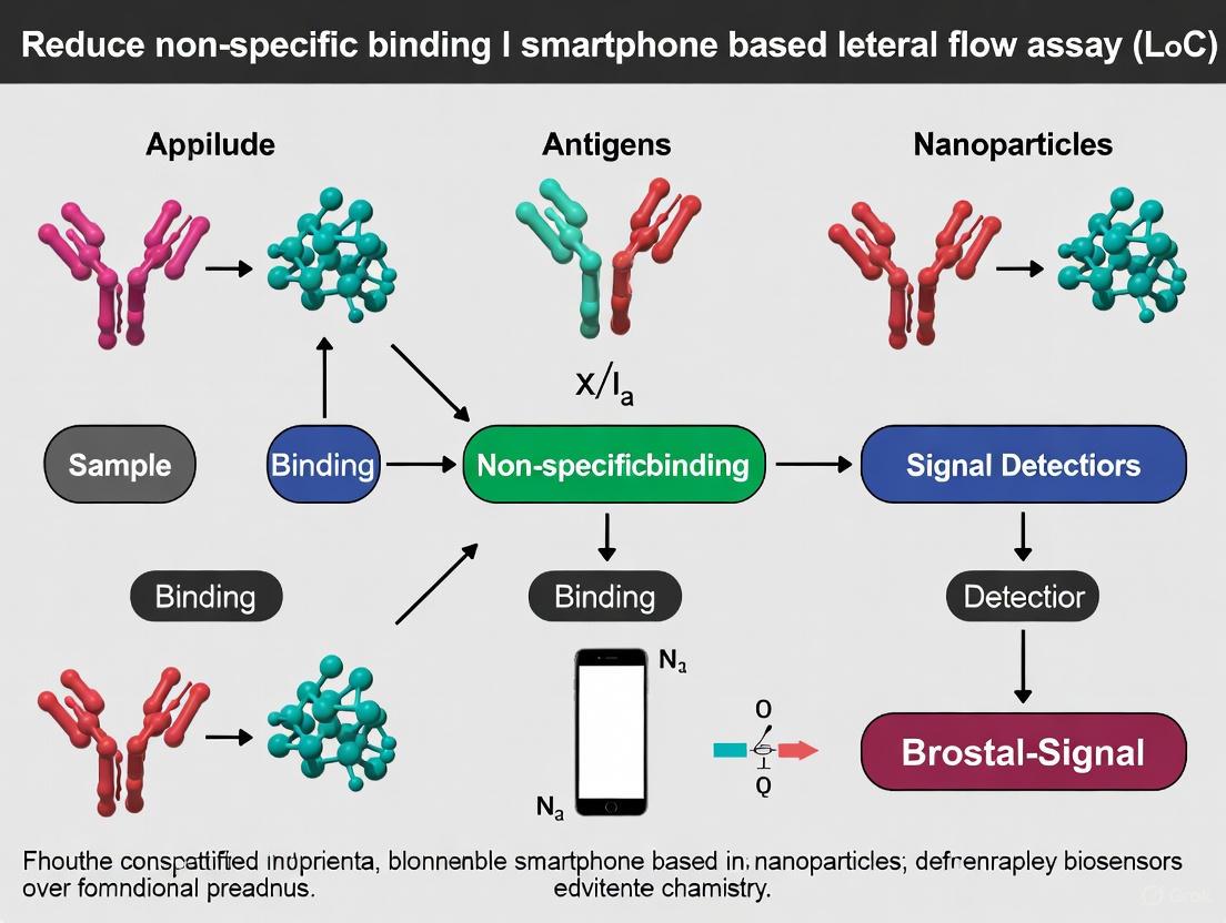
Non-Specific Binding (NSA or NSB) is a fundamental challenge in the development and deployment of biosensors, particularly for smartphone-based Lab-on-a-Chip (LoC) devices. NSB occurs when molecules present in a sample adhere to the sensor's surface through non-targeted interactions, rather than through specific recognition by the bioreceptor [17]. In the context of diagnostic biosensors, this phenomenon leads to elevated background signals that are often indistinguishable from the specific binding signal of the target analyte, resulting in diagnostic inaccuracies such as false positives and false negatives [17] [8].
For smartphone-based LoC biosensors, which aim to provide rapid, point-of-care testing, the implications of NSB are particularly severe. These devices are often designed for use with complex biological samples (e.g., blood, saliva, urine) which contain a multitude of proteins and other biomolecules that can contribute to NSB [17]. The presence of NSB can compromise the sensitivity, specificity, and reproducibility of these devices, ultimately creating significant barriers to their clinical validation and widespread adoption [17] [3]. Understanding and mitigating NSB is therefore not merely an optimization step, but a critical requirement for ensuring the reliability of diagnostic results.
Troubleshooting Guide: Identifying and Resolving NSB
Frequently Asked Questions (FAQs)
Q1: Our smartphone-based LoC biosensor consistently shows high background signal in control samples that do not contain the target analyte. What is the most likely cause? A1: A consistently high background signal is a classic indicator of significant Non-Specific Binding. The cause is likely the adsorption of non-target molecules (e.g., other proteins, lipids, or cellular debris from the sample matrix) onto the sensing surface. This can occur on the bioreceptor itself, on the substrate between bioreceptors, or on the sensor's transducer elements [17] [8].
Q2: We observe a strong signal, but subsequent validation with a reference method (e.g., ELISA) does not confirm the presence of the target. What does this suggest? A2: This discrepancy strongly suggests that your sensor is producing false-positive results. The signal is likely generated by NSB, where other components in the sample are binding to the sensor surface and generating a response similar to that of the specific target analyte [18] [3].
Q3: After initial successful testing with purified samples, the performance of our biosensor degrades significantly when using complex clinical samples (e.g., serum). Why? A3: Complex clinical samples like serum contain a high concentration and diversity of proteins (such as albumin and immunoglobulins) and other biomolecules. These samples greatly increase the potential for NSB, which can mask the specific signal, reduce the sensor's dynamic range, and raise its effective limit of detection [17] [19]. Your surface passivation method may be insufficient for real-world samples.
Q4: Can the physical design of our microfluidic LoC device influence NSB? A4: Yes. The materials used in the device (e.g., PDMS, plastics) can be inherently prone to protein adsorption. Furthermore, areas with low flow rates or stagnant zones can allow molecules to settle and adsorb non-specifically. Surface roughness at the micro-scale can also increase the available surface area for NSB [17].
Troubleshooting Flowchart: Diagnosing NSB Issues
The following diagram outlines a systematic workflow for diagnosing the root cause of NSB in your biosensing experiments.
Diagram 1: A systematic workflow for diagnosing the root cause of NSB.
Experimental Protocols for NSB Mitigation
This section provides detailed methodologies for the most effective and commonly used strategies to reduce NSB in biosensor development.
Surface Passivation with Protein Blockers
Principle: This passive method involves coating the sensor surface with a protein that adsorbs to non-specific binding sites, thereby "blocking" them and preventing the non-specific adsorption of other sample components [17] [8].
Detailed Protocol:
- After immobilizing your specific bioreceptor (e.g., antibody, aptamer) onto the sensor surface, rinse the surface with an appropriate buffer (e.g., Phosphate Buffered Saline - PBS).
- Prepare a 1-5% (w/v) solution of Bovine Serum Albumin (BSA) or casein in your running buffer. Filter sterilize the solution if necessary.
- Incubate the sensor surface with the BSA solution for 30-60 minutes at room temperature.
- Thoroughly rinse the surface with buffer to remove any unbound BSA.
- The sensor is now ready for use. The treated surface should be kept hydrated.
Considerations: BSA is a globular protein with domains of varying charge, making it effective at shielding a range of non-specific interactions [8]. However, ensure that the blocking protein does not interfere with the activity of your immobilized bioreceptor.
Optimization of Buffer Conditions
Principle: Adjusting the chemical environment of the sample and running buffer can minimize NSB driven by electrostatic and hydrophobic interactions [8].
Detailed Protocol:
- Adjust pH: Determine the isoelectric point (pI) of your target analyte and potential interfering proteins. Adjust your buffer to a pH that neutralizes the overall charge of your analyte or the sensor surface to reduce charge-based NSB. A common starting point is a pH near 7.4 (physiological), but optimization is required [8].
- Add Surfactants: Introduce a non-ionic surfactant like Tween 20 at a concentration of 0.01-0.1% (v/v) to your running buffer and sample diluent. This disrupts hydrophobic interactions [8].
- Increase Ionic Strength: Add salts such as NaCl (150-500 mM) to your buffer. The ions will shield electrostatic charges on proteins and the sensor surface, reducing charge-based NSB [8].
- Test these conditions systematically using a negative control to quantify the reduction in background signal.
Signal Discrimination via Sensor Design
Principle: Advanced sensor designs and data analysis can help distinguish the electronic or physical signature of specific binding from that of NSB.
Detailed Protocol (based on chemiresistive PEDOT sensors):
- Fabricate a conductometric transducer, for example using a vapor-phase polymerized interpenetrating network of P(EDOT-3TE) on a fabric substrate [3].
- Functionalize the sensor with your specific bioreceptor (e.g., Avidin for Biotin detection).
- Record the resistance (R) of the sensor in buffer to establish a baseline.
- Introduce the analyte solution and monitor the percent change in resistance (ΔR%) over time.
- Key Observation: In this specific system, specific binding (e.g., Biotin-Avidin) typically produces a negative ΔR% (resistance decrease), while non-specific binding (e.g., from Gliadin or Casein) produces a positive ΔR% (resistance increase) [3]. This opposite response can be used to flag and discount NSB events.
- This data can be further processed with machine learning classifiers (e.g., Random Forest) to automatically identify and filter out NSB-related signals [3].
Research Reagent Solutions for NSB Reduction
The table below summarizes key reagents used to combat NSB, their mechanisms of action, and their typical applications.
Table 1: Essential Reagents for Mitigating Non-Specific Binding in Biosensor Research.
| Reagent | Function & Mechanism | Typical Application & Concentration |
|---|---|---|
| Bovine Serum Albumin (BSA) | Protein blocker; physically adsorbs to vacant sites on the sensor surface, creating a hydrophilic barrier against NSB [17] [8]. | Incubation as a 1-5% (w/v) solution in buffer after bioreceptor immobilization [8]. |
| Tween 20 | Non-ionic surfactant; disrupts hydrophobic interactions between analytes and the sensor surface [8]. | Added to running buffers and sample diluents at 0.01-0.1% (v/v) [8]. |
| Sodium Chloride (NaCl) | Ionic salt; shields electrostatic charges via its ions, reducing charge-based attraction between proteins and the surface [8]. | Used in buffers at concentrations of 150-500 mM to suppress NSB [8]. |
| Casein | Milk protein blocker; similar to BSA, it passivates surfaces by covering sticky sites with an inert protein layer [17]. | Used as an alternative to BSA at 1-3% (w/v) concentration, especially in immunoassays [17]. |
| (3-Glycidyloxypropyl)trimethoxysilane (GOPS) | Crosslinker; provides a controlled chemical method to immobilize bioreceptors, reducing random orientation and denaturation that can exacerbate NSB [3]. | Used in vapor-phase or solution-phase functionalization to create a stable, oriented receptor layer [3]. |
Visualizing the NSB Mitigation Strategy Workflow
A holistic approach to tackling NSB involves a combination of strategies, as illustrated in the workflow below.
Diagram 2: A multi-faceted approach is required to effectively mitigate NSB and achieve a reliable biosensor.
For smartphone-based LoC biosensors aiming for clinical relevance, addressing Non-Specific Binding is not an optional refinement but a core component of the development process. The consequences of unmitigated NSB—diagnostic inaccuracies, false positives, and ultimately, a failure to achieve clinical validation—are severe [18] [19]. By systematically diagnosing the sources of NSB and implementing a combination of robust surface passivation, buffer optimization, and intelligent sensor design or data analysis, researchers can overcome these barriers. The protocols and reagents detailed in this guide provide a foundational toolkit for developing next-generation, field-deployable biosensors that are both sensitive and reliable.
Advanced Materials and Engineering Solutions to Minimize Non-Specific Interactions
Non-specific adsorption (NSA), also known as biofouling, presents a persistent challenge in the development of smartphone-based lab-on-chip (LoC) biosensors. NSA occurs when biomolecules irreversibly adsorb to sensing surfaces through physisorption, resulting in high background signals that are indiscernible from specific binding. This phenomenon decreases sensitivity, specificity, and reproducibility—critical parameters for point-of-care diagnostic devices [17]. For smartphone-based biosensors intended for use in resource-limited settings, effective antifouling strategies are particularly essential as these platforms aim to provide reliable analytical performance outside controlled laboratory environments [20] [21].
This technical support center article addresses specific experimental issues researchers encounter when implementing three leading antifouling surface chemistries: polyethylene glycol (PEG), poly(oligo(ethylene glycol) methacrylate) (POEGMA) brushes, and zwitterionic polymers. The guidance is framed within the context of reducing non-specific binding in smartphone-based LoC biosensors for healthcare monitoring [21] [22].
Antifouling Chemistry Comparison
The table below summarizes key characteristics of the three primary antifouling chemistries discussed in this guide.
Table 1: Comparison of Key Antifouling Surface Chemistries
| Chemistry | Antifouling Mechanism | Optimal Application Method | Key Advantages | Reported Performance Data |
|---|---|---|---|---|
| PEG | Forms a hydrated steric barrier that reduces protein adsorption [17] | "Grafting to" approach with linear polymers [23] | Well-established, commercially available, good antifouling properties | Significantly lower immobilized IgG density compared to pSBMA; >2-fold lower signal in wearable antigen capture [23] |
| POEGMA Brushes | Extended brush architecture creates thicker hydrated layer | Surface-initiated polymerization (ATRP) | Higher grafting density, enhanced steric hindrance, tunable properties | Information not available in search results |
| Zwitterionic Polymers (e.g., pSBMA) | Strong electrostatic interaction with water molecules creates a super-hydrophilic interface [17] | "Grafting to" approach [23] | Superior hydration, higher biomarker capture capacity, stability | Highest IgG density; >2-fold increase in signal for dengue NS1 capture vs. PEG [23] |
Troubleshooting Guide: FAQs and Solutions
Q1: My PEG-coated biosensor shows high non-specific adsorption despite proper surface functionalization. What could be causing this?
Potential Causes and Solutions:
- Oxidative Degradation: PEG chains are susceptible to oxidative degradation in ambient conditions. Ensure all procedures are performed in an oxygen-free environment when possible, and use fresh PEG solutions prepared immediately before functionalization.
- Inadequate Surface Density: Suboptimal grafting density creates gaps where nonspecific adsorption can occur. For "grafting to" approaches, increase polymer concentration and reaction time. Consider switching to surface-initiated polymerization for brush formations like POEGMA to achieve higher packing density.
- Molecular Weight Issues: The hydrodynamic radius of your PEG must be appropriate for your application. Compare polymers of equivalent molecular weight and hydrodynamic radius for accurate assessment, as performance varies significantly with these parameters [23].
Q2: Why does my zwitterionic polymer coating (pSBMA) show excellent antifouling but poor biomarker capture efficiency?
Investigation and Resolution:
- Verify Immobilization Density: This is the most likely cause. Interestingly, research shows that pSBMA surfaces can achieve significantly higher densities of immobilized capture antibodies (e.g., IgG) compared to PEG surfaces while maintaining comparable nonspecific adsorption levels [23].
- Check Bioconjugation Strategy: Ensure your method for attaching recognition elements (antibodies, aptamers) to the zwitterionic coating does not compromise the antifouling properties. Use site-specific conjugation chemistry to orient recognition elements properly.
- Confirm Antifouling Performance: Re-test the antifouling efficacy in complex media (e.g., diluted plasma). If NSA has increased, the conjugation process may have damaged the polymer layer.
Q3: How do I select between PEG and zwitterionic polymers for my specific smartphone-based biosensor?
Decision Framework:
- For Maximum Signal Intensity: Choose zwitterionic polymers (e.g., pSBMA). Direct comparisons show pSBMA-coated devices capture significantly more target analyte (>2-fold increase in signal for dengue NS1) compared to PEG-coated devices with equivalent antifouling performance [23].
- For Established Protocols: PEG remains a viable option with extensive literature support, though it may yield lower signal intensity.
- Consider the Sensing Environment: Both chemistries demonstrate comparable antifouling behavior in complex environments including single protein solutions, diluted plasma, and when applied to biological tissues [23].
- Sample Type: The choice might be influenced by your specific sample matrix (sweat, tears, saliva, ISF). Test both coatings with your actual sample to determine the best performer [22].
Q4: What are the best practices for characterizing antifouling performance on my sensor surface?
Validation Protocol:
- Use Complex Media for Testing: Beyond single-protein solutions (e.g., BSA), validate performance in diluted plasma or serum to simulate real-world conditions [23].
- Benchmark Against Standards: Compare your results against well-characterized surfaces like bare gold/silicon and known effective coatings.
- Employ Multiple Techniques:
- Surface Plasmon Resonance (SPR): Label-free, real-time monitoring of adsorption.
- Fluorescence Microscopy: After exposure to fluorescently-tagged proteins, measure nonspecific adhesion.
- Electrochemical Impedance Spectroscopy (EIS): Monitor changes in charge transfer resistance due to fouling.
Detailed Experimental Protocols
Protocol 1: Functionalization with Zwitterionic pSBMA via "Grafting To" Approach
This protocol is adapted from methods used to create wearable biosensors with superior antigen capture capability [23].
Research Reagent Solutions:
Table 2: Essential Reagents for pSBMA Grafting
| Reagent | Function | Notes |
|---|---|---|
| Sulfobetaine methacrylate (SBMA) monomer | Polymer building block | Provides zwitterionic properties |
| Amine-reactive crosslinkers | Links polymer to surface | EDC/NHS chemistry common |
| Amine-modified substrate | Surface for functionalization | Polycarbonate, gold, or silicon |
| Oxygen scavengers | Prevents polymerization inhibition | Required for free radical polymerization |
Procedure:
- Surface Activation: Create amine groups on your substrate (e.g., polycarbonate array or sensor chip). For polycarbonate, this may involve hydrolysis or plasma treatment.
- Polymer Synthesis: Prepare poly(sulfobetaine methacrylate) (pSBMA) with controlled molecular weight and hydrodynamic radius. Purify thoroughly.
- Surface Coupling: React the pre-formed pSBMA polymer with the activated surface using a "grafting to" approach. Utilize amine-reactive end groups on the polymer and amine groups on the surface with EDC/NHS chemistry.
- Washing and Validation: Rinse extensively with deionized water and appropriate buffers to remove physisorbed polymer. Characterize the coating thickness (e.g., with ellipsometry) and validate antifouling performance against negative controls.
Protocol 2: Integrating Antifouling Coatings into Smartphone-based Biosensors
This protocol outlines the workflow for developing a complete smartphone-based biosensing platform incorporating antifouling surface chemistry, drawing from recent advances in the field [20] [21] [22].
Procedure:
- Substrate Preparation: Select and clean your sensor substrate (e.g., SPR chip, electrode). Common substrates include gold for SPR sensors and carbon or gold for electrochemical sensors.
- Antifouling Coating Application: Follow Protocol 1 (for pSBMA) or established protocols for PEG/POEGMA to apply the chosen antifouling chemistry to the sensor surface.
- Biorecognition Element Immobilization: Immobilize specific capture probes (antibodies, aptamers) onto the functionalized surface. Ensure orientation is controlled to maximize binding site availability.
- Microfluidic Integration and Smartphone Coupling: Integrate the functionalized sensor into a microfluidic system, often 3D-printed [20]. Couple this with the smartphone-based detection system (optical or electrochemical) [21].
- Validation: Test the complete system with target analytes in relevant biological fluids (e.g., sweat, saliva, plant-based milk models) to establish sensitivity (LOD) and confirm reduction of NSA [20].
The Scientist's Toolkit: Essential Research Reagents
Table 3: Key Reagent Solutions for Antifouling Biosensor Research
| Category | Specific Examples | Function in Research |
|---|---|---|
| Antifouling Polymers | Polyethylene Glycol (PEG), Poly(sulfobetaine methacrylate) (pSBMA), Poly(carboxybetaine methacrylate) (pCBMA) | Form hydrated layers that resist non-specific protein adsorption [23] [17] |
| Surface Activation Reagents | EDC, NHS, Sulfo-SMCC, (3-Aminopropyl)triethoxysilane (APTES) | Create functional groups (-COOH, -NH₂) on sensor surfaces for polymer attachment |
| Characterization Tools | Surface Plasmon Resonance (SPR), Ellipsometry, Fluorescence Microscopy | Quantify coating thickness, density, and antifouling performance [17] |
| Blocking Agents | Bovine Serum Albumin (BSA), Casein, Milk Proteins | Traditional physical blockers for passive NSA reduction, often used as benchmarks [17] |
| Smartphone Integration Components | 3D-printed microfluidic chips, dark boxes, optical filters, portable potentiostats | Enable portable, point-of-care operation of the biosensing platform [20] [21] |
Frequently Asked Questions (FAQs)
Q1: What are the most effective nanomaterials for minimizing non-specific binding in smartphone-based LoC biosensors? Non-specific binding (NSB) is a major challenge that can severely compromise the accuracy of your biosensor. The most effective nanomaterials act as both sensitive transducers and physical or chemical shields against interference. Key materials include:
- Graphene and its derivatives: Their large, delocalized π-electron system allows for efficient functionalization with biorecognition elements, creating a uniform surface that leaves fewer sites for non-specific interactions [24]. The choice between pristine graphene (Gr), graphene oxide (GrO), and reduced graphene oxide (rGrO) is critical, as their surface chemistry and conductivity differ significantly [25].
- Metal-Organic Frameworks (MOFs): MOFs offer tunable porosity and high surface area, enabling precise molecular sieving that can physically block larger interferents from reaching the transducer surface while allowing the target analyte to interact [26].
- Functionalized Gold Nanoparticles (AuNPs): AuNPs provide a versatile platform for creating dense layers of biorecognition elements (e.g., antibodies, aptamers). This high packing density sterically hinders the adsorption of non-target molecules. Their strong optical properties also enhance signal-to-noise ratios in optical detection schemes [16].
Q2: How can I functionalize a graphene surface to improve its selectivity for my target biomarker? A controlled, multi-step functionalization process is essential for creating a selective and low-fouling graphene surface [24]. The standard workflow is as follows:
- Pre-treatment: Clean the graphene surface with acetone or phosphate-buffered saline (PBS) to remove manufacturing residues and contaminants [24].
- Functionalization: Introduce linker molecules to the surface. This can be achieved via:
- π–π stacking: Using molecules like 1-pyrenebutyric acid N-hydroxysuccinimide ester (PBSE) that adsorb onto the graphene lattice via π-electron interactions [24].
- Covalent bonding: For graphene oxide (GrO), exploit its oxygen-containing groups (e.g., carboxyl) to form covalent bonds with linkers using EDC/NHS chemistry [24].
- Immobilization: Covalently attach the specific bioreceptors (e.g., antibodies, DNA aptamers) to the activated linker molecules on the surface [24].
- Blocking: This is a critical step for reducing NSB. Passivate any remaining unreacted, reactive sites on the graphene and linker molecules with inert proteins (e.g., Bovine Serum Albumin - BSA) or small molecules like ethanolamine [24].
- Washing: Finally, wash the functionalized sensor with PBS or deionized water to remove any unbound molecules [24].
Q3: My smartphone-based optical biosensor shows high background noise. Is this an issue with the nanomaterial or the device? High background noise can originate from either or both areas. A systematic troubleshooting approach is recommended:
- Nanomaterial & Chemistry:
- Insufficient Blocking: This is a primary cause of NSB. Re-optimize your blocking step by testing different blocking agents (BSA, casein, commercial blocking buffers) and incubation times [24].
- Non-optimized Nanomaterial Density: An overly dense layer of nanomaterials (e.g., AuNPs) can cause light scattering, while a sparse layer leads to poor signal. Titrate the concentration of your nanomaterial during sensor fabrication [16].
- Smartphone & Device Integration:
- Ambient Light Leakage: Ensure your microfluidic chip or sensor cartridge is perfectly aligned and sealed within the smartphone dongle or attachment to prevent external light from interfering with the camera's readout [16].
- Inconsistent Calibration: Implement a calibration protocol that uses a built-in standard or reference channel for every measurement to account for variations between different smartphones and ambient conditions [16].
Q4: Which blocking agent should I use for a sensor designed to detect proteins in human serum? The choice of blocking agent depends on the sensor surface and the sample matrix. For complex biofluids like serum, a multi-pronged approach is often best.
- BSA (1-5% w/v): A universal standard, effective at blocking a wide range of hydrophobic and some hydrophilic interactions. It is a good first choice [24].
- Casein (1-3% w/v): Excellent for blocking non-specific protein interactions, particularly in immunoassays. It can be more effective than BSA in some cases due to its different protein structure.
- Synergistic Mixtures: Commercial blocking buffers often contain a mix of proteins (like BSA and casein) along with polymers and detergents to provide broader protection against various NSB mechanisms.
- Pro-Tip: Always prepare your blocking solution in the same buffer used for your assays (e.g., PBS) and filter it (0.2 µm) before use to remove aggregates that could deposit on your sensor.
Troubleshooting Guides
Issue 1: Low Signal-to-Noise Ratio in Electrochemical Graphene FET (GFET) Sensors
A low signal-to-noise ratio (SNR) indicates either a weak target signal, high background interference, or both.
| Probable Cause | Diagnostic Steps | Solution & Recommended Protocols |
|---|---|---|
| High NSB on Graphene Surface | Measure sensor response in pure analyte buffer vs. spiked serum/plasma. A large signal in blank serum indicates NSB. | Re-optimize the functionalization protocol. Ensure the blocking step is performed after bioreceptor immobilization. Test the efficacy of different blocking agents (e.g., BSA vs. casein) using a control sensor without the bioreceptor [24]. |
| Poor Bioreceptor Immobilization | Characterize the surface after each functionalization step using techniques like Raman spectroscopy or XPS to confirm the presence of bioreceptors. | Standardize the immobilization conditions (pH, ionic strength, time). For GFETs, ensure the bioreceptor is immobilized close enough to the surface to effectively gate the channel upon binding [25] [24]. |
| Inhomogeneous Nanomaterial Film | Inspect the graphene/electrode surface under SEM or AFM. Check for cracks or aggregations. | Switch to a more reproducible deposition method like electrospraying or spin-coating. Use surfactants or functionalization to improve nanomaterial dispersion prior to deposition [25]. |
Issue 2: Signal Drift and Inconsistent Readings Between Runs in Smartphone LoC
Signal drift makes calibration unreliable and results non-reproducible.
| Probable Cause | Diagnostic Steps | Solution & Recommended Protocols |
|---|---|---|
| Unstable Nanomaterial Immobilization | Perform a stability test by continuously measuring the baseline signal in buffer over several hours. | Improve the adhesion between the nanomaterial and the transducer surface. Use stronger linkers like silane chemistry for oxide surfaces or dopamine-based anchors for versatile substrates [27]. |
| Calibration Drift | Regularly measure a standard solution with a known concentration. Track the signal output for this standard over time and across different smartphone devices. | Implement a dual-referencing strategy:1. Internal Reference: Use a functionalized channel that lacks the specific bioreceptor to measure and subtract NSB.2. On-board Calibrant: Incorporate a calibration standard within the microfluidic chip that is analyzed with every run [16] [28]. |
| Biofouling in Complex Media | Run multiple assay cycles using the same sensor without regeneration. A gradual performance decline indicates fouling. | Integrate anti-fouling nanomaterials like zwitterionic polymer coatings or hydrophilic hydrogels (e.g., PEG-based) around the sensing area. These materials create a hydration layer that repels proteins [29] [27]. |
Table 1: Performance Comparison of Nanomaterials for NSB Reduction in Biosensing.
| Nanomaterial | Key Mechanism for NSB Reduction | Typical LoD Improvement | Best Suited Sensing Modality | Key Challenge |
|---|---|---|---|---|
| Graphene (Gr) | High surface area; efficient bioreceptor packing [25] [24]. | Up to 10-fold vs. bare electrode [24]. | GFET, Electrochemical [25] [24] | No intrinsic bandgap; requires functionalization [25]. |
| Graphene Oxide (GrO) | Abundant oxygen groups for covalent bioreceptor attachment [25] [24]. | High (picomolar range for fluorescence) [16]. | Fluorescence, Colorimetric [25] | Low electrical conductivity [25]. |
| Gold Nanoparticles (AuNPs) | Steric hindrance from dense bioreceptor layers; plasmonic enhancement [16]. | ~50% signal amplification efficiency [16]. | Optical (SPR, Colorimetric), Electrochemical [16] | Potential aggregation; long-term stability [16]. |
| MOFs | Tunable porosity for size-exclusion of interferents [26]. | Picomolar LODs demonstrated [26]. | Fluorescent, Electrochemical [26] | Stability in aqueous/biological media [26]. |
| MXenes | Hydrophilic surface with tunable termination groups [25] [27]. | High sensitivity in wearable enzymatic sensors [25]. | Electrochemical, Wearable Sensors [25] [27] | Susceptible to oxidation in aqueous media [25]. |
Table 2: Efficacy of Common Blocking Agents Against Different Biofluids.
| Blocking Agent | Mechanism of Action | Recommended For (Sample Matrix) | Notes & Limitations |
|---|---|---|---|
| Bovine Serum Albumin (BSA) | Adsorbs to hydrophobic and charged sites [24]. | Serum, Plasma, Buffer | Universal standard; may contain trace impurities that cause NSB. |
| Casein | Forms a protein layer that masks surfaces. | Serum, Milk, Cellular Lysates | Very effective for immunoassays; can be less soluble than BSA. |
| Skim Milk | Complex mixture of proteins (mostly casein). | Serum, General Use | Inexpensive and effective; but can introduce bacterial contamination if not used fresh. |
| Poly(ethylene glycol) (PEG) | Creates a hydrated, steric barrier ("brush" layer). | Serum, Plasma, Whole Blood | Excellent for reducing protein adsorption; requires covalent grafting. |
| Ethanolamine | Blocks reactive NHS-ester groups. | Any, following NHS-ester chemistry | Small molecule; used specifically to deactivate succinimide esters. |
Experimental Protocol: MOF-Enhanced Fluorescence Sensor for Serum Analysis
This protocol details the creation of a biosensor using a Zeolitic Imidazolate Framework-8 (ZIF-8) MOF to reduce NSB in the fluorescent detection of a DNA biomarker.
1. Synthesis of ZIF-8 Nanoparticles:
- Prepare two separate solutions: Solution A (50 mL methanol with 5 mmol 2-methylimidazole) and Solution B (50 mL methanol with 1.25 mmol zinc acetate dihydrate).
- Rapidly pour Solution A into Solution B under vigorous stirring. Allow the reaction to proceed for 1 hour at room temperature.
- Centrifuge the resulting white precipitate at 10,000 rpm for 10 minutes. Wash the pellet three times with fresh methanol and then re-disperse in deionized water [26].
2. Functionalization of MOF with DNA Probe:
- Incubate the aqueous ZIF-8 nanoparticle solution (1 mg/mL) with a 5' amino-modified DNA probe (5 µM) for 12 hours at 4°C on a rotator.
- The DNA probes will coordinate with the Zn²⁺ sites on the MOF surface, creating a dense, oriented layer of capture probes [26].
3. Sensor Assembly and Blocking:
- Spot the DNA-functionalized ZIF-8 solution onto the sensing area of your smartphone LoC device.
- After drying, incubate the sensor with a 1% BSA solution in PBS for 1 hour at 37°C to block any remaining non-specific sites on the MOF and device substrate [26] [24].
- Rinse thoroughly with a washing buffer (e.g., PBS with 0.05% Tween 20) to remove unbound BSA.
4. Detection and Signal Acquisition:
- Introduce your sample (e.g., serum spiked with target DNA) to the sensor. The target will hybridize with the probe, and a fluorescent intercalating dye (e.g., SYBR Green) can be added.
- Use the smartphone camera housed in a dark box to capture the fluorescence emission. The ZIF-8 MOF will concentrate the reaction locally and its porous structure will help exclude larger serum proteins, thereby enhancing the signal and reducing background from NSB [26] [16].
Experimental Workflow Visualization
Sensor Fabrication and Assay Workflow
The Scientist's Toolkit: Essential Research Reagents
Table 3: Key Reagent Solutions for Fabricating NSB-Resistant Nanobiosensors.
| Reagent / Material | Function / Purpose | Example in Protocol |
|---|---|---|
| Graphene Oxide (GrO) Dispersion | Provides a highly functionalizable 2D nanomaterial platform with oxygen groups for covalent chemistry [25] [24]. | Used as the transducer layer in electrochemical or FET sensors. |
| Gold Nanoparticle (AuNP) Colloid | Serves as a core for bioreceptor immobilization and provides plasmonic signal enhancement [16]. | Functionalized with thiolated aptamers for optical detection. |
| ZIF-8 MOF Nanoparticles | Creates a porous shield around the sensing element, enabling size-selective analyte access [26]. | Synthesized as a nano-carrier for DNA probes in fluorescence assays. |
| EDC/NHS Coupling Kit | Activates carboxyl groups (-COOH) on nanomaterials/surfaces for covalent conjugation to amine-bearing bioreceptors [24]. | Used to immobilize antibodies on GO surfaces. |
| PBSE (1-Pyrenebutyric Acid NHS Ester) | A π-π stacking linker for non-covalent functionalization of pristine graphene surfaces [24]. | Applied to GFET sensors before antibody attachment. |
| BSA (Bovine Serum Albumin) | A standard blocking agent to passivate unreacted surface sites and minimize NSB [24]. | Used as a 1-5% solution in PBS after bioreceptor immobilization. |
| PEG-SH (Thiolated Polyethylene Glycol) | Forms an anti-fouling self-assembled monolayer on gold surfaces to resist protein adsorption [27]. | Mixed with thiolated probes on AuNP surfaces to create a bio-inert background. |
Frequently Asked Questions (FAQs)
Q1: What are the most effective surface chemistries to minimize non-specific binding (NSB) in my microfluidic biosensor?
Non-specific binding (NSB) can be mitigated through several surface chemistry approaches. A combination of techniques often yields the best results. Silanization using reagents like APTES introduces functional groups (e.g., -NH₂, -SH) that create a stable, reactive surface for subsequent bioconjugation [30]. PEGylation is highly effective for creating a hydrophilic, anti-fouling surface that reduces non-specific protein and cell attachment [30]. Furthermore, click chemistry offers a highly efficient and specific method for attaching biomolecules to surfaces, creating stable linkages that minimize unwanted interactions [30]. For the highest specificity, consider combining these methods—for example, using a silanized surface as a base for a PEGylated layer that is further functionalized with capture probes via click chemistry [30].
Q2: My hydrophobic barriers are failing, leading to cross-contamination between assay zones. What could be the cause?
Hydrophobic barrier failure can stem from several issues in the fabrication process. First, inadequate surface preparation can prevent the hydrophobic polymer from properly adhering. Ensure the substrate (e.g., glass) is thoroughly cleaned with piranha solution or plasma treatment to remove all contaminants [30]. Second, improper functionalization is a common culprit. The polymerization mixture for the hydrophobic stripes (e.g., containing butyl methacrylate (BMA) and ethylene dimethacrylate (EDMA)) must be precisely formulated and UV-cured correctly [31]. Finally, verify that the post-patterning treatment, such as a brief immersion in NaOH to remove unwanted methacrylate groups, has been performed successfully, as this is critical for final barrier integrity and subsequent PDMS-glass bonding [31].
Q3: How can I integrate sample preparation to reduce NSB from complex biological samples like blood?
Integrating sample preparation is crucial for handling complex samples. Magnetic bead-based extraction is a prominent method suitable for microfluidic platforms. Magnetic beads coated with functional groups can bind nucleic acids or other targets from a lysed sample. By applying an external magnetic field, the beads—and the purified target—can be moved through different washing droplets to remove impurities, proteins, and other NSB-causing agents before elution into a detection zone [32]. Silica-based solid-phase extraction is another effective method, where a silica membrane or pillar array within the microchip binds nucleic acids in the presence of a chaotropic salt, allowing contaminants to be washed away before clean elution [32]. Both methods concentrate the target analyte and significantly reduce background interferents.
Q4: The signal-to-noise ratio on my smartphone-based sensor is poor. How can I improve it for multiplexed detection?
Improving the signal-to-noise ratio involves optimizing both the chemistry and the optics. To reduce background noise, ensure you have a robust non-fouling surface coating, such as commercial polymers (e.g., SuSoS AziGrip4), which form a stable hydrostatic barrier to prevent unspecific protein attachment [30]. For optical clarity in smartphone detection, use a glass substrate for your microfluidic chip due to its superior optical clarity and low autofluorescence compared to many plastics [30]. Structurally, designs based on hydrophobic patterning can achieve an interaction area greater than 95%, minimizing dead space and unwanted surface interactions that contribute to noise [31]. For your smartphone, utilize its built-in capabilities like the camera for colorimetric detection and wireless peripherals (Bluetooth, NFC) for data transfer to streamline the sensing platform and reduce external electronic noise [33].
Troubleshooting Guides
Troubleshooting Hydrophobic Barrier Patterning
| Problem | Possible Cause | Solution |
|---|---|---|
| Incomplete or non-uniform hydrophobic patterning | Pre-polymer solution not filling the PDMS mold via capillary action. | Ensure the PDMS slab has a clean, open channel structure. Check for debris blocking the channels. Use fresh pre-polymer solution [31]. |
| Poor adhesion of hydrophobic polymer to substrate | Glass surface not properly methacrylated. | Follow the methacrylation protocol rigorously: treat with NaOH and HCl, then incubate with the silane mixture (e.g., 3-(trimethoxysilyl)propyl methacrylate) for a full hour [31]. |
| Barriers are not sufficiently hydrophobic | Incorrect UV curing time or intensity. | Cure the filled channels with UV light (e.g., 265 nm, 15 mW/cm²) for the recommended duration (e.g., 10 minutes), ensuring the coverslip faces the light source [31]. |
Troubleshooting High Non-Specific Binding
| Problem | Possible Cause | Solution |
|---|---|---|
| High background signal across the entire channel | Lack of a passivating, non-fouling surface layer. | Apply a PEGylation treatment to the entire surface before patterned functionalization. This creates a hydrophilic, anti-fouling background [30]. |
| NSB even after PEGylation | Non-specific protein or biofilm attachment on specific regions. | Integrate specialized non-fouling polymer coatings like SuSoS AziGrip4 or PAcrAm, which form a highly durable hydrostatic barrier that withstands mechanical stress and ethanol sterilization [30]. |
| NSB from sample impurities | Unpurified sample contains interferents. | Integrate an on-chip sample preparation module, such as a magnetic bead-based nucleic acid purification system, to isolate the target analyte before it enters the detection zone [32]. |
Experimental Protocols & Data
Protocol 1: Fabricating Hydrophobic Barriers via Soft Lithography
This protocol is adapted from a method for creating 3D microfluidic cell culture platforms with >95% interaction area [31].
- Substrate Preparation: Treat a glass coverslip with 1M NaOH for 1 hour, rinse with DI water, then immerse in 1M HCl for 30 minutes. Rinse and dry with N₂ gas.
- Methacrylation: Immediately functionalize the clean glass by incubating with a mixture of ethanol, 3-(trimethoxysilyl)propyl methacrylate, and glacial acetic acid (5:2:3 ratio) for 1 hour at room temperature. Rinse with acetone and dry with N₂.
- Prepare Hydrophobic Pre-polymer: Mix 30 wt% butyl methacrylate (BMA), 20 wt% ethylene dimethacrylate (EDMA), 50 wt% 1-decanol, and 1-6 wt% (relative to BMA/EDMA) photoinitiator DMPAP.
- Patterning: Place a PDMS slab with your desired channel pattern onto the methacrylated glass. Fill the channels with the hydrophobic pre-polymer via capillary action.
- UV Curing: Irradiate the assembly with UV light (265 nm, 15 mW/cm²) for 10 minutes with the glass side facing the source.
- Post-processing: Rinse the patterned coverslip thoroughly with ethanol. Immerse in 1M NaOH for 5 minutes to remove excess methacrylate, then wash with DI water and dry with N₂. The substrate is now ready for bonding to a secondary PDMS slab.
Protocol 2: Surface PEGylation for NSB Reduction
This protocol outlines a general method for applying anti-fouling PEG coatings [30].
- Surface Activation: Clean the substrate (glass or silicon) using an oxygen plasma treatment or piranha solution to generate reactive hydroxyl groups.
- Silane Priming (Optional but recommended): Apply an aminosilane (e.g., APTES) or an epoxysilane to the surface to introduce functional groups for covalent PEG attachment.
- PEG Application: Utilize wet-chemistry methods such as spin-coating or immersion to apply the PEG solution (e.g., biotin-PEG or azide-PEG) to the activated surface.
- Curing: Allow the PEG layer to form a stable coating. This may involve incubation at a specific temperature or exposure to UV light for photoinduced PEGylation if using a photosensitive PEG derivative.
Quantitative Data on Performance
Table 1: Comparison of Surface Functionalization Methods for NSB Reduction
| Functionalization Method | Key Reagent(s) | Primary Function | Reported Outcome/Performance |
|---|---|---|---|
| Silanization | APTES, Epoxysilanes | Introduces reactive groups (-NH₂, epoxy) for biomolecule immobilization [30]. | Creates a stable, reactive surface for subsequent conjugation. Foundation for further functionalization [30]. |
| PEGylation | Polyethylene glycol (PEG) | Creates a hydrophilic, anti-fouling surface to reduce non-specific binding [30]. | Significantly reduces background noise in diagnostics; enhances signal-to-noise ratio [30]. |
| Click Chemistry | Azides, Alkynes | Enables highly efficient and specific biomolecule attachment via cycloaddition [30]. | Provides precise and stable functionalization, improving assay specificity [30]. |
| Structured Silanization (IMT/CSEM) | Not Specified | Wafer-scale patterned functionalization on glass [30]. | Higher functionalization capacity than conventional plastics; superior optical performance [30]. |
| Hydrophobic Patterning | BMA-EDMA polymer | Forms physical barriers to contain hydrogels and define flow paths [31]. | Achieves >95% effective interaction area between cells/ECM, minimizing artificial obstructions [31]. |
| Magnetic Bead DNA Cleanup | DNA-binding magnetic beads | On-chip buffer exchange and sample purification [34]. | Average DNA recovery efficiency of 80% ± 4.8%, effectively removing enzymes and salts [34]. |
Workflow Visualizations
Diagram 1: Hydrophobic Patterning and Assay Integration
Diagram 2: Surface Chemistry Strategy for NSB Reduction
The Scientist's Toolkit: Research Reagent Solutions
Table 2: Essential Materials for Microfluidic NSB Reduction
| Reagent/Material | Function | Example Use Case |
|---|---|---|
| APTES ((3-Aminopropyl)triethoxysilane) | Aminosilane used to introduce primary amine (-NH₂) groups onto glass/silica substrates for covalent biomolecule binding [30]. | Foundation for building functionalized surfaces; enables EDC/NHS coupling of proteins or DNA [30]. |
| PEG Derivatives (e.g., Biotin-PEG, Azide-PEG) | Polyethylene glycol polymers create a hydrophilic, anti-fouling layer that resists non-specific protein adsorption [30]. | Applied after silanization to passivate the surface and drastically reduce background signal [30]. |
| BMA and EDMA (Butyl Methacrylate, Ethylene Dimethacrylate) | Monomers used to create a hydrophobic polymer matrix for patterning physical barriers within microchannels [31]. | Formulated into a pre-polymer solution to create hydrophobic walls that define hydrogel regions or fluidic paths [31]. |
| Magnetic Beads (e.g., silica-coated) | Solid-phase support for binding nucleic acids or proteins from a complex sample lysate, enabling purification and concentration [32]. | Integrated into microfluidic systems for automated sample prep (lysis, binding, washing, elution) to remove NSB contaminants [32]. |
| Non-fouling Polymer Coatings (e.g., SuSoS AziGrip4) | Commercial coatings that form a highly durable hydrostatic barrier to prevent unspecific protein, cell, and biofilm attachment [30]. | Used in critical sensing regions of the chip to prevent clogging and improve signal-to-noise ratios in complex media [30]. |
This technical support center provides targeted guidance for researchers integrating high-affinity biorecognition elements into smartphone-based Lab-on-a-Chip (LoC) biosensors. A primary challenge in this field is reducing non-specific binding (NSB)—the unwanted adsorption of non-target molecules to sensor surfaces—which can severely compromise detection sensitivity and specificity, particularly in complex sample matrices [13] [35]. This resource offers troubleshooting guides and FAQs focused on the use of aptamers and phages to achieve enhanced specificity, framed within the context of a thesis dedicated to advancing point-of-care diagnostic platforms.
Biosensors are defined as analytical devices that combine a biorecognition element with a physicochemical transducer to produce a measurable signal [36] [37]. The selection of the biorecognition element is paramount, as it directly influences key performance characteristics such as sensitivity, selectivity, and reusability [36]. The following table summarizes the core characteristics of major biorecognition elements.
Table 1: Comparison of Key Biorecognition Elements for Biosensors
| Biorecognition Element | Type | Target Examples | Key Advantages | Key Limitations |
|---|---|---|---|---|
| Antibody [36] | Natural (Protein) | Proteins, Peptides | High specificity and affinity; well-established protocols. | Animal-based production (costly, time-consuming); batch-to-batch variation; sensitive to environment. |
| Aptamer [36] [38] [39] | Synthetic (Nucleic Acid) | Ions, small molecules, proteins, cells | Chemically synthesized (low cost, high batch uniformity); small size for better penetration; can be regenerated. | Susceptible to nuclease degradation; requires specialized selection process (SELEX). |
| Phage [36] | Pseudo-Natural | Proteins, Peptides | Can display a vast diversity of peptide binders; relatively stable. | Limited to protein-protein interactions; propagation requires host bacteria. |
| Molecularly Imprinted Polymer (MIP) [36] | Synthetic (Polymer) | Small molecules, proteins | High physical/chemical stability; synthetic production. | Can suffer from heterogeneity in binding sites; complex optimization. |
| Enzyme [36] | Natural (Protein) | Substrates, Inhibitors | Catalytic amplification of signal. | Specific to enzyme substrates; stability can be an issue. |
Troubleshooting Guide: FAQs on Reducing Non-Specific Binding
FAQ 1: Why is non-specific binding (NSB) a particularly critical issue for smartphone-based LoC biosensors?
NSB leads to a high background signal, which obscures the specific signal from the target analyte. In smartphone-based LoC systems, which often rely on miniaturized optics and simplified electronics, this signal-to-noise ratio is especially critical. A small degree of NSB can significantly reduce the sensor's apparent sensitivity and limit of detection (LOD), making it unreliable for detecting low-abundance biomarkers in real-world samples like blood or serum [13] [35]. Furthermore, in a microfluidic LoC format, the high surface-to-volume ratio amplifies the impact of any surface fouling.
FAQ 2: What are the primary strategies to suppress NSB on electrode surfaces in electrochemical biosensors?
Strategies to minimize NSB are broadly classified into physical and chemical surface modifications [13].
- Physical Modifications: These involve attaching molecules directly to the surface to create a physical barrier.
- Blocking Buffers: Solutions of inert proteins (e.g., Bovine Serum Albumin - BSA) or surfactants are used to occupy any remaining reactive sites on the sensor surface after immobilization of the biorecognition element.
- Chemical Modifications: These create a more controlled, covalently bound surface chemistry that resists protein adsorption.
- Self-Assembled Monolayers (SAMs): Ordered layers of molecules (e.g., alkanethiols on gold) can be engineered to present functional groups that resist protein adhesion.
- Polymer Coatings: Polymers like polyethylene glycol (PEG) or oligo(ethylene glycol) are the gold standard for creating a non-fouling, "brush-like" surface that repels proteins through steric and hydrative forces [13].
- Diazonium Salt Chemistry: Provides a stable carbon-based layer on electrode surfaces, which can be further functionalized.
FAQ 3: How do aptamers help in reducing NSB compared to traditional antibodies?
Aptamers offer several inherent advantages that can be leveraged to minimize NSB:
- Synthetic Control: Aptamers are chemically synthesized, allowing for precise and reproducible modification with specific functional groups (e.g., thiol, amino) that enable oriented and dense immobilization on sensor surfaces. A well-ordered monolayer is less prone to NSB than the random orientation often seen with adsorbed antibodies [35] [39].
- Size and Stability: Their small size reduces steric hindrance and the "footprint" for non-specific interactions. Furthermore, their stability allows them to undergo rigorous regeneration using harsh conditions (e.g., low pH, denaturants) to strip off any non-specifically bound contaminants without permanent damage, which is often not possible with antibodies [38] [39].
- Tailored Selection: The SELEX process used to select aptamers can be designed to include counter-selection steps against common interferents present in the sample matrix. This proactively enriches for aptamers with high specificity and low cross-reactivity [40] [41].
FAQ 4: What are common issues with aptamer stability and how can they be mitigated?
A primary concern is the susceptibility of natural DNA/RNA aptamers to degradation by nucleases present in biological samples [42] [39].
Mitigation Strategies:
- Chemical Modification: Incorporate modified nucleotides into the aptamer sequence during synthesis, such as 2'-fluoro or 2'-O-methyl ribose substitutions in RNA aptamers, or use locked nucleic acids (LNA), which dramatically increase resistance to nucleases [36] [41].
- Post-SELEX Optimization: After selection, the full-length aptamer can be "truncated" to its minimal functional sequence, which often improves not only stability but also binding affinity and kinetics [41].
- Use of Peptide Nucleic Acids (PNA): PNAs, with their uncharged peptide backbone, are completely resistant to nucleases and can form very stable duplexes, though they require their own specialized selection processes [36].
Experimental Protocols
Protocol 1: Immobilization of Thiol-Modified Aptamers on Gold Surfaces with Backfilling for NSB Suppression
This is a standard and highly effective method for creating a well-ordered, low-fouling aptasensor surface [13] [39].
Principle: A thiol-modified aptamer chemisorbs onto a gold electrode/surface via a strong Au-S bond. The remaining gold surface is then "backfilled" with a short-chain PEG- or OEG-terminated alkanethiol to create a non-fouling monolayer that prevents NSB.
Materials:
- Gold disk electrode or gold-coated sensor chip
- Thiol-modified DNA aptamer
- 6-mercapto-1-hexanol (MCH) or similar PEG-thiol
- Tris-EDTA (TE) buffer or phosphate buffer (pH 7.4)
- Ultrapure water
Method:
- Surface Cleaning: Clean the gold surface rigorously with oxygen plasma or piranha solution (Caution: extremely corrosive), followed by rinsing with copious amounts of ethanol and ultrapure water. Dry under a stream of nitrogen.
- Aptamer Immobilization: Spot or incubate the cleaned gold surface with a 1-10 µM solution of the thiol-modified aptamer in an appropriate buffer (e.g., TE or phosphate) for 1-2 hours at room temperature. This allows the thiol group to covalently attach to the gold.
- Rinsing: Gently rinse the surface with immobilization buffer to remove any physisorbed aptamers.
- Backfilling (Critical Step for NSB): Incubate the surface with a 1-2 mM solution of MCH for 30-60 minutes. MCH molecules will occupy any vacant sites on the gold, displacing weakly bound aptamers and forming a dense, hydrophilic monolayer that resists protein adsorption.
- Final Rinsing and Storage: Rinse the functionalized sensor thoroughly with buffer. It can now be used immediately or stored briefly in buffer at 4°C.
The following diagram illustrates this immobilization and backfilling workflow:
Systematic Evolution of Ligands by EXponential enrichment (SELEX) is the iterative process used to isolate aptamers with high affinity and specificity for a target molecule [36] [38] [41].
Principle: A vast library of random single-stranded DNA or RNA sequences (10^13 - 10^15 different molecules) is incubated with the target. Bound sequences are partitioned from unbound ones, amplified by PCR (for DNA) or RT-PCR (for RNA), and used as the library for the next round. Over 5-20 rounds, sequences with the highest affinity and specificity are enriched.
Materials:
- Synthetic ssDNA or RNA library with random regions
- Immobilized purified target (for ease of separation)
- Binding buffer
- Materials for PCR/RT-PCR and purification
- Negative selection targets (e.g., related proteins, sample matrix components)
Method:
- Incubation: The oligonucleotide library is incubated with the target.
- Partitioning: Target-bound sequences are separated from unbound sequences. This can be achieved via filtration, magnetic beads, or capillary electrophoresis (CE-SELEX).
- Washing: Weakly or non-specifically bound sequences are removed by stringent washing.
- Elution: Specifically bound sequences are eluted from the target.
- Amplification: Eluted sequences are amplified by PCR/RT-PCR to create an enriched library for the next round.
- Counter-Selection (for specificity): In later rounds, the enriched library is first incubated with non-target molecules (e.g., a closely related protein or a blank bead) before exposure to the real target. Sequences that bind to the non-target are discarded, positively selecting for specific binders.
- Cloning and Sequencing: After the final round, the enriched pool is cloned and sequenced to identify individual aptamer candidates.
The following diagram illustrates the SELEX process with a critical counter-selection step for enhanced specificity:
The Scientist's Toolkit: Essential Reagents and Materials
Table 2: Key Research Reagent Solutions for Aptamer-Based Biosensor Development
| Reagent/Material | Function | Example Application/Note |
|---|---|---|
| Thiol-Modified Aptamer [13] [39] | Enables covalent, oriented immobilization on gold transducer surfaces. | Creates a stable foundation for the biosensing interface. |
| 6-Mercapto-1-hexanol (MCH) [13] | A backfilling molecule to form a SAM on gold, minimizing NSB. | Critical for creating a non-fouling surface after aptamer immobilization. |
| Polyethylene Glycol (PEG) [13] | A polymer used in coatings to create a non-adhesive surface that resists protein adsorption. | Can be used as a bulk coating or as a terminus on a SAM (e.g., PEG-thiol). |
| Bovine Serum Albumin (BSA) [13] | A blocking protein used to passivate any remaining reactive sites on the sensor surface. | A common and cost-effective physical blocking agent. |
| Magnetic Beads [38] [41] | Used for efficient separation of target-bound aptamers during the SELEX process and in magnetic bead-based assays. | Can be coated with the target for easy partitioning. |
| Nuclease-Free Aptamers (LNA/PNA) [36] [41] | Chemically modified aptamers resistant to degradation in biological samples. | Crucial for maintaining sensor stability and performance in complex, nuclease-rich samples like serum. |
The strategic selection and engineering of biorecognition elements, particularly aptamers, are fundamental to overcoming the pervasive challenge of non-specific binding in next-generation biosensors. By applying the detailed troubleshooting advice, optimized protocols, and material strategies outlined in this guide, researchers can significantly enhance the specificity, reliability, and clinical viability of their smartphone-based LoC diagnostic devices.
Core Concepts and Technical Foundation
Understanding the Signal Discrimination Problem
What is the fundamental challenge? In biosensing, the target-specific binding signal must be distinguished from non-specific binding (NSB) noise. NSB occurs when molecules adsorb to sensor surfaces through physisorption (weak intermolecular forces) rather than specific biorecognition, generating false-positive signals that obscure true detection events. [17]
Why is this critical for smartphone-based LoC biosensors? These systems operate with minimal laboratory controls and complex sample matrices (e.g., blood, plasma), where NSB from abundant non-target proteins can be orders of magnitude higher than target biomarker concentrations. Without effective discrimination, NSB severely compromises sensitivity, specificity, and reproducibility. [17] [43]
The Role of AI and Machine Learning
Traditional approaches rely on surface chemistry to physically prevent NSB. AI/ML offers a complementary data-driven strategy: instead of solely preventing NSB, machine learning models learn to identify and computationally subtract its characteristic patterns from the composite sensor signal.
Key Insight: A comprehensive study evaluating 26 regression models found that stacked ensemble learning, Gaussian Process Regression, and tree-based models achieved exceptional performance (RMSE ≈ 0.1465, R² = 1.00) in predicting and interpreting electrochemical biosensor responses, significantly outperforming classical linear methods. [44]
Troubleshooting Guides
Poor Model Generalization
Problem: Your ML model performs well on training data but poorly on new experimental data or different sample types.
| Potential Cause | Diagnostic Steps | Solution |
|---|---|---|
| Non-representative training data | Audit dataset diversity in key parameters (enzyme amount, pH, analyte concentration) | Apply SHAP analysis to identify feature influences; augment training with data covering expected operational ranges [44] |
| Overfitting | Compare training vs. validation performance metrics (RMSE, MAE) | Implement 10-fold cross-validation; use simpler models or regularization; try tree-based models known for hardware efficiency and accuracy balance [44] |
| Covariate shift | Statistical testing of feature distribution differences between training and deployment data | Incorporate domain adaptation techniques; include diverse sample matrices in training |
Inadequate NSB Discrimination
Problem: The AI system fails to adequately distinguish between specific binding and NSB patterns.
Diagnostic Procedure:
- Confirm ground truth labeling: Verify that training data accurately labels specific vs. non-specific binding events.
- Analyze feature importance: Use SHAP or permutation feature analysis to identify which parameters most influence predictions. [44]
- Check for signal separation: Examine whether raw signal features (kinetics, amplitude, spatial distribution) provide sufficient discriminative information.
Solutions:
- Integrate physical NSB reduction: Combine ML with active removal methods like surface acoustic wave (SAW) streaming, which uses Rayleigh waves to generate shear forces that remove weakly-bound NSB proteins. [43]
- Expand feature set: Incorporate temporal signal features (binding kinetics, dissociation rates) that often differ between specific and non-specific interactions.
- Ensemble methods: Implement stacked ensemble models combining GPR, XGBoost, and ANN for improved stability and generalization across different conditions. [44]
Performance Degradation in Complex Matrices
Problem: Model performance decreases when analyzing real biological samples (e.g., plasma, blood) compared to buffer solutions.
Solutions:
- Augment with matrix-specific data: Train models with data from complex matrices spiked with known target concentrations.
- Signal enhancement techniques: Combine ML with plasmon-enhanced fluorescence using silver nanostructures, which can increase sensitivity by nearly 50-fold. [43]
- Multi-modal sensing: Incorporate additional sensing modalities (e.g., electrochemical, acoustic) to provide complementary features for better discrimination.
Frequently Asked Questions (FAQs)
Q1: What types of machine learning models are most effective for biosensor signal discrimination?
A: Research comparing 26 regression models across six methodological families found that tree-based models (Random Forests, XGBoost), Gaussian Process Regression (GPR), and wide artificial neural networks (ANN) consistently achieve near-perfect performance. For optimal results, stacked ensemble models that combine multiple approaches (e.g., GPR, XGBoost, and ANN) provide the best prediction stability and generalization across different conditions. [44]
Q2: How can I make my ML model more interpretable for regulatory approval?
A: Employ model interpretability techniques like SHAP (SHapley Additive exPlanations) analysis and permutation feature importance. These methods quantitatively identify which input parameters (e.g., enzyme amount, pH, analyte concentration) most influence predictions, providing actionable guidance for experimental optimization and transparent decision-making for regulatory review. [44]
Q3: What are the most effective physical methods to reduce NSB that can be combined with ML?
A: Both passive and active methods show promise:
- Passive: Surface coatings with bovine serum albumin (BSA), casein, or specialized polymers create hydrophilic boundaries that resist protein adsorption. [17]
- Active: Surface acoustic wave (SAW) streaming generates fluid shear forces that remove non-specifically bound proteins without affecting specific bindings. Dielectrophoretic (DEP) forces can selectively rupture weaker NSB interactions based on their binding strength differences. [45] [43]
Q4: How much training data is typically required for effective signal discrimination?
A: While requirements vary by application, one comprehensive framework successfully trained models using a systematically generated dataset encompassing variations in key parameters: enzyme amount, glutaraldehyde concentration, pH, conducting polymer scan number, and analyte concentration. The models were rigorously validated using 10-fold cross-validation to ensure robustness with limited data. [44]
Q5: Can AI assistance introduce new forms of bias or discrimination in biosensing?
A: Yes, AI systems can potentially exacerbate disparities if uncertainty is unevenly distributed across different sample types or demographic groups. Research shows that seemingly neutral uncertainty thresholds can trigger discriminatory impacts. Mitigation strategies include regular bias auditing, diverse training datasets, and considering "selective friction" approaches that provide uncertainty warnings rather than withholding predictions entirely. [46] [47]
Experimental Protocols & Methodologies
Protocol: ML-Assisted Signal Discrimination with Acoustic NSB Removal
This protocol combines surface acoustic wave (SAW) technology for physical NSB reduction with machine learning for computational signal discrimination.
Principle: Rayleigh SAW streaming generates fluid shear forces to remove non-specifically bound proteins, while ML models distinguish residual NSB from specific signals based on multidimensional signal patterns. [43]
Materials:
- SAW device fabricated on lithium niobate substrate
- Silver nanoparticle-coated sensing region (fabricated via rapid thermal annealing)
- Fluorophore-labeled detection antibodies
- Smartphone-based imaging system or portable detector
Procedure:
- Surface Functionalization: Immobilize capture antibodies on the silica-coated AgNP region of the SAW device.
- Sample Incubation: Apply the complex sample (e.g., plasma spiked with target analyte) to the sensing region and incubate for 15-30 minutes.
- SAW NSB Removal: Activate Rayleigh SAW streaming at optimized frequency and power (typically 10-30 MHz) for 2-5 minutes to remove non-specifically bound proteins.
- Signal Acquisition: Image the fluorescence signal using smartphone camera or detector.
- Feature Extraction: Extract multi-dimensional features including signal intensity, binding kinetics, spatial distribution patterns, and environmental parameters (pH, temperature).
- ML Discrimination: Process features through pre-trained ensemble model to classify specific vs. non-specific binding events and quantify target analyte concentration.
Validation: Spiked recovery experiments in complex matrices with known target concentrations.
Protocol: Dielectrophoretic (DEP) Discrimination of Specific Binding
Principle: This approach exploits differences in binding strength between specific and non-specific interactions. Applied dielectrophoretic forces selectively rupture weaker NSB bonds while specific bindings remain intact. [45]
Materials:
- Microfabricated electrodes integrated with magnetoresistive sensors
- Magnetic nanoparticles functionalized with recognition elements
- AC signal generator for DEP field application
Procedure:
- Assay Assembly: Conduct magnetic bead-based sandwich assay on chip surface.
- DEP Application: Apply repulsive DEP forces via integrated electrodes using optimized frequency and voltage parameters.
- Signal Measurement: Detect remaining magnetic particles using giant-magnetoresistive (GMR) sensors after DEP application.
- Data Analysis: Correlate retained signal with specifically bound particles; use ML to optimize DEP parameters for different assay conditions.
Performance Data & Benchmarking
Comparative Performance of ML Models for Biosensor Signal Prediction
Table: Evaluation of 26 regression models for electrochemical biosensor response prediction (10-fold cross-validation) [44]
| Model Category | Best Performing Specific Models | RMSE | R² Score | Key Advantages |
|---|---|---|---|---|
| Tree-based Models | Decision Tree Regressors, XGBoost | ≈0.1465 | 1.00 | Balance accuracy, interpretability, and hardware efficiency |
| Gaussian Process | Gaussian Process Regression (GPR) | ≈0.1465 | 1.00 | Provides uncertainty estimates |
| Neural Networks | Wide Artificial Neural Networks | ≈0.1465 | 1.00 | Captures complex nonlinear relationships |
| Stacked Ensemble | GPR + XGBoost + ANN | 0.143 | 1.00 | Best prediction stability and generalization |
| Kernel-based | Support Vector Regression (SVR) | Higher than above | <1.00 | Limited performance for this application |
| Linear Models | Linear Regression, Ridge Regression | Highest | <1.00 | Poor handling of nonlinear relationships |
Effectiveness of Physical NSB Reduction Methods
Table: Performance comparison of NSB reduction techniques for biosensing applications
| Method | Principle | Reported Effectiveness | Limitations |
|---|---|---|---|
| Surface Acoustic Waves (SAW) | Rayleigh wave streaming generates shear forces | Reduced LOD of CEA in plasma to 11.81 ng/mL [43] | Requires piezoelectric substrate, power source |
| Dielectrophoresis (DEP) | Electrically-induced forces rupture weak NSB bonds | Successful discrimination in on-chip magnetic bio-assays [45] | Requires microfabricated electrodes |
| Surfactant Modification | Electrostatic blocking of external functional groups | Eliminated NSB in molecularly imprinted polymers [48] | Surfactant compatibility with biorecognition elements |
| Tyramide Signal Amplification (TSA) | Enzymatic amplification of specific signals | >6× signal intensity increase, broader dynamic ranges [49] | Additional processing steps required |
| Plasmon Enhancement | Metal-enhanced fluorescence amplification | 49.99-fold sensitivity increase for CEA detection [43] | Nanofabrication complexity |
The Scientist's Toolkit: Essential Research Reagents & Materials
Table: Key reagents and materials for AI-assisted signal discrimination experiments
| Category | Specific Items | Function/Purpose | Example Applications |
|---|---|---|---|
| Surface Chemistry | Bovine Serum Albumin (BSA), casein, PEG-based blockers | Passive reduction of NSB via surface coating [17] | General biosensor surface preparation |
| Surfactants | Sodium dodecyl sulfate (SDS), cetyl trimethyl ammonium bromide (CTAB) | Electrostatic modification to eliminate NSB in MIPs [48] | Molecularly imprinted polymer sensors |
| Signal Amplification | Tyramide reagents, horseradish peroxidase (HRP) | Enzymatic signal amplification for low-abundance targets [49] | Single extracellular vesicle detection |
| Plasmonic Materials | Silver nanoparticles, gold nanoparticles, graphene | Metal-enhanced fluorescence for signal enhancement [50] [43] | Improving sensitivity in smartphone-based detection |
| Physical Removal | Surface acoustic wave devices, dielectrophoresis chips | Active removal of NSB through shear forces [45] [43] | Point-of-care sensors for complex matrices |
| ML Training | SHAP analysis tools, permutation importance algorithms | Model interpretability and feature importance analysis [44] | Understanding key biosensor parameters |
Troubleshooting NSB: A Practical Guide for Optimization and Performance Enhancement
FAQs: Understanding and Identifying NSB
Q1: What is non-specific adsorption (NSA) and why is it a problem in biosensing? Non-specific adsorption (NSA), also known as non-specific binding (NSB), occurs when molecules adsorb to a sensor's surface through physisorption rather than specific, targeted interactions. This phenomenon leads to high background signals that are often indistinguishable from specific binding, resulting in decreased sensitivity, specificity, dynamic range, and reproducibility of the biosensor. It is a persistent challenge that complicates data analysis and can cause false-positive signals, especially when working with complex samples like serum or plasma [1].
Q2: How can I confirm that the signal I'm seeing is due to NSB and not specific binding? The most direct method is to run control experiments where the specific binding interaction is absent. This can involve using sensor surfaces without the immobilized capture ligand (e.g., an unfunctionalized electrode or waveguide), using a ligand that is known not to bind the analyte, or analyzing samples that do not contain the target analyte. A signal that persists in these control experiments is indicative of NSB. For techniques like BLI, this is formalized in the "double-referencing" method, which subtracts signals from a reference sensor and a buffer blank [7].
Q3: Why are my NSB problems worse when studying weak protein-protein interactions? The study of weak interactions (with KD > 1 µM) requires the use of high analyte concentrations (often >10 µM) to accurately determine binding affinity and kinetics. The magnitude of NSB is proportional to the analyte concentration. Therefore, the high analyte concentrations necessary for characterizing weak interactions substantially increase NSB signals, which can overwhelm the specific binding signal and complicate data analysis [7].
Q4: What are the most common physicochemical causes of NSB? NSA is primarily driven by physisorption, which is governed by several intermolecular forces:
- Hydrophobic interactions: A major driver for the adsorption of many proteins.
- Electrostatic interactions: Occur between charged residues on proteins and charged surfaces.
- van der Waals forces: Universal attractive forces between atoms and molecules.
- Hydrogen bonding: Can occur between surface functionalities and polar groups on biomolecules [13] [1].
Troubleshooting Guide: Diagnosing and Resolving NSB
This guide helps you systematically identify the source of NSB and select appropriate countermeasures.
| Observation | Possible Cause | Recommended Solution |
|---|---|---|
| High background in blank/control channels | Stickiness of the bare sensor substrate (e.g., polymer, metal, oxide). | Passivate the surface with a blocking agent (e.g., BSA, casein) or a chemical coating (e.g., PEG, SAMs) [13] [1]. |
| NSB increases with analyte concentration | Expected behavior, but problematic for weak interactions. | Employ combinatorial blocker admixtures (e.g., BSA + sucrose + imidazole for BLI) to enhance suppression at high concentrations [7]. |
| NSB persists after using common blockers | Inefficient or incompatible blocker for your specific protein-surface combination. | Screen alternative or combinatorial blockers. Saccharides like sucrose have been identified as potent, compatible NSB blockers [7]. |
| Inconsistent NSB between experiments | Variable sample composition or surface conditioning. | Standardize sample preparation and buffer composition. Use a rigorous referencing method (e.g., double-referencing in BLI) [7]. |
| High NSB on electrochemical immunosensors | Denaturation or mis-orientation of capture antibodies, leading to "sticky" surfaces. | Optimize the surface immobilization chemistry (e.g., use SAMs, orienting proteins like Protein A/G, or avidin-biotin) to ensure a well-ordered, non-fouling layer [13]. |
Experimental Protocols for NSB Quantification
Protocol 1: Quantifying NSB in Biolayer Interferometry (BLI)
Purpose: To measure the intrinsic NSB of a protein analyte to the biosensor tip, which is critical for designing and interpreting quantitative binding assays, especially for weak interactions.
Materials:
- BLI instrument (e.g., Octet, BLItz)
- Appropriate biosensors (e.g., Ni-NTA for His-tagged studies)
- Purified protein analyte
- Assay Buffer (e.g., PBS or HEPES with 150 mM NaCl)
- NSB Blocking Admixture: 1% BSA, 20 mM Imidazole, and 0.6 M Sucrose in assay buffer [7]
Method:
- Hydration: Hydrate the biosensors in assay buffer for at least 10 minutes.
- Baseline (60 s): Establish a baseline in assay buffer.
- Loading (Optional): This step is omitted to measure analyte binding to the bare biosensor.
- Baseline 2 (60 s): Return to baseline in the assay buffer.
- Association (300 s): Dip the sensor into a well containing the analyte dissolved in the NSB Blocking Admixture. Use a concentration range relevant to your assay (e.g., 1-40 µM).
- Dissociation (300 s): Transfer the sensor back to the assay buffer to monitor dissociation.
- Data Analysis: The response (nm shift) during the association and dissociation steps is the NSB signal. A well-suppressed NSB should have a minimal signal compared to your specific binding signal.
Protocol 2: Assessing NSB on Electrode Surfaces for Immunosensing
Purpose: To evaluate the non-specific adsorption of proteins onto an electrode surface and the efficiency of anti-fouling coatings.
Materials:
- Screen-Printed Electrodes (SPEs) or other electrode of interest
- Blocking agents (e.g., BSA, PEG-based coatings)
- Non-target protein (e.g., serum albumin, lysozyme)
- Electrochemical cell and potentiostat
- Redox probe (e.g., Ferrocene, Potassium Ferricyanide)
Method:
- Surface Preparation: Modify the electrode surface with your intended anti-fouling coating (e.g., SAM, polymer brush).
- Blocking: Incubate the electrode with a blocking agent (e.g., 1% BSA) for 1 hour.
- Challenge: Expose the electrode to a solution containing a high concentration of non-target proteins (e.g., 10% serum, 1 mg/mL BSA) for 30-60 minutes.
- Washing: Rinse the electrode thoroughly with buffer to remove loosely bound proteins.
- Electrochemical Interrogation:
- Method A (Impedance): Measure the electrochemical impedance spectroscopy (EIS) signal in a redox probe solution before and after the challenge. A large increase in charge-transfer resistance (Rct) indicates significant protein fouling.
- Method B (Amperometry): If using an enzyme-linked assay, measure the amperometric current generated by an enzyme substrate (e.g., H2O2 for HRP) after the challenge. A significant signal indicates NSB of the enzyme conjugate.
- Data Analysis: Compare the signal from the challenged electrode to a control electrode that was not exposed to proteins. A effective anti-fouling surface will show minimal change.
Research Reagent Solutions for NSB Suppression
A toolkit of common reagents used to mitigate non-specific binding.
| Reagent | Function & Mechanism | Example Application |
|---|---|---|
| Bovine Serum Albumin (BSA) | Protein blocker; occupies vacant sites on the surface via physical adsorption. | A common component in blocking buffers for immunoassays and biosensors [7] [13]. |
| Tween-20 | Non-ionic detergent; reduces hydrophobic interactions and disrupts protein-surface adhesion. | Used in washing buffers for ELISA and SPR, though can interfere with some assays like phage display [51] [7]. |
| Polyethylene Glycol (PEG) | Polymer brush; creates a hydrophilic, steric barrier that repels proteins. | Grafted onto surfaces (e.g., electrodes, waveguides) to create non-fouling surfaces [51] [13]. |
| Sucrose | Osmolyte & NSB blocker; enhances protein solvation, reducing aggregation and surface adhesion. | Highly effective as a component of a blocker admixture for BLI, especially at high analyte concentrations [7]. |
| Self-Assembled Monayers (SAMs) | Chemical coating; forms a dense, ordered layer on metals (e.g., gold) that can be tailored with terminal anti-fouling groups (e.g., oligoethylene glycol). | Used to functionalize electrode and SPR sensor chip surfaces to minimize NSB [13] [1]. |
| Casein | Protein blocker; derived from milk, it effectively blocks interstitial spaces on surfaces. | Used as an alternative to BSA in some commercial blocking buffers [7]. |
Signaling Pathways and Workflow Diagrams
NSB Diagnostic and Mitigation Workflow
Biosensor NSB in Smartphone-Based LoC Context
Troubleshooting Guides
Troubleshooting High Background Signal
Problem: High, uniform background signal across the sensor surface, reducing the signal-to-noise ratio.
| Potential Cause | Diagnostic Steps | Recommended Solutions |
|---|---|---|
| Incomplete Blocking | Inspect signal in negative control and non-sensing areas. | Increase blocking time or concentration of blocker (e.g., BSA, Casein) [52] [53]. Use engineered blocking buffers designed for specific sensor types [54]. |
| High Antibody Concentration | Perform an antibody titration assay. | Decrease the concentration of the primary or secondary antibody [52]. |
| NSB to Surface | Test with a sample lacking the target analyte. | Incorporate non-ionic detergents (e.g., Tween-20 at 0.01-0.1%) into wash buffers [52]. Apply surface acoustic waves (SAWs) to physically remove NSB proteins [55]. |
| Contaminated Reagents | Prepare fresh buffers and use fresh plastics. | Prepare fresh substrate and buffer solutions. Avoid reusing plastics to prevent HRP contamination [52]. |
Troubleshooting Non-Specific Bands/Binding
Problem: Non-specific bands on Western blots or unexpected binding on sensor surfaces.
| Potential Cause | Diagnostic Steps | Recommended Solutions |
|---|---|---|
| Low Antibody Specificity | Test different antibody lots or vendors. | Increase the dilution of the primary antibody [54]. Perform primary antibody incubation at 4°C to decrease non-specific binding [54]. |
| Ineffective Blocking Agent | Compare different blocking buffers on the same sensor. | Switch from milk or BSA to an engineered blocking buffer [54]. For fluorescent detection, use a protein-free blocking buffer if cross-reactivity occurs [54]. |
| NSB to Active Sensing Layer | Use fluorescence microscopy to confirm binding location. | Treat the sensor surface with blocking agents like Bovine Serum Albumin (BSA) to occupy vacant sites [56] [53]. For electrochemical sensors, use chemical surface modifications like PEG or SAMs [53] [13]. |
Troubleshooting Weak or No Signal
Problem: The signal from the target analyte is absent or too weak to detect.
| Potential Cause | Diagnostic Steps | Recommended Solutions |
|---|---|---|
| Insufficient Antigen or Antibody | Check sample concentration and standard curve. | Increase the concentration of the primary or secondary antibody [52]. For biological samples, start with a more concentrated sample or spike with a known antigen concentration [52]. |
| Antibody Incompatibility | Verify the species of primary and secondary antibodies. | Ensure the secondary antibody was raised against the host species of the primary antibody (e.g., anti-mouse for a mouse primary) [52]. |
| Improper Immobilization | Check the binding capacity of the sensor surface/plate. | For ELISA, extend the coating step duration to 4°C overnight [52]. Use a plate validated for biosensing, not tissue culture [52]. |
| Buffer Inhibitors | Check buffer composition. | Ensure sodium azide is not present in buffers, as it inhibits Horseradish Peroxidase (HRP) activity [52]. |
Frequently Asked Questions (FAQs)
Q1: What are the primary strategies to reduce Non-Specific Binding (NSA) in biosensors? NSA reduction strategies are broadly categorized into two groups:
- Passive Methods (Blocking): These aim to prevent NSA by coating the surface. This includes physical adsorption of blocker proteins like BSA or casein [53], and chemical modification of surfaces with polymers like PEG (polyethylene glycol) or Self-Assembled Monolayers (SAMs) to create a hydrophilic, non-fouling boundary [53] [13].
- Active Methods (Removal): These dynamically remove adsorbed molecules after functionalization. A prominent example is using Surface Acoustic Waves (SAWs) to generate shear forces that shear away weakly adhered biomolecules without damaging the sensor [55] [53].
Q2: How does buffer composition influence non-specific binding? The buffer composition is critical for minimizing NSA. Key considerations include:
- pH and Ionic Strength: These affect the electrostatic interactions between proteins and the sensor surface. An optimal pH and ionic strength can reduce non-specific electrostatic binding.
- Detergents: Adding non-ionic detergents like Tween-20 (typically at 0.01-0.1%) to wash buffers helps reduce hydrophobic interactions, a major driver of NSA [52].
- Blocking Agents: Incorporating proteins like BSA (1-5%) or casein into buffers blocks vacant binding sites on the sensor surface [56] [53].
Q3: What is the impact of incubation time and temperature on assay specificity?
- Temperature: Incubating the primary antibody at 4°C (instead of room temperature) can significantly decrease non-specific binding while maintaining specific interactions [54].
- Time: Increasing incubation times (e.g., overnight for coating or antibody binding) can improve specific signal strength, which may allow for higher antibody dilutions to be used, thereby reducing background caused by excessive antibody concentration [54] [52].
Q4: For a smartphone-based LoC biosensor, what specific considerations exist for blocking? Smartphone-based LoC biosensors require robust and integrated solutions:
- Material Compatibility: Blocking agents and surface modifications must be compatible with the diverse materials (e.g., polymers, metals, 2D materials like MoS2) used in microfluidic and sensor fabrication [56] [53].
- Integration with Active Removal: For reusable or continuous monitoring sensors, active NSA removal methods like SAWs are ideal. Substrates like ST-Quartz allow integration of SAW-based removal with shear-horizontal SAW (SH-SAW) sensing on a single chip [55].
- Minimizing Manual Steps: The blocking protocol should be simple and amenable to automation within the microfluidic device.
Table 1: Comparison of Common Blocking Agents
| Blocking Agent | Typical Concentration | Mechanism | Best For | Limitations |
|---|---|---|---|---|
| Bovine Serum Albumin (BSA) | 1-5% [56] | Physical adsorption to occupy vacant sites on the surface. | General purpose; ELISA; electrochemical sensors [53] [13]. | May not be sufficient for highly sticky surfaces; can cross-react with some antibodies [54]. |
| Casein | 1-3% [52] | Physical adsorption; effective in milk-based buffers. | Immunoassays; blotting. | Can be less effective in some buffer systems compared to engineered blockers. |
| Non-Ionic Detergents (Tween-20) | 0.01-0.1% [52] | Reduces hydrophobic interactions. | Adding to wash buffers to reduce background in all assay types. | Not a standalone blocking agent; used in conjunction with protein blockers. |
| Engineered Blocking Buffers | As per manufacturer | Proprietary formulations designed to enhance specific binding and reduce NSA. | Fluorescent and chemiluminescent Western blotting; challenging applications [54]. | Higher cost than traditional blockers. |
| Polyethylene Glycol (PEG) | Varies | Chemical grafting creates a hydrated, steric repulsion layer. | Modifying electrode surfaces in electrochemical immunosensors [13]. | Requires chemical modification of the surface. |
Table 2: Optimization of Key Assay Conditions
| Parameter | Optimization Goal | Typical Range | Effect on NSA |
|---|---|---|---|
| Primary Antibody Concentration | Find the dilution that maximizes signal-to-noise. | Titration from 1:100 to 1:100,000+ | Too high a concentration is a common cause of high background and non-specific bands [54] [52]. |
| Blocking Time | Ensure complete coverage of all non-specific sites. | 1 hour to overnight [52] | Incomplete blocking is a primary cause of high background [54]. |
| Incubation Temperature | Favor specific binding over non-specific binding. | 4°C or Room Temperature | Incubating at 4°C can help decrease non-specific binding of antibodies [54]. |
| Wash Buffer Stringency | Remove loosely bound molecules without disrupting specific binding. | 1X to 5X washes per step; with 0.01-0.1% Tween-20 [52] | Insufficient washing is a common cause of high, uniform background [52]. |
Experimental Protocols
Protocol: Surface Blocking and Antibody Immobilization for an Electrochemical Immunosensor
This protocol is adapted from research on MoS2-based biosensors and general practices for suppressing NSA [56] [13].
Objective: To immobilize capture antibodies onto a sensor surface while minimizing non-specific adsorption for subsequent antigen detection.
Materials:
- Sensor substrate (e.g., IDT electrode with MoS2 thin film [56] or Screen-Printed Electrode (SPE) [13]).
- Capture antibody solution.
- Blocking agent: e.g., 3% BSA in PBS [56].
- Phosphate Buffered Saline (PBS), pH 7.4.
- Wash buffer: PBS with 0.05% Tween-20 (PBST).
Procedure:
- Antibody Preparation: Dilute the capture antibody in PBS to a concentration of 1-10 µg/mL. Some protocols add a blocking agent like BSA (e.g., 3%) directly to the antibody solution during this step [56].
- Antibody Immobilization: Apply the antibody solution to completely cover the sensor surface. Incubate for 1 hour at room temperature on a mechanical shaker to ensure even coverage and physisorption of antibodies into the porous sensing layer [56].
- Washing: Rinse the sensor surface three times with ample PBST to remove any non-immobilized antibodies.
- Blocking: Incubate the sensor with a 3% BSA solution (or other selected blocking agent) for at least 1 hour at room temperature. This step blocks the remaining vacant sites on the sensor surface [56].
- Final Wash: Rinse the sensor again with PBST to prepare it for the antigen detection step.
Protocol: Active Removal of NSA using Surface Acoustic Waves (SAWs)
This protocol is based on research using Rayleigh waves on ST-Quartz substrates to remove non-specifically bound proteins [55].
Objective: To use acoustic streaming forces to physically remove non-specifically bound proteins from a biosensor surface without disrupting specifically bound capture antibodies.
Materials:
- Functionalized SAW device (e.g., ST-Quartz substrate with IDTs).
- RF signal generator and amplifier.
- Microfluidic chamber or setup to contain liquid over the sensor.
Procedure:
- Exposure to Complex Fluid: Expose the sensor surface to a complex biological fluid (e.g., serum) containing both the target analyte and interfering proteins, allowing NSB to occur.
- Initial Rinse: Gently rinse the surface with buffer to remove loosely adhered molecules. Note that this will not remove strongly physisorbed NSB proteins.
- SAW Activation: Apply an amplified RF signal to the input IDT to excite Rayleigh SAWs. The frequency and power must be optimized for the specific device (e.g., 50-100 MHz frequencies have been studied) [55].
- NSB Removal: The generated SAWs travel across the delay path, creating acoustic streaming in the fluid. This produces lift and drag forces that overcome the van der Waals adhesion forces holding NSB proteins to the surface, shearing them away [55].
- Sensing: After NSA removal, the sensor surface has a reduced background, allowing for more specific and sensitive detection of the target analyte. The same ST-Quartz substrate can often be used with Shear-Horizontal (SH)-SAW mode for the actual sensing step [55].
Signaling Pathways and Workflows
NSA Reduction Strategies Workflow
This diagram illustrates the logical decision process for selecting and applying NSA reduction methods in biosensor development.
Mechanism of Acoustic NSA Removal
This diagram details the forces involved in the active removal of non-specifically bound proteins using Surface Acoustic Waves (SAWs).
The Scientist's Toolkit: Research Reagent Solutions
Table 3: Essential Materials for NSA Reduction in Biosensing
| Item | Function/Description | Example Application |
|---|---|---|
| Bovine Serum Albumin (BSA) | A protein blocker that physisorbs to surfaces, occupying sites that would otherwise bind non-specifically. | General-purpose blocking agent for ELISA, Western blot, and electrochemical sensor preparation [56] [53]. |
| Tween-20 | A non-ionic detergent that reduces hydrophobic interactions, a major contributor to NSA. | Added to wash buffers (0.01-0.1%) to reduce background signal in virtually all solid-phase assays [52]. |
| Polyethylene Glycol (PEG) | A polymer that can be chemically grafted to surfaces to form a hydrated, steric repulsion layer that resists protein adsorption. | Creating non-fouling surfaces on electrodes and waveguides for electrochemical and optical biosensors [53] [13]. |
| Self-Assembled Monolayers (SAMs) | Ordered molecular assemblies that form spontaneously on surfaces, providing a well-defined chemical interface for specific immobilization and blocking. | Modifying gold surfaces in SPR sensors or electrodes to present specific functional groups (e.g., oligo(ethylene glycol)) that resist NSA [53] [13]. |
| ST-Quartz SAW Device | A piezoelectric substrate that supports both Rayleigh waves (for NSA removal) and Shear-Horizontal (SH) waves (for sensing in liquids) on a single chip. | Integrated "lab-on-a-chip" devices where acoustic streaming actively cleans the sensor surface between measurements [55]. |
| Engineered Blocking Buffers | Commercial, proprietary formulations designed to maximize specific signal and minimize NSA for particular detection methods. | Achieving low background in sensitive fluorescent or chemiluminescent Western blotting and immunoassays [54]. |
FAQs: Troubleshooting Non-Specific Binding in Smartphone-Based LoC Biosensors
FAQ 1: What are the primary causes of non-specific binding in smartphone-based LoC biosensors, and how can I identify them?
Non-specific binding occurs when assay components interact with surfaces or other molecules in an unintended manner, leading to elevated background signal and reduced detection accuracy. In a smartphone-based context, this can manifest as inconsistent signal intensity between device models or fluctuating readings under different environmental conditions. Key causes include suboptimal assay stringency, variations in the optical path of different smartphone cameras, and temperature-dependent reaction kinetics. Identification involves running negative controls (samples without the target analyte) on each smartphone platform and under various environmental conditions to establish a baseline for non-specific signal [57].
FAQ 2: How can I optimize my assay to minimize non-specific binding across different smartphone cameras?
Assay optimization is critical for cross-platform compatibility. Key strategies include:
- Stringency Optimization: Systematically adjust the chemical stringency of your washing buffers (e.g., by modifying ionic strength or adding detergents like Tween-20) to disrupt weak, non-specific interactions without eluting the specific signal.
- Surface Passivation: Treat the biosensor surface with blocking agents (e.g., BSA, casein, or commercial blocking blends) to occupy reactive sites before the assay is run.
- Thermal Control: Implement a consistent incubation temperature. Using a simple, portable thermoelectric heater or incubator can minimize performance drift caused by ambient temperature fluctuations, which is crucial when deploying tests in the field [58].
FAQ 3: My biosensor signal is stable in the lab but becomes variable when used with different smartphones. What should I investigate?
This points to variability introduced by the detection system. Focus your investigation on:
- Camera Sensor Variations: Different smartphone models use different image sensors (CMOS variants), lenses, and built-in image processing algorithms (e.g., auto-white balance, sharpening). To counteract this, use a standardized imaging setup: employ a dark box to eliminate ambient light, and use a reference color card or intensity standard in every image to allow for post-capture color and intensity normalization [59].
- Consistent Focus and Exposure: Ensure the phone's camera is locked to a fixed focus distance and manual exposure settings. Relying on automatic modes can lead to significant signal intensity variations between runs and devices.
FAQ 4: What experimental controls are essential for troubleshooting non-specific binding in a multi-device study?
Implementing a rigorous control scheme is non-negotiable for reliable data.
- Negative Controls: Include samples that lack the target analyte or a key assay component (e.g., the primary detection antibody) in every experiment on every device. This directly measures the degree of non-specific binding [57].
- Positive Controls: Use a known concentration of the target analyte to verify that the assay is functioning correctly on all platforms.
- Background/Blank Controls: Measure the signal from a bare, unfunctionalized region of the LoC device or a well with only buffer to account for any inherent background fluorescence or color from the device material itself.
Troubleshooting Guide: Common Issues and Solutions
The following table outlines specific issues, their potential causes, and actionable solutions to ensure consistent biosensor performance.
| Problem | Possible Causes | Recommended Solutions |
|---|---|---|
| High & Variable Background Signal | Inadequate surface blocking; Inconsistent washing; Contaminated reagents. | Test different blocking agents (BSA, casein, synthetic blockers); Standardize wash buffer volume, incubation time, and number of washes; Aliquot reagents to prevent degradation and contamination [57]. |
| Signal Drift Over Acquisition Time | Temperature instability affecting reaction kinetics; Photobleaching of fluorescent labels. | Use a portable incubator for temperature control; Reduce light exposure time or use more photostable dyes [58]. |
| Inconsistent Results Between Smartphone Models | Variations in camera sensor spectral response; Different built-in image processing; Auto-exposure/focus changes. | Use a uniform imaging accessory (dark box); Include a color/intensity reference in frame; Use a manual camera control app on all devices. |
| Poor Reproducibility in Field Conditions | Fluctuations in ambient temperature and humidity; Variable ambient light. | Characterize assay performance across a range of temperatures/humidities; Always use a light-isolated imaging environment [59]. |
Experimental Protocols for Key Investigations
Protocol 1: Systematic Characterization of Non-Specific Binding
Objective: To quantify and minimize non-specific binding signals across different smartphone models and environmental conditions.
Materials:
- Smartphone-based LoC biosensor platform
- At least three different smartphone models
- Assay reagents (buffers, detection antibodies, labels)
- Blocking agents (e.g., 1% BSA, 1% casein in PBS)
- Negative control samples (without analyte)
- Portable temperature incubator
- Light-tight imaging box
Methodology:
- Functionalize your LoC devices according to your standard protocol.
- Blocking Optimization: Divide the devices into groups and block each group with a different blocking agent (e.g., Group A: 1% BSA, Group B: 1% Casein, Group C: a commercial blocker). Incubate for 1 hour at room temperature.
- Apply Negative Controls: Apply the negative control sample to all devices and run the full assay protocol.
- Imaging: Image the results using each smartphone model housed in the light-tight box. Ensure the same external lighting conditions and include a reference color card.
- Environmental Testing: Repeat steps 1-4 under different controlled environmental conditions (e.g., 18°C, 25°C, 32°C) using the portable incubator.
- Data Analysis: Quantify the signal from the negative controls for each combination of blocking agent, smartphone model, and temperature. The optimal condition is the one that yields the lowest, most consistent background signal across all devices and temperatures.
Protocol 2: Cross-Platform Smartphone Imaging Validation
Objective: To develop a normalization pipeline that ensures consistent color/intensity readings across various smartphone cameras.
Materials:
- Multiple smartphone models
- A standardized color and grayscale reference card (e.g., X-Rite ColorChecker)
- Light-tight imaging box
- Image analysis software (e.g., ImageJ, Python with OpenCV)
Methodology:
- Setup: Place the reference card inside the imaging box. Position each smartphone in turn to image the card under identical conditions.
- Image Acquisition: Using a manual camera app, set a fixed ISO, shutter speed, white balance (using a custom white balance setting from the gray card), and focus distance. Capture an image of the reference card with each phone.
- Profile Generation: In your analysis software, extract the Red, Green, and Blue (RGB) values from the known color patches on the reference card for each smartphone image.
- Transformation Matrix: Calculate a color transformation matrix that maps the measured RGB values from each smartphone to the standard reference values of the card.
- Application: For all subsequent biosensor images, apply the corresponding transformation matrix to normalize the colors and intensities before performing quantitative analysis. This corrects for the different characteristics of each camera system.
Visualizing the Workflow: Experimental and Analysis Pathways
Experimental Optimization Workflow
Smartphone Variability Assessment Logic
The Scientist's Toolkit: Key Research Reagent Solutions
The following table details essential materials and their functions for developing robust smartphone-based LoC biosensors.
| Item | Function in the Context of Reducing Non-Specific Binding |
|---|---|
| Blocking Agents (BSA, Casein) | Proteins used to passivate unused binding sites on the biosensor surface, preventing non-specific adsorption of assay components [58]. |
| Aliquoted Reagents | Small, single-use volumes of critical reagents (e.g., antibodies, enzymes) to prevent freeze-thaw degradation and contamination, which is a common source of assay variability and increased background [57]. |
| Negative Control Samples | Samples that mimic the test matrix but lack the target analyte. They are essential for quantifying the level of non-specific binding and setting a threshold for positive signal detection [57]. |
| Stringency Wash Buffers | Buffers containing salts (to adjust ionic strength) and mild detergents (e.g., Tween-20) that help dissociate weakly bound, non-specific molecules during washing steps without affecting the specific target-binding complex. |
| Portable Temperature Incubator | A small, Peltier-based device to maintain a consistent assay temperature in field settings, critical as reaction kinetics and binding affinities are highly temperature-dependent [59]. |
| Standardized Reference Card | A card with known color and grayscale values included in every image to correct for variations in smartphone camera color response, white balance, and exposure during post-processing analysis. |
| Light-Tight Imaging Box | A simple enclosure that eliminates the variable of ambient light, ensuring that the illumination conditions are identical for every reading, regardless of the external environment [59]. |
Overcoming Scalability and Fabrication Hurdles in Antifouling Layer Deposition
A technical guide for integrating advanced antifouling solutions into your next-generation smartphone-based biosensors.
FAQs: Core Concepts & Definitions
Q1: Why is overcoming nonspecific binding (NSB) particularly critical for smartphone-based Lab-on-Chip (LoC) biosensors?
Nonspecific binding refers to the undesired adhesion of molecules or cells to surfaces other than the intended target binding sites. In smartphone-based LoC biosensors, which are designed for use in resource-limited settings, NSB can severely compromise the accuracy, sensitivity, and reliability of the diagnostic result. The miniaturized scale of these devices and the use of complex biological samples (like blood or saliva) make them especially vulnerable to fouling, which can lead to false signals and inaccurate readings [60] [21] [61].
Q2: What are the primary mechanisms by which antifouling layers work?
Antifouling strategies can be broadly classified into several mechanistic categories, as outlined in the table below [62]:
Table: Primary Anti-fouling Mechanisms
| Mechanism | Description | Key Materials |
|---|---|---|
| Anti-adhesion | Creates a steric and/or hydration barrier to prevent the initial attachment of proteins or cells. | PEG, PEO, Zwitterionic polymers (e.g., sulfobetaine) [62]. |
| Fouling-release | Creates a surface with low adhesion strength, allowing attached fouling to be easily removed by flow or shear forces. | Silicone elastomers, hydrogels, polysiloxane copolymers [62]. |
| Cytocidal | Kills or inhibits the growth of fouling organisms (e.g., bacteria) through the release of toxic substances. | Copper compounds, zinc pyrithione, organotin compounds (heavily regulated) [62]. |
| Cytostatic | Inhibits the proliferation or growth of fouling organisms without necessarily killing them, reducing environmental impact. | Specific polymer coatings that disrupt cell division [62]. |
| Physical/Mechanical | Uses micro- or nanoscale topography to impose physical stress on adhering cells, causing membrane deformation and lysis. | Nanopillars, nanostructured surfaces [63]. |
| Immobilized Liquid (IL) Layers | Provides a smooth, dynamic, and self-healing liquid interface that prevents contaminants from finding a stable anchoring point. | Medical-grade perfluorocarbons, silicone oil infused into porous or polymer surfaces [64]. |
Q3: What are the most significant scalability challenges when moving an antifouling method from a lab prototype to mass production for point-of-care devices?
The key challenges include:
- Process Uniformity & Reproducibility: Achieving a consistent, defect-free coating over large surface areas and across thousands of devices is complex. Any defect can become a nucleation site for fouling [64].
- Material Cost & Availability: Some high-performance materials, such as specific fluorinated compounds or custom-synthesized polymers, can be prohibitively expensive or difficult to source in large quantities.
- Integration with Device Architecture: Many antifouling strategies are developed on flat, pristine surfaces. Integrating them with the complex microfluidic channels, electrodes, and optical components of a LoC sensor without compromising functionality is non-trivial.
- Curing & Processing Time: Techniques that require long curing times, high temperatures, or specialized equipment (e.g., for layer-by-layer deposition) can create bottlenecks in a high-throughput manufacturing setting.
Troubleshooting Guide: Fabrication Hurdles
Table: Common Fabrication Issues and Solutions
| Problem | Potential Causes | Solutions & Mitigation Strategies |
|---|---|---|
| Inconsistent Coating Coverage | Unoptimized deposition parameters (e.g., spin speed, dip rate); improper surface pre-treatment; solution viscosity issues. | - Standardize and tightly control deposition parameters.- Implement rigorous surface cleaning and activation protocols (e.g., oxygen plasma treatment).- Include a dye or tracer in the coating solution for quick visual inspection of uniformity. |
| Poor Adhesion & Delamination | Mismatch in surface energy between substrate and coating; mechanical stress during device assembly or operation; incomplete curing. | - Use primers or adhesion promoters (e.g., silanes for glass/oxide surfaces).- Explore cross-linking strategies to strengthen the coating matrix.- Optimize the curing process (time, temperature, UV intensity). |
| High Non-specific Binding Persists | Coating chemistry is unsuitable for the specific biofluid; coating is too thin or has defects; the analyte itself is prone to NSB (e.g., hydrophobic). | - Screen multiple antifouling chemistries (e.g., PEG vs. zwitterions) using a Design of Experiments (DOE) approach [61].- Increase coating thickness within the constraints of your device.- Add NSB mitigators to the assay buffer (e.g., surfactants, carrier proteins). |
| Clogging of Microfluidic Channels | Coating solution is too viscous; drying occurs during the coating process; the coating method is not suited for high-aspect-ratio channels. | - Dilute the coating solution or find a solvent that reduces viscosity without compromising film quality.- Maintain a humid environment during processing.- Switch to a coating method better suited for internal channels, such as pressure- or vacuum-driven flow of the coating solution. |
| Loss of Optical Clarity | Coating introduces light scattering (e.g., due to roughness or crystallization); coating is too thick. | - Use polymer formulations that form smooth, amorphous films.- For optical detection zones, optimize coating thickness to balance antifouling performance and transparency. |
Experimental Protocols for Key Antifouling Strategies
Protocol 1: Deposition of a Zwitterionic Polymer Coating via Co-deposition
This protocol is adapted from research on enhancing anti-fouling performance in ultrafiltration membranes and represents a robust method for creating a stable, hydrophilic surface [65].
Objective: To form a uniform polydopamine (PDA)-polyethyleneimine (PEI) coating integrated with zwitterionic molecules on a biosensor substrate to resist protein adsorption.
Materials:
- Research Reagent Solutions:
- Dopamine hydrochloride: Precursor for the adhesive PDA layer.
- Polyethyleneimine (PEI): Enhances deposition uniformity and surface hydrophilicity.
- Zwitterionic compound (e.g., sulfobetaine methacrylate): Provides the primary antifouling functionality via a hydration layer.
- Tris-HCl buffer (10 mM, pH 8.5): Reaction buffer for dopamine polymerization.
- Oxygen Plasma Cleaner: For substrate activation and cleaning.
Procedure:
- Substrate Preparation: Clean the sensor substrate (e.g., glass, gold, or polymer) thoroughly. Treat with oxygen plasma for 5-10 minutes to activate the surface and ensure good wettability.
- Preparation of Co-deposition Solution: Dissolve dopamine hydrochloride (2 mg/mL) and PEI (2 mg/mL) in the Tris-HCl buffer. Add the zwitterionic compound to a final concentration of 1-2 mg/mL.
- Co-deposition: Immerse the activated substrate in the co-deposition solution. Allow the reaction to proceed for 4-24 hours at room temperature with gentle shaking. The solution will gradually darken as PDA forms.
- Rinsing and Drying: Carefully remove the substrate and rinse it extensively with deionized water to remove any loosely adsorbed particles. Dry the coated substrate under a stream of nitrogen or in a vacuum desiccator.
- Validation: Characterize the coating by measuring the water contact angle (should be significantly reduced, e.g., by ~43% as reported) and testing against a protein solution (e.g., humic acid) to confirm a high rejection rate (>90%) [65].
Protocol 2: Creating an Immobilized Liquid (SLIPS) Layer
This protocol is based on the Slippery Liquid-Infused Porous Surfaces (SLIPS) technology, which provides a versatile, self-healing antifouling interface [64].
Objective: To fabricate a dynamic, liquid-repellent surface on a plastic substrate common in microfluidics (e.g., PDMS) by infusing a lubricant into a porous or swollen polymer matrix.
Materials:
- Research Reagent Solutions:
- Porous or Polymer Substrate: Expanded Polytetrafluoroethylene (ePTFE) or Polydimethylsiloxane (PDMS).
- Lubricant Fluid: Medical-grade silicone oil or perfluorinated fluids (e.g., perfluorodecalin).
- Fluorosilanization Agent: (Optional) For enhancing chemical affinity between substrate and lubricant.
Procedure:
- Substrate Functionalization (Optional but Recommended): To enhance lubricant retention, vapor-deposit or solution-treat the substrate with a fluorosilane to create a low-surface-energy layer that matches the chemistry of the lubricant.
- Lubricant Infusion: Place the substrate in a vacuum desiccator. Introduce the lubricant fluid to fully cover the substrate. Apply a vacuum for 30-60 minutes to draw air out of the pores/polymer network.
- Saturation: Release the vacuum, allowing the lubricant to infiltrate the entire porous structure. Continue to soak the substrate for several hours to ensure complete saturation.
- Draining: Remove the substrate and tilt it to drain excess lubricant. Gently wipe the surface with a lint-free cloth to leave a smooth, immobile liquid film.
- Validation: Test the omniphobicity by placing droplets of water and various oils on the surface. They should slide off easily at low tilt angles. Test antifouling performance by exposing the surface to blood plasma or bacterial culture and observe a significant reduction in adhesion compared to an uncoated control [64].
The Scientist's Toolkit: Essential Research Reagents
Table: Key Materials for Antifouling Research
| Reagent / Material | Function in Antifouling Deposition |
|---|---|
| Poly(ethylene glycol) (PEG) & Derivatives | A gold-standard polymer that forms a hydrophilic, steric barrier to prevent protein adsorption [62]. |
| Zwitterionic Compounds (e.g., SBMA, CBMA) | Provides a super-hydrophilic surface via a bound water layer, offering exceptional resistance to nonspecific protein and cell adhesion [65] [62]. |
| Polydopamine (PDA) | A bio-inspired adhesive polymer used as a primer layer to facilitate the subsequent deposition of functional coatings onto diverse substrates [65]. |
| Medical-Grade Silicone Oils | Used as the lubricant in immobilized liquid (SLIPS) coatings, providing a dynamic, self-healing, and biocompatible antifouling interface [64]. |
| Perfluorinated Liquids (e.g., Perfluorodecalin) | Lubricants for SLIPS coatings, especially useful for repelling both aqueous and oily contaminants; some grades are approved for clinical use [64]. |
| Polyethyleneimine (PEI) | A polymer often used in co-deposition with PDA to improve coating uniformity, stability, and hydrophilicity [65]. |
Experimental Workflow & Decision Pathway
The following diagram outlines a logical workflow for selecting and optimizing an antifouling strategy for a smartphone-based LoC biosensor, based on the specific constraints of the application.
Diagram Title: Antifouling Strategy Selection Workflow
Core Concepts: Sensitivity and Specificity in Biosensing
For researchers developing smartphone-based Lab-on-Chip (LoC) biosensors, achieving an optimal balance between sensitivity and specificity is a fundamental challenge, primarily due to the pervasive issue of non-specific adsorption (NSA). The table below defines these core performance metrics and their relationship to NSA.
| Metric | Definition | Impact of Non-Specific Adsorption (NSA) |
|---|---|---|
| Sensitivity | The ability of a biosensor to correctly identify positive samples, minimizing false negatives. [66] | Decreased sensitivity as NSA can mask the specific signal from low-concentration target analytes, causing them to be missed. [1] |
| Specificity | The ability of a biosensor to correctly identify negative samples, minimizing false positives. [66] | Decreased specificity as NSA leads to false-positive signals that are indistinguishable from specific binding. [1] |
| Reproducibility | The consistency of sensor performance across multiple tests or production batches. | Severely compromised because NSA is often an uncontrolled, variable phenomenon. [1] |
| Limit of Detection (LoD) | The lowest concentration of an analyte that can be reliably detected. | Elevated (worsened) due to increased background signal and noise from non-specifically bound molecules. [1] |
The Central Problem: NSA, also known as non-specific binding or biofouling, occurs when non-target molecules in a sample (e.g., proteins, cells) adsorb onto the sensor surface. [1] This generates a high background signal that obscures the specific signal from the target analyte, directly compromising both sensitivity and specificity. For miniaturized smartphone-based LoC platforms, where signal-to-noise ratios are paramount, controlling NSA is especially critical. [1]
Experimental Protocols for NSA Reduction
A robust experimental workflow is essential for developing effective anti-NSA strategies. The following protocols detail both passive and active methods.
Protocol 1: Passive Surface Coating with Blocking Proteins
This is a foundational method to prevent NSA by creating a physical or chemical barrier on the sensor surface. [1]
- Objective: To passivate unreacted sites on the sensor surface to prevent the adsorption of non-target molecules.
- Materials:
- Bovine Serum Albumin (BSA)
- Phosphate-Buffered Saline (PBS)
- Purified water
- Incubation chamber
- Procedure:
- After immobilizing the bioreceptor (e.g., antibody, aptamer) on the sensor surface, rinse the surface with PBS. [67]
- Prepare a 1-5% (w/v) solution of BSA in PBS.
- Incubate the sensor surface with the BSA solution for 30-60 minutes at room temperature.
- Thoroughly wash the sensor with PBS and purified water to remove any unbound BSA molecules. [67]
- The sensor is now ready for exposure to the sample analyte.
Protocol 2: Active Removal via Hydrodynamic Shearing in Microfluidics
This method uses controlled fluid flow within a microchannel to generate shear forces that remove weakly adhered biomolecules post-functionalization. [1]
- Objective: To dynamically remove non-specifically adsorbed molecules after sample introduction using fluid-induced shear forces.
- Materials:
- Integrated microfluidic chip
- Precision syringe pump
- PBS buffer or designated wash buffer
- Procedure:
- After the sample has been introduced and allowed to incubate, initiate a controlled flow of wash buffer through the microfluidic channel using the syringe pump.
- The flow rate must be optimized to generate sufficient shear stress to remove physisorbed non-target molecules without detaching the specifically bound target analytes or the immobilized bioreceptors.
- Continue the washing process for a predetermined duration (e.g., 5-10 minutes) while monitoring the signal output. A stable, low background signal indicates successful removal of NSA.
Troubleshooting Guides and FAQs
FAQ 1: Why does my biosensor have a high background signal even with a negative control sample?
- Problem: This is a classic symptom of significant non-specific adsorption. [1]
- Solution:
- Re-optimize blocking: Ensure the concentration and incubation time of your blocking agent (e.g., BSA) are sufficient. Test alternative blockers like casein or specialized commercial blocking mixtures. [1]
- Increase wash stringency: Increase the number of wash cycles or the shear force during washing (e.g., higher flow rate, inclusion of a mild detergent like Tween-20). [1]
- Check surface functionalization: Verify that your bioreceptors are properly oriented and densely packed to leave fewer vacant sites for NSA. [1]
FAQ 2: Why is the signal from my target analyte inconsistent between experimental runs?
- Problem: Poor reproducibility is often linked to variable NSA, which can be influenced by slight changes in sample composition or surface preparation. [1]
- Solution:
- Standardize protocols: Strictly control sample preparation, incubation times, and wash steps.
- Implement active removal: Incorporate an active removal method (e.g., hydrodynamic shearing) to achieve more consistent and complete removal of NSA compared to passive washing alone. [1]
- Characterize with complex samples: Always validate sensor performance using samples that contain a matrix of possible non-target analytes, not just purified targets in buffer. [68]
FAQ 3: My sensor has good specificity but poor sensitivity for low-concentration targets. How can I improve it?
- Problem: Low sensitivity can result when the specific signal from a low-abundance analyte is lost in the noise created by residual NSA. [1]
- Solution:
- Enhance signal transduction: Integrate nanomaterials like graphene, which offers high electrical conductivity and a large surface area, to improve the signal strength from each binding event. [67]
- Reduce NSA further: Employ advanced antifouling coatings such as self-assembled monolayers (SAMs) of poly(ethylene glycol) (PEG) or polymer brushes like poly(oligo(ethylene glycol) methacrylate) (POEGMA), which create a hydrophilic, non-charged barrier. [1] [69]
The Scientist's Toolkit: Research Reagent Solutions
The table below lists key materials essential for developing and optimizing smartphone-based LoC biosensors with minimized NSA.
| Reagent/Material | Function in Biosensor Development |
|---|---|
| Bovine Serum Albumin (BSA) | A common protein-based blocking agent used to passivate uncoated surfaces and reduce NSA. [1] |
| Graphene & Derivatives (GO, rGO) | Nanomaterial platform providing exceptional electrical conductivity for signal transduction and a high surface area for bioreceptor immobilization. [67] |
| Self-Assembled Monolayers (SAMs) | Chemical layers (e.g., alkanethiols on gold) that provide a well-defined, functionalizable surface for controlled bioreceptor attachment and to which antifouling molecules like PEG can be tethered. [1] |
| Poly(Ethylene Glycol) (PEG) | A widely used polymer for creating hydrophilic, antifouling surfaces that resist protein adsorption through steric repulsion and hydration. [1] |
| Poly(oligo(ethylene glycol) methacrylate) (POEGMA) | A polymer brush coating with superior antifouling properties, physically preventing NSA and eliminating the need for traditional blocking steps in some assays. [69] |
| Phosphate-Buffered Saline (PBS) | A standard buffer used for rinsing sensors, preparing solutions, and as a washing agent to remove unbound molecules. [67] |
Workflow Visualization: Optimizing Biosensor Performance
The following diagram outlines the logical decision-making process for diagnosing and addressing sensitivity and specificity issues related to NSA in biosensor development.
Biosensor NSA Troubleshooting Workflow
Validation Frameworks and Comparative Analysis of NSB Reduction Strategies
FAQs: Troubleshooting Non-Specific Binding (NSB) in Biosensor Development
FAQ 1: What are the primary strategies to eliminate non-specific binding in my biosensor assays?
Non-specific binding can be mitigated through several buffer and surface modification strategies [70]:
- Adjust buffer pH: Modifying the pH of your running buffer and analyte solution to match the isoelectric point of your protein can neutralize charge-based interactions.
- Increase salt concentration: Higher salt concentrations (e.g., NaCl) can shield charged proteins from interacting with surfaces.
- Use buffer additives: Adding bovine serum albumin (BSA) at concentrations around 1% can shield the analyte from charged surfaces and non-specific protein interactions.
- Add surfactants: Introducing low concentrations of non-ionic surfactants (e.g., Tween) can disrupt hydrophobic interactions between the analyte and sensor surface.
The optimal method depends on the characteristics of your analyte and ligand, such as isoelectric point, charge, size, and composition [70].
FAQ 2: In a complex sample, how can I distinguish a specific binding signal from a non-specific one?
Advanced sensing platforms combined with data analysis can differentiate these events. One study on a chemiresistive biosensor demonstrated that specific binding between complementary pairs (e.g., Biotin/Avidin) resulted in a negative ΔR (percent change in resistance), and this response increased with analyte concentration. In contrast, non-specific binding produced a positive ΔR. This fundamental difference in signal direction enabled the use of machine learning classifiers to predict the presence of a specific target in a dual-protein solution with 75% accuracy [3].
FAQ 3: For validating my smartphone-based biosensor, why should I benchmark against SPR instead of just ELISA?
While ELISA is a proven gold standard, Surface Plasmon Resonance (SPR) offers critical advantages for validation that can guide and improve your ELISA results [71] [72]:
- Real-Time Kinetics: SPR provides real-time data on association and dissociation rates, which ELISA, as an endpoint assay, cannot.
- Accurate Affinity Measurement: ELISA can significantly underestimate binding affinity if the incubation time is insufficient for the system to reach equilibrium. SPR can determine the precise time required for equilibrium, ensuring accurate measurement of the affinity constant (KD) [72].
- Detection of Low-Affinity Binders: SPR is more effective at characterizing low-affinity interactions, which are often lost during the multiple washing steps in an ELISA, leading to false negatives [71].
- Label-Free Detection: SPR's label-free nature streamlines assay design and avoids potential issues with label interference [71].
Benchmarking against SPR provides a more comprehensive kinetic profile, helping you optimize incubation times and interpret results from your smartphone-based biosensor more accurately.
FAQ 4: My analyte is a large molecule (e.g., protein, nucleic acid). What special considerations are needed to prevent NSB?
Large molecules like peptides, proteins, and nucleic acids are more prone to NSB due to strong electrostatic and hydrophobic effects [73]. Strategies include:
- Screening Solvents and pH: Adjusting the pH of the dissolution solvent can improve compound solubility, reducing adsorption [73].
- Using Low-Adsorption Consumables: Employ tubes and plates specifically designed for proteins and nucleic acids.
- Passivating Liquid Flow Paths: For nucleic acid drugs, adding chelating agents like EDTA to the mobile phase and using surface-passivated chromatographic columns can drastically reduce adsorption to metal surfaces and improve signal [73].
Benchmarking Data: SPR vs. ELISA Performance Comparison
The following tables summarize key comparative data from studies evaluating SPR and ELISA, which should inform your validation protocols.
Table 1: General Method Comparison between SPR and ELISA [71]
| Parameter | SPR | ELISA |
|---|---|---|
| Data Measurement | Real-time kinetics (affinity & kinetics) | End-point (affinity only) |
| Label Requirement | Label-free | Requires labeled antibody |
| Experiment Length | Short (minutes to hours) | Long (often > 1 day) |
| Low-Affinity Interaction Detection | Effective | Poor (can lead to false negatives) |
| Throughput | High (with multi-channel systems) | Moderate |
| Sample Consumption | Low | Relatively higher |
Table 2: Quantitative Performance in Selected Studies
| Study Context | SPR Performance | ELISA Performance | Key Finding |
|---|---|---|---|
| Detection of Paralytic Shellfish Toxins [74] | N/A | N/A | Results comparable to HPLC and mouse bioassay. SPR highlighted for reduced manual labor, simplicity, and superior real-time analysis. |
| Detection of ALCAM in Human Sera [75] | Detection limit: < 1 ng/mL. Excellent correlation with ELISA. | Detection limit: < 1 ng/mL. Excellent correlation with SPR. | Both methods showed similar sensitivity and could distinguish cancer from control sera. |
| Anti-Drug Antibody (ADA) Detection [72] | Detected 8 additional ADA-positive patients missed by ELISA. ADA levels 7-490x higher. | Failed to detect ADA in 8 patients. | SPR provides a more accurate and comprehensive measurement of antibody binding, crucial for clinical assessment. |
| Affinity Measurement (Alpaca Antibodies) [72] | Reported true KD for clones R4 and R9. | KD values 43.7-fold (R4) and 14.1-fold (R9) higher than SPR, underestimating affinity. | SPR is essential for determining true affinity, which can then be used to optimize ELISA protocols. |
Experimental Protocols for Key Cited Studies
This protocol outlines the methodology for a chemiresistive biosensor that can differentiate binding types.
- Sensor Fabrication:
- Substrate Preparation: Use a polypropylene-cellulose fabric as a high-surface-area substrate.
- Polymer Deposition: Employ Vapor-Phase Polymerization (VPP).
- Soak the fabric in a 40 wt.% solution of Iron(III) p-toluenesulfonate hexahydrate (Fe(PTS)₃) in butanol (oxidant).
- Polymerize EDOT monomer at 70°C for 1 hour to form a PEDOT layer.
- Rinse in ethanol to remove unreacted components.
- Polymerize 3-thiopheneethanol (3TE) at 70°C for 1 hour to form an interpenetrating P3TE network.
- Surface Functionalization:
- Linker Attachment: Covalently attach (3-Glycidyloxypropyl)trimethoxysilane (GOPS) to the sensor surface at 120°C for 2 hours.
- Receptor Immobilization: Immobilize Avidin (the capture molecule) overnight in a PBS solution.
- Blocking: Wash with a 1:1 ratio of Bovine Serum Albumin (BSA) to PBS to minimize protein adsorption on unoccupied sites.
- Resistance Measurement:
- Submerge the functionalized sensor in PBS.
- Apply a constant DC current (e.g., 950 µA).
- Monitor resistance for 30 minutes. At the 15-minute mark, add the analyte (e.g., Biotin for specific binding, Gliadin for non-specific).
- Calculate the percent change in resistance (ΔR%):
ΔR% = (R₀ - R₁)/R₁ × 100, where R₁ is the resistance before analyte addition and R₀ is the final resistance. - Expected Outcome: Specific binding yields a negative ΔR; non-specific binding yields a positive ΔR.
This protocol uses SPR kinetics to determine the correct incubation time for an ELISA to reach equilibrium binding.
- Step 1: Determine Kinetics and Equilibrium Time (tₑqᵤᵢₗ) with SPR
- Immobilize the ligand (e.g., an antigen) onto an SPR sensor chip.
- Inject the analyte (e.g., an antibody) at multiple concentrations over the sensor surface.
- From the real-time sensorgram, obtain the association rate (kₒₙ) and dissociation rate (kₒff).
- Calculate the equilibrium dissociation constant:
K_D = k_off / k_on. - Calculate the time to equilibrium (tₑqᵤᵢₗ), which is the minimum incubation time required for the system to reach stable binding.
- Step 2: Apply tₑqᵤᵢₗ to ELISA Development
- Design your ELISA protocol as usual (coat plate with capture molecule, block, etc.).
- During the step where the analyte is incubated with the capture molecule, ensure the incubation period is at least as long as the tₑqᵤᵢₗ determined by SPR (this could be several hours).
- Proceed with washing, detection, and signal measurement.
- Expected Outcome: The ELISA-reported K_D value will be much closer to the true affinity measured by SPR, preventing significant underestimation.
The Scientist's Toolkit: Key Research Reagent Solutions
Table 3: Essential Materials for Biosensor Development and NSB Mitigation
| Reagent / Material | Function | Example Application |
|---|---|---|
| Bovine Serum Albumin (BSA) | Protein-based blocking agent. Shields charged surfaces and occupies non-specific protein binding sites [3] [70]. | Added to buffer and sample solutions at ~1% to reduce NSB in immunoassays and biosensor experiments [70]. |
| Non-Ionic Surfactants (e.g., Tween 20) | Disrupt hydrophobic interactions that cause NSB. Helps to uniformly disperse analytes [70] [73]. | Added at low concentrations (e.g., 0.05%) to running buffers or sample solutions. |
| Low-Adsorption Consumables | Tubes and plates with specially treated polymer surfaces to minimize analyte adsorption [73]. | Critical for handling sensitive samples, especially proteins, peptides, and nucleic acids. |
| Chelating Agents (e.g., EDTA) | Binds metal ions, reducing NSB of analytes (like nucleic acids) to metal liquid phase lines and columns [73]. | Added to mobile phases in liquid chromatography systems. |
| Surface-Passivated Chromatography Columns | HPLC/UPLC columns with treated inner surfaces to minimize interaction with challenging analytes [73]. | Used for analyzing compounds prone to adsorption (e.g., phosphorylated compounds, nucleic acids) to improve peak shape and signal. |
| Octet Kinetics Buffer | A commercially available buffer optimized to minimize NSB in biosensor assays like BLI [61]. | Used as a running or dilution buffer to improve data quality in label-free kinetic assays. |
Signaling Pathways and Experimental Workflows
Sensor Fabrication and NSB Identification Workflow
SPR-Guided ELISA Optimization Logic
Troubleshooting Guide: Resolving Non-Specific Binding in Smartphone-Based LoC Biosensors
This technical support center provides targeted solutions for a central challenge in biosensor development: mitigating non-specific binding (NSB). NSB occurs when non-target molecules adhere to the sensor surface, causing false positives and reducing diagnostic accuracy. The following guides address common issues researchers encounter when developing smartphone-based Lab-on-a-Chip (LoC) biosensors.
Frequently Asked Questions (FAQs)
FAQ 1: Our smartphone-based electrochemical biosensor shows high background noise in complex biological samples like serum. What surface chemistry strategies can we employ to reduce fouling?
Answer: High background noise is frequently caused by proteins and other biomolecules non-specifically adsorbing to your electrode surface. We recommend implementing an anti-fouling coating.
- Polymer Brush Solution: Graft dense polymer brushes like poly(ethylene glycol) (PEG) or zwitterionic polymers onto your electrode surface. These brushes form a highly hydrated layer that creates a physical and energetic barrier against non-target molecules. The "grafting from" technique, such as surface-initiated atom transfer radical polymerization (SI-ATRP), is particularly effective for producing high-density brushes that offer superior anti-fouling performance [76] [77].
- Nanomaterial Solution: Modify your electrode with nanostructures like reduced graphene oxide (rGO) or gold nanoparticles (AuNPs). These materials can improve electron transfer and allow for further functionalization with recognition elements, but they may require a passivation layer (like a short PEG molecule) to prevent NSB on the nanomaterial itself [37].
- Hybrid Approach: The most robust solution is to combine these methods. For example, first coat your electrode with a nanomaterial like AuNPs to enhance the surface area and conductivity, then grow a dense polymer brush from the nanoparticle surface to provide excellent anti-fouling properties [37].
FAQ 2: The sensitivity of our optical LoC biosensor has dropped after immobilizing the capture probe. How can we improve probe orientation and binding efficiency?
Answer: Poor probe orientation can sterically hinder the binding of your target analyte.
- Polymer Brush Solution: Use a polymer brush layer as a 3D scaffold. The high surface area of the brush allows for a greater density of probe immobilization. Furthermore, you can functionalize the brush with specific chemical groups (e.g., NHS esters for amines) that facilitate a more uniform, oriented attachment of your antibodies or aptamers, improving their accessibility to the target [76] [78].
- Nanomaterial Solution: Nanomaterials like AuNPs can be functionalized with probes via simple thiol chemistry, which often provides good orientation. The local surface plasmon resonance of AuNPs can also enhance optical signals, boosting overall sensitivity [37].
- Hybrid Approach: Create a composite surface where polymer brushes are grown from a nanomaterial-modified electrode. This combines the superior loading capacity and controlled chemistry of brushes with the enhanced electrical or optical properties of the nanomaterials [77].
FAQ 3: For a portable impedimetric biosensor, what surface modification offers the best stability and reproducibility for detecting protein markers in blood?
Answer: Reproducibility is key for point-of-care diagnostics.
- Polymer Brush Solution: Brushes synthesized via controlled "grafting from" methods (e.g., SI-ATRP or SI-RAFT) offer highly reproducible thickness and density, leading to consistent sensor-to-sensor performance. Their stability is derived from covalent attachment to the substrate [76] [79].
- Nanomaterial Solution: While nanomaterials can boost signal, their deposition can sometimes lack uniformity, affecting reproducibility. Layer-by-layer assembly or electrodeposition can improve consistency.
- Hybrid Approach: A hybrid system using a thin, well-defined polymer brush as a base layer can provide a uniform platform for the subsequent, controlled attachment of nanomaterials, ensuring both stability and enhanced signal transduction [79].
FAQ 4: We are using PNA (Peptide Nucleic Acid) probes for DNA detection. Why is this beneficial for our smartphone-based electrochemical platform, and how can we optimize the surface further?
Answer: PNA probes are an excellent choice for portable biosensors due to their electrically neutral 2-(N-aminoethyl)glycine backbone [80]. This neutrality:
- Reduces Non-Specific Binding: Unlike negatively charged DNA backbones, the neutral PNA backbone minimizes electrostatic repulsion with its DNA target, leading to stronger and more stable hybridization. It also reduces non-specific adsorption of charged interferents present in real samples [80].
- Confers Enzymatic Stability: PNA is resistant to degradation by nucleases and proteases, enhancing the shelf-life and robustness of your sensor [80]. To further optimize the surface, immobilize your PNA probes on a polymer brush layer that has been tailored with anti-fouling properties. This two-pronged approach—using a specific probe and a non-fouling surface—synergistically minimizes NSB.
Experimental Protocols for Key Methodologies
Protocol 1: Creating an Anti-Fouling Surface via the "Grafting From" Method (SI-ATRP)
This protocol details the synthesis of a poly(oligo(ethylene glycol) methacrylate) (POEGMA) brush on a gold electrode, a common surface in LoC devices [76] [78].
- Surface Preparation: Clean gold electrodes with oxygen plasma or piranha solution to remove organic contaminants. (Caution: Piranha solution is highly corrosive.)
- Initiator Immobilization: Incubate the clean gold substrates in a 1 mM ethanolic solution of a thiolated ATRP initiator (e.g., 11-(2-Bromo-2-methylpropanoyloxy)undecyl-1-thiol) for 12-24 hours. This forms a self-assembled monolayer of initiators.
- Rinsing: Thoroughly rinse the substrates with ethanol and dry under a stream of nitrogen.
- Polymerization Mixture: In a Schlenk flask, prepare the polymerization mixture. Deoxygenate a solution of OEGMA monomer, deionized water, and methanol by bubbling with nitrogen for 30 minutes.
- Polymerization: Transfer the initiator-functionalized electrodes to the flask. Add the ATRP catalyst (e.g., CuBr) and ligand under a nitrogen atmosphere. Allow the reaction to proceed for a predetermined time (e.g., 1-4 hours) to control brush thickness.
- Termination: Remove the electrodes and immerse them in tetrahydrofuran to terminate the reaction.
- Characterization: Characterize the resulting polymer brush using ellipsometry (thickness) and water contact angle measurement (hydrophilicity).
Protocol 2: Assembling a Hybrid Nanomaterial-Polymer Brush Surface
This protocol creates a composite surface that leverages the signal enhancement of nanomaterials and the anti-fouling properties of polymer brushes [37] [77].
- Nanomaterial Deposition: Electrodeposit gold nanoparticles (AuNPs) onto a clean carbon or gold electrode by cycling the potential in a solution of HAuCl4.
- Initiator Functionalization: Incubate the AuNP-modified electrode in the thiolated ATRP initiator solution, as in Protocol 1, Step 2. The initiators will attach to the AuNPs.
- Brush Synthesis: Follow Protocol 1, Steps 4-7, to grow the POEGMA brush directly from the surface of the AuNPs.
Protocol 3: Evaluating Non-Specific Binding Using Electrochemical Impedance Spectroscopy (EIS)
This is a standard method to quantify the anti-fouling performance of your modified surfaces [79].
- Baseline Measurement: Measure the EIS spectrum of your modified electrode in a 5 mM
[Fe(CN)6]^(3-/4-)redox probe solution prepared in a clean buffer (e.g., PBS). Record the charge transfer resistance (R_ct) from the Nyquist plot. - Challenge Sample: Incubate the electrode in a complex, fouling solution (e.g., 10% blood serum or 1 mg/mL bovine serum albumin in PBS) for 30-60 minutes.
- Rinsing: Gently rinse the electrode with PBS to remove loosely adsorbed molecules.
- Post-Fouling Measurement: Measure the EIS spectrum again in the fresh
[Fe(CN)6]^(3-/4-)solution. - Analysis: Calculate the percentage change in R_ct. A smaller change indicates a superior anti-fouling surface, as fewer proteins have adsorbed to block electron transfer.
Quantitative Data Comparison
The table below summarizes the key characteristics of each approach for reducing NSB.
Table 1: Comparative Efficacy of Strategies for Reducing Non-Specific Binding
| Feature | Polymer Brushes | Nanomaterials | Hybrid Approach |
|---|---|---|---|
| Primary Anti-Fouling Mechanism | Formation of a hydrated, steric barrier [76] | Enhanced electron transfer; can be functionalized with passivating ligands [37] | Combined steric barrier and enhanced signal transduction |
| Typical % Reduction in NSB (vs. bare gold) | >90% (for high-density PEG brushes) [77] | 50-80% (highly dependent on material and functionalization) [37] | >95% (superior performance in complex media) |
| Impact on Sensor Signal | Can insulate electrode if too thick; requires optimization | Generally enhances signal (electrical/optical) [37] | Optimized to minimize insulation while maximizing signal |
| Reproducibility | High (with controlled polymerization) [76] | Moderate to Low (dependent on deposition uniformity) | High (with controlled synthesis) |
| Ease of Integration with LoC | Moderate (requires in-situ reaction or pre-modification) | High (easy to deposit or mix in channels) | Moderate to Complex (multi-step process) |
Workflow and System Diagrams
The following diagrams illustrate the core experimental workflow and the functional mechanism of a hybrid biosensor.
Surface Modification Workflow
Hybrid Biosensor Mechanism
The Scientist's Toolkit: Essential Research Reagents
Table 2: Key Reagents for Developing Low-NSB Biosensors
| Reagent | Function in the Experiment | Key Characteristic |
|---|---|---|
| Thiolated ATRP Initiator | Covalently attaches to gold surfaces to initiate "grafting from" polymerization [76]. | Enables controlled growth of polymer brushes directly from the sensor surface. |
| OEGMA Monomer | Forms poly(OEGMA) brushes that create a highly hydrated, protein-repellent surface [76]. | Provides excellent anti-fouling properties and biocompatibility. |
| Gold Nanoparticles (AuNPs) | Enhances electrochemical and optical signals; provides high surface area for probe immobilization [37]. | Can be functionalized with thiols for easy conjugation. |
| Reduced Graphene Oxide (rGO) | Improves electrical conductivity on the electrode; increases active surface area [37]. | Its 2D structure and functional groups aid in biomolecule attachment. |
| PNA (Peptide Nucleic Acid) Probe | Acts as a synthetic, neutral-charged recognition element for DNA/RNA targets [80]. | Reduces electrostatic repulsion, leading to stronger hybridization and lower NSB. |
FAQs: POEGMA Technology and Assay Design
Q1: What is POEGMA and why is it used in high-sensitivity assays? POEGMA, or Poly(oligo(ethylene glycol) methacrylate, is a brush-like polymer known for its exceptional protein resistance and antifouling properties [81]. Its structure features short oligo(ethylene glycol) side chains attached to a polymer backbone, which creates a dense, hydrophilic brush surface that effectively repels non-specific biomolecule adsorption [82] [83]. This property is crucial for achieving femtogram-per-mL sensitivity, as it minimizes background noise from non-specific binding without requiring multiple washing steps [82].
Q2: How does POEGMA coating help achieve minimal-wash protocols? POEGMA coatings eliminate the need for traditional bead blocking and extensive washing steps by creating a surface that inherently resists non-specific protein adsorption [82]. In the Magnetic Proximity Extension Assay (MagPEA) workflow, POEGMA-coated beads perform this function passively. This simplifies the assay, reduces procedural errors, and prevents the loss of beads that contain target immunocomplexes, which is critical for detecting low-abundance proteins [82].
Q3: What are the key considerations for immobilizing capture antibodies on POEGMA surfaces? The recommended method is physical entanglement within the porous POEGMA polymer brush under vacuum [82]. This approach avoids complex covalent chemistry. Key parameters for successful immobilization include:
- Vacuum Application: Essential for driving antibody embedding into the polymer matrix.
- Polymer Brush Density: Controlled by the ARGET-ATRP synthesis time, it affects the porosity available for antibody entanglement.
- Antibody Concentration: Must be optimized to ensure sufficient capture capacity without overloading the polymer structure.
Q4: Can POEGMA be used on various biosensor substrates? Yes, POEGMA brushes can be patterned on multiple substrates, including ultra-thin gold and plain glass, using techniques like micro-contact printing [81]. The stability of these brushes, as confirmed by ellipsometry and surface plasmon resonance (SPR), is excellent during storage and cell culture, making them suitable for different biosensor platforms [81].
Troubleshooting Guides
Table 1: Troubleshooting POEGMA-based Assays
| Problem Phenomenon | Potential Root Cause | Recommended Solution |
|---|---|---|
| High Background Signal | Incomplete POEGMA polymerization or low brush density [82] | Optimize ARGET-ATRP reaction time (typically 30-60 mins); confirm initiator attachment on beads [82]. |
| Insufficient antibody embedding | Ensure proper vacuum application during antibody immobilization [82]. | |
| Low Signal Intensity | Antibody denaturation during immobilization | Avoid harsh chemical reactions; use the recommended physical entanglement method [82]. |
| POEGMA brush is too dense, hindering antigen access | Fine-tune the polymerization time to balance antifouling and analyte accessibility [82]. | |
| Inconsistent Results Between Runs | Variation in polymer brush thickness | Standardize the synthesis protocol (glucose concentration, reaction temperature, time) [82]. |
| Bead loss during manual handling | Use the minimal-wash protocol to reduce bead loss; employ consistent magnetic separation times [82]. |
Experimental Protocols
Protocol 1: Synthesis of POEGMA Brushes on Magnetic Beads via ARGET-ATRP
This protocol outlines the procedure for coating magnetic beads with POEGMA polymer brushes using an oxygen-tolerant ARGET-ATRP method [82].
Key Reagents:
- Amine-terminated magnetic beads (e.g., Dynabeads M-270)
- α-Bromoisobutyryl bromide (initiator)
- Oligo(ethylene glycol) methacrylate (OEGMA) monomer
- Glucose Oxidase (GOx), L-Ascorbic acid, CuBr₂, HMTETA ligand
Step-by-Step Procedure:
- Initiator Attachment: Wash 100 µL of amine-terminated beads with PBS-Br buffer and dry under vacuum for 2 hours. Resuspend the dried beads in 1.25 mL anhydrous dichloromethane (DCM) in a glass vial. Add 700 µL triethylamine and 370 µL α-Bromoisobutyryl bromide sequentially. React on an end-over-end rotator for 12 hours at room temperature. Wash thoroughly with DCM, isopropyl alcohol, and milli-Q water. Resuspend in PBS-Br buffer [82].
- Polymerization Mixture Preparation:
- Prepare a 2x Glucose Mixture: Combine 120 µL of 30% glucose, 110 µL of 10% sodium pyruvate, 10 µL of 5.0 kU/mL GOx, and 260 µL of PBS-Br buffer for a total volume of 500 µL.
- Prepare a 2x Monomer Mixture: Combine 176 µL OEGMA, 62 µL of 0.3 mg/mL L-Ascorbic acid, 11.8 µL of 10 mg/mL CuBr₂, 0.2 µL HMTETA ligand, and 250 µL PBS-Br buffer for a total volume of 500 µL [82].
- POEGMA Polymerization: Mix the 500 µL 2x Glucose Mixture and 500 µL 2x Monomer Mixture with 50 µL of initiator-attached beads. React on an end-over-end rotator at room temperature for the desired duration (e.g., 30-60 minutes) to control brush thickness.
- Post-Polymerization Wash: After reaction, pellet the beads on a magnetic rack and remove the supernatant. Wash the beads four times with 200 µL of a 50% isopropanol/water solution, then resuspend in an appropriate storage buffer [82].
Protocol 2: Antibody Immobilization on POEGMA-Coated Beads
This protocol describes a wash-free method for immobilizing capture antibodies onto POEGMA-coated beads [82].
Key Reagents:
- POEGMA-coated magnetic beads
- Capture antibody (e.g., anti-IL-8)
- Phosphate-buffered saline (PBS)
Step-by-Step Procedure:
- Antibody Preparation: Dilute the capture antibody to the desired concentration in PBS.
- Immobilization: Incubate the POEGMA-coated beads with the antibody solution under vacuum for a defined period. This process drives the physical embedding of antibody molecules into the porous POEGMA brush layer without the need for covalent chemistry [82].
- Ready-to-Use Beads: The beads can be used directly in the MagPEA workflow without a separate blocking or washing step to remove unbound antibody, streamlining the assay process [82].
The Scientist's Toolkit: Research Reagent Solutions
Table 2: Essential Materials for POEGMA-Based MagPEA
| Item | Function/Role in the Assay |
|---|---|
| Oligo(ethylene glycol) methacrylate (OEGMA) | The monomer unit used to synthesize the POEGMA polymer brush via ARGET-ATRP [82]. |
| Amine-terminated Magnetic Beads | The solid support (e.g., Dynabeads M-270) for initiator attachment and subsequent polymer growth [82]. |
| α-Bromoisobutyryl bromide | The ATRP initiator, which is covalently attached to the bead surface to kick-start polymerization [82]. |
| Glucose Oxidase (GOx) / L-Ascorbic Acid | Components of the ARGET-ATRP system that scavenge oxygen, allowing the polymerization to proceed under ambient conditions [82]. |
| Capture Antibody | The protein (e.g., anti-IL-8) immobilized on the POEGMA beads to specifically capture the target analyte from the sample [82]. |
| PEA Probes (Antibody-Oligo Conjugates) | A pair of antibodies conjugated to DNA oligonucleotides; they bind the captured target and, upon proximity, their DNA tails hybridize and are extended to create a template for qPCR [82]. |
Workflow and Signaling Pathway Diagrams
Foundational Performance Metrics: Frequently Asked Questions (FAQs)
Q1: What are the key analytical performance parameters I need to verify for a clinical biosensor? For clinical translation, biosensors must be characterized by three core parameters: Reproducibility (precision), Limit of Detection (LoD), and Dynamic Range. Reproducibility, often expressed as the Coefficient of Variation (CV%), indicates the precision of the measurement across multiple runs. The LoD is the lowest analyte concentration that can be reliably distinguished from a blank, while the Dynamic Range defines the span of concentrations over which the sensor provides a quantifiable and linear response [84] [85].
Q2: How are the Limit of Blank (LoB) and Limit of Detection (LoD) specifically calculated? The Clinical and Laboratory Standards Institute (CLSI) provides standardized protocols (EP17) for these calculations [84]. The formulas are based on testing multiple replicates of blank and low-concentration samples, assuming a Gaussian distribution of results [84].
- Limit of Blank (LoB): The highest apparent analyte concentration expected from a blank sample.
- Formula:
LoB = mean_blank + 1.645(SD_blank)
- Formula:
- Limit of Detection (LoD): The lowest concentration that can be reliably distinguished from the LoB.
- Formula:
LoD = LoB + 1.645(SD_low concentration sample)
- Formula:
Q3: Why is my biosensor's Limit of Detection (LoD) worse in complex samples like serum? This is a classic symptom of Non-Specific Binding (NSB). In a clean buffer, your sensor may have a low LoD. However, in serum or plasma, proteins and other biomolecules can bind non-specifically to the sensor surface. This NSB creates a background signal that masks the specific signal from your target analyte, effectively raising the LoD. One study on an impedance biosensor found the lowest detectable streptavidin concentration was an order of magnitude higher in the presence of 0.1% fetal calf serum compared to a clean buffer [86].
Q4: What CV% is typically required for a biosensor to be suitable for point-of-care (POC) use? According to the Clinical and Laboratory Standards Institute (CLSI) guidelines, a coefficient of variation (CV) of less than 10% is required for reproducibility, accuracy, and stability to meet POC standards [87].
Q5: Is achieving an ultra-low LoD always the most important goal for a clinical biosensor? Not always. This is known as the "LOD paradox" [85]. While a low LoD is crucial for detecting low-abundance biomarkers in early disease, an intense focus on pushing LoD lower can come at the expense of other critical factors like a wide dynamic range, robustness, cost-effectiveness, and ease of use. The biosensor's performance must be "fit for purpose," meaning its LoD and dynamic range should align with the clinically relevant concentration window of the target analyte [85].
Q6: How can I systematically optimize my biosensor to reduce NSB and improve reproducibility? A Design of Experiments (DoE) approach is a powerful chemometric tool for this purpose. Instead of testing one variable at a time, DoE allows you to systematically vary multiple factors simultaneously (e.g., buffer pH, ionic strength, blocking agents, surface roughness) to identify optimal conditions that minimize NSB and maximize signal-to-noise ratio while also revealing interactions between variables [88] [61].
Troubleshooting Guides
Troubleshooting Guide 1: High Background Signal and Non-Specific Binding
| Symptom | Potential Cause | Recommended Action |
|---|---|---|
| High signal in negative controls/blank samples. | Inadequate blocking of the sensor surface. | Incorporate a blocking step with agents like BSA, casein, or specialized commercial blockers. Systematically test different blockers using a DoE approach [61]. |
| LoD in serum is significantly higher than in buffer. | Serum proteins binding non-specifically to the sensor surface [86]. | Optimize buffer composition. Use additives like fetal calf serum or specialized kinetics buffer to compete with or mitigate NSB [86] [61]. |
| Poor reproducibility (High CV%) between sensor chips. | Inconsistent surface chemistry or electrode fabrication. | Calibrate semiconductor manufacturing settings. Ensure electrode thickness >0.1 μm and surface roughness <0.3 μm for improved consistency [87]. |
| Inaccurate kinetic measurements in BLI or SPR. | Analyte hydrophobicity or charge leading to NSB. | Modify buffer conditions (pH, salt concentration). Use a DOE to screen various NSB mitigators [61]. |
Troubleshooting Guide 2: Poor Reproducibility and Failing CLSI Criteria
| Symptom | Potential Cause | Recommended Action |
|---|---|---|
| High CV% (>10%) across different production batches. | Uncontrolled variation in electrode manufacturing. | Implement and calibrate Semiconductor Manufacturing Technology (SMT) production settings to ensure consistent electrode thickness and roughness [87]. |
| High CV% across multiple assay runs with the same chip. | Unstable bioreceptor immobilization. | Improve the immobilization strategy. Use a streptavidin-biotin system with an optimized linker (e.g., a GW linker) to provide ideal flexibility and rigidity for better orientation and stability [87]. |
| Inconsistent performance between different reagent lots. | Variation in bioreceptor or chemical reagent quality. | Source reagents from qualified suppliers and establish strict quality control specifications for all critical materials. |
Essential Metrics and Performance Data
Table 1: Key Performance Metrics for Clinical Biosensors
Table summarizing the core parameters, their definitions, calculation methods, and target values based on CLSI guidelines and recent literature.
| Metric | Definition | Typical Calculation / Standard | Target for POC Use |
|---|---|---|---|
| Reproducibility | The precision of the measurement under varied conditions (e.g., between days, operators, lots). | Coefficient of Variation (CV%) = (Standard Deviation / Mean) x 100 [87]. | CV < 10% [87] |
| Limit of Blank (LoB) | The highest apparent analyte concentration expected from a blank sample. | LoB = mean_blank + 1.645(SD_blank) [84]. |
Established via CLSI EP17 protocol [84]. |
| Limit of Detection (LoD) | The lowest analyte concentration reliably distinguished from the LoB. | LoD = LoB + 1.645(SD_low concentration sample) [84]. |
Should be below the clinical decision point for the target analyte [85]. |
| Dynamic Range | The range of analyte concentrations over which the sensor provides a quantitative response. | From the LoQ to the upper limit of linearity. | Must encompass the physiologically and clinically relevant concentrations [85]. |
Table 2: Impact of NSB Mitigation Strategies on Biosensor Performance
Table based on data from recent optimization studies, showing how addressing NSB improves key metrics.
| Optimization Strategy | Performance Metric | Before Optimization | After Optimization |
|---|---|---|---|
| Using GW linker with streptavidin biomediator [87] | Reproducibility (CV%) | >10% (Fails POC standard) | <10% (Meets POC standard) |
| Calibrating SMT settings (Thickness >0.1μm, Roughness <0.3μm) [87] | Accuracy (CV%) | >10% (Fails POC standard) | <10% (Meets POC standard) |
| Employing blocking agents & buffer additives [86] [61] | LoD in complex media | 5 μg/mL (with 0.1% serum) [86] | Improved relative to unoptimized state (specific gain depends on system) |
Experimental Protocols
Protocol 1: Determining LoB and LoD According to CLSI EP17
This protocol is used to establish the fundamental detection capabilities of your biosensor [84].
Materials:
- Biosensor platform
- Matrix-matched blank sample (contains all components except the analyte)
- Low-concentration analyte sample (prepared in the same matrix)
Method:
- Test the Blank Sample: Measure at least 20 replicates of the blank sample.
- Calculate LoB: Compute the mean and standard deviation (SD) of the blank measurements. Apply the formula:
LoB = mean_blank + 1.645(SD_blank). - Test the Low-Concentration Sample: Measure at least 20 replicates of a sample containing a low concentration of the analyte.
- Calculate LoD: Compute the mean and SD of the low-concentration sample. Apply the formula:
LoD = LoB + 1.645(SD_low concentration sample). - Verification: Confirm the LoD by testing a sample at the calculated LoD concentration. No more than 5% of the results should fall below the LoB.
Protocol 2: Systematic Optimization of Biosensor Surface to Minimize NSB Using DoE
This protocol uses a factorial Design of Experiments (DoE) to efficiently find the best conditions to reduce NSB [88] [61].
Materials:
- Functionalized biosensor chips
- DoE software (e.g., MODDE [61]) or statistical package
- Buffer components (e.g., salts, detergents, blocking agents like BSA, casein)
- Target analyte and interfering substances (e.g., serum proteins)
Method:
- Identify Factors: Select variables that may influence NSB (e.g., pH, Ionic Strength, Type of Blocking Agent, Concentration of Blocking Agent).
- Define Ranges: Choose a high (+1) and low (-1) level for each factor.
- Create Experimental Matrix: Use a 2^k factorial design to define the set of experiments. For 3 factors, this requires 8 unique experiments.
- Run Experiments: Conduct each experiment in the matrix, measuring the response (e.g., signal from a negative control containing interfering substances but no target analyte).
- Analyze Data: Fit a mathematical model to the data to identify which factors significantly reduce the NSB signal and if there are any interactions between them.
- Validate Model: Run a confirmation experiment at the optimal conditions predicted by the model to verify the reduction in NSB.
The Scientist's Toolkit: Research Reagent Solutions
Table 3: Essential Reagents for Biosensor Development and NSB Mitigation
A list of key materials used in advanced biosensor research for improving performance.
| Reagent / Material | Function in Biosensor Development |
|---|---|
| Streptavidin Biomediator | Provides a high-affinity bridge for immobilizing biotinylated bioreceptors (e.g., antibodies, DNA), enhancing stability [87]. |
| GW Linker | A specific peptide linker fused to streptavidin. It offers an optimal balance of flexibility and rigidity, improving bioreceptor orientation and function, which boosts accuracy and reduces NSB [87]. |
| Poly-l-lysine-polyethylene glycol-biotin (PLL-PEG-biotin) | A copolymer used to create non-fouling surfaces on gold electrodes. PEG resists protein adsorption, while biotin allows for specific capture of streptavidin-linked molecules [86]. |
| Octet Kinetics Buffer | A commercially available buffer formulation designed to minimize NSB in label-free biosensor assays like BLI [61]. |
| NHS-PEG4-Biotinylation Kit | A kit used to introduce biotin groups onto proteins (e.g., antibodies) for subsequent immobilization onto streptavidin-coated sensor surfaces [87]. |
Optimization Workflows and Signaling Pathways
Systematic NSB Optimization Workflow
How NSB Impacts Key Metrics
Cost-Benefit Analysis of NSB Reduction Strategies for Low-Cost, Point-of-Care Applications
Frequently Asked Questions (FAQs)
1. What is non-specific binding (NSB) and why is it a critical issue in smartphone-based LoC biosensors? Non-specific binding (NSB) refers to the unwanted adhesion of analyte molecules or other sample components to surfaces other than the intended target recognition site, such as the sensor surface or the biosensor chamber [8]. In smartphone-based Lab-on-a-Chip (LoC) biosensors, NSB is particularly detrimental as it can lead to false-positive signals, reduced sensitivity, and inaccurate quantitative results [7] [61]. Since these platforms are designed for portability and use in non-laboratory settings, minimizing NSB is essential to ensure reliability and diagnostic accuracy without the need for complex, centralized laboratory equipment [21].
2. What are the primary cost drivers when developing NSB reduction strategies for point-of-care (POC) applications? The primary cost drivers include the price and stability of blocking reagents (e.g., BSA, surfactants), the complexity of surface chemistry modification required on the sensor, and the need for additional manufacturing or quality control steps [7]. For low-cost POC applications, strategies must balance effectiveness with the constraints of disposable, single-use devices. Reagents that are highly effective but expensive or require special storage conditions (like some fluorophores) can significantly increase the overall cost, making simpler additives like saccharides more attractive [7].
3. How does the choice of detection method (optical vs. electrochemical) in a smartphone biosensor influence NSB mitigation? The detection method dictates the sensing interface and the nature of interfering signals. Optical methods (e.g., colorimetric, fluorescent) can suffer from NSB that causes background scattering or autofluorescence [21]. Electrochemical methods are susceptible to interferents that undergo non-specific redox reactions at the electrode surface [21]. Therefore, the NSB mitigation strategy must be tailored to the transduction mechanism. For instance, blocking agents that work for an optical sensor might not be suitable for an electrochemical sensor if they insulate the electrode.
4. Can NSB be completely eliminated, or is the goal to manage it to an acceptable level? For most practical applications, especially in complex biological samples like blood or saliva, the goal is to reduce NSB to a level where the specific binding signal is overwhelmingly dominant [8] [61]. Complete elimination is often impossible and economically unfeasible for low-cost devices. The acceptable level is determined by the clinical or analytical requirement for the detection limit and signal-to-noise ratio.
Troubleshooting Guide: Common NSB Issues in Smartphone LoC Biosensors
Problem 1: High Background Signal in Negative Control Samples
- Potential Causes:
- Inadequate blocking of the sensor surface.
- Hydrophobic or charge-based interactions between sample proteins and the sensor.
- Sample matrix effects (e.g., high lipid content in blood).
- Solutions:
- Optimize the blocking buffer: Systematically test combinations of blockers. Research shows that a mixture of 1% BSA, 0.6 M sucrose, and 20 mM imidazole can be significantly more effective than BSA alone [7].
- Adjust buffer conditions: Increase the salt concentration (e.g., 150-200 mM NaCl) to shield charge-based interactions, or add a mild non-ionic surfactant like Tween-20 (0.005-0.01%) to disrupt hydrophobic binding [7] [8].
- Include a negative control: Always run a sample without the target analyte to quantify and subtract the NSB background [89].
Problem 2: Inconsistent Results Between Different Production Batches of Sensors
- Potential Causes:
- Variability in surface chemistry during sensor chip fabrication.
- Inconsistent application of the blocking layer during manufacturing.
- Solutions:
- Standardize the surface activation protocol: Ensure rigorous quality control for steps like plasma treatment or chemical functionalization.
- Implement a robust blocking step: Use a blocking agent that is easy to dispense and stable over time. Consider the cost-effectiveness of sucrose-BSA admixtures for large-scale production [7].
Problem 3: Loss of Specific Signal Sensitivity After Implementing NSB Blockers
- Potential Causes:
- The blocking agent is sterically hindering the binding site of the immobilized capture molecule (e.g., antibody).
- The blocker is interacting with the target analyte itself.
- Solutions:
- Test different blockers: If BSA causes issues, try casein or synthetic blocking polymers.
- Optimize the order of assembly: Immobilize the capture ligand before applying the blocking agent to ensure the binding site is exposed.
- Dilute the blocker: Find the minimum effective concentration that suppresses NSB without affecting specific binding.
Cost-Benefit Analysis of Common NSB Reduction Strategies
Table 1: Comparison of common NSB reduction reagents and their properties.
| Strategy / Reagent | Estimated Cost | Mechanism of Action | Effectiveness | Best Used For | Key Considerations |
|---|---|---|---|---|---|
| Bovine Serum Albumin (BSA) | Low | Protein blocker; shields surfaces and occupies nonspecific sites [8]. | Moderate | General purpose; protein-based assays [7]. | Inexpensive and widely available, but may not be sufficient for highly charged or hydrophobic surfaces [7]. |
| Tween-20 | Very Low | Non-ionic surfactant; disrupts hydrophobic interactions [8]. | Moderate to High | Systems prone to hydrophobic binding [7]. | Very low cost and effective at low concentrations. Can potentially disrupt some protein complexes at high concentrations. |
| Sucrose (in admixture) | Very Low | Osmolyte; enhances protein solvation and stabilizes biomolecules [7]. | High (in combination) | A potent, low-cost additive for combination blockers [7]. | Excellent for POC; cheap, stable, and compatible with biosensors. Most effective when combined with BSA and imidazole [7]. |
| Casein | Low | Protein blocker from milk; effective at masking surfaces. | Moderate | Optical assays like colorimetric detection. | Can sometimes increase NSB for certain analytes; requires empirical testing [7]. |
| High Salt (e.g., NaCl) | Very Low | Shields electrostatic interactions by increasing ionic strength [8]. | Moderate | Systems where NSB is driven by charge. | Very low cost, but high salt can disrupt specific electrostatic protein-protein interactions. |
| Imidazole | Low | Competes with His-tagged proteins for binding to Ni-NTA surfaces [7]. | High for specific cases | BLI systems with Ni-NTA biosensors and His-tagged ligands [7]. | Specific to a particular sensor chemistry. Can reduce ligand immobilization efficiency if concentration is too high [7]. |
Table 2: Summary of experimental findings from key studies on NSB reduction.
| Study Focus | Key Experimental Protocol | Quantitative Outcome | Implications for POC Cost-Benefit |
|---|---|---|---|
| Efficacy of Saccharide Blockers | Testing NSB of protein analytes at high concentrations (up to 40 μM) on Ni-NTA biosensors with various blocker admixtures [7]. | A tri-component admixture (1% BSA, 0.6 M sucrose, 20 mM imidazole) suppressed NSB more effectively than BSA alone or with Tween-20 [7]. | Sucrose is a very low-cost, stable sugar. Its use can dramatically improve data quality without significantly increasing reagent cost, ideal for POC. |
| Common Additives Marginally Suppress NSB | Comparing NSB signals of p85β analyte with common additives like BSA (1%), Tween-20 (0.005%), and casein (0.2%) [7]. | Common additives showed only marginal NSB suppression at high analyte concentrations; casein sometimes increased NSB [7]. | Relying solely on traditional, low-cost blockers may be insufficient. Suggests a need for optimized, combination strategies. |
| Buffer pH Adjustment | Adjusting the pH of the running buffer to be near the isoelectric point (pI) of the analyte to neutralize its charge [8]. | Can significantly reduce charge-based NSB, as demonstrated by the reduction in NSB of rabbit IgG with pH adjustment and added salt [8]. | A very low-cost intervention, but requires knowledge of the analyte's pI. Easy to implement in a manufactured assay buffer. |
The Scientist's Toolkit: Key Research Reagent Solutions
Table 3: Essential materials and reagents for developing NSB reduction strategies.
| Item | Function in NSB Reduction | Typical Working Concentration |
|---|---|---|
| Bovine Serum Albumin (BSA) | A general-purpose protein blocker that adsorbs to surfaces, preventing non-specific protein binding [7] [8]. | 1% (w/v) |
| Tween-20 | A non-ionic surfactant that reduces hydrophobic interactions between analytes and the sensor surface [7] [8]. | 0.005% - 0.01% (v/v) |
| Sucrose | An osmolyte that enhances protein solvation and stabilizes biomolecules, effectively reducing NSB as part of a blocker admixture [7]. | 0.6 M |
| NaCl | Salt used to shield charge-based nonspecific interactions by increasing the ionic strength of the buffer [8]. | 150 - 200 mM |
| Imidazole | Competes with His-tagged molecules for binding sites on Ni-NTA biosensors, reducing NSB to the sensor surface itself [7]. | 20 - 50 mM |
Experimental Protocol: Testing a Novel NSB Blocker Admixture
This protocol is adapted from research on biolayer interferometry (BLI) and can be adapted for characterizing surfaces in smartphone LoC biosensors [7].
Objective: To evaluate the effectiveness of a sucrose-BSA-imidazole admixture in suppressing NSB on a sensor surface.
Materials:
- Sensor chips (e.g., Ni-NTA, streptavidin, or the specific surface used in your LoC).
- Your protein analyte of interest.
- Blocking agents: BSA, sucrose, imidazole.
- Assay buffer (e.g., phosphate-buffered saline).
- (Optional) A reference ligand for positive control.
Method:
- Prepare Buffer Solutions:
- Buffer A: Base assay buffer.
- Buffer B: Base assay buffer + 1% BSA.
- Buffer C: Base assay buffer + 1% BSA + 0.6 M Sucrose.
- Buffer D: Base assay buffer + 1% BSA + 0.6 M Sucrose + 20 mM Imidazole.
Baseline Measurement:
- Hydrate the sensors in Buffer A for at least 10 minutes.
- Take a baseline reading in Buffer A.
NSB Test:
- Transfer the sensor to a well containing your analyte at a high concentration (e.g., 10-40 μM) prepared in Buffer A.
- Monitor the binding response. This is your "high NSB" reference.
- Repeat this process with the same concentration of analyte prepared in Buffers B, C, and D.
Data Analysis:
- Compare the binding response (e.g., change in reflectance, wavelength, or current) for the analyte in different buffers.
- A significant reduction in signal in Buffers C and D, especially D, indicates effective NSB suppression by the sucrose-based admixture.
Conceptual Workflow for NSB Troubleshooting
The following diagram illustrates a logical, step-by-step approach to diagnosing and mitigating NSB in your experiments.
Key Takeaways for POC Applications
For researchers and professionals developing low-cost, point-of-care smartphone biosensors, the most promising NSB reduction strategies are those that are highly effective, low-cost, and easy to integrate into mass manufacturing. The evidence suggests that moving beyond single-agent blockers like BSA and adopting combinatorial admixtures that include cheap osmolytes like sucrose offers a superior cost-benefit profile [7]. Systematic testing using a structured DOE approach can efficiently identify the optimal buffer conditions, saving time and resources while ensuring the reliability of the final diagnostic device [61].
Conclusion
Reducing non-specific binding is not merely an incremental improvement but a fundamental requirement for the clinical translation of smartphone-based LoC biosensors. The convergence of advanced antifouling materials, intelligent microfluidic design, and AI-powered data analytics provides a powerful toolkit to overcome this persistent challenge. Future research must focus on standardizing validation protocols, developing scalable and sustainable fabrication methods, and creating integrated systems that combine multiple NSB mitigation strategies. Success in this endeavor will unlock the full potential of these portable platforms, enabling reliable, lab-quality diagnostics at the point of need and transforming personalized medicine, environmental monitoring, and global health security.
References
- 1. Non-Specific Adsorption Reduction Methods in Biosensing [https://www.mdpi.com/1424-8220/19/11/2488]
- 2. Surmodics - Non - Specific : What You Need to Know Binding [https://shop.surmodics.com/non-specific-binding]
- 3. Isolating Specific vs. Non-Specific Binding Responses in ... [https://pmc.ncbi.nlm.nih.gov/articles/PMC8512428/]
- 4. AI-Enhanced Surface Functionalization in Biosensors [https://www.sciencedirect.com/science/article/abs/pii/S0165993625003887]
- 5. Recent advancements in microfluidic-based biosensors for ... [https://www.sciencedirect.com/science/article/pii/S2590137024000530]
- 6. Overcoming non-specific binding to measure the active ... [https://www.sciencedirect.com/science/article/abs/pii/S0956566318304433]
- 7. Suppressing Nonspecific Binding in Biolayer Interferometry ... [https://pmc.ncbi.nlm.nih.gov/articles/PMC8945191/]
- 8. 4 Ways to Reduce Non-Specific Binding in SPR Experiments [https://nicoyalife.com/blog/4-ways-reduce-non-specific-binding-spr/]
- 9. Smartphone-driven centrifugal microfluidics for diagnostics in ... [https://pmc.ncbi.nlm.nih.gov/articles/PMC11512838/]
- 10. Troubleshooting Stelo Glucose Biosensor | Stelo by Dexcom [https://www.stelo.com/en-us/support/faqs/troubleshooting?srsltid=AfmBOoq56PWqJAYnpyrgBbgRhIfPyEtFpA4zD1JiZijxULIa0tCT20pH]
- 11. Sub-nanoliter, real-time flow monitoring in microfluidic ... [https://www.nature.com/articles/s41598-018-28983-w]
- 12. Biofouling control strategy through denatured extracellular ... [https://www.sciencedirect.com/science/article/abs/pii/S0043135425004518]
- 13. Suppressing Non-Specific Binding of Proteins onto ... [https://pmc.ncbi.nlm.nih.gov/articles/PMC6468902/]
- 14. Promising Solutions to Address the Non-Specific ... [https://www.mdpi.com/2227-9040/13/3/92]
- 15. Selected physical and chemical cleanings remove biofilm ... [https://www.nature.com/articles/s41545-023-00278-2]
- 16. Smartphone-Based Biosensors: Current Trends ... [https://www.mdpi.com/2673-4591/106/1/10]
- 17. Non-Specific Adsorption Reduction Methods in Biosensing [https://pmc.ncbi.nlm.nih.gov/articles/PMC6603772/]
- 18. Biosensors, Artificial Intelligence Biosensors, False Results ... [https://pmc.ncbi.nlm.nih.gov/articles/PMC12025796/]
- 19. Biosensors for rapid detection of bacterial pathogens in ... [https://www.sciencedirect.com/science/article/pii/S0160412022002847]
- 20. A portable smartphone-based imaging surface plasmon ... [https://www.sciencedirect.com/science/article/pii/S0039914023001170]
- 21. Smartphone-based mobile biosensors for the point-of-care ... [https://pmc.ncbi.nlm.nih.gov/articles/PMC9034388/]
- 22. Field effect transistor based wearable biosensors for ... [https://jnanobiotechnology.biomedcentral.com/articles/10.1186/s12951-023-02153-1]
- 23. Comparison between polyethylene glycol and zwitterionic ... [https://pubmed.ncbi.nlm.nih.gov/26446192/]
- 24. Graphene-Based Biosensors: Enabling the Next ... [https://www.mdpi.com/2079-6374/15/9/586]
- 25. A Review of Graphene-Integrated Biosensors for Non-Invasive ... [https://pmc.ncbi.nlm.nih.gov/articles/PMC12608150/]
- 26. The Versatility of Metal-Organic Frameworks-based ... [https://www.sciencedirect.com/science/article/pii/S2666831925001675]
- 27. Recent Advances in Flexible Materials for Wearable ... [https://pmc.ncbi.nlm.nih.gov/articles/PMC12467128/]
- 28. POC Sensor Systems and Artificial Intelligence—Where We ... [https://pmc.ncbi.nlm.nih.gov/articles/PMC12467669/]
- 29. Green nanotechnology for implantable biosensors [https://www.sciencedirect.com/science/article/pii/S2590137025001050]
- 30. Microfluidics in Focus: The Power of Structured Functionalization [https://blog.hseag.com/microfluidics-in-focus-the-power-of-structured-functionalization]
- 31. Hydrophobic patterning-based 3D microfluidic cell culture ... [https://pmc.ncbi.nlm.nih.gov/articles/PMC6342489/]
- 32. Integrated microfluidic systems with sample preparation and ... [https://pmc.ncbi.nlm.nih.gov/articles/PMC8876602/]
- 33. Smartphone-Based Multiplexed Biosensing Tools for Health ... [https://pmc.ncbi.nlm.nih.gov/articles/PMC9406027/]
- 34. Automated Digital Microfluidic Sample Preparation for Next ... [https://www.sciencedirect.com/science/article/pii/S2211068211001057]
- 35. Aptamers and antibodies in optical biosensing [https://link.springer.com/article/10.1007/s44371-025-00094-2]
- 36. Guide to Selecting a Biorecognition Element for Biosensors [https://pmc.ncbi.nlm.nih.gov/articles/PMC6416154/]
- 37. Smartphone-Integrated Electrochemical Devices for ... [https://pmc.ncbi.nlm.nih.gov/articles/PMC12467198/]
- 38. Screening, identification, and application of nucleic acid ... [https://www.sciencedirect.com/science/article/abs/pii/S0924224422001169]
- 39. New Insights into Aptamers: An Alternative to Antibodies in ... [https://www.mdpi.com/1422-0067/25/13/6833]
- 40. Aptamer-based biosensors: from SELEX to biomedical ... [https://www.sciencedirect.com/science/article/pii/S0165993625002171]
- 41. Recent Advances in the Optimization of Nucleic Acid ... [https://www.mdpi.com/2079-6374/15/10/641]
- 42. Emerging trends: Smartphone-assisted aptasensors ... [https://www.sciencedirect.com/science/article/abs/pii/S0026265X24018484]
- 43. Combining plasmon-enhanced fluorescence with Rayleigh ... [https://www.sciencedirect.com/science/article/abs/pii/S0956566322008624]
- 44. Machine learning-based prediction and interpretation of ... [https://www.sciencedirect.com/science/article/abs/pii/S0026265X25030048]
- 45. Discrimination of specific and non-specific bindings by ... [https://www.sciencedirect.com/science/article/abs/pii/S0956566308006337]
- 46. Unequal Uncertainty: Rethinking Algorithmic Interventions ... [https://arxiv.org/html/2508.07872v1]
- 47. Bias in AI: How to Identify and Prevent Discrimination [https://emtrain.com/blog/ethics-and-compliance/bias-in-ai-how-to-identify-and-prevent-discrimination/]
- 48. Innovative approaches to suppress non-specific adsorption ... [https://www.sciencedirect.com/science/article/abs/pii/S0956566324000563]
- 49. Signal Amplification for Fluorescent Staining of Single ... [https://pmc.ncbi.nlm.nih.gov/articles/PMC12508258/]
- 50. Advanced Waveguide Based LOC Biosensors: A Minireview [https://pmc.ncbi.nlm.nih.gov/articles/PMC9323137/]
- 51. non-specific binding nsb: Topics by Science.gov [https://www.science.gov/topicpages/n/non-specific+binding+nsb]
- 52. ELISA Troubleshooting Guide | Bio-Techne [https://www.bio-techne.com/resources/protocols-troubleshooting/elisa-troubleshooting]
- 53. Non-Specific Adsorption Reduction Methods in Biosensing - PMC [https://www.ncbi.nlm.nih.gov/pmc/articles/PMC6603772/]
- 54. Western Blot Troubleshooting: Non-specific bands [https://azurebiosystems.com/blog/western-blot-troubleshooting-what-to-do-about-non-specific-bands/]
- 55. Removal of Non-Specifically Bound Proteins Using ... [https://www.mdpi.com/1424-8220/22/11/4096]
- 56. All-printed highly sensitive 2D MoS2 based multi-reagent ... [https://pmc.ncbi.nlm.nih.gov/articles/PMC5517636/]
- 57. 4 PCR Tips When Encountering Nonspecific Binding [https://www.goldbio.com/blogs/articles/troubleshooting-pcr-part-1-4-pcr-tips-when-encountering-nonspecific-binding?srsltid=AfmBOor8rQi8VDFqkJlugfKiR4vVPf3w4eyTBdHhaqeRCybw1jfp5-sK]
- 58. Troubleshooting Non-Specific Amplification [https://bento.bio/resources/bento-lab-advice/troubleshooting-non-specific-amplification-with-bento-lab/]
- 59. Eco Rating | evaluating environmental impact of smartphones [https://www.ecoratingdevices.com/]
- 60. The smartphone biosensors for point-of-care detection ... [https://www.sciencedirect.com/science/article/pii/S2451910321002398]
- 61. Overcoming Non-Specific Binding in Biolayer ... [https://www.sartorius.com/en/products/biolayer-interferometry/bli-resources/overcoming-non-specific-binding-in-bli-article]
- 62. Polymers for anti-fouling applications: a review - RSC Publishing [https://pubs.rsc.org/en/content/articlehtml/2025/va/d5va00034c]
- 63. Bioinspired nanostructured surfaces for antimicrobial and ... [https://www.sciencedirect.com/science/article/pii/S2215038225000470]
- 64. Immobilized liquid layers: A new approach to anti-adhesion ... [https://pmc.ncbi.nlm.nih.gov/articles/PMC4950346/]
- 65. Enhanced anti-fouling performance through integrated ... [https://www.sciencedirect.com/science/article/pii/S1383586625000048]
- 66. Balancing Sensitivity and Specificity Enhances Top and ... [https://www.mdpi.com/2223-7747/14/3/308]
- 67. Graphene-Based Biosensors: Enabling the Next Generation of ... [https://pmc.ncbi.nlm.nih.gov/articles/PMC12467994/]
- 68. The Example of Blood Glucose Meters [https://www.mdpi.com/2227-9040/13/8/300]
- 69. Insights in biosensors and biomolecular electronics 2024 ... [https://pmc.ncbi.nlm.nih.gov/articles/PMC12423070/]
- 70. How to eliminate non-specific binding? [https://www.aatbio.com/resources/faq-frequently-asked-questions/how-to-eliminate-non-specific-binding]
- 71. SPR vs ELISA | Comparing techniques for biomolecular ... [https://nicoyalife.com/blog/spr-vs-elisa-comparing-techniques-for-biomolecular-detection/]
- 72. SPR vs. ELISA: SPR Guides Accurate ELISA Results [https://www.rapidnovor.com/spr-vs-elisa/]
- 73. Nonspecific Binding: Main Factors of Occurrence and ... [https://dmpkservice.wuxiapptec.com/articles/44-nonspecific-binding-main-factors-of-occurrence-and-strategies/]
- 74. Comparison of ELISA and SPR biosensor technology for ... [https://pubmed.ncbi.nlm.nih.gov/19926541/]
- 75. Comparative study of SPR and ELISA methods based on ... [https://www.sciencedirect.com/science/article/abs/pii/S0956566308006143]
- 76. Responsive Polymer Brush Design and Emerging ... [https://pmc.ncbi.nlm.nih.gov/articles/PMC11468394/]
- 77. Applications of Polymer Brushes in Nanotechnology [https://www.azonano.com/article.aspx?ArticleID=6475]
- 78. Synthesis and specific biomedical applications of polymer ... [https://www.sciencedirect.com/science/article/pii/S2666523923001782]
- 79. Hybrid Impedimetric Biosensors for Express Protein ... [https://www.mdpi.com/2072-666X/15/2/181]
- 80. PNA-Based Biosensors for Portable Electrochemical ... [https://www.sciencedirect.com/science/article/pii/S2590137025001293]
- 81. Exploiting the superior protein resistance of polymer brushes ... [https://pmc.ncbi.nlm.nih.gov/articles/PMC3712172/]
- 82. A streamlined proximity extension assay using POEGMA ... [https://www.frontiersin.org/journals/bioengineering-and-biotechnology/articles/10.3389/fbioe.2024.1462203/full]
- 83. Polyethylene Glycol‐Like Brush Polymer Conjugate of a ... [https://pmc.ncbi.nlm.nih.gov/articles/PMC9008788/]
- 84. Limit of Blank, Limit of Detection and Limit of Quantitation [https://pmc.ncbi.nlm.nih.gov/articles/PMC2556583/]
- 85. When lower isn't always better in biosensor research and ... [https://www.sciencedirect.com/science/article/abs/pii/S0956566324006766]
- 86. Effect of the nonspecific binding in differential impedance ... [https://pubmed.ncbi.nlm.nih.gov/30744382/]
- 87. Improving the reproducibility, accuracy, and stability of an ... [https://www.sciencedirect.com/science/article/abs/pii/S0956566320301081]
- 88. Perspectives on systematic optimization of ultrasensitive ... [https://pubs.rsc.org/en/content/articlehtml/2024/tc/d4tc02131b]
- 89. 4 PCR Tips When Encountering Nonspecific Binding [https://www.goldbio.com/blogs/articles/troubleshooting-pcr-part-1-4-pcr-tips-when-encountering-nonspecific-binding?srsltid=AfmBOorUXsS-mvUhC_Yz6Qe7lzbejS9r-c6PAeKG9QsCksiZQFFoWoG9]