Ultimate Guide to Smartphone Colorimetric Calibration: Methods for Precise Quantitative Analysis in Biomedical Research
This comprehensive guide explores advanced calibration methodologies for smartphone-based quantitative colorimetric analysis, tailored for researchers and drug development professionals.
Ultimate Guide to Smartphone Colorimetric Calibration: Methods for Precise Quantitative Analysis in Biomedical Research
Abstract
This comprehensive guide explores advanced calibration methodologies for smartphone-based quantitative colorimetric analysis, tailored for researchers and drug development professionals. It covers foundational principles of smartphone colorimetry, detailed calibration protocols using specialized apps and software, strategies to overcome illumination and hardware variability, and rigorous validation against reference spectrophotometers. The article provides practical frameworks for implementing robust, field-deployable colorimetric sensors for applications spanning clinical diagnostics, therapeutic drug monitoring, and environmental analysis, addressing both current capabilities and future directions in mobile sensing technology.
Smartphone Colorimetry Fundamentals: Principles, Advantages, and Core Components
Core Principles of Smartphone-Based Colorimetry and CIELAB Color Space
Core Principles and Frequently Asked Questions
Fundamental Concepts
Q1: What is the key advantage of using the CIELAB color space over standard RGB in smartphone colorimetry?
The CIELAB color space (also referred to as Lab*) provides significant advantages for scientific colorimetric analysis. Unlike RGB, which is device-dependent and highly sensitive to lighting changes, CIELAB is designed to approximate human vision and is a device-independent, standardized color model. Its a* and b* chromatic coordinates exhibit inherent resistance to illumination changes, a phenomenon explained by the concept of "equichromatic surfaces." This makes CIELAB particularly valuable for quantitative analysis, as it is intended to be a perceptually uniform space where a given numerical change corresponds to a similar perceived change in color. While no space is perfectly uniform, CIELAB is highly effective for detecting small color differences. In practice, this enables much broader measurement ranges compared to absorbance-based techniques, with comparable limits of detection, but without the need for complex, controlled lighting housings [1] [2] [3].
Q2: What are the common connection issues with colorimeter apps and how are they solved?
Connection problems often stem from incorrect pairing procedures and permission settings.
- Incorrect Pairing Method: Do not pair the colorimeter directly from your phone's main Bluetooth settings. Instead, open the dedicated application (e.g., LScolor app) and select your device's Serial Number (SN) from within the app's connection interface [4].
- Location Services Not Enabled: For both iOS and Android devices, the "Location" permission must be enabled for the app to scan for and connect to Bluetooth Low Energy (BLE) devices. This can be enabled via your phone's Settings menu [4].
- Insufficient App Permissions: If the app stalls on the initial screen, it may lack necessary permissions. Reinstalling the app and granting all requested permissions (especially "Location" and "Access/Modify Internal Storage") upon first launch typically resolves this [4].
Calibration and Measurement
Q3: Why is calibration critical and what methods improve accuracy?
Calibration is essential to overcome biases introduced by variable smartphone hardware and environmental factors. Research has systematically quantified that lighting conditions and viewing angles can introduce substantial bias, with color deviation (ΔE) increasing by up to 64% at oblique angles [3]. Advanced calibration methods use a color reference chart (e.g., a Spyder Color Checkr) to implement a matrix-based color correction. This methodology can reduce inter-device and lighting-dependent variations by 65–70% [3]. For the highest accuracy, an augmented reality-guided approach can be used. This system directs the user to capture an image at an optimal angle to minimize non-Lambertian reflectance, and when combined with a novel color correction algorithm, can reduce color variance by up to 90% [5].
Q4: What is a fundamental limitation of RGB-based colorimetry?
A key limitation is the artificial discontinuities created when highly saturated colors exceed the sRGB color gamut. During kinetic monitoring, for example, this can manifest as "shouldering" effects in the data that are not present in reference spectrophotometric measurements. This occurs because the RGB color space cannot accurately represent all visible colors, leading to clipping and distortion for colors outside its gamut [3].
Experimental Protocols and Data
Detailed Methodology for Illumination-Invariant Colorimetric Sensing
This protocol is based on research demonstrating that careful optimization of color space boosts performance [1].
- Sample Preparation: Prepare samples or sensors that produce monotonal shadings with spectral compositions covering a wide range of the visible spectrum.
- Image Acquisition: Place the sample adjacent to a standardized color reference chart. Capture images using a smartphone camera. For optimal results, use an app that guides the user to a consistent viewing angle to minimize reflective effects [5].
- Color Data Extraction: Use a region of interest (ROI) selection algorithm to automatically extract raw color data from both the sample and the reference chart.
- Color Space Conversion: Convert the raw image data (typically sRGB) first to CIE XYZ values, and then to the CIELAB color space using standard formulas. The most common conversion uses the D65 standard illuminant as the reference white point [2] [3].
- ( L^* = 116 \, f(Y/Y_n) - 16 )
- ( a^* = 500 \left( f(X/Xn) - f(Y/Yn) \right) )
- ( b^* = 200 \left( f(Y/Yn) - f(Z/Zn) \right) ) Where ( f(t) = t^{1/3} ) if ( t > (\frac{6}{29})^3 ), else ( f(t) = \frac{1}{3}(\frac{29}{6})^2 t + \frac{4}{29} ).
- Color Correction: Apply a matrix-based color correction algorithm using the known reference values from the color chart to the measured values. This corrects for device-specific and lighting-specific biases [3] [5].
- Quantitative Analysis: Use the corrected ( a^* ) and ( b^* ) coordinates for quantitative analysis, as they provide the highest illumination-invariance. Construct calibration curves by plotting these chromatic coordinates against analyte concentration.
Performance Comparison of Color Spaces
The table below summarizes key performance characteristics of different color spaces used in smartphone-based colorimetry, based on research findings.
Table 1: Quantitative Comparison of Color Spaces in Smartphone Colorimetry
| Color Space | Illumination Invariance | Perceptual Uniformity | Typical Measurement Range | Key Advantage |
|---|---|---|---|---|
| RGB | Low [1] | Low [2] | Limited by gamut clipping [3] | Simple to acquire, direct from sensor |
| sRGB | Low [1] | Low [2] | Limited, prone to "shouldering" at high saturation [3] | Standard for consumer digital images |
| CIELAB | High (a, b coordinates) [1] | High (Intended) [2] | Broad, outperforms absorbance-based techniques [1] | Device-independent, illumination-resistant |
The Scientist's Toolkit: Essential Research Reagents and Materials
Table 2: Key Materials for Smartphone-Based Colorimetric Experiments
| Item | Function / Application |
|---|---|
| Color Reference Chart (e.g., Spyder Color Checkr, RAL Classic charts) | Provides known color values for calibrating and correcting color data from smartphone cameras, critical for reducing inter-device variability [3] [5]. |
| Paper-Based Microfluidics / Lateral Flow Assays | Serve as low-cost, portable, and disposable platforms for colorimetric reactions in point-of-care diagnostics and environmental testing [6]. |
| Polymeric Dye Films (e.g., with nitrophenol or azobenzene moieties) | Provide reversible, continuous color changes in response to analytes like pH; are robust for long-term monitoring as the dye is covalently fixed [7]. |
| Nanoparticle-Based Sensors (e.g., Gold, Silver NPs) | Act as colorimetric probes; color changes occur due to aggregation or specific reactions, enabling detection of various chemical and biological targets [6]. |
| Standard Illuminant Data (D65 or D50) | Used as the reference white point (( Xn, Yn, Z_n )) for accurate conversion from CIE XYZ to the CIELAB color space [2] [3]. |
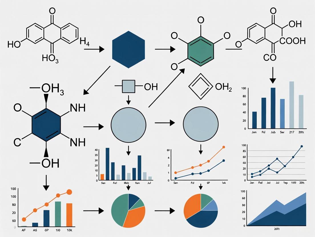
Workflow Visualization
The following diagram illustrates the complete workflow for achieving accurate, illumination-invariant colorimetric measurements using a smartphone.
The core logical relationship in optimizing smartphone colorimetry is summarized below.
Smartphone-based quantitative colorimetric analysis represents a significant shift in diagnostic and environmental testing, moving from traditional centralized laboratories to portable, point-of-need applications. This methodology leverages the ubiquitous smartphone as a powerful analytical tool, combining optical sensors with sophisticated software to perform quantitative chemical analysis. The core principle involves using the smartphone's camera to capture images of colorimetric reactions—where a change in color indicates the presence or concentration of a target analyte—and then using image processing algorithms to convert color intensity into quantitative data.
This technical support center provides researchers and scientists with essential troubleshooting guides, detailed protocols, and FAQs to overcome common challenges in implementing these systems, with a specific focus on robust calibration methods essential for obtaining research-grade data.
★ Technical Troubleshooting Guide: FAQs & Solutions
FAQ 1: How can I minimize the impact of varying ambient lighting on measurement accuracy?
- Problem: Inconsistent lighting conditions cause significant signal variance, leading to poor data reproducibility.
- Solution: Implement a reference color correction system directly within your sensor design.
- Procedure: Integrate three reference cells (e.g., with low-, medium-, and high-blue intensity) on the same sensor strip as your test zone [8]. Capture an image of the entire sensor. For analysis, first convert the RGB values of the reference cells to absorbance. Then, use the following relationship to correct the sensing area's signal:
Corrected Abs = (Abs of Sensing Area) / (Correlation Slope of Blue References)[8] - This method digitally normalizes the image, effectively canceling out the effects of variable illumination and different camera qualities [8].
- Procedure: Integrate three reference cells (e.g., with low-, medium-, and high-blue intensity) on the same sensor strip as your test zone [8]. Capture an image of the entire sensor. For analysis, first convert the RGB values of the reference cells to absorbance. Then, use the following relationship to correct the sensing area's signal:
FAQ 2: What smartphone camera settings are critical for reproducible results?
- Problem: Automatic camera processing (white balance, auto-focus, color enhancement) introduces unpredictable variability.
- Solution: Always use the manual or "Pro" mode and capture images in RAW format if possible [8].
- Essential Settings:
- Manual White Balance: Set to a fixed value (e.g., "Daylight" or a specific color temperature).
- Manual Focus: Ensure the sensor is in sharp focus.
- Disable Filters: Turn off all automatic color enhancement, filters, and HDR modes.
- RAW Format: Using RAW image capture bypasses the phone's built-in JPEG processing pipeline, providing unprocessed data from the sensor that is ideal for quantitative analysis [8].
- Essential Settings:
FAQ 3: My colorimetric data is noisy. How can I improve signal stability?
- Problem: High coefficient of variation in replicate measurements.
- Solution: Ensure you are analyzing the correct color channel and using a standardized image processing workflow.
- Channel Selection: For reactions that produce a red complex (e.g., the thiocyanatoiron(III) complex), the blue channel often provides the most sensitive and inverse relationship for quantification [9] [8]. Confirm this by analyzing the RGB deconvolution of your specific reaction.
- Standardized Analysis: Use a consistent region of interest (ROI) size and location when analyzing images with software like ImageJ. Calculate the absolute absorbance for the most relevant color channel using the formula:
A = -log(I/I₀), whereIis the mean intensity of the test zone andI₀is the mean intensity of an on-sensor white reference area [8].
FAQ 4: How can I validate the accuracy of my smartphone method against a gold standard?
- Problem: Uncertainty about the reliability of a novel smartphone-based assay.
- Solution: Perform a method comparison study using a standard laboratory instrument, such as a UV-Vis spectrophotometer.
- Validation Protocol: Prepare a series of standard concentrations. Analyze each sample with both the smartphone method and the UV-Vis spectrometer. Use statistical tests, such as a dependent samples t-test, to determine if there is a significant difference between the results at a 95% confidence level (p < 0.05). Successful validation is achieved when no statistically significant difference is found [9].
★ Detailed Experimental Protocol: Determining a Chemical Equilibrium Constant (Kc)
This protocol outlines the methodology for determining the equilibrium constant (Kc) of the thiocyanatoiron(III) complex, [Fe(SCN)]²⁺, adapting a published inquiry-based activity for researchers [9].
Materials and Equipment
Table: Essential Research Reagents and Equipment
| Item | Specification / Function |
|---|---|
| Iron(III) Nitrate | Provides the Fe³⁺ ions for complex formation [9]. |
| Potassium Thiocyanate (KSCN) | Provides the SCN⁻ ions for complex formation [9]. |
| Nitric Acid | Provides an acidic medium to prevent iron hydrolysis [9]. |
| White Well-Plate | Provides a uniform white background for consistent imaging, replacing traditional test tubes or cuvettes [9]. |
| Autopipettes | For accurate and precise liquid handling (e.g., volumes from 10–1000 µL) [9]. |
| Smartphone | Must have a camera with manual control capabilities [8]. |
| Light Control Box | Optional but recommended to create consistent, uniform illumination for image capture [9]. |
| ImageJ Software | Open-source image processing software for quantitative color intensity analysis [9]. |
Procedure
Step 1: Preparation of Standard Solutions
- Prepare a stock solution of 2.00 × 10⁻² M Fe(NO₃)₃ in 0.25 M HNO₃.
- Prepare a stock solution of 2.00 × 10⁻² M KSCN.
- Create a series of 5-6 standard solutions with known concentrations of [Fe(SCN)]²⁺ by mixing varying volumes of the two stock solutions and diluting with 0.25 M HNO₃ to a constant final volume.
Step 2: Image Acquisition
- Place the well-plate containing the standard solutions and your test samples inside the light control box.
- Using a smartphone mounted on a stand, capture images of the well-plate with all camera settings fixed in manual mode, as detailed in the troubleshooting guide above [8].
Step 3: Image Analysis with ImageJ
- Open the image in ImageJ.
- For each well, use the "Oval" selection tool to define a consistent Region of Interest (ROI) within the solution.
- Use the "Analyze > Measure" function to obtain the mean intensity values for the Red, Green, and Blue (RGB) channels.
- Also measure the mean intensity of a white reference area on the well-plate (I₀).
- Calculate the absorbance for the most relevant color channel (typically Blue for the red complex):
A = -log( I_well / I_whiteReference ).
Step 4: Data Analysis and Kc Calculation
- Plot the absorbance (A) of the standard solutions against the known concentration of [Fe(SCN)]²⁺ to create a calibration curve and obtain a linear regression equation.
- For the equilibrium samples, use this calibration curve to determine the equilibrium concentration of [Fe(SCN)]²⁺ from the measured absorbance.
- Using an ICE (Initial, Change, Equilibrium) table and the stoichiometry of the reaction
Fe³⁺ + SCN⁻ ⇌ [Fe(SCN)]²⁺, calculate the equilibrium concentrations of all species. - Calculate the equilibrium constant:
Kc = [Fe(SCN)²⁺] / ([Fe³⁺][SCN⁻]).
Table: Example Data Structure for Kc Determination
| Initial [Fe³⁺] (M) | Initial [SCN⁻] (M) | Absorbance (Blue) | Equilibrium [[Fe(SCN)]²⁺] (M) | Equilibrium [Fe³⁺] (M) | Equilibrium [SCN⁻] (M) | Kc |
|---|---|---|---|---|---|---|
| 2.00 x 10⁻³ | 2.00 x 10⁻⁴ | 0.15 | Calculated from Calibration | Initial - Equilibrium | Initial - Equilibrium | Calculated |
| 2.00 x 10⁻³ | 4.00 x 10⁻⁴ | 0.28 | ... | ... | ... | ... |
| ... | ... | ... | ... | ... | ... | ... |
★ Visual Workflows for Experimental and Calibration Processes
Smartphone Colorimetry Workflow
Three-Reference Calibration System
Frequently Asked Questions (FAQs)
Q1: My colorimetric results are inconsistent across different smartphones. What is the primary cause and how can it be mitigated? The primary cause is the automatic image processing (e.g., auto-white balance, color enhancement) performed by smartphone cameras and the variability in ambient lighting [8]. To mitigate this:
- Use Manual/Pro Mode: Operate the smartphone camera in manual or "Pro" mode to disable all automatic enhancements [8].
- Capture RAW Images: Use RAW image format capture where available, as it provides unprocessed sensor data. This can be enabled on Samsung phones in "Pro Mode" or on iPhones using third-party apps like Halide Mark II [8].
- Incorporate a Reference: Use a sensor design that includes an internal reference area or reference cells (e.g., a white blotting paper or colored reference cells) within the same image to allow for post-capture correction of lighting variations [8].
Q2: How can I control lighting conditions without an expensive laboratory setup? A low-cost, controlled imaging environment can be created using a simple light-diffusing imaging box. This box shields the sensor from ambient daylight and improves the signal-to-noise ratio [10]. For more advanced control, an ambient ring light-based smartphone platform can be used to provide consistent, uniform illumination [11].
Q3: What are the essential features to look for in a clip-on accessory for smartphone colorimetry? An effective clip-on accessory should:
- Provide Controlled Illumination: Integrate a stable light source, such as the phone's own LED flash channeled through optical fibers, to ensure consistent excitation [12].
- Ensure Optical Alignment: Precisely align optical components (lenses, diffraction gratings) with the phone's camera and flash using a custom-fabricated cradle [12].
- Standardize Sample Positioning: Include a cartridge or holder to maintain a fixed distance and angle between the sample, the light source, and the camera lens [12] [13].
Troubleshooting Guides
Issue 1: Inconsistent Color Values Under Varying Ambient Light
Problem: Analyte concentration results vary significantly when the same sample is imaged in different locations (e.g., in a bright vs. a dim lab).
Solution: Implement a multi-reference cell correction method.
- Step 1: Sensor Design. Incorporate a three-reference-cell system directly onto the sensor strip. These cells should have varying intensities of a stable color, with blue reference cells demonstrated to perform well [8].
- Step 2: Image Capture. Capture the sensor image under your experimental conditions, ensuring the reference cells are in the frame.
- Step 3: Calculate Correction Factor.
- Use image processing software (e.g., ImageJ) to convert the RGB values of the reference cells to absorbance values:
Abs = -log(I/I₀), whereIis the reference cell intensity andI₀is the white reference intensity [8]. - A linear correlation is established between the absorbance values of the reference cells captured under uncontrolled conditions and their values from a controlled condition (e.g., inside a light box) [8].
- The slope of this correlation is your correction factor.
- Use image processing software (e.g., ImageJ) to convert the RGB values of the reference cells to absorbance values:
- Step 4: Apply Correction. Correct the absorbance of your sensing area using the formula:
Corrected Abs = (Abs of Sensing Area) / (Correlation Slope from Blue References)[8].
Issue 2: Non-Linear or Unreliable Calibration Curves
Problem: The calibration curve plotted from RGB values has a poor correlation coefficient, making quantitative analysis unreliable.
Solution: Optimize the image analysis workflow and color model.
- Step 1: Validate Imaging Setup. Ensure you have followed the protocols in FAQ A1 and A2. Consistency is key.
- Step 2: Convert Color Spaces. RGB "gray values" are inversely related to color intensity. Convert RGB to CMY (Cyan, Magenta, Yellow) values using the formula
CMY = 255 - RGBto obtain values proportional to the color intensity developed in the assay [10]. - Step 3: Select the Optimal Color Channel. Test all RGB and CMY channels against your calibration standards. The channel that shows the most significant and consistent change with analyte concentration should be selected for building your final calibration curve. For example, the blue (B) channel may be optimal for a blue-colored product [10] [14].
- Step 4: Use Open-Source Software. Utilize robust, scientific image processing software like ImageJ for color quantification, as it can provide more precise and reproducible results than simple mobile apps [10] [11].
Experimental Protocols & Data Presentation
Protocol: Quantitative Iron Detection in Whole Blood
This protocol is adapted from a peer-reviewed method for smartphone-based iron quantification [8].
1. Sensor Fabrication and Assembly:
- Materials: A 3D-printed top and bottom sensor frame, four membrane layers (general nylon, fiberglass, asymmetric polysulfone, hydrophilic nylon), white blotting paper for the reference area, and three blue reference cells.
- Assembly: Laser-cut membranes into 6x6 mm squares. Assemble them between the 3D-printed frames, with the hydrophilic nylon (4th layer) impregnated with iron-capturing reagents. Include the white reference and blue reference cells on the side. Individually pack finished sensors in aluminized Mylar bags with desiccant [8].
2. Sensor Testing and Image Acquisition:
- Sample Application: Apply a liquid sample to the sampling port with the reading side face down. After a 10-minute reaction time, flip the sensor so the reading side is face up [8].
- Smartphone Imaging:
- Use smartphones with cameras set to manual/Pro mode (e.g., iPhone XR, Samsung Galaxy S10+, Samsung Note 8).
- Disable all filters and color enhancements.
- If supported, enable RAW image capture.
- Capture the image in a consistent manner, ensuring the entire sensing and reference area is in frame [8].
3. Image Analysis and Data Correction:
- Software: Use ImageJ (version 1.54g or later).
- ROI Analysis: Select regions of interest (ROIs) for the sensing area, white reference (I₀), and the three blue reference cells.
- Calculate Absorbance: For each ROI, obtain the mean intensity (I) and calculate absolute absorbance:
Abs = -log(I/I₀). - Apply In-Image Correction: Use the calculated slope from the blue reference cells to correct the sensing area absorbance, as detailed in the troubleshooting guide above [8].
The table below summarizes performance data for this method across different phone models and lighting conditions [8].
Table 1: Performance of Smartphone-Based Iron Quantification
| Smartphone Model | Lighting Condition (Lux) | Coefficient of Variation (CV) | Improvement vs. Previous Method |
|---|---|---|---|
| Samsung Galaxy S10+ | Controlled (1316 ± 3) | ~5% | Baseline |
| iPhone XR | Variable (6 - 1693) | ~5% | Consistent performance across lights |
| Samsung Note 8 | Variable (6 - 1693) | ~5% | Consistent performance across lights |
| Average across platform | Mixed | 5.13% | Absorbance results improved by 8.80% |
The Scientist's Toolkit: Key Research Reagent Solutions
Table 2: Essential Reagents and Materials for Colorimetric Assays
| Item | Function / Application | Example from Literature |
|---|---|---|
| Citric Acid, Ascorbic Acid, Thiourea (Reagent A) | Component of a reducing agent mixture for iron quantification; facilitates the colorimetric reaction [8]. | Used with Ferene to detect iron in blood [8]. |
| Ferene (Reagent B) | Chromogenic agent that reacts with iron to produce a colored complex [8]. | Used for iron quantification, measured at 590 nm [8]. |
| Phosphotungstate Reagent | Oxidizing agent used in the detection of uric acid; produces a blue-colored product in an alkaline medium [10]. | Detection of uric acid in artificial and real urine samples [10]. |
| Hydrophilic Nylon Membrane | The fourth layer in a sensor stack; impregnated with capturing reagents for the target analyte [8]. | Serves as the reaction site in the iron sensor [8]. |
| Paper-based Test Strips | A solid support for dry reagent pads that change color upon exposure to a liquid analyte. | Used in urinalysis for glucose, ketones, pH, etc. [12] [11]. |
Workflow Visualization
Diagram Title: Smartphone Colorimetric Analysis Workflow
Diagram Title: Troubleshooting Guide for Data Consistency
Diagram Title: Clip-on Spectrometer Assembly Path
Frequently Asked Questions (FAQs)
Q1: Why do I get different RGB values when using different smartphones to analyze the same sample?
Smartphone cameras undergo device-specific processing (demosaicing, gamma correction, sharpening, and compression) that alters raw sensor data, leading to inter-device variability [15]. This is compounded by differences in camera sensors, lenses, and built-in image processing algorithms [16]. Furthermore, ambient lighting conditions and the camera angle relative to the sample can introduce significant bias [16] [17]. A study measuring urine samples with five different smartphones found that without color correction, agreement between devices was poor, particularly for the Red channel [17].
Q2: My colorimetric assay shows inconsistent results between replicates. What are the common causes?
Inconsistent replicates often stem from three main areas:
- Sample Handling: Variability in pipetting accuracy or inconsistent preparation of biological samples can alter results [18].
- Reagent Issues: Using expired or improperly stored reagents can change their reactivity. Reagents must be prepared according to instructions and stored at recommended temperatures, protected from light if necessary [18].
- Assay Conditions: Non-optimized incubation times and temperatures can lead to variable reaction rates and background formation [18]. Always include blank and positive controls to account for non-specific absorbance and assess assay performance.
Q3: How can I improve the reliability of my smartphone-based colorimetric measurements?
Implement these key steps:
- Use a Controlled Imaging Environment: Capture images in a light-box or customized photo box to block ambient light and standardize lighting conditions [10] [17].
- Employ Color Correction: Use a color calibration card (e.g., Datacolor SpyderCHECKR) in your images. A matrix-based correction method can reduce inter-device and lighting-dependent variations by 65–70% [16] [17].
- Explore Robust Color Spaces: While RGB is common, the Saturation channel from HSV (Hue-Saturation-Value) color space can provide more reliable results for intensity-based assays and is less susceptible to ambient lighting noise [15].
- Ensure Proper Calibration: Always calibrate your system with standard solutions across the expected concentration range to create a reliable calibration curve [18].
Q4: What does it mean if my calculated absorbance value is greater than 2.00?
Absorbance readings above 2.00 typically indicate that your sample solution is too concentrated or too dark [19]. In this range, very little light passes through the sample, making the signal unreliable and difficult to distinguish from noise. For accurate readings, you should dilute your sample so that its absorbance falls within the useful range of 0.05 to 1.0 [19].
Q5: My assay's color is very intense, but the RGB values seem to max out and don't change with higher concentrations. What is happening?
This indicates that you are likely dealing with a highly saturated color that exceeds the gamut, or reproducible color range, of the standard sRGB color space [16]. This can create artificial discontinuities in your data. The solution is to dilute your samples to bring the color intensity back into a range where the RGB values change proportionally with concentration.
Troubleshooting Guides
Problem: High Background Noise or Signal
Possible Causes and Solutions:
- Cause 1: Non-specific reactions or contaminated reagents.
- Solution: Use high-purity reagents and include a blank control to subtract the background signal [18].
- Cause 2: Sub-optimal assay conditions.
- Solution: Carefully optimize incubation times and temperatures according to manufacturer guidelines to minimize non-specific background formation [18].
- Cause 3: Complex sample matrix (e.g., lipids, proteins in biological fluids).
- Solution: Dilute the sample, or use pre-clearing techniques like centrifugation or filtration to remove interfering particulates [18].
Problem: Poor Agreement Between Smartphones
Possible Causes and Solutions:
- Cause 1: Differing built-in image processing pipelines across smartphone brands.
- Cause 2: Varying lighting conditions and camera angles.
- Cause 3: Relying on the native RGB color channels which are highly sensitive to ambient noise.
- Solution: Convert images to HSV color space and use the Saturation channel for analysis, which has been shown to be more robust to lighting variations [15].
Problem: Non-Linear or Poor Correlation in Calibration Curve
Possible Causes and Solutions:
- Cause 1: Sample concentrations are outside the linear dynamic range of the assay.
- Solution: Dilute or concentrate your samples to fit within the established linear range [19].
- Cause 2: Improver color value extraction or region of interest (ROI) selection.
- Solution: Use consistent ROI size and location. Employ software like Image J to quantify average color intensity across a defined area rather than a single point [10].
- Cause 3: Using an inappropriate color channel for analysis.
Experimental Protocols
1. Principle: In an alkaline medium, uric acid reduces phosphotungstate reagent, producing a blue-colored tungsten complex. The intensity of this blue color is proportional to the concentration of uric acid.
2. Materials and Reagents:
- Uric acid standard solution
- Phosphotungstate reagent (Follin reagent)
- Sodium carbonate (Na₂CO₃) solution, 10%
- Artificial urine or diluted real urine sample
- Glass cuvettes
- Smartphone (e.g., Samsung Galaxy A52)
- Imaging box with white background
- Computer with Image J software
3. Procedure:
- Color Development: Transfer aliquots of uric acid standard (1-5 mL of a 30 µg/mL solution) into a series of 10 mL volumetric flasks. Add 3.0 mL of 10% Na₂CO₃ to each flask and let stand for 10 minutes. Then, add 1.0 mL of phosphotungstate reagent, mix well using a vortex, and dilute to the mark with distilled water. The final concentration range should be 3.0–15 µg/mL.
- Imaging: Place the solutions in glass cuvettes. Position them in an imaging box to eliminate external light interference. Capture an image using the smartphone camera against a white background. Ensure the image includes all samples and a color calibration card if used.
- Image Analysis with Image J:
- Open the image file (TIFF format is preferred) in Image J.
- Crop the image to remove blank edges, ensuring each sample segment is included.
- For each sample, select a consistent Region of Interest (ROI) within the colored solution.
- Use Image J's analysis tools to measure the mean gray value for the Red, Green, and Blue (RGB) channels within the ROI.
- Convert the RGB values to CMY (Cyan-Magenta-Yellow) values using the formula: CMY = 255 - RGB [10]. The CMY value is proportional to the color intensity.
4. Data Analysis:
- Plot the CMY values (or the values from the most responsive channel) against the known uric acid concentrations to generate a calibration curve.
- Use the regression equation of this curve to determine the concentration of unknown samples.
Workflow: Smartphone-Based Colorimetric Analysis
The following diagram illustrates the general workflow for a quantitative smartphone-based colorimetric analysis, from sample preparation to concentration determination.
Data Presentation
Table 1: Comparison of Colorimetric Analysis Methods for Quantitative Determination
This table summarizes key performance characteristics of different analytical methods as demonstrated in the determination of uric acid [10].
| Method | Linear Range (µg/mL) | Correlation Coefficient (R²) | Key Advantages | Key Limitations |
|---|---|---|---|---|
| DIC / Image J | 3.0 – 15 | ~0.99 (CMY values) | High precision; uses free, open-source software; good for static analysis [10]. | Requires transfer to computer for analysis. |
| DIC / RGB Color Detector App | 3.0 – 15 | ~0.97 (B channel) | Direct on-phone analysis; portable and rapid [10]. | Lower correlation than Image J; primarily semi-quantitative [10]. |
| UV/VIS Spectrophotometry | 3.0 – 15 | ~0.99 (Absorbance) | Gold standard; high accuracy and precision [10]. | Requires expensive, non-portable laboratory equipment. |
| HSV Saturation Method | Varies by assay | High (as per study) | Robust to ambient lighting; improved limit of detection; enables equipment-free analysis [15]. | Performance is application-specific; requires validation. |
Table 2: Research Reagent Solutions for Smartphone Colorimetry
This table details essential materials and their functions for setting up a smartphone-based colorimetric experiment, as referenced in the search results.
| Item | Function / Application | Example from Literature |
|---|---|---|
| Color Calibration Card | Standardizes color measurements across different devices and lighting conditions by providing reference colors for correction [16] [17]. | Datacolor SpyderCHECKR 24 [17]. |
| Imaging Box / Light Box | Provides a controlled environment with consistent, diffuse lighting, shielding the sample from variable ambient light [10] [17]. | Custom-built polystyrene foam box with LED light source [17]. |
| Image Analysis Software | Used to quantitatively extract color intensity values from digital images. | Image J (open-source) [10], Adobe Photoshop [17]. |
| Open-Source Mobile Apps | Allows for direct, on-device color extraction, useful for rapid or field-based screening. | RGB Color Detector [10], Color Picker [11]. |
| Standard Cuvettes / Petri Dishes | Hold liquid samples for imaging; ensure consistent optical path length and placement. | Glass cuvettes [10], disposable Petri dishes [17]. |
Technical Specifications & Methodologies
For high-precision requirements, especially in kinetic studies, a more advanced color correction is needed:
- Image Capture with References: Always capture the sample alongside a color calibration chart that includes known reference colors.
- Reference Color Processing: Use a spectrophotometer to measure the visible transmission/reflectance spectra of the color chart patches. Convert these spectra to reference CIE XYZ values using standard illuminants (e.g., D65) and observer functions.
- Matrix Calculation: Extract the smartphone's RGB values for each corresponding color patch. Calculate a correction matrix (e.g., using linear least squares) that best maps the smartphone RGB values to the reference XYZ values.
- Application: Apply this correction matrix to all subsequent sample images taken with the same smartphone and lighting setup to obtain standardized color values.
Logical Relationship: From Sample to Quantitative Result
The following diagram maps the logical pathway of converting a physical sample's property into a quantitative analytical result using smartphone colorimetry, highlighting critical transformation steps.
The Role of Standard Illuminants and Observers in Color Measurement
Frequently Asked Questions
1. What are standard illuminants and observers, and why are they critical for smartphone colorimetry? Standard illuminants are published theoretical sources of visible light with defined spectral power distributions (SPDs), providing a basis for comparing colors under different lighting [20] [21]. Standard observers are mathematical functions representing the average human eye's color response under a specific field of view [22] [23]. In smartphone-based colorimetry, they are fundamental for transforming device-specific camera responses into standardized, reproducible color values, ensuring your quantitative results are accurate and comparable across different devices, locations, and times.
2. I'm setting up my smartphone imaging system. Which standard illuminant should I use to simulate daylight? You should use a D-series illuminant, specifically CIE Standard Illuminant D65 [20] [21]. It is intended to represent average daylight with a correlated color temperature (CCT) of approximately 6500 K and is the standard representative daylight illuminant for colorimetric calculations [20] [24]. While D50 (5003 K) is also used in some industries like photography, D65 is the canonical choice for scientific applications requiring representative daylight [21].
3. My color measurements don't match visual assessments. Could the standard observer be the issue? Yes. The CIE 1931 2° Standard Observer is based on a narrow field of view and may not correlate well with visual assessments, especially if your sample is large or viewed peripherally [22]. For a wider field of view, the CIE 1964 10° Supplementary Standard Observer is more representative of how the human eye perceives color in such contexts and is recommended for instrumental color measurement [22] [23]. Ensure your color analysis software is configured for the correct observer.
4. How can I achieve a consistent D65 illuminant in my smartphone setup? Achieving a perfect artificial source for D65 is challenging [20]. The most practical approach is to use a high-quality daylight-simulating LED panel with a high Color Rendering Index (CRI > 95). Characterize the LED's SPD with a spectrometer if possible, and use the CIE's metamerism index to assess its quality as a daylight simulator [20]. Alternatively, for less critical applications, you can perform a white balance calibration on your smartphone using a standard white reference tile under your chosen light source.
5. What causes two samples to match under my phone's flash but look different outdoors? This is a classic case of metamerism [25]. Two colors with different spectral compositions are metamers if they match under one illuminant but not under another [25]. Your phone's flash (which may be similar to illuminant A) and daylight (D65) have different SPDs. If your samples are metameric, they will appear different under these two light sources. This underscores the importance of using and reporting a standard illuminant in your analysis.
Essential Concepts for Experiment Design
The following tables summarize the core components you must define for your colorimetric experiments.
Table 1: Common CIE Standard Illuminants [20] [21] [24]
| Illuminant | Correlated Color Temperature (CCT) | Represents | Key Application in Smartphone Colorimetry |
|---|---|---|---|
| A | 2856 K | Typical incandescent / tungsten-filament lighting. | Use when the phone's built-in flash is the primary light source. |
| D50 | 5003 K | "Horizon" daylight. | Common in photography and graphic arts; a daylight reference. |
| D55 | 5500 K | Mid-morning / mid-afternoon daylight. | An alternative daylight reference. |
| D65 | 6504 K | Noon daylight (standard). | The default for representing average daylight. |
| F Series | Varies (e.g., F2: 4230 K) | Various fluorescent lamps. | Use when measuring under typical office or lab fluorescent lighting. |
Table 2: CIE Standard Colorimetric Observers [22] [23] [25]
| Standard Observer | Field of View | Description | Recommended Use |
|---|---|---|---|
| CIE 1931 | 2° (≈ thumbnail at arm's length) | First standardized function; based on the belief color-sensing cones were in a 2° foveal arc [22]. | Colorimeters; quality control for small samples [22]. |
| CIE 1964 | 10° (≈ palm at arm's length) | Supplementary standard; more representative of typical human color perception [22]. | Recommended for spectrophotometry and formulating color for larger samples [22]. |
Experimental Protocol: Smartphone Color Measurement Calibration
This protocol provides a methodology for calibrating a smartphone-based colorimetric analysis system using standard illuminants and observers.
1. Objective To establish a standardized workflow for capturing and analyzing color data with a smartphone, ensuring measurements are traceable to CIE standards.
2. Materials and Reagents Table 3: Research Reagent Solutions & Essential Materials
| Item | Function / Specification |
|---|---|
| Smartphone | Fixed in a stand; camera settings locked (ISO, shutter speed, white balance). |
| Controlled Light Box | Equipped with high-CRI D65-simulating LEDs. |
| Color Calibration Chart | Chart with known colorimetric values (e.g., X-Rite ColorChecker). |
| Standard White Reference Tile | Provides a consistent white point for white balance and reflectance calibration. |
| Image Analysis Software | Software capable of converting RGB to CIE XYZ and CIELAB values (e.g., Python, Matlab, ImageJ with plugins). |
3. Workflow Diagram The following diagram illustrates the logical workflow for a calibrated smartphone color measurement experiment.
4. Step-by-Step Procedure
- System Setup: Place the smartphone on a stable stand inside the light box, ensuring the camera lens is parallel to the sample plane. Power on the D65-simulating LED light source and allow it to warm up for a consistent output.
- Reference Capture: Place the color calibration chart within the field of view. Capture an image in RAW format (DNG) if supported, otherwise use the highest quality JPEG without compression.
- Sample Capture: Replace the calibration chart with your test sample and capture an image under identical settings and geometry.
- Image Pre-processing: Use software to correct for lens distortion and extract the average RGB values from each color patch of the chart and the region of interest in your sample. Perform a white balance correction using the known white reference tile.
- Colorimetric Transformation: Build a transformation matrix that maps the device's calibrated RGB values from the chart image to the known CIE XYZ values of the chart patches. The calculation of XYZ tristimulus values for a color with a spectral power distribution
S(λ)is defined as: X = ∫ S(λ) * x̄(λ) dλ Y = ∫ S(λ) * ȳ(λ) dλ Z = ∫ S(λ) * z̄(λ) dλ where x̄, ȳ, and z̄ are the color-matching functions for the chosen standard observer [25]. Apply this matrix to the RGB values from your sample image to convert them to standardized CIE XYZ, and subsequently to a perceptually uniform color space like CIELAB. - Data Analysis: Perform your quantitative analysis (e.g., concentration determination) using the calibrated CIELAB values, particularly the lightness (L), and chromaticity (a, b*) coordinates.
Troubleshooting Guide
| Problem | Potential Cause | Solution |
|---|---|---|
| Poor repeatability | Inconsistent lighting geometry or camera settings. | Use a fixed light box and mount. Lock all camera settings (ISO, shutter speed, white balance). |
| Measurements differ from benchtop spectrometer | Mismatch in standard illuminant or observer definitions. | Verify and align the illuminant (e.g., D65) and observer (e.g., 1964 10°) settings in all analysis software. |
| Colors look different under another phone | Uncalibrated device-dependent RGB space. | Implement the calibration protocol using a standard chart to transform to device-independent color spaces (XYZ, CIELAB). |
| Samples match in app but not visually | Metamerism or use of the 2° observer for a large sample [25]. | Check for metamerism by comparing under a second standard illuminant. Switch to the 1964 10° standard observer for analysis [22]. |
Step-by-Step Calibration Protocols: From Basic Apps to Advanced Reference Systems
Troubleshooting Guides and FAQs
Frequently Asked Questions
Q1: My application is giving inconsistent RGB values for the same sample. What could be the cause? Inconsistent readings are often due to variable lighting conditions. Ensure all measurements are taken in a controlled, uniform lighting environment. Avoid shadows and direct light on the sample. Furthermore, use a fixed-distance holder or a 3D-printed jig to maintain a consistent distance and angle between the smartphone camera and the sample for every measurement [26].
Q2: How can I validate the accuracy of my smartphone colorimeter setup? A reliable method is to use certified colorimetric tiles or standards with known reference values [26]. Measure these standards with your setup and calculate the CIELab color difference (ΔE) between your measured values and the certified values. A lower ΔE indicates higher accuracy. For greater precision, consider using a clip-on dispersive grating, which has been shown to improve colorimetric performance compared to using the smartphone camera alone [26].
Q3: What is the difference between the RGB Detector and PhotoMetrix Pro apps for calibration? While both apps can be used for color detection, their calibration approaches differ. The RGB Detector app used in research has auto-calibrating capabilities, converting camera output to RGB coordinates that are independent of the camera model [26]. PhotoMetrix Pro is known for providing more advanced analytical functionalities, allowing for the construction of calibration curves for quantitative analysis. The choice depends on whether you need basic color detection (RGB Detector) or a more comprehensive analytical tool (PhotoMetrix Pro).
Q4: Why is a "sandwich-type" lateral flow assay mentioned in the context of smartphone colorimetry? The sandwich-type Lateral Flow Immunoassay (LFA) represents an advanced application of smartphone-based colorimetry. It combines immunochromatographic test strips with a smartphone application for automated image acquisition, calibration, and classification [27]. This integration allows for semi-quantitative analysis of specific biomarkers, such as 25-hydroxyvitamin D, by converting the color intensity of a test line into a quantitative result, demonstrating the move towards decentralized diagnostics [27].
Troubleshooting Common Experimental Issues
| Problem | Possible Cause | Solution |
|---|---|---|
| High variation between replicate measurements | Inconsistent camera focus or sample illumination. | Use a fixed-focus setting on the camera app and a dedicated light source (e.g., built-in white LED) [26]. |
| Calibration curve has poor linearity (low R² value) | Improper color space usage or sample concentration outside dynamic range. | Ensure RGB values are correctly transformed into a suitable color space (e.g., CIELab) for analysis [26]. |
| App cannot distinguish between similar colors | Limited color resolution of the smartphone camera or insufficient contrast. | Use a clip-on dispersive grating to enhance measurement precision [26]. |
| Results are not reproducible across different smartphone models | Variances in camera sensors and built-in image processing. | Use an app with auto-calibrating capability or perform a device-specific calibration with certified standards [26]. |
Experimental Protocols and Data Presentation
Protocol: Smartphone Colorimetry with Certified Standards
This methodology outlines the steps for performing a reliable colorimetric measurement and calibration using a smartphone and certified reference tiles [26].
- Equipment Setup: Smartphone, a 3D-printed or fixed holder to maintain a constant sample distance, certified color tiles (e.g., red, green, blue, yellow), and a white reference tile (e.g., Spectralon) [26].
- Application Configuration: Install a color detection application (e.g., RGB Detector). Ensure the smartphone's flash (white LED) is enabled as a consistent light source [26].
- Image Acquisition: Place the white reference tile in the holder and capture an image for white balancing. Then, place each certified color tile in the holder and capture multiple images (e.g., 5 replicates).
- Data Extraction: Use the application to obtain the average RGB values for each tile from the captured images.
- Data Conversion: Convert the averaged RGB values to tristimulus XYZ coordinates using a predefined conversion matrix. An example matrix from literature is [26]:
[X, Y, Z] = [0.412, 0.358, 0.180; 0.213, 0.715, 0.072; 0.019, 0.119, 0.950] * [R, G, B]These XYZ values are then transformed into the CIELab color space to calculate the color difference ΔE [26]. - Validation: Calculate the ΔE between your measured CIELab values and the certified values for the tiles to quantify the accuracy of your setup [26].
Quantitative Performance Data from Literature
The table below summarizes the colorimetric performance achievable with different smartphone setups, as reported in research. The color difference (ΔE) and resolution (δE) are key metrics for assessing accuracy and precision [26].
Table 1: Performance of Smartphone Colorimetry on Certified Color Tiles
| Color Tile | Color Difference using RGB Detector (ΔE) | Color Difference using GoSpectro Grating (ΔE) |
|---|---|---|
| Yellow (YW) | Data not specified in results | Smallest difference observed [26] |
| Cyan (CY) | Data not specified in results | Smallest difference observed [26] |
| Purple (PU) | Data not specified in results | Biggest difference observed [26] |
| Red (RD) | Data not specified in results | Biggest difference observed [26] |
| All Tiles (Average) | Acceptable results for quick evaluation [26] | Best results, highest precision [26] |
Workflow Visualization
The Scientist's Toolkit: Essential Research Reagents and Materials
Table 2: Key Materials for Smartphone-Based Quantitative Colorimetric Analysis
| Item | Function in the Experiment |
|---|---|
| Certified Colorimetric Tiles (e.g., from Labsphere) | Provide a set of colors with known, certified tristimulus values. They are essential for validating the accuracy and precision of the smartphone colorimeter by calculating the color difference ΔE [26]. |
| White Reference Tile (e.g., Spectralon) | Serves as a high-reflectance standard for white balancing and calibration before sample measurement, ensuring consistent and accurate color capture [26]. |
| Fixed-Distance Holder / 3D-Printed Jig | A physical fixture that maintains a consistent distance and angle between the smartphone camera and the sample. This is critical for achieving reproducible results by eliminating variability from hand-held operation [26]. |
| Clip-On Dispersive Grating (e.g., GoSpectro) | An accessory that clips onto the smartphone camera, turning it into a basic spectrophotometer. It significantly enhances colorimetric precision over using the camera alone by dispersing light and allowing wavelength-based analysis [26]. |
| Lateral Flow Assay (LFA) Strips | Used in advanced applications for detecting specific analytes (e.g., vitamins, pathogens). The smartphone app quantitatively reads the color intensity of the test line, enabling semi-quantitative analysis at the point of care [27]. |
| Anti-Idiotype Antibody | A specialized reagent used in a "sandwich-type" LFA for small molecules like Vitamin D. It allows for a more sensitive and reproducible assay format compared to traditional competitive assays [27]. |
Troubleshooting Guides
FAQ 1: How can I convert my RGB immunofluorescence images to a colorblind-friendly format in ImageJ?
Problem: A user needs to convert RGB immunofluorescence images to a cyan/magenta/yellow (CMY) format to make them colorblind-friendly but finds that the color scheme is not retained when saving and reopening the files.
Solution: The most reliable method involves splitting the original channels and then re-merging them with the desired CMY color assignments, rather than converting an existing RGB image.
Detailed Protocol:
- Open your original image in ImageJ/Fiji.
- Split the channels: Run
Image > Color > Split Channels. This creates separate grayscale images for each channel (e.g., "red," "green," "blue"). - Re-merge with new colors: Run
Image > Color > Merge Channels....- In the dialog box, assign the red channel to the cyan channel, the green channel to the magenta channel, and the blue channel to the yellow channel.
- For example, in the "C5 (cyan)" dropdown, select your original image's "(red)" channel.
- In the "C6 (magenta)" dropdown, select your original image's "(green)" channel.
- In the "C7 (yellow)" dropdown, select your original image's "(blue)" channel.
- Ensure the "Create composite" box is unchecked to generate a true RGB image with the new color mapping [28].
- Save the new image: Use
File > Save As > Tiffto preserve the color scheme. This method creates an RGB image that should retain the CMY colors when reopened in other software like Photoshop or Illustrator [28].
Important Consideration: Note that simply recoloring channels as cyan, magenta, and yellow may not be fully effective for all types of color blindness, as dichromatic observers cannot discern colors from three mixed channels. A more robust alternative is to use the Colorblind Action Bar plugin for Fiji, which performs a semi-CYM conversion designed to address oversaturation and be more perceptible [28].
FAQ 2: Why does my image appear all black after importing, even though the data is valid?
Problem: After loading an image, it displays as entirely black or very dark, but the user knows the data is present.
Solution: This is a common issue with high-bit-depth images (e.g., 12-bit, 14-bit, or 16-bit) where the actual data occupies only a small portion of the full display range.
Troubleshooting Steps:
- Verify Data Presence: Move your mouse over the image and observe the status bar at the bottom of the main ImageJ window. If pixel values other than zero are displayed as you move the cursor, your data is intact [29].
- Auto-adjust Contrast:
- Go to
Image > Adjust > Brightness/Contrast...(or pressShift+C). - In the dialog that opens, click the Auto button. This will rescale the display to map the minimum and maximum intensity values in your image data to black and white, making the features visible [29].
- Go to
- Prevent Future Issues (during import): If you are using the Bio-Formats Importer for specific file types, you can disable autoscaling upon import:
- Use
File > Import > Bio-Formats. - Select your file.
- In the import options, uncheck the "Autoscale" box.
- Click OK. The data will then be scaled to the maximum value of the bit depth instead of being autoscaled to the image's actual intensity range [29].
- Use
FAQ 3: How do I convert an intensity-based color scale to a concentration-based scale?
Problem: A user has an 8-bit image with a color scale from 0 to 255 and needs to convert this to a concentration scale, for example, 0 to 150 mmol/liter.
Solution: Use ImageJ's calibration tool to establish a mathematical relationship between pixel intensity and concentration.
Experimental Protocol:
- Open your image in ImageJ.
- Open the Calibrate Tool: Navigate to
Analyze > Calibrate.... - Perform Calibration:
- In the Calibrate dialog, you will see a plot of Intensity vs. Value.
- From the "Function" dropdown, select the calibration model that best fits your data (e.g., Linear, Polynomial).
- You need known standard concentrations to create the calibration curve. Enter the known concentration values in the "Value" column corresponding to their measured intensity values.
- Click "OK" to apply the calibration. Once calibrated, ImageJ will display concentration values instead of raw intensity values when you use tools like the mouse pointer to probe the image [30].
- Alternative Method for Image Conversion: If you need to create a new image where the pixel values directly represent concentration:
- First, convert your image to 32-bit float using
Image > Type > 32-bit. This prevents data clipping during mathematical operations. - Then, use
Process > Math > Macro...to apply the calibration function (e.g.,v=(v/255)*150) to convert the 0-255 intensity range to a 0-150 concentration range [30].
- First, convert your image to 32-bit float using
FAQ 4: What should I do if ImageJ freezes or becomes unresponsive during processing?
Problem: ImageJ stops responding to inputs during an operation.
Solution: Generate a "thread dump" or "stack trace" to capture the program's state, which is invaluable for developers to diagnose the problem.
Detailed Protocol:
- The Easy Way (Within ImageJ):
- Press
Shift + \(backslash) while ImageJ is the active window. - If successful, a new window containing the stack trace will open.
- Press
Ctrl+A(orCmd+Aon Mac) to select all text, thenCtrl+C(orCmd+C) to copy it. You can then paste this into a bug report [29].
- Press
- The Fallback Method (Using the Console):
- If the first method fails, launch ImageJ from the system console/terminal to capture log output.
- Linux/macOS: Open a terminal and run
DEBUG=1 /path/to/ImageJ(adjust the path as needed). - Windows: Make a copy of
ImageJ-win64.exe, rename it todebug.exe, and run it. This launches ImageJ with an attached command prompt. - Reproduce the freeze.
- On Linux/macOS: Press
Ctrl + \in the terminal window to print the stack trace. Select the text with your mouse and copy it. - On Windows: Press
Ctrl + Pause(the Break key) in the command prompt. Click the icon in the upper left corner of the window, choose Edit > Mark, select the stack trace with your mouse, and press Enter to copy it [29].
Experimental Protocols & Data Presentation
Quantitative CMY Conversion Workflow
The following diagram illustrates the core methodology for converting an RGB image to a quantitative, colorblind-friendly CMY format in ImageJ.
Comparison of Colorblind-Friendly Conversion Methods
The table below summarizes the two primary methods for creating accessible images, helping you choose the right approach for your research.
| Method | Key Feature | Best Use Case | Limitation |
|---|---|---|---|
| Manual Channel Merge [28] | Direct reassignment of original channels to Cyan, Magenta, and Yellow during merge. | Full control over channel-color mapping; requires a specific, consistent color scheme. | May not be effective for all forms of color blindness; can lead to oversaturation. |
| Colorblind Action Bar Plugin [28] | Semi-automated plugin designed specifically for color accessibility. | General-purpose creation of colorblind-friendly figures; handles oversaturation better. | Less granular control than manual method; requires plugin installation. |
Essential Research Reagent Solutions for Colorimetric Analysis
This table lists key materials and software tools essential for conducting quantitative colorimetric analysis, particularly in the context of smartphone-based and ImageJ-driven research.
| Item | Function in Research | Example Application |
|---|---|---|
| TCS3200 Color Sensor [31] | A programmable RGB color light-to-frequency converter that captures raw color data from samples. | Integrating with a Raspberry Pi to create a low-cost, portable colorimetric sensor for protein assays (e.g., BCA, Bradford) [31]. |
| Micro-BCA/Bradford Assay Kits [31] | Standardized chemical reagents that produce a color change proportional to protein concentration. | Generating calibration curves for quantitative protein analysis using image-based or sensor-based colorimetry [31]. |
| Standardized Color Charts | Provides a known reference for color correction and white balancing across different lighting conditions. | Essential for calibrating smartphone cameras or scanners to ensure reproducible color data acquisition. |
| Fiji/ImageJ Software | Open-source image analysis platform with built-in tools and plugins for color space conversion, calibration, and quantification. | Performing CMY conversion, intensity-to-concentration calibration, and colocalization analysis [29] [30] [28]. |
| Colorblind Action Bar Plugin [28] | A specialized Fiji plugin that transforms images into colorblind-friendly color spaces. | Preparing scientific figures and microscopy images that are accessible to a wider audience, including those with color vision deficiencies [28]. |
Troubleshooting Guides and FAQs
Frequently Asked Questions
1. What is the primary purpose of a multi-cell color reference sticker in smartphone colorimetry? The primary purpose is to achieve color constancy. These stickers contain patches of known colors, allowing software to mathematically model and correct for the variable illumination conditions (color temperature, brightness) present when an image is captured [32]. This transformation ensures the colors in the image accurately represent the true colors of the scene, independent of the lighting, which is critical for quantitative measurements [32].
2. My color-corrected results are still inconsistent. What could be wrong? Inconsistent results after correction can stem from several issues:
- Localized Shadows or Glare: The color correction algorithm assumes even illumination across the reference sticker. If shadows or glare affect only part of the sticker, as indicated by diverging color measurements from oppositely placed chips, the correction will be unreliable [32].
- Low-Quality Reference Sticker: The printing quality of the color stickers is critical. High variation in the printed color patches (with a high ΔE00 score, e.g., >5.3) will directly impair the system's precision [32].
- Incorrect Camera Settings: Using automatic settings or JPEG processing can introduce unpredictable color shifts [33] [34]. Whenever possible, use a manual camera mode and capture in a raw image format to minimize automated processing.
3. How do I validate that my smartphone sensor and reference system are working correctly? A validation procedure should test each component of your system [32]:
- Phone Sensor Consistency: Photograph a known color patch (e.g., from a Pantone library) multiple times under identical lighting. Convert the images to the CIELAB color space and calculate the mean ΔE00 between all pairwise comparisons. A mean ΔE00 of ≤1.0 indicates excellent sensor repeatability [32].
- Sticker Quality: Measure the color values of patches from several randomly selected stickers from your print batch. The ΔE00 between the measured color and the known reference value should be as low as possible (e.g., <3 on average) [32].
4. Are there alternatives to using a physical color card for illumination correction? Yes, retrospective computational methods like BaSiCPy exist. These software-based approaches derive an illumination correction function directly from a set of your images, without needing a reference object captured in every shot [35]. This is particularly useful for correcting uneven illumination (vignetting) in fields like fluorescence microscopy [35].
Troubleshooting Common Problems
| Problem | Possible Cause | Solution |
|---|---|---|
| High variation in corrected color values | 1. Inconsistent lighting conditions.2. Shadows or glare on the reference sticker.3. Low-quality printed color stickers. | 1. Use a controlled, diffuse light source.2. Reposition the setup to avoid shadows/glare. Check for color divergence in mirrored patches [32].3. Source stickers from a printer that guarantees low ΔE00 variation (e.g., <5.3) [32]. |
| Corrected image colors look unnatural | The color transfer algorithm is too aggressive or using an inappropriate method. | Ensure the software uses a localized transfer, applying corrections only within the region of interest defined by the reference card to avoid over-correction of the entire scene [32]. |
| Different results from different smartphones | Proprietary JPEG processing and auto-white-balance algorithms vary by manufacturer and model [33] [34]. | 1. Use a manual camera mode and disable all auto-features (white balance, exposure, saturation).2. Use a machine learning classifier trained on data from multiple phone models to improve inter-phone repeatability [34]. |
| The system fails to correct for extreme color temperatures | The color gamut of the reference sticker may not be broad enough to cover the transform required for extreme lighting. | Use an enhanced color sticker design that includes a wider range of brightness and hues, extending beyond the original ColorChecker palette to capture a broader range of possible transforms [32]. |
Experimental Protocols and Data
Protocol: Validating a Smartphone-Based Color Correction Pipeline
This protocol outlines the key experiments to validate a system like the HueDx platform, which uses a multi-cell reference sticker [32].
1. Objective To empirically measure the performance and limitations of a smartphone-based color correction system, including the phone hardware, reference sticker quality, and software correction capabilities.
2. Materials and Reagents
- Smartphone with a manual camera mode (e.g., iPhone 11 or newer).
- Custom multi-cell color reference sticker (e.g., HueCard).
- Controlled lighting environment with variable color temperature and brightness.
- Colorimetric assay of interest (e.g., paper-based total protein diagnostic assay).
- Standardized color patches (e.g., Pantone Cool Gray 1C, Neutral Black).
3. Methodology
Step 1: Phone Sensor Quantification
- Place a standardized color patch in a controlled, evenly lit environment.
- Capture ten images of the patch without moving the phone or the patch.
- Manually isolate the patch in each image and convert the pixels to the CIELAB color space.
- Calculate the average color value for each image and then compute the ΔE00 (Delta E 2000) pairwise between all images.
- Report the mean ΔE00 and max ΔE00. A mean ΔE00 ≤ 1.0 indicates the sensor has imperceptible variation and is sufficiently consistent [32].
Step 2: Sticker Manufacturing Quality Control
- Randomly select a batch of printed color stickers (e.g., 10 from 400).
- Image each sticker under standard laboratory illumination conditions.
- For each color patch on each sticker, measure the average color value and calculate the ΔE00 against its known reference value.
- Report the mean ΔE00, max ΔE00, and standard deviation across all patches and all stickers. High-quality prints should have a mean ΔE00 < 3 and a max ΔE00 < 5.3 [32].
Step 3: Color Correction Pipeline Efficacy
- Capture images of the color reference sticker and a colorimetric assay under a wide range of illumination conditions (varying brightness and color temperature).
- Process the images through the color correction pipeline (e.g., using white-balancing, multivariate Gaussian distributions, and dynamic lookup tables).
- Calculate the ΔE00 between the corrected images and a reference image taken under standard lab conditions.
- A ΔE00 < 3 after correction indicates the system has successfully restored the images to near-imperceptible levels of difference [32].
Step 4: Real-World Application in Diagnostics
- Perform a colorimetric assay (e.g., total protein test) with samples of known concentration.
- Capture and analyze images with and without using the color correction pipeline.
- Compare the coefficient of variation (CV) in precision testing and calculate the limits of blank (LoB), detection (LoD), and quantitation (LoQ) for both methods. A well-functioning system will show a lower CV and lower LoB/LoD/LoQ with color correction [32].
The following table summarizes expected performance metrics for a robust system based on the HueDx study [32].
Table 1: Key Performance Indicators (KPIs) for System Validation
| Validation Target | Metric | Expected Outcome | Industry Standard |
|---|---|---|---|
| Phone Sensor | Mean ΔE00 (pairwise) | ≤ 1.0 | ≤ 1.0 (Imperceptible) [32] |
| Reference Sticker | Max ΔE00 (vs. reference) | < 5.3 | ≤ 5.0 (Small perceptible difference) [32] |
| Color Correction | ΔE00 after correction | < 3.0 | N/A |
| Diagnostic Assay (Precision) | Coefficient of Variation (CV) | Almost 2x lower with correction | Lower is better [32] |
The Scientist's Toolkit: Essential Research Reagents and Materials
Table 2: Key Research Reagent Solutions for Smartphone Colorimetry
| Item | Function | Example/Specification |
|---|---|---|
| Multi-Cell Color Reference Card | Provides known color values for the software to model and correct for prevailing illumination conditions. | HueCard; contains 48 color patches and 2 black/white references, mirroring an enhanced ColorChecker palette [32]. |
| Smartphone with Manual Controls | Acts as the image acquisition device. Requires control over focus, white balance, ISO, and shutter speed. | iPhone 11 or newer; or Android phones supporting third-party camera apps with manual mode [32]. |
| Controlled Light Source | Provides consistent, diffuse illumination to minimize shadows and glare, which are challenging to correct. | D65 standard illuminant (6500K color temperature) is a common calibration target [36] [37]. |
| Colorimetric Assay | The test medium that produces a color change proportional to the analyte concentration. | Paper-based total protein diagnostic assay; hydrogen peroxide test strips [32] [34]. |
| Calibration Software | Applies color transfer algorithms to transform the image based on the reference sticker. | HueTools; utilizes algorithms like multivariate Gaussian distributions and dynamic lookup tables [32]. |
| Colorimeter | A device used for high-accuracy color measurement to validate the system and create reference values. | X-Rite i1 Display Pro; used for calibrating displays and measuring color patches [36]. |
Workflow and System Diagrams
Illumination Correction Workflow
System Components and Algorithms
Frequently Asked Questions (FAQs)
Q1: Why should I use CIELAB over RGB for smartphone-based colorimetric analysis?
CIELAB offers significant advantages for quantitative analysis because it is designed to be perceptually uniform, meaning numerical changes correspond more closely to perceived color changes. Crucially, research shows that the CIELAB color space—specifically its a* and b* chromatic coordinates—exhibits inherent resistance to illumination changes. This makes it superior to RGB models, which are highly sensitive to lighting variations and limit reliability. Using CIELAB can enable housing-free, illumination-invariant detection, simplifying your field setup [1].
Q2: My CIELAB results seem perceptually inaccurate, especially for highly saturated colors. Why?
You are likely observing the limitations of CIELAB in accounting for the Helmholtz-Kohlrausch effect. This phenomenon describes how strongly saturated colors can appear brighter than their measured L* (lightness) value suggests. For example, a saturated red may look significantly brighter than a gray with the same L* value. This is a known issue where CIELAB undervalues the contribution of saturation to perceived lightness [38].
Q3: How can I optimize the median cut algorithm when working in CIELAB color space?
Standard median cut in CIELAB does not always improve results because it may not prioritize lightness (L) error sufficiently. To optimize it, consider scaling the L channel more aggressively than the a* and b* channels. Experimental evidence shows that weighting the L* channel, for instance, by a factor of two, forces the algorithm to reduce perceptual lightness error more effectively, leading to visibly better results, such as the removal of color blotches in image quantizations [39].
Q4: What are the common pitfalls when measuring color data for analysis?
Common mistakes include:
- Relying on visual assessment: Human perception is subjective and prone to fatigue. Always use instrument-based measurement [40].
- Ignoring environmental factors: Ambient light, temperature, and humidity can affect color measurements. Control these factors where possible [40].
- Misunderstanding device output: Ensure you know whether your smartphone app or spectrophotometer provides data in RGB, CIELAB, or another space, and understand the required conversions [2] [40].
Troubleshooting Guides
Issue: Results Are Inconsistent Under Different Lighting Conditions
Problem: Color measurements taken with a smartphone vary unpredictably when lighting changes. Solution:
- Switch to CIELAB Chromaticity Coordinates: Base your analysis on the a* and b* channels of CIELAB instead of RGB values. These coordinates form "equichromatic surfaces" that are inherently more resilient to illumination changes [1].
- Use a Controlled Lighting Box: For critical measurements, use a simple light control box with adjustable LEDs to ensure consistent, uniform illumination during image capture [41].
- Employ a Reference Chart: Include a standardized color reference chart (e.g., X-Rite ColorChecker) within your captured image. This allows for post-hoc color correction and calibration across different lighting scenarios.
Issue: Poor Color Quantization or Palette Generation
Problem: When reducing an image to a limited color palette (e.g., for analysis), the results are perceptually poor. Solution: This is common when using algorithms like median cut in an unoptimized color space.
- Implement Lightness-Scaled CIELAB: Apply median cut in CIELAB, but scale the L* channel relative to a* and b. A relative weight of 2:1 for L versus a/b has been shown to improve results by better prioritizing perceptual lightness [39].
- Verify with Pixel Mapping: Ensure the final pixel mapping step (assigning the original colors to the nearest palette color) is also performed in a perceptual color space like CIELAB or Oklab for greatest accuracy [39].
Issue: Converting Between RGB and CIELAB is Causing Errors
Problem: Converted colors seem incorrect or out of gamut. Solution:
- Linearize RGB Data: RGB values from cameras and displays are typically gamma-encoded. You must first linearize these RGB values (remove the gamma correction) before applying the transformation to CIEXYZ and then to CIELAB [2].
- Specify the Correct White Point: CIELAB is calculated relative to a reference white point (e.g., D65 for typical displays, D50 for printing). Using the wrong white point will lead to incorrect conversions. Ensure your conversion math uses the one appropriate for your context [2].
- Use Floating-Point Calculations: An 8-bit integer implementation of CIELAB can lead to significant quantization errors and clipping. For research purposes, always perform conversions using floating-point arithmetic [2].
Experimental Protocols & Data Presentation
Protocol: Smartphone-Assisted Colorimetric Determination of an Equilibrium Constant (Kc)
This protocol is adapted for determining the Kc of the thiocyanatoiron(III) complex and serves as a model for quantitative analysis [41].
1. Principle The concentration of the red [Fe(SCN)]²⁺ complex ion at equilibrium is determined by analyzing the blue color intensity (B-channel) of smartphone-captured images. A calibration curve is created from standards of known concentration, which is then used to find unknown concentrations in test mixtures for Kc calculation [41].
2. Key Research Reagent Solutions
| Reagent / Equipment | Function / Specification |
|---|---|
| Iron(III) Nitrate Solution | Provides the Fe³⁺ reactant ion. |
| Potassium Thiocyanate Solution | Provides the SCN⁻ reactant ion. |
| Nitric Acid (Aqueous) | Provides an acidic medium to prevent iron precipitation. |
| White 20-Well Acrylic Plate | Serves as a reusable, multi-sample container for reaction mixtures. |
| Adjustable Autopipettes (10-200 µL, 100-1000 µL) | Ensures accurate and precise liquid handling. |
| Light Control Box | Provides consistent, uniform illumination for reproducible image capture. |
| ImageJ Software | Used to process images and determine the mean blue intensity in a Region of Interest (ROI). |
3. Step-by-Step Methodology
- Step 1: Preparation of Standard Solutions. Prepare a series of standard solutions with known concentrations of the [Fe(SCN)]²⁺ complex.
- Step 2: Sample Loading and Image Capture. Pipette each standard and unknown test solution into separate wells of the white well-plate. Place the plate inside the light control box. Using a smartphone mounted on a stand, capture an image of the entire plate, ensuring the camera settings (white balance, focus, exposure) are fixed in manual mode.
- Step 3: Image Analysis in ImageJ.
- Open the image in ImageJ.
- Select the "Rectangle" tool and draw a ROI of consistent size within the first well.
- Go to
Analyze > Tools > ROI Managerand click "Add" to save the ROI. - Go to
Image > Color > Split Channels. You will use the "blue" channel image for analysis. - With the ROI selected in the blue channel image, press
Ctrl+M(orAnalyze > Measure) to record the mean gray value. This value represents the blue color intensity. - Repeat for all wells by applying the saved ROI to each corresponding location.
- Step 4: Data Processing and Kc Calculation.
- Plot a calibration curve of the mean blue intensity (y-axis) against the known concentration of [Fe(SCN)]²⁺ (x-axis) for the standard solutions. Perform a linear regression.
- Use the linear equation from the calibration curve to determine the equilibrium concentration of [Fe(SCN)]²⁺ in your test mixtures based on their measured blue intensity.
- Using the initial concentrations of Fe³⁺ and SCN⁻ and the equilibrium concentration of [Fe(SCN)]²⁺, calculate the equilibrium concentrations of all species.
- Apply the formula for Kc:
Kc = [Fe(SCN)²⁺] / ([Fe³⁺] * [SCN⁻])[41].
Quantitative Data on Color Space Performance
The table below summarizes key findings from research on color space performance, which can inform the selection of an optimal color space for your application.
| Study Focus | Key Finding | Quantitative Result / Advantage | Citation |
|---|---|---|---|
| Illumination Invariance | CIELAB a* and b* channels are more resistant to lighting changes than RGB. | Enables housing-free detection; provides a broader measurement range than absorbance-based techniques. | [1] |
| Heart Rate Monitoring (IPPG) | The Green channel in RGB space was optimal for pulse signal extraction. | Green channel showed the lowest mean squared error in heart rate estimation compared to other color spaces like YCbCr, HSV, and LAB. | [42] |
| Median Cut Algorithm | Scaling the L* channel in CIELAB improves palette quality. | Scaling L* by a factor of 2 reduced perceptual lightness error more effectively than unscaled CIELAB or sRGB. | [39] |
| Color Measurement | CIELAB is device-independent and designed for perceptual uniformity. | Useful for detecting small differences in color, though not perfectly uniform. Euclidean distance ΔE approximates perceptual difference. | [2] |
Workflow Visualization
The following diagram illustrates the logical workflow for a smartphone-based colorimetric analysis, from sample preparation to data interpretation.
Smartphone Colorimetry Workflow
The diagram below details the critical color space conversion and optimization process that occurs during the image analysis phase.
Color Space Conversion Process
Smartphone-based quantitative colorimetric analysis represents a transformative approach in biomedical and environmental monitoring, offering a cost-effective, portable, and accessible alternative to traditional laboratory instruments. This method leverages smartphone cameras as analytical tools to quantify color changes in chemical and biological assays, converting visual information into digital data for precise measurement. The core principle involves capturing images of colorimetric reactions under controlled conditions and using software to analyze color intensity values (typically in RGB, HSV, or CIE Lab* color spaces) that correlate with analyte concentration [43] [8].
The reliability of these analyses hinges on robust calibration methods that account for variables including ambient lighting conditions, camera sensor differences between smartphone models, and sample preparation consistency. Proper calibration ensures that the color data extracted from smartphone images provides accurate, reproducible, and quantitative results comparable to those obtained from standard laboratory equipment like UV-Vis spectrophotometers [8] [44]. This technical support document outlines standardized protocols and troubleshooting guidance for implementing these methods effectively in research and development settings, particularly for professionals in drug development and environmental science.
Frequently Asked Questions (FAQs) & Troubleshooting Guides
Q1: Our colorimetric readings vary significantly between different smartphone models. How can we standardize results across multiple devices?
A1: Device-specific variation is a common challenge caused by differences in camera sensors and built-in image processing algorithms. Implement these standardization strategies:
- Use RAW Image Format: Configure smartphone cameras to capture images in RAW format (or use third-party camera apps like Halide Mark II) to bypass automatic post-processing like color enhancement and white balance adjustment [8].
- Employ a Color Correction Algorithm: Utilize a standardized color card or reference chart within every image. Advanced mobile applications like SMP-CC use Root Polynomial-based Correction Algorithms (RPCC) to map device-dependent RGB values to a device-independent color space (e.g., CIE Lab*). This can reduce the color difference (ΔE) to less than 4.36, minimizing impacts from both lighting and device model [44].
- Internal Reference System: For specific assays, design sensors with embedded reference color cells. Research on iron quantification demonstrated that using three blue reference cells (low-, medium-, and high-intensity) on the sensor itself allows for in-image digital correction. A linear model derived from these references corrects the sensing area's absorbance, achieving an average coefficient of variation of 5.13% across different phones [8].
Q2: How can we minimize the impact of fluctuating ambient light on measurement accuracy?
A2: Inconsistent lighting is a major source of error. Solutions range from simple hardware to advanced software corrections.
- Use a Light Control Box: A simple, portable light box with consistent LED illumination (e.g., 1316 ± 3 lux) creates uniform lighting conditions, eliminating shadows and intensity variations. One study showed this reduced the relative standard deviation (RSD) of green intensity measurements to less than 4.90% [45].
- Software-Based Correction: If a light box is not feasible, the color card or reference system mentioned in A1 is essential. The algorithm uses the reference colors to computationally normalize the image, effectively subtracting the variability introduced by the light source [44].
- Standardize Imaging Geometry: Maintain a fixed distance and angle between the smartphone camera and the sample. Using a custom viewfinder or a fixed-position stand ensures reproducible imaging geometry [44].
Q3: Our assay lacks sensitivity and has a high limit of detection. What approaches can improve this?
A3: Several methodological and material enhancements can improve sensitivity.
- Optimize Colorimetric Reagents: The choice of reagents fundamentally determines sensitivity. For example, in tetracycline detection, using natural phenolic compounds from rubber tree bark to synthesize gold nanoparticles (AuNPs) provided a low limit of detection of 15 ng mL⁻¹. The growth of AuNPs induces a strong surface plasmon resonance (SPR) shift, yielding a pronounced color change [45].
- Explore Alternative Signal Generation Mechanisms: For detecting biomolecules like Alkaline Phosphatase (ALP), moving beyond traditional chromogens can be beneficial. A Tyndall effect-based assay uses enzyme-triggered in-situ assembly of Cu-GMP coordination polymers, which scatter light. This scattering intensity, quantified by a smartphone, provided a detection limit of 0.184 U/mL for ALP [46].
- Utilize the Optimal Color Channel: During image analysis with software like ImageJ, deconvolute the RGB image and test the correlation of the Red, Green, and Blue channels with concentration. Often, one channel (e.g., Green for the purple-red AuNPs) shows a more sensitive and linear response [45].
Q4: How can we validate that our smartphone-based method produces reliable quantitative data?
A4: Validation against a gold standard method is crucial for establishing credibility.
- Cross-Correlation with Spectrophotometry: Prepare a series of standard concentrations and analyze them using both the smartphone method and a laboratory UV-Vis spectrophotometer. Statistical analysis (e.g., a dependent samples t-test) should show no significant difference between the two methods at a 95% confidence level (p < 0.05) [43].
- Determine Linear Range and LOD/LOQ: Establish a calibration curve with the smartphone method. The coefficient of determination (R²) should be high (e.g., >0.99). The limit of detection (LOD) and quantification (LOQ) can be calculated from this curve [45].
- Recovery Studies in Complex Matrices: Spike the analyte into real-world samples (e.g., serum, wastewater) and measure the recovery rate. Recovery rates between 85-115% (e.g., 86.4%-114.4% for tetracyclines in water, 102.6%-109.0% for ALP in serum) indicate good accuracy and minimal matrix interference [46] [45].
Experimental Protocol: Determination of Iron Concentration in Aqueous Solution
This protocol is adapted from a published method for quantifying iron in blood, optimized here for environmental water testing [8].
Principle
Iron (III) ions in a sample react with a colorimetric reagent containing ferene in an acidic environment to form a stable blue-colored complex. The intensity of the blue color, proportional to the iron concentration, is quantified by analyzing the green channel absorbance of a smartphone image.
Research Reagent Solutions & Essential Materials
Table 1: Key Reagents and Materials for Iron Quantification Assay
| Item Name | Function / Specification |
|---|---|
| Iron (III) Nitrate Nonahydrate (INN) | Primary standard for preparing calibration solutions. |
| Citric Acid, Ascorbic Acid, Thiourea (Reagent A) | Creates acidic environment and reduces interfering substances. |
| Ferene (Reagent B) | Chromogenic agent that complexes with Fe²⁺/Fe³⁺ to form a blue product. |
| Sensor Strip | Disposable strip with asymmetric polysulfone & hydrophilic nylon membranes for sample separation and reaction [8]. |
| White 96-Well Microplate | Provides a uniform white background for consistent image capture. |
| Smartphone with RAW Capture | Primary imaging device (e.g., Samsung Galaxy S10+, iPhone XR with Halide app). |
| Light Control Box | Portable box with uniform LED lights to standardize illumination. |
Step-by-Step Procedure
- Sensor Preparation: Fabricate sensor strips as described in Figure 1. The sensing area should be impregnated with the mixed colorimetric reagents (Reagent A and B in a 3:1 ratio). A dedicated white reference area must be included on the strip [8].
- Standard and Sample Preparation: Prepare iron standard solutions in deionized water at concentrations of 0, 50, 100, 150, and 300 μg/dL. For environmental samples, filter water to remove particulate matter.
- Reaction Execution: Pipette 10 μL of each standard or sample solution onto the sensor's sampling port. Allow the reaction to proceed for exactly 10 minutes at room temperature to ensure complete color development.
- Image Acquisition:
- Place the sensor strip inside the light control box.
- Position the smartphone on a fixed stand above the sensor.
- Ensure the camera is in manual/Pro mode, with RAW capture enabled and all auto-enhancements disabled.
- Capture the image, ensuring the entire sensor, including the white reference and sensing areas, is in frame.
- Image and Data Analysis:
- Transfer the image to a computer and open it with ImageJ or similar software.
- Use the "Rectangular" selection tool to define regions of interest (ROIs) for the sensing area and the white reference area.
- Perform RGB deconvolution (Image > Color > Split Channels). Use the Green channel for analysis.
- Measure the mean intensity value for both the sensing area (I) and the white reference (I₀).
- Calculate the absolute absorbance: Absorbance = -log (I / I₀) [8].
- Calibration and Quantification:
- Plot the absorbance values of the standard concentrations to generate a linear calibration curve.
- Use the equation of the calibration curve (y = mx + c) to calculate the unknown iron concentration in the sample based on its measured absorbance.
The workflow for this experimental protocol is outlined in the diagram below.
Figure 1. Workflow for smartphone-based iron quantification.
Experimental Protocol: Determination of Tetracycline Antibiotics in Water
This protocol details a green chemistry approach for detecting tetracycline residues in water samples using gold nanoparticle growth [45].
Principle
Tetracycline antibiotics facilitate the reduction of gold ions (Au³⁺) to gold nanoparticles (AuNPs) in the presence of natural phenolic compounds (e.g., from rubber tree bark) which act as reducing and stabilizing agents. The resulting AuNPs exhibit a characteristic purple-red color with an absorption peak around 540 nm, the intensity of which is proportional to the tetracycline concentration.
Research Reagent Solutions & Essential Materials
Table 2: Key Reagents and Materials for Tetracycline Quantification Assay
| Item Name | Function / Specification |
|---|---|
| Tetracycline Standard | Analytical standard for calibration (e.g., Oxytetracycline, Chlortetracycline). |
| Natural Phenolic Compound Extract | Reducing/Stabilizing agent; extracted from para rubber tree bark waste. |
| Gold Chloride (HAuCl₄) | Precursor for the synthesis of gold nanoparticles. |
| Alkaline Buffer (pH ~9) | Provides optimal pH conditions for AuNP formation. |
| 96-Well Microwell Plate | Transparent plate for holding reaction mixtures for imaging. |
| Smartphone & Light Control Box | For consistent image capture under controlled illumination. |
Step-by-Step Procedure
- Reagent Preparation: Prepare a 0.61 mM solution of natural phenolic extract in an alkaline buffer (e.g., Tris-HCl, pH ~9). Prepare a 0.66 mM solution of gold chloride (HAuCl₄) in deionized water.
- Standard and Sample Preparation: Prepare tetracycline standard solutions in the concentration range of 0.05 to 0.50 μg mL⁻¹ in deionized water. Prepare environmental water samples by filtering through a 0.45 μm membrane.
- Reaction Execution:
- In a reaction vial, mix 400 μL of natural phenolic extract solution with 100 μL of the standard or sample solution.
- Add 400 μL of gold chloride solution to the mixture.
- Vortex mix thoroughly and incubate at room temperature for 15-20 minutes to allow for full color development (AuNP growth).
- Image Acquisition:
- Transfer 200 μL of each reaction mixture to a 96-well microwell plate.
- Place the plate inside the light control box and capture an image with the smartphone, ensuring consistent placement and settings as in the previous protocol.
- Image and Data Analysis:
- Open the image in ImageJ. Select the Green channel from the deconvoluted RGB image for analysis, as it provides the best sensitivity for the purple-red AuNPs.
- Measure the mean green intensity for each well.
- The intensity value can be used directly, or converted to a form of absorbance [45].
- Calibration and Quantification:
- Plot the green intensity (or absorbance) against the known tetracycline concentrations to generate a calibration curve.
- This curve typically shows a linear range from 0.05 to 0.50 μg mL⁻¹, with a limit of detection (LOD) as low as 15 ng mL⁻¹ [45].
- Calculate the concentration of tetracycline in unknown samples using this calibration curve.
The logical relationship of the chemical reaction and analysis is shown below.
Figure 2. Tetracycline detection logic via AuNP formation.
Core Calibration Data and Performance Metrics
The following table summarizes key quantitative performance data from the case studies and related methods, providing benchmarks for method validation.
Table 3: Performance Metrics of Smartphone-Based Colorimetric Assays
| Analyte | Linear Range | Limit of Detection (LOD) | Key Calibration Method | Reference Method Correlation |
|---|---|---|---|---|
| Iron (Fe) | 0 - 300 μg/dL | Not Specified | Embedded Blue Reference Cells & Absorbance Calculation [8] | High correlation with UV-Vis spectrophotometer (R² not specified) [8] |
| Tetracycline | 0.05 - 0.50 μg mL⁻¹ | 15 ng mL⁻¹ | Green Channel Intensity in a Light Control Box [45] | R² = 0.9940 vs. concentration; Recovery: 86.4% - 114.4% in water [45] |
| Alkaline Phosphatase (ALP) | 0.375 - 3.75 U/mL | 0.184 U/mL | Tyndall Scattering Intensity [46] | Recovery: 102.6% - 109.0% in serum samples [46] |
| Chemical Equilibrium Constant (Kc) | N/A | N/A | Blue Color Intensity vs. -log[Concentration] [43] | No statistical difference from UV-Vis method (p < 0.05) [43] |
Overcoming Technical Challenges: Illumination, Hardware Variability, and Environmental Factors
Troubleshooting Guides
Guide 1: Addressing Ambient Lighting Inconsistencies
Problem: Variations in ambient light intensity and color temperature cause inconsistent color values, leading to inaccurate quantitative results.
Solutions:
- Use Built-in Flash: Enlist the smartphone's built-in LED flash to dominate the lighting conditions. This makes the light source consistent and reproducible across different imaging sessions, effectively mitigating the influence of variable ambient light [47].
- Employ a Reference Color: Include a reference or control assay zone on the same paper-based device. Machine learning models can use the color information from this reference to partially factor out variations due to ambient lighting and camera parameters [48].
- Implement Background Rescaling: Use a standardized background, such as a white background, in your imaging setup. Software can then rescale the colors of the assay region based on this known background to compensate for different environmental lighting conditions [47].
Guide 2: Correcting Image Capture Artifacts
Problem: Images suffer from distortions, incorrect colors, or blurriness due to camera optics, sensor misalignment, or suboptimal capture parameters.
Solutions:
- System Calibration: Use calibration software to correct for lens distortions (e.g., curved lines appearing straight) and misaligned sensors. One method reduced the root-mean-square deviation of distorted lines from 23–65 pixels to about 1 pixel [49].
- Control Distance and Angle: Maintain a consistent distance and a 90-degree angle between the smartphone camera and the sample. This minimizes perspective errors and ensures the entire region of interest is in focus. Real-time calibration techniques can help dynamic systems adapt to changes [15] [49].
- Bypass Auto-Corrections: Be aware that smartphone cameras automatically apply preprocessing algorithms like auto-white balance, gamma correction, and sharpening, which are optimized for appearance, not quantitative measurement. Where possible, use professional camera apps that allow manual control over settings like focus, white balance, and ISO [15].
Guide 3: Improving Computational Analysis and Model Generalization
Problem: Analytical models perform well on training data but fail to generalize to new images taken under different conditions (different phones, users, or lighting).
Solutions:
- Select Robust Color Spaces: Transform images from the native RGB color space to more perceptually uniform spaces like HSV or CIE L*a*b* (LAB). The saturation channel in HSV space has been shown to be robust to ambient lighting variations and can enable equipment-free evaluation [15].
- Incorporate Reference Data: When training machine learning models, use the color data from both the sample and a reference zone as input features. This significantly improves the model's prediction accuracy for analyte concentrations [48].
- Choose Appropriate ML Models: Evaluate different machine learning models for your specific application. For colorimetric assays, models like Artificial Neural Networks (ANN) and Support Vector Machines (SVM) have demonstrated high accuracy, particularly when used with the LAB color space [48].
Frequently Asked Questions (FAQs)
Q1: What is the single most effective way to reduce the impact of variable lighting? A: The most effective and accessible method is to use the smartphone's built-in LED flash as a consistent, dominant light source during image capture. This approach, combined with background rescaling, has been proven to be effective across various phone models and manufacturers without requiring external accessories [47].
Q2: Which color space should I use for the most reliable analysis? A: While the choice can be application-specific, the HSV (Hue, Saturation, Value) color space is often highly effective. The saturation channel is particularly robust for analyzing assays that undergo an intensity change, as it is less susceptible to variations in ambient light intensity compared to native RGB channels [15]. The LAB color space has also shown top performance in machine learning models for concentration prediction [48].
Q3: My machine learning model works poorly on new data. How can I improve it? A: This is typically a generalization issue. Ensure your training dataset includes images captured under a wide variety of conditions (different phones, lighting, users). Furthermore, improve your model's input by including color data from a reference assay zone alongside the sample data. This allows the model to learn to factor out unwanted variations [48].
Q4: How often should I recalibrate my imaging setup? A: For high-precision work, it is recommended to establish a periodic validation schedule. Recalibrate every 6 to 12 months, or more frequently if the system is used intensively or if its components are subject to environmental stress. Regular checks using reference objects ensure long-term accuracy [49].
Q5: Can I really perform quantitative analysis without any external hardware attachments? A: Yes, accessory-free quantitative imaging is achievable. By combining a controlled light source (the built-in flash), a standardized imaging setup (fixed distance/angle), and robust computational methods (like analysis in the HSV color space or using ML models trained with reference colors), you can obtain accurate results suitable for many field-deployment scenarios [15] [47].
Experimental Protocols for Key Methodologies
Protocol 1: Accessory-Free Smartphone Imaging with Built-in Flash
Objective: To acquire quantitative colorimetric images using only a smartphone, without external hardware, by leveraging the built-in flash.
- Preparation: Place the paper-based analytical device (PAD) on a neutral, consistent background (e.g., solid white paper).
- Setup: Position the smartphone in a fixed holder or stand, ensuring the camera lens is parallel to the PAD surface at a distance of 15-20 cm.
- Lighting Control: Conduct the imaging in a dimly lit environment. Activate the smartphone's built-in LED flash to function as the primary, controlled light source [47].
- Image Capture: Use the smartphone's native camera app to take a photograph. Ensure the entire PAD and the reference background are within the frame.
- Image Processing:
- Extract the Region of Interest (ROI) for both the sample and the reference/background areas.
- Rescale the color values of the sample ROI based on the known reference background to compensate for any residual lighting variations [47].
Protocol 2: Machine Learning-Assisted Colorimetric Analysis
Objective: To train a robust machine learning model for predicting analyte concentrations from images taken under variable conditions.
- Data Collection: Capture a large and diverse dataset of PAD images. Vary the conditions systematically: different smartphones, users, ambient lighting (indoor, outdoor, fluorescent, incandescent), and times of day [48].
- Feature Extraction: For each image, extract the mean pixel intensity values from the sample zone and a reference assay zone. Perform this extraction in multiple color spaces: RGB, HSV, and LAB [48].
- Model Training and Selection:
- Divide the dataset into training and testing sets.
- Train multiple ML models (e.g., Logistic Regression, Support Vector Machine, Random Forest, Artificial Neural Network) using the extracted color features.
- Evaluate the models via cross-validation and on the separate test dataset to select the best-performing one. Studies have shown ANNs and SVMs with LAB color space can achieve accuracies over 90% [48].
- Validation: Validate the final model's performance using a completely independent set of PAD images not used during the training phase.
Table 1: Performance of Machine Learning Models in Different Color Spaces for Food Color Assay Prediction (10 concentration classes) [48]
| Machine Learning Model | RGB Color Space | HSV Color Space | LAB Color Space |
|---|---|---|---|
| Logistic Regression (LR) | 0.684 | 0.663 | 0.664 |
| Support Vector Machine (SVM) | 0.673 | 0.672 | 0.680 |
| Random Forest (RF) | 0.691 | 0.804 | 0.780 |
| Artificial Neural Network (ANN) | 0.721 | 0.698 | 0.709 |
Table 2: Best-Achieved Prediction Accuracies for Different Assay Types [48]
| Assay Type | Best Model & Color Space | Prediction Accuracy |
|---|---|---|
| Food Color | Artificial Neural Network (ANN) with LAB | 0.966 |
| Enzyme Inhibition (Pesticide) | Support Vector Machine (SVM) with LAB | 0.908 |
Experimental Workflows
Workflow 1: Accessory-Free Smartphone Imaging Process
Workflow 2: Computational Analysis Pipeline for Colorimetric Images
The Scientist's Toolkit: Essential Research Reagents & Materials
Table 3: Key Reagents and Materials for Smartphone-Based Colorimetric Analysis
| Item | Function in the Experiment |
|---|---|
| Paper-Based Analytical Device (PAD) | The platform for the colorimetric assay; typically contains zones for the sample and a reference or control. |
| Smartphone with Camera and Flash | The primary image acquisition device. The built-in flash provides a consistent, dominant light source for accessory-free imaging [47]. |
| Color Calibration Card | A card with known color values used to standardize colors across different images and correct for white balance and color shifts. |
| Neutral Background | A uniform, non-reflective background (e.g., matte white) that facilitates automated image analysis and background rescaling [47]. |
| Smartphone Holder / Stand | A fixed mount to maintain a consistent distance and a 90-degree angle between the camera and the sample, minimizing perspective errors [49]. |
| Open-Source Software Libraries (e.g., OpenCV) | Software libraries used for image processing tasks, including color space transformation, ROI extraction, and feature analysis [15]. |
In smartphone-based quantitative colorimetric analysis, a primary challenge is ensuring consistent and accurate results across diverse camera hardware. Differences in sensors, lenses, and built-in image processing algorithms between smartphone models can lead to significant variations in measured color data. This guide details established calibration strategies to mitigate these hardware-dependent effects, enabling reproducible scientific measurements.
Troubleshooting Guides
Guide 1: Diagnosing Color Measurement Inconsistencies Across Smartphones
Problem: Color intensity values (RGB, Lab*) for the same sample differ significantly when captured by different smartphones.
Symptoms:
- Inconsistent concentration readings from the same colorimetric assay across devices.
- A high coefficient of variation (CV) in data collected from multiple phone models.
- Systematic color shifts (e.g., all images have a red or blue tint).
Solution Steps:
- Verify Capture Settings: Ensure all smartphones are set to manual camera mode (or "Pro" mode) with auto-white balance, auto-exposure, and auto-focus disabled. Use RAW image capture if available to bypass built-in processing [50] [3].
- Inspect the Reference: Check that a physical color reference chart (e.g., a 24-patch ColorChecker) or on-sensor reference cells are clearly visible, in focus, and evenly illuminated in all images [3] [51].
- Apply Color Correction: Implement a color correction algorithm using the reference chart to transform device-dependent RGB values from each phone into a standardized color space like CIE Lab* or CIE XYZ [44] [51].
- Validate the Workflow: After correction, recalculate the color values. The difference (ΔE) between the measured reference colors and their known values should be minimized, typically to a ΔE of less than 4.36 for accurate work [44].
Guide 2: Resolving Issues with Saturated Colors and Gamut Limitations
Problem: Color values "clip" or plateau at high analyte concentrations, creating artificial discontinuities in data that are not present in spectrophotometric measurements.
Symptoms:
- "Shouldering" effects in kinetic profiles or calibration curves at high concentrations [3].
- Inability to distinguish between deeply saturated samples.
Solution Steps:
- Identify Gamut Mismatch: This occurs when the color produced by the reaction is more saturated than the sRGB color space (the standard for most displays and images) can represent [3].
- Shift Color Space: For analyses involving highly saturated colors, consider using a color space with a wider gamut, such as Adobe RGB or ProPhoto RGB, during the image processing stage [52] [53].
- Dilute the Sample: If a color space change is not feasible, dilute the sample to bring its color intensity back within the measurable range of the sRGB gamut [3].
- Verify with Spectrophotometer: Cross-check results with a laboratory spectrophotometer to confirm that the shouldering effect is an artifact of the camera and not the chemical reaction [3].
Frequently Asked Questions (FAQs)
Q1: Why can't we just use a standard light box to control lighting instead of complex color correction? While a light box provides consistent illumination, it does not fully compensate for the inherent differences in camera sensors and image signal processors (ISPs) between phone models. Color correction algorithms actively translate the color data from each specific device to a standardized reference, addressing both lighting and hardware variations for greater accuracy across diverse devices [50] [3] [44].
Q2: What is the simplest color correction method I can implement? A matrix-based transformation is a robust and relatively simple method. It involves:
- Capturing an image of a reference color chart with known values.
- Measuring the average RGB values for each color patch from your image.
- Calculating a correction matrix that best maps your device's RGB values to the standard Lab* or sRGB values of the chart.
- Applying this matrix to all subsequent sample images [3] [44].
Q3: We are developing a low-cost diagnostic test. Is it necessary to use an expensive, commercial color chart? No. Several studies have successfully used custom-printed color charts on high-quality photo paper. The critical factors are the stability and consistency of the printed colors. You must empirically validate that your custom chart's colors are reproducible and stable over time [44] [51].
Q4: What does the ΔE value represent, and what is a good target value? Delta E (ΔE) is a metric for quantifying the perceived difference between two colors. A lower ΔE indicates a better color match.
- ΔE < 1.0: Imperceptible to the human eye.
- ΔE < 3.0: Acceptable for most critical applications.
- ΔE < 5.0: Acceptable for general colorimetric analysis [44] [51]. Aim for a post-correction ΔE of less than 4.36 for your reference colors to ensure reliable quantitative results [44].
The following table summarizes the performance of various cross-device calibration strategies as reported in recent literature.
Table 1: Performance Metrics of Different Calibration Methods
| Calibration Strategy | Key Methodology | Reported Performance Metric | Value | Citation |
|---|---|---|---|---|
| Three Reference Cell System | Uses low/medium/high intensity blue reference cells on the sensor for in-image correction. | Average Coefficient of Variation (across phone models) | 5.13% | [50] |
| Matrix-based Color Correction | Employs a polynomial-based correction algorithm using a 24-color reference chart. | Average Color Difference (ΔE) after correction | < 4.36 | [44] |
| HueDx Color Correction Pipeline | Utilizes multivariate gaussian distributions and dynamic lookup tables (LUTs). | Coefficient of Variation (with vs. without correction in an assay) | Almost 2x lower with correction | [51] |
| sRGB Gamut Limitation | Observation of signal distortion with highly saturated colors. | Manifestation of the artifact | "Shouldering" in kinetic profiles | [3] |
Experimental Protocols
Protocol 1: Implementing a Matrix-Based Color Correction for Cross-Device Analysis
This protocol allows for the accurate transformation of device-dependent RGB values to the standardized CIE Lab* color space [3] [44].
Materials:
- Standard 24-patch color reference chart (e.g., Datacolor Spyder Checkr, X-Rite ColorChecker).
- Smartphones to be calibrated.
- A stable, diffuse light source (a light box is ideal, but uniform indoor lighting can suffice).
- A tripod or mount to fix the smartphone's position.
- Image processing software (e.g., Python with OpenCV, MATLAB, ImageJ).
Image Acquisition:
- Place the color reference chart on a flat surface under stable lighting.
- Mount the smartphone and frame the shot so the chart fills most of the image without shadows.
- Set the camera to manual mode. Disable auto-white balance, auto-exposure, and auto-focus. Use the highest resolution and save in RAW format if possible.
- Capture the image.
Data Extraction:
- Load the image into your processing software.
- For each of the 24 color patches, extract the average RGB pixel values.
- Record the known reference Lab* values for each patch (provided by the chart manufacturer).
Calculation of Correction Matrix:
- The goal is to find a transformation matrix
Mthat satisfies:[L*, a*, b*]' ≈ M * [R, G, B, 1]'. - This is typically solved using linear least-squares regression. The RGB values (with a added column of 1s to account for an offset) form the predictor matrix, and the Lab* values form the response matrix.
- The matrix
M(size 3x4) is calculated to minimize the difference between the transformed device RGB values and the reference Lab* values.
- The goal is to find a transformation matrix
Validation:
- Apply the matrix
Mto the RGB values of the color patches you just captured. - Convert the corrected Lab* values back to RGB for visualization, or use them directly.
- Calculate the ΔE between the corrected values and the reference values. The average ΔE should be acceptably low (e.g., < 5.0).
- Apply the matrix
Protocol 2: On-Sensor Reference Calibration for Vertical Flow Assays
This method embeds calibration directly onto the sensor strip, making it robust against ambient lighting changes [50].
Sensor Fabrication:
- Integrate three reference cells with varying intensities of blue dye (low, medium, high) directly onto the sensor strip, adjacent to the sensing area. Blue is chosen for its low coefficient of variation compared to other colors [50].
- A white reference area (e.g., white blotting paper) must also be present.
Image Acquisition and Analysis:
- After running the assay, capture an image of the sensor with a smartphone.
- Using software like ImageJ, define regions of interest (ROIs) for the sensing area and the three blue reference cells.
- For each ROI, calculate the absolute absorbance (A) for the green channel (if the sensor color is measured in the green spectrum) using the formula:
A = -log(I/I₀), whereIis the intensity of the sensing/reference area andI₀is the intensity of the white reference area [50].
Correction Calculation:
- A correlation plot is generated by comparing the absorbance values of the blue reference cells captured under uncontrolled conditions against the values obtained from a controlled, standard environment (e.g., in a light box).
- The slope of this correlation line is calculated, which quantifies the deviation caused by the environment and camera.
- The corrected absorbance of the sensing area is then calculated as:
Corrected Abs = Abs_sensing / Correlation_Slope_Blue_Ref[50].
Quantification:
- This corrected absorbance value is used with your pre-established calibration curve to determine the analyte concentration.
Visual Workflows
Diagram 1: Cross-Device Color Calibration Workflow
Diagram 2: On-Sensor Reference Calibration Process
The Scientist's Toolkit: Essential Research Reagents & Materials
Table 2: Key Materials for Cross-Device Calibration Experiments
| Item Name | Function / Explanation |
|---|---|
| Standard Color Reference Chart (e.g., X-Rite ColorChecker Classic) | Provides a set of scientifically formulated, stable colors with known reference values under standard illuminants (D50, D65). Serves as the ground truth for calculating color correction matrices. |
| Spectrally Tunable Light Source / Light Box | Provides consistent, uniform, and spectrally controllable illumination during image capture, minimizing one major variable (lighting) in the calibration process. |
| Neutral Density Filters | Allows for the reduction of light intensity without altering its color temperature, useful for testing camera performance and calibration under different illumination intensities. |
| RAW Image Capture App (e.g., Halide for iOS) | Enables capture of unprocessed sensor data (RAW files), bypassing the manufacturer's lossy compression and color enhancement algorithms that can distort quantitative analysis. |
| Color Measurement Software | Software like ImageJ, MATLAB, or Python with OpenCV/Scikit-image is essential for precise extraction of RGB/Lab* values from image regions of interest and for implementing correction algorithms. |
Frequently Asked Questions (FAQs)
Q1: Why is CIELAB often considered superior to RGB for smartphone-based colorimetric sensing?
CIELAB is a device-independent color space designed to approximate human vision. Its key advantage lies in its perceptual uniformity, meaning a numerical change in color value corresponds to a similar perceived change to the human eye. This makes it particularly effective for quantifying color changes in sensing applications. Crucially, research shows that the a* and b* chromatic coordinates in CIELAB exhibit inherent resistance to illumination changes, making the sensing system more robust under varying lighting conditions. In contrast, models based on the RGB color space are highly sensitive to illumination changes, limiting their reliability for quantitative analysis [1].
Q2: What is a common method for quantifying the accuracy of smartphone color measurements?
A standard method for quantifying accuracy is the calculation of the CIELAB color difference, ΔE. This metric represents the Euclidean distance between two points in the Lab color space, effectively measuring the total perceived difference between a certified reference color and the color measured by the smartphone system [26]. It is calculated as: ΔE* = √( (ΔL)^2 + (Δa)^2 + (Δb*)^2 ) A smaller ΔE value indicates a closer match to the reference and higher measurement accuracy. This value is also used to compute the resolution of the color measurements [26].
Q3: How can I convert my smartphone's camera output from RGB to CIELAB?
Converting RGB to CIELAB requires a two-step process. First, you must convert the smartphone's RGB values to CIE XYZ tristimulus coordinates using a conversion matrix. The specific coefficients of this matrix can vary depending on the camera sensor. One study used the following matrix [26]:
Second, you convert the XYZ values to CIELAB (Lab*) using the standardized formulas [2] [26]. This involves using a specified reference white illuminant (like D65) and a piecewise function to account for perceptual nonlinearities.
Q4: What are the standard illuminants used in CIELAB calculations, and which one is common for sensing?
CIELAB is calculated relative to a reference white. The CIE recommends the use of the D65 standard illuminant, which approximates average daylight, and this is used in most industries [2]. A notable exception is the printing industry, which often uses the D50 illuminant [2]. The choice of illuminant affects the final L, a, and b* values.
Troubleshooting Guides
Issue 1: High Signal Variability Under Different Lighting Conditions
Problem: Your colorimetric measurements show significant variation when the lighting (illumination) changes, leading to poor reproducibility.
Diagnosis: This is a classic symptom of over-reliance on the RGB color space. RGB values are highly dependent on the characteristics of the light source and the camera sensor, making them unsuitable for illumination-invariant sensing [1].
Solution:
- Switch from RGB to CIELAB Color Space: Reprocess your captured images using the CIELAB model. The a* and b* channels are more resilient to illumination changes [1].
- Control the Imaging Environment: Use a simple imaging box to shield your sample from ambient daylight and improve the signal-to-noise ratio [10]. This provides consistent lighting during image capture.
- Use a Reference White Tile: Always include a reference white (e.g., a Spectralon tile) in your images. This allows for calibration and normalization of the color values during processing [26].
Issue 2: Poor Correlation Between Color Intensity and Analyte Concentration
Problem: The relationship between your measured color values and the target analyte's concentration is weak or non-linear.
Diagnosis: The chosen color channel or metric may not be optimally sensitive to the specific color change occurring in your assay.
Solution:
- Explore Different Color Channels: Do not assume the R, G, or B channel is best. For instance, in an assay developing a blue color, the B value in RGB space or the b* value in CIELAB space might show the strongest correlation with concentration [10].
- Convert to Complementary Color Values (CMY): If you are using RGB and the color intensity causes a decrease in the RGB values, convert them to Cyan-Magenta-Yellow (CMY) values using the formula CMY = 255 - RGB. This can linearize the relationship with concentration [10].
- Validate with CIELAB ΔE: Use the overall color difference (ΔE) as a composite metric, as it incorporates changes in lightness, red-green, and blue-yellow components, potentially offering a more robust correlation [26].
Issue 3: Low Accuracy Compared to Reference Spectrophotometer
Problem: Your smartphone-based results have a significant and consistent error when compared to a conventional laboratory spectrophotometer.
Diagnosis: This can be caused by several factors, including the limited color gamut of the RGB model, improper color management, or quantization errors in 8-bit image formats.
Solution:
- Use a High Bit-Depth Format: Avoid 8-bit per channel image formats (e.g., JPEG) that can lead to significant quantization errors. Capture and process images in a raw or 16-bit format (e.g., TIFF) to minimize data loss [2].
- Employ a Clip-On Spectrometer: For higher precision, use a commercially available clip-on dispersive grating (e.g., GoSpectro) that turns your smartphone camera into a compact spectrometer. This provides spectral data that can be converted to more accurate tristimulus (XYZ) values [26].
- Calibrate with Certified Color Tiles: Use certified colorimetric tiles to build a device-specific calibration profile. This helps correct for the unique characteristics of your smartphone's camera and LED flash [26].
Experimental Protocol: Comparing RGB and CIELAB Performance
Objective: To quantitatively evaluate and compare the illumination invariance of RGB and CIELAB color spaces using a smartphone-based colorimeter.
Materials:
- Smartphone with camera and a color detection/analysis application.
- Certified color tiles or prepared liquid samples with a range of colors.
- A simple imaging box to control lighting.
- Reference white tile (Spectralon or equivalent).
- Computer with image processing software (e.g., ImageJ) capable of CIELAB conversion.
Methodology:
- Setup: Place the reference white tile and color samples inside the imaging box.
- Image Acquisition: Capture images of the samples under two different, controlled light sources (e.g., smartphone LED and ambient room light). Ensure the reference white is in every image.
- Color Extraction: For each sample under each light condition:
- Data Analysis:
- For each color space (RGB and CIELAB), calculate the mean and standard deviation for each sample across the different lighting conditions.
- A lower standard deviation for a given sample under varying light indicates better illumination invariance.
Expected Outcome: The chromaticity channels of CIELAB (a* and b*) will demonstrate significantly lower variability (standard deviation) across lighting conditions compared to the individual R, G, and B channels, confirming their superior robustness for sensing applications [1].
The following table summarizes key performance differences between RGB and CIELAB color spaces based on published research.
Table 1: Performance Comparison of RGB and CIELAB Color Spaces for Smartphone Sensing
| Feature | RGB Color Space | CIELAB Color Space | Reference |
|---|---|---|---|
| Illumination Invariance | Highly sensitive to changes | Inherently resistant (a, b channels) | [1] |
| Perceptual Uniformity | Not perceptually uniform | Designed to be perceptually uniform | [2] |
| Device Dependence | Device-dependent | Device-independent | [2] |
| Typical Use Case | Qualitative/semi-quantitative, display-referred | Quantitative analysis, illumination-invariant detection | [1] [10] |
| Color Difference Metric | Euclidean distance in RGB space | ΔE* (CIELAB color difference) | [26] |
Workflow and Signaling Diagrams
Diagram 1: Colorimetric Analysis Workflow
Research Reagent Solutions
Table 2: Essential Materials for Smartphone Colorimetry Experiments
| Item Name | Function / Application | Reference |
|---|---|---|
| Certified Colorimetric Tiles | Provide a reference standard with known color values for calibrating the smartphone colorimeter and validating its accuracy. | [26] |
| Spectralon Reference White Tile | Serves as the reference white illuminant in CIELAB calculations, essential for normalizing color values and achieving device independence. | [26] |
| Clip-On Dispersive Grating | Attaches to the smartphone camera, converting it into a simple spectrometer for more precise spectral measurements beyond standard RGB. | [26] |
| Imaging Box | A simple enclosure that shields the sample from variable ambient light, providing a controlled environment and improving signal-to-noise ratio. | [10] |
| ImageJ Software | An open-source image processing program used to extract quantitative color data (RGB values) from images for subsequent analysis and conversion. | [10] |
Troubleshooting Guides
Troubleshooting Guide for Environmental Control
| Problem Category | Specific Issue | Possible Cause | Solution | Preventive Measure |
|---|---|---|---|---|
| Temperature Fluctuations | Inconsistent analytical results between runs. | - Uncontrolled ambient lab temperature.- Heat generation from equipment.- Direct sunlight on the experimental setup. | - Perform experiments in a climate-controlled laboratory.- Use an environmental chamber for precise temperature regulation [54].- Allow equipment to warm up and stabilize before use. | - Establish a standard pre-experiment stabilization period.- Monitor room temperature logs daily. |
| Humidity Variations | Unstable color development in reagents. | - High humidity causing reagent deliquescence or hydrolysis.- Low humidity leading to solvent evaporation in open vessels. | - Use a sealed incubation chamber with controlled humidity [55].- For precise control, use an electropneumatic humidistat to mix dry and humid air flows [56].- Prepare fresh reagents and store them with desiccants. | - Standardize reagent storage protocols.- Use parafilm or sealed plates during incubation steps. |
| Image Capture & Lighting | Varying color intensity values for the same sample. | - Changes in ambient light color or intensity.- Camera settings (white balance, ISO) not fixed.- Glare or shadows on the sample. | - Use a dedicated, portable light control box with consistent LED lighting (e.g., 5500 K) for all image capture [57].- Set smartphone camera to manual/pro mode with fixed settings.- Use an imaging box with white interior to homogenize light [10]. | - Create a standard operating procedure (SOP) for smartphone camera settings.- Include a color card in every captured image for post-hoc correction. |
| Sample Preparation | High background noise or precipitation. | - Contaminated artificial urine or buffer components.- Incorrect pH affecting color reaction.- Inconsistent vortexing or reaction time. | - Filter artificial urine samples before use [55].- Optimize and control the pH of the reaction medium [10].- Adhere strictly to optimized vortex and incubation times (e.g., 10 minutes) [10]. | - Validate new batches of artificial urine [10].- Use calibrated pipettes and timers. |
Troubleshooting Guide for Image Analysis
| Problem Category | Specific Issue | Possible Cause | Solution | Preventive Measure |
|---|---|---|---|---|
| Software Output | High variability in RGB/CMY values. | - Image format with high compression (e.g., JPEG).- Region of Interest (ROI) selection is inconsistent.- Background subtraction not applied. | - Save images in an uncompressed format like TIFF (Tagged Image File Format) [10].- Use Image J's ROI manager to analyze the exact same area each time.- Measure and subtract the background gray value from a blank area [10]. | - Define a standardized ROI size and apply it to all samples.- Incorporate a blank control in every imaging session. |
| Calibration & Data | Poor linearity (R²) in calibration curves. | - Concentration range is too wide.- Signal saturation at high concentrations.- Environmental factors not stabilized. | - Prepare calibration standards within the linear dynamic range (e.g., 3.0–15 μg/mL for uric acid) [10].- Use a dilution series to ensure measurements are within the detectable range.- Re-optimize color development steps (reagent volume, reaction time) [10]. | - Perform a linearity test when establishing a new assay.- Run a fresh calibration curve with each experimental batch. |
Frequently Asked Questions (FAQs)
Q1: Why is controlling temperature and humidity so critical in smartphone-based colorimetric analysis? Colorimetric reactions are often temperature-sensitive, and humidity can affect both reagent stability and the rate of solvent evaporation. Uncontrolled environmental factors introduce significant variability, reducing the accuracy, reproducibility, and reliability of your quantitative data [55] [54].
Q2: What is the simplest way to control lighting for smartphone image capture? The most effective and simple method is to use a portable light control box. These boxes provide consistent, diffuse LED lighting (e.g., 5500 K color temperature) and shield the sample from ambient light, ensuring that all images are captured under identical conditions [57].
Q3: My lab doesn't have a high-end environmental chamber. What is a cost-effective alternative for humidity control? You can build an affordable (e.g., €500), open-source humidistat. These devices use proportional solenoid valves and flow sensors to mix dry and humid air, providing stable, PID-based closed-loop humidity control for laboratory-scale applications [56].
Q4: I am using ImageJ for analysis. Why should I convert RGB values to CMY, and how is it done? RGB gray values increase as color becomes lighter, which is counter-intuitive for measuring color intensity. The CMY (Cyan, Magenta, Yellow) values are proportional to the amount of light absorbed and thus to the color intensity. Convert using the formula: CMY = 255 − RGB [10].
Q5: How can I validate that my smartphone-based colorimetric method is producing accurate results? Validate your method by comparing its results with a reference method, such as UV/VIS spectrophotometry. Use statistical tests (e.g., a dependent samples t-test) to confirm there is no significant difference between the results obtained from both methods at a 95% confidence level [10] [57].
Experimental Protocols
Detailed Protocol: Determination of Uric Acid Using Smartphone and ImageJ
This protocol is adapted from a published method for the quantitative determination of uric acid in artificial and real urine [10].
1. Principle In an alkaline medium, uric acid reduces phosphotungstate reagent, producing a stable blue-colored complex (tungsten blue). The intensity of this blue color is directly proportional to the concentration of uric acid and can be quantified by analyzing images of the solution [10].
2. Materials and Reagents
- Uric acid standard solution (30 μg/mL): Prepared in diluted artificial urine (10:90 with distilled water) [10].
- Phosphotungstate reagent (Follin reagent) [10].
- Sodium carbonate solution (10% aqueous) [10].
- Artificial urine: Composed of Sodium chloride, Potassium chloride, Potassium dihydrogen phosphate, Sodium citrate, Sodium sulfate, and Magnesium sulfate in distilled water (pH ~5.9) [10].
- Glass cuvettes or a white 20-well acrylic plate [10] [57].
- Smartphone with a high-resolution camera (e.g., 64 MP) [10].
- Light control box or imaging box with a white interior and consistent lighting [10] [57].
- Computer with ImageJ software installed.
3. Step-by-Step Procedure A. Color Development
- Prepare a series of standard solutions by transferring aliquots (1.0–5.0 mL) of the 30 μg/mL uric acid stock into a series of 10 mL volumetric flasks.
- To each flask, add 3.0 mL of 10% sodium carbonate solution. Allow the mixtures to stand for 10 minutes.
- Add 1.0 mL of phosphotungstate reagent to each flask. Vortex the mixtures well for a standardized time (e.g., 10 minutes).
- Make up the volume in each flask to 10 mL with distilled water. The final concentrations will range from 3.0 to 15 μg/mL.
- Transfer the solutions to glass cuvettes or a white well-plate.
B. Image Acquisition
- Place all samples against a white background inside the light control box to eliminate external light interference.
- Capture an image of all samples using a smartphone camera. Ensure the camera settings (white balance, ISO, focus) are set to manual and kept constant.
- Save the captured image in an uncompressed format, preferably TIFF.
C. Image Analysis with ImageJ
- Open the image in ImageJ.
- If the image contains multiple samples, crop it to remove blank edges and ensure each sample segment is clear.
- Use the Rectangular Selection tool to select a consistent Region of Interest (ROI) over the first sample.
- Open the Analyze > Set Measurements menu and ensure Mean gray value is selected.
- Run Analyze > Measure to record the mean gray value for the red, green, and blue (RGB) channels.
- Move the ROI to the next sample and repeat. Use the ROI Manager (Analyze > Tools > ROI Manager) to streamline this process for multiple samples.
- For each measurement, calculate the CMY value using the formula: CMY = 255 - Mean Gray Value.
- Plot the CMY values (y-axis) against the corresponding uric acid concentrations (x-axis) to generate the calibration curve.
Workflow Diagram
The Scientist's Toolkit: Essential Research Reagent Solutions
The following table lists key materials and reagents used in smartphone-based quantitative colorimetric analysis, with a specific focus on uric acid determination as a model system [10].
| Item | Function/Application in the Assay |
|---|---|
| Artificial Urine | A synthetic matrix that mimics the chemical composition of real urine. It is used for preparing calibration standards and validating methods to minimize matrix effects from real samples [10]. |
| Phosphotungstate Reagent (Follin Reagent) | The color-developing agent. It is reduced by analytes like uric acid in an alkaline medium to produce a blue-colored complex (tungsten blue), which is the basis for the measurement [10]. |
| Sodium Carbonate (Na₂CO₃) | Provides the necessary alkaline medium (pH ~10) for the color-forming reaction between uric acid and phosphotungstate to proceed efficiently [10]. |
| ImageJ Software | An open-source image processing program. It is used to quantify color intensity by measuring the RGB gray values from images of colored solutions, which are then converted to analyte concentration [10] [57]. |
| Light Control Box | A portable enclosure with controlled, consistent LED lighting. It eliminates variability in ambient light during image capture, which is critical for obtaining reproducible and accurate color data [10] [57]. |
| White Well-Plate / Cuvettes | The vessel for holding samples during imaging. A white background is preferred as it helps in providing a uniform, non-interfering background for consistent color analysis [10] [57]. |
Technical Support Center
Frequently Asked Questions (FAQs)
FAQ 1: What are the most critical factors to ensure consistent results in smartphone-based colorimetric assays? Consistency relies on standardizing three key areas: reagent preparation, sample handling, and imaging conditions. Reagents must be fresh and stored properly, samples should be prepared with calibrated pipettes to minimize variation, and images must be captured in a controlled, uniform lighting environment to reduce external interference. [18]
FAQ 2: How can I reduce high background noise in my colorimetric readouts? High background can be mitigated by using high-purity reagents to minimize non-specific interactions, carefully optimizing incubation times and temperatures as per manufacturer guidelines, and always including a blank control in your assay setup to allow for accurate background subtraction. [18]
FAQ 3: My positive control fails in a colorimetric LAMP assay. What could be wrong? Failure of a positive control is often due to improper pipetting during reaction setup or poor mixing of reagents. Ensure proper pipetting technique is used and that all reagents are thoroughly mixed after thawing and again prior to incubation. [58]
FAQ 4: Why is the selectivity of my assay insufficient for my complex biological sample? Sample matrices can contain interfering substances. To improve selectivity, employ sample preparation techniques such as dilution, centrifugation (pre-clearing), or filtration. Using a matrix-specific assay kit that has been validated for your sample type (e.g., serum, urine) is also critical. [18]
FAQ 5: How can I make my smartphone-based detection platform more robust across different devices and lighting conditions? Incorporate a color correction algorithm and use a standard color card for calibration. Using a device-independent color space (like L*a*b*) and a polynomial-based correction algorithm (RPCC) can significantly minimize the impact of different cameras and external lighting. [44]
Troubleshooting Guide
| Problem | Possible Cause(s) | Solution(s) |
|---|---|---|
| Inconsistent results between replicates | Variability in pipetting; inconsistent sample preparation or reagent handling. [18] | Use calibrated pipettes; standardize sample handling protocols; perform assays in multiple replicates. [18] |
| Colorimetric reaction is the wrong color prior to amplification | Incompatible nucleic acid sample input causing pH shift; repeated exposure of master mix to atmosphere. [58] | Dilute sample in nuclease-free water or adjust pH to ~8.0; avoid extended air exposure of master mix. [58] |
| No color change in positive control | Improper pipetting; poor mixing of reagents. [58] | Ensure proper pipetting techniques are used; mix all reagents thoroughly after thawing and before incubation. [58] |
| High background signal | Non-specific reactions; contaminated or impure reagents; suboptimal incubation conditions. [18] | Use high-purity reagents; optimize incubation time/temperature; include a blank control. [18] |
| Low sensitivity in smartphone detection | Uncontrolled lighting conditions; differences in smartphone cameras; lack of color calibration. [44] | Use a uniform light box for imaging; employ a color correction algorithm with a standard color card. [44] |
| Negative control turns positive | Reagent contamination with target analyte. [58] | Replace all reagent stocks; clean equipment and work area with an appropriate decontaminant (e.g., 10% chlorine bleach). [58] |
Experimental Protocols & Data
Detailed Protocol: Smartphone-Based Uric Acid Quantification with ImageJ
This protocol details the quantitative determination of uric acid using a smartphone and ImageJ software, suitable for analysis in artificial or real urine. [10]
1. Materials and Reagents
- Artificial Urine: Prepare by dissolving 1.5 g NaCl, 0.96 g KCl, 1.0982 g KH₂PO₄, 1.0 g Sodium citrate, 0.6 g Na₂SO₄, and 0.1155 g MgSO₄·7H₂O in 250 mL distilled water (pH ~5.9). [10]
- Uric Acid Stock Solution (30 μg/mL): Dissolve 3.0 mg uric acid in diluted artificial urine (10:90 with distilled water). [10]
- Phosphotungstate Reagent (Folin reagent) [10]
- Sodium Carbonate Solution (10%): Aqueous solution. [10]
- Equipment: Smartphone (e.g., Samsung Galaxy A52), computer with ImageJ software, glass cuvettes, volumetric flasks, vortex mixer. [10]
2. Procedure
- Sample Preparation: Transfer aliquots (1.0-5.0 mL) of uric acid stock solution into a series of 10 mL volumetric flasks. [10]
- Color Development: To each flask, add 3.0 mL of 10% Na₂CO₃ solution. Allow to stand for 10 minutes. Add 1.0 mL of phosphotungstate reagent, mix well using a vortex, and dilute to volume with distilled water. The final concentration range will be 3.0–15 μg/mL. [10]
- Imaging: Place the solutions in glass cuvettes. Capture images using a smartphone in an imaging box with a white background to control lighting. Save images in TIFF format. [10]
- Image Analysis with ImageJ:
- Open the image in ImageJ.
- Crop the image to remove blank edges and ensure each sample segment is analyzed.
- Use ImageJ to measure the RGB gray values for each sample segment.
- Convert RGB values to CMY (Cyan, Magenta, Yellow) values using the formula: CMY = 255 - RGB. The CMY values are proportional to the color intensity. [10]
- Calibration Curve: Plot the CMY values against the known uric acid concentrations to generate a linear calibration curve. [10]
3. Optimization Parameters The following conditions were found to be optimal for the color reaction between uric acid and phosphotungstate. [10]
| Parameter | Optimal Value |
|---|---|
| Phosphotungstate Reagent Volume | 1.0 mL |
| Sodium Carbonate Volume | 3.0 mL |
| Vortex Time after Reagent Addition | 10 minutes |
Quantitative Data from Comparative Studies
Table 1: Analytical Performance of Different Colorimetric Methods for Uric Acid Detection [10]
| Method | Linear Range (μg/mL) | Correlation Coefficient (R) | Key Reagent/Substrate |
|---|---|---|---|
| DIC / Image J | 3.0 – 15 | ~0.99 | Phosphotungstate |
| Spectrophotometry (Reference) | 3.0 – 15 | ~0.99 | Phosphotungstate |
| DIC / RGB Color Detector App | 3.0 – 15 | 0.97 | Phosphotungstate |
Table 2: Comparison of Commercial Thrombin Generation Assays [59]
| Method | Analysis Method | Substrate | Detection Wavelength | Plasma Volume |
|---|---|---|---|---|
| Technothrombin TGA | Fluorogenic | Z-Gly-Gly-Arg-AMC | 390/460 nm (Ex/Em) | 40 μL |
| Thrombinoscope | Fluorogenic | Z-Gly-Gly-Arg-AMC | 390/460 nm (Ex/Em) | 80 μL |
| Innovance ETP (BCS) | Chromogenic | H-β-Ala-Gly-Arg-pNA | 405 nm (Absorbance) | 135 μL |
The Scientist's Toolkit: Key Research Reagent Solutions
Table 3: Essential Materials for Colorimetric Assay Development
| Item | Function/Benefit |
|---|---|
| Phosphotungstate Reagent | Used in the colorimetric detection of uric acid; reacts in alkaline medium to produce a blue color (tungsten blue). [10] |
| Z-Gly-Gly-Arg-AMC (ZGGR-AMC) | A fluorogenic substrate used in thrombin generation assays. Thrombin cleavage releases the AMC fluorophore, detected at 460 nm. [59] |
| H-β-Ala-Gly-Arg-pNA | A chromogenic substrate for thrombin. Thrombin cleavage releases p-nitroaniline (pNA), detected by absorbance at 405 nm. [59] |
| Standard Color Card | Used for color calibration in smartphone-based detection to correct for variations in lighting and camera models, improving accuracy. [44] |
| Image J Software | An open-source image processing program used to quantify color intensities from images by measuring RGB values and converting to CMY. [10] |
| Artificial Urine | A simulated urine matrix used for method development and calibration to mimic the chemical composition of real urine samples. [10] |
Workflow and Pathway Diagrams
Smartphone Colorimetric Analysis Workflow
Color Correction Algorithm Pathway
HPLC Method Development Strategy
Validation Frameworks and Performance Benchmarking Against Reference Methods
Frequently Asked Questions (FAQs)
Q1: What is the key advantage of using CIEDE2000 (ΔE₀₀) over simpler color difference formulas like CIE76 (ΔE*ₐ₆)?
CIEDE2000 provides a more accurate measure of perceived color difference by accounting for the non-uniformities of human vision. It incorporates weighting functions for lightness (L), chroma (C), and hue (h), and includes corrections for perceptual sensitivity at different color regions (e.g., greater tolerance in the 560 nm range). This makes it superior to the simpler Euclidean calculation of CIE76, especially for saturated colors where human vision is less sensitive to chroma changes [60] [61] [62]. A ΔE₀₀ value of approximately 1.0 is generally considered the threshold for a just-noticeable difference [60].
Q2: How do I experimentally determine the Limit of Detection (LoD) and Limit of Quantitation (LoQ) for my colorimetric assay?
LoD and LoQ are determined through a multi-step process involving the analysis of blank and low-concentration samples [63].
- Limit of Blank (LoB) is first established as the highest apparent signal from a blank sample (containing no analyte): LoB = meanblank + 1.645(SDblank) [63].
- Limit of Detection (LoD) is the lowest concentration that can be reliably distinguished from the LoB: LoD = LoB + 1.645(SD_low concentration sample) [63].
- Limit of Quantitation (LoQ) is the lowest concentration at which the analyte can be quantified with acceptable precision and bias (e.g., a specific CV target like 20%). It is always greater than or equal to the LoD and is determined by testing samples with concentrations at or above the LoD until your predefined precision and bias goals are met [63].
Q3: My smartphone-based colorimetric results are inconsistent between different phones and lighting conditions. How can I improve reproducibility?
This is a common challenge due to automatic image processing (white balance, gamma correction), varying camera sensors, and ambient light [64] [15]. Key solutions include:
- Use Manual Camera Controls: Operate the smartphone camera in "Pro" or manual mode and capture images in RAW format to bypass automatic processing [8].
- Employ Color Space Transformation: Convert native RGB values to Hue-Saturation-Value (HSV) color space. Using the Saturation channel as your metric has been shown to be more robust to ambient lighting variations than raw RGB values [15].
- Incorporate an Internal Reference: Design your sensor to include internal reference color cells. The RGB values from these references can be used to compute a correction factor that normalizes the signal from the sensing area, compensating for different lighting conditions and camera variabilities [8].
- Standardize Imaging Geometry: Use a simple light box or attachment to maintain consistent distance, angle, and illumination relative to the sample [15].
Troubleshooting Guides
Problem 1: High Variance in Color Difference (ΔE) Measurements
Potential Causes and Solutions:
- Cause: Instrumental Drift: Colorimeters and spectrophotometers require regular calibration. Their internal light sources age and white reference tiles can become contaminated or degrade [64].
- Solution: Adhere to a strict calibration schedule as per the manufacturer's instructions. For smartphones, calibrate with the device's built-in white reference before every use. Return instruments to the factory for annual calibration [64].
- Cause: Non-Standardized Imaging Conditions: Slight changes in camera angle, distance, or ambient light can significantly alter RGB values [15].
Problem 2: Unable to Achieve a Low LoD/LoQ
Potential Causes and Solutions:
- Cause: High Background Noise: Non-specific reactions or impurities in reagents can cause a high signal in blank samples, raising the LoB and consequently the LoD [18].
- Solution: Always include blank controls. Use high-purity reagents and optimize incubation times and temperatures to minimize non-specific binding. For complex samples like serum, employ sample preparation techniques like dilution, filtration, or centrifugation to remove interferents [18].
- Cause: Insufficient Signal-to-Noise Ratio: The colorimetric signal at low analyte concentrations may be too weak.
Problem 3: Non-Linear or Narrow Dynamic Range
Potential Causes and Solutions:
- Cause: Sensor Saturation: At high analyte concentrations, the color may become too dark for the camera to distinguish, or the chemical reaction may reach its maximum capacity.
- Solution: Dilute samples suspected of having high analyte concentrations so they fall within the middle of the dynamic range. Ensure your analysis uses a color model that does not saturate [65].
- Solution: Explore alternative color spaces. The non-linear response of the HSV Saturation channel can sometimes provide a wider effective linear range compared to RGB intensity values [15] [34].
Experimental Protocols
Protocol 1: Determining LoB, LoD, and LoQ for a Smartphone Colorimetric Assay
This protocol is adapted from CLSI guideline EP17 [63].
Sample Preparation:
- Prepare a minimum of 20 replicates of a blank sample (contains all reagents but no analyte).
- Prepare a minimum of 20 replicates of a low-concentration sample (e.g., a dilution of the lowest non-zero calibrator).
Image Acquisition and Processing:
Calculation:
- Calculate the LoB: meanblank + 1.645(SDblank).
- Calculate the Provisional LoD: LoB + 1.645(SD_low concentration sample).
- Verify the LoD: Test 20 replicates of a sample at the provisional LoD concentration. No more than 5% of the results (≤1 out of 20) should fall below the LoB. If this fails, test a higher concentration sample and re-estimate the LoD [63].
- Determine the LoQ: Test samples at concentrations at or above the verified LoD. The LoQ is the lowest concentration at which the assay meets pre-defined goals for bias and imprecision (e.g., CV ≤ 20%) [63].
Protocol 2: Validating the Linear Dynamic Range
- Calibration Curve: Prepare and analyze a series of samples across the entire expected concentration range (e.g., from 0% to 150% of the target). Analyze each concentration in triplicate [65].
- Data Fitting: Plot the measured signal (e.g., Saturation) against the analyte concentration. Perform linear regression analysis.
- Assessment: The linear range is the interval over which the instrument's signal is directly proportional to the concentration of the analyte. This is typically identified by a high coefficient of determination (R²) and a random distribution of residuals around zero [65].
Reference Data Tables
Table 1: Comparison of Color Difference Formulas
| Metric | Formula (Simplified) | Key Features | Best Use Case |
|---|---|---|---|
| CIE76 (ΔE*ₐ₆) | √(ΔL*² + Δa*² + Δb*²) |
Simple Euclidean distance in CIELAB space. Not perceptually uniform. | Quick, rough estimates where high accuracy is not critical. |
| CIE94 (ΔE*₉₄) | √( (ΔL*/SL)² + (ΔC*/Sc)² + (ΔH*/Sh)² ) Weighting functions: SL=1, Sc=1+0.045C, Sh=1+0.015C |
Introduces weighting functions for chroma (C*) to improve perceptual uniformity. | Better accuracy than CIE76; suitable for many industrial applications. |
| CIEDE2000 (ΔE₀₀) | Complex, includes lightness, chroma, and hue weighting, plus rotation term for blue regions. | The most perceptually accurate formula. Accounts for non-uniformity in hue and chroma perception. | Industries requiring high precision (e.g., printing, packaging, pharmaceutical branding) [60] [61] [62]. |
| Parameter | Sample Type | Recommended Replicates (Verification) | Key Characteristic | Statistical Definition |
|---|---|---|---|---|
| Limit of Blank (LoB) | Sample containing no analyte | 20 | Highest apparent signal from a blank sample | LoB = meanblank + 1.645(SDblank) [63] |
| Limit of Detection (LoD) | Sample with low concentration of analyte | 20 | Lowest concentration reliably distinguished from LoB | LoD = LoB + 1.645(SD_low concentration sample) [63] |
| Limit of Quantitation (LoQ) | Sample at or above the LoD | 20 | Lowest concentration measurable with defined precision and bias | LoQ ≥ LoD; determined by meeting precision (e.g., CV) and bias goals [63] |
Methodologies and Workflows
Smartphone Colorimetric Analysis Workflow
Validation Metrics Logical Relationship
The Scientist's Toolkit: Research Reagent Solutions
Table 3: Essential Materials for Smartphone-Based Colorimetric Analysis
| Item | Function | Example/Note |
|---|---|---|
| Smartphone with Manual Camera Controls | Image sensor for data acquisition. | Use "Pro" mode on Android or apps like Halide for iOS to capture RAW images and disable auto-processing [8]. |
| Internal Reference Cells | For in-image correction of lighting and camera variations. | Integrated colored cells (e.g., varying intensities of blue) on the sensor strip used to compute a normalization factor [8]. |
| Standardized Imaging Setup | Controls distance, angle, and ambient light. | A 3D-printed jig or a simple light box ensures consistent imaging geometry [15] [8]. |
| Color Calibration Target | Verifies color accuracy of the imaging system. | A standardized color chart (e.g., X-Rite ColorChecker). |
| Image Processing Software | Automates ROI extraction and color space transformation. | OpenCV (programmatic) or ImageJ (GUI) can be used for batch processing images [15] [8]. |
| Assay-Specific Reagents | Produces the colorimetric reaction. | e.g., For iron quantification: Citric acid, Ascorbic acid, Thiourea, and Ferene [8]. |
This technical support resource is designed for researchers conducting quantitative colorimetric analysis, framed within a broader thesis on calibration methods for smartphone-based systems. It provides performance benchmarks, detailed protocols, and troubleshooting guidance to help you navigate the transition from traditional to smartphone-based spectrophotometry.
Performance Comparison Tables
Colorimetric Accuracy (CIEDE2000 ΔE) Benchmarking
Table 1: Colorimetric performance comparison across device types on standardized color targets.
| Device Category | Example Devices | Average ΔE00 | Key Performance Notes | Citation |
|---|---|---|---|---|
| Traditional Benchtop | Konica Minolta CM-700d, X-Rite Ci64 | ~0.2 | Reference standard; high inter-instrument agreement | [66] |
| Portable Spectrophotometers | Nix Spectro 2 | 0.5 - 1.05 | Matched 99% of RAL+ colors; best low-cost performer | [66] |
| Portable Spectrophotometers | Spectro 1 Pro, ColorReader | 1.07 - 1.39 | Matched ~85% of RAL+ colors | [66] |
| Portable Spectrophotometers | Pico | ~1.85 | Matched 54-77% of RAL+ colors | [66] |
| Smartphone + Grating | GoSpectro | Varies by color (e.g., Yellow: 2.1, Red: 11.5) | Performance depends on color; requires spectral calibration | [67] |
| Smartphone Camera Only | RGB Detector App | Varies by color (e.g., Yellow: 6.5, Red: 23.2) | Subject to illuminant error; suitable for quick assessments only | [67] |
Technical and Operational Characteristics
Table 2: Key technical specifications and operational factors influencing measurement quality.
| Characteristic | Traditional Spectrophotometers | Smartphone-Based Systems | Citation |
|---|---|---|---|
| Spectral Resolution | High (e.g., 31 channels, 10nm steps) | Lower (e.g., 15nm with dedicated sensor; RGB-only with camera) | [66] [68] |
| Typical Cost | USD 5,000 - 10,000+ | USD 100 - 1,200 (add-ons); uses existing smartphone | [66] [67] |
| Key Strengths | High accuracy, controlled geometry, standardized illuminants | Portability, cost-effectiveness, connectivity, spatial measurement | [66] [67] |
| Primary Error Sources | Calibration drift, lamp aging | Lighting conditions, camera sensor variability, lens geometry, angle | [3] [67] |
| Typical LOD (Metals) | Not applicable for direct imaging | Copper: ~0.59 mg/L, Iron: ~0.48 mg/L (with specific setup) | [69] |
Experimental Protocols
Standardized Calibration Protocol for Smartphone Cameras (SPECTACLE Method)
This protocol, based on the SPECTACLE methodology, enables reliable calibration without specialized equipment [70].
Materials:
- Smartphone with RAW image capability
- Standardized color reference chart (e.g., Spyder Color Checkr, RAL Classic)
- Uniform light source (e.g., computer screen or diffused daylight)
- Neutral gray background
- Tape and a sheet of plain white printer paper
Procedure:
- Enable RAW Capture: Configure your smartphone camera app to save images in RAW (DNG) format to bypass automatic processing.
- Flat-Field Correction: Tape the white printer paper over the phone's camera lens. Capture an image of a uniformly illuminated surface (e.g., a computer screen displaying white or the sun on a cloudy day). This measures the lens's light distribution pattern [70].
- Spectral Response Calibration: Photograph the entire color reference chart under consistent, diffuse lighting. Ensure the chart fills most of the frame.
- Color Chart Analysis: Use image analysis software (e.g., ImageJ) to extract average RGB values from each color patch in the RAW image.
- Correction Matrix Calculation: Calculate a 3x3 correction matrix that maps the measured RGB values from your phone to the certified XYZ or CIELab values of the chart using linear regression [3] [67].
- Database Upload (Optional): Contribute your calibration data to the open-source SPECTACLE database to aid community efforts [70].
Workflow for Quantitative Colorimetric Analysis
The following diagram illustrates the core workflow for a quantitative smartphone-based colorimetric experiment, from setup to data analysis.
The Scientist's Toolkit: Essential Research Reagents and Materials
Table 3: Key materials and their functions in smartphone colorimetry.
| Item | Function | Example Use Case | Citation |
|---|---|---|---|
| Color Reference Chart | Provides certified colors for calibrating the camera's response. | SPECTACLE calibration; converting device-dependent RGB to standard color spaces (CIELab) [3] [70]. | [3] [70] |
| Polyvinylpyrrolidone (PVP) | Capping agent for stabilizing silver nanoparticles used as colorimetric probes. | Biomedical sensing (e.g., determining doxorubicin in plasma) [71]. | [71] |
| Light Control Box | Provides consistent, uniform illumination to minimize ambient light variations. | Essential for reproducible image capture in quantitative analysis [72] [3]. | [72] [3] |
| ImageJ Software | Open-source image processing tool for analyzing color intensity (RGB) from defined regions. | Used to quantify color changes in samples for concentration determination [72] [71]. | [72] [71] |
| GoSpectro Grating | Clip-on dispersive grating that turns a smartphone camera into a compact spectrometer. | Enables spectral measurements beyond simple RGB analysis, improving precision [67]. | [67] |
Troubleshooting Guides and FAQs
FAQ 1: My smartphone system shows good repeatability but poor agreement with the benchtop spectrophotometer. What is the cause?
Answer: This is a common calibration issue. The primary causes and solutions are:
- Cause: Uncalibrated RGB Response. Smartphone cameras have unique, non-linear RGB spectral response functions and apply automatic white balance and compression, distorting color data [3] [73] [67].
- Solution: Implement a rigorous calibration protocol. Use a certified color chart and shoot in RAW mode to bypass in-camera processing. Calculate and apply a color correction matrix to map your camera's RGB output to standard CIE color values [3] [70].
- Cause: Uncontrolled Illumination. The color of light reflected from a sample is a product of the illuminant and the sample itself. Varying ambient light leads to irreproducible results [3].
- Solution: Use a standardized light control box with a consistent color temperature (e.g., D65 daylight simulator) for all measurements [72] [3].
FAQ 2: I observe artificial "shouldering" or discontinuities in my kinetic profiles when using smartphone video. Why?
Answer: This is a fundamental limitation known as sRGB gamut limitation.
- Cause: The sRGB color space, used by most smartphone cameras and displays, has a limited gamut. Highly saturated colors produced in reactions (e.g., during dye degradation) can fall outside this gamut. When the color correction algorithm tries to map these "out-of-gamut" colors back into the sRGB space, it creates artificial discontinuities or "shouldering" in the kinetic profile that are not present in the actual chemical process [3].
- Solution: This is a challenging problem. The most robust solution is to use a system that measures full spectra, such as a clip-on grating spectrometer (e.g., GoSpectro), rather than relying solely on RGB values [3] [67]. Alternatively, diluting the sample to reduce color saturation may help keep measurements within the sRGB gamut.
FAQ 3: My results are inconsistent, even when measuring the same sample multiple times. How can I improve reproducibility?
Answer: Poor repeatability is often tied to variable measurement geometry and environmental factors.
- Cause: Inconsistent Viewing Angle and Distance. Slight changes in the angle or distance between the phone, sample, and light source significantly alter the measured color values [3].
- Solution: Use a 3D-printed jig or mount to fix the position of the smartphone and sample for every measurement. This ensures a consistent geometry [67].
- Cause: Automatic Camera Settings. Auto-focus, auto-exposure, and auto-white balance can change between captures, introducing variance [73] [68].
- Solution: Use a dedicated camera app that allows full manual control over focus, exposure (ISO, shutter speed), and white balance. Lock these settings once optimized [70].
FAQ 4: For my specific application (e.g., educational, field screening, or high-precision research), which system is most appropriate?
Answer: The choice depends on your required accuracy, budget, and portability needs. The following decision pathway can help guide your selection.
Low-Cost Portable Spectrophotometers as Validation Tools
FAQ: Instrument Selection and Operation
Q1: What are the key differences between portable spectrophotometers and smartphone-based colorimetric analysis?
Portable spectrophotometers are dedicated instruments designed for precise color measurement and quality control. They use established geometric structures (like 45/0° or d/8°) to ensure measurements correlate with human visual assessment and are reproducible. Their key strengths include high inter-instrument agreement (e.g., average ΔE*ab as low as 0.15) and excellent repeatability (e.g., standard deviation of 0.03 on white tile measurements) [74]. In contrast, smartphone-based colorimetric (SBC) methods utilize the smartphone's camera and a dedicated app to capture images of colored samples. The image is then processed into Red, Green, and Blue (RGB) histograms, which are converted to absorbance values for quantitative analysis. While SBC methods offer exceptional portability and cost-effectiveness, they may have higher limits of detection compared to dedicated spectrophotometers [75].
Q2: How do I select the right portable spectrophotometer for validating smartphone-based assays?
Your choice should be guided by the specific requirements of your validation protocol. Consider the following factors based on your assay's characteristics:
- Wavelength Range: Ensure the instrument covers the absorbance peaks of your target analytes. UV-Vis models offer the broadest flexibility [76].
- Measurement Geometry: For solid samples or materials where appearance is critical, a instrument with 45/0° geometry is ideal as it closely matches human visual assessment. For liquids and transparent samples, a d/8° sphere geometry is often used [74].
- Performance Metrics: For rigorous validation, prioritize instruments with high inter-instrument agreement and low read-to-read repeatability to ensure your reference data is reliable [74].
- Connectivity: Features like USB-C, Bluetooth, and Wi-Fi facilitate easy data transfer to computers or cloud services for further analysis [74].
Q3: What are the foundational best practices for ensuring accurate spectrophotometer readings?
Adhering to basic operational protocols prevents many common issues:
- Warm-Up Time: Always allow the instrument's lamp to warm up for at least 15-30 minutes before use to stabilize the light source [77].
- Cuvette Handling: Handle cuvettes by their frosted sides and wipe the clear optical surfaces with a lint-free cloth before measurement. Ensure they are clean, unscratched, and free of air bubbles [77].
- Proper Blanking: Use the exact same solvent or buffer as your sample for the blank measurement. For the highest precision, use the same cuvette for both the blank and the sample [77].
- Regular Calibration: Calibrate the instrument daily, at the start of a shift, or before any critical measurement work using certified calibration standards [74].
Troubleshooting Guide
This guide helps diagnose and resolve common problems encountered with portable spectrophotometers during experimental validation work.
| Problem | Possible Causes | Recommended Solutions |
|---|---|---|
| Unstable or Drifting Readings | • Insufficient lamp warm-up [77]• Sample concentration too high (Absorbance >1.5 AU) [77]• Air bubbles in the sample cuvette [77]• Environmental vibrations or drafts [77] | • Let the instrument warm up for 15-30 mins [77].• Dilute the sample to bring absorbance into the optimal 0.1-1.0 AU range [77].• Gently tap the cuvette to dislodge bubbles [77].• Move the instrument to a stable, vibration-free surface [77]. |
| Instrument Fails to "Zero" or "Blank" | • Sample compartment lid not closed [77]• Light source (lamp) is near end of life [76]• Incorrect blank solution [77]• Dirty optics or cuvette holder [76] | • Ensure the compartment lid is fully shut [77].• Check the lamp's usage hours and replace if necessary [76].• Re-prepare the blank with the correct solvent [77].• Inspect and clean the optics and cuvette holder as per manufacturer instructions [76]. |
| Negative Absorbance Readings | • The blank solution was "dirtier" (higher absorbance) than the sample [77].• The cuvette was dirty during blank measurement [77].• Using different cuvettes for blank and sample [77]. | • Re-clean the cuvette and perform a new blank measurement [77].• Always use the same cuvette for both blank and sample measurements [77]. |
| Inconsistent Readings Between Replicates | • Cuvette orientation changed between measurements [77].• Sample is evaporating or degrading over time [77].• Sample is light-sensitive [77]. | • Always place the cuvette in the holder with the same orientation [77].• Minimize time between measurements and keep the cuvette covered [77].• Work quickly with light-sensitive samples [77]. |
| Instrument Status/Error Codes | • White calibration tile is outdated (>12 months) [74].• Control measurement during calibration is outside tolerance (e.g., dE > 0.5) [74]. | • Replace the white calibration tile annually [74].• Check that the correct aperture is selected in the software and on the instrument. Re-calibrate. If the issue persists, contact technical support [74]. |
Experimental Protocol: Method Validation with a Portable Spectrophotometer
The following workflow outlines the key steps for using a portable spectrophotometer to validate a smartphone-based colorimetric method, using the determination of Atenolol as an example from the literature [75].
Objective: To validate a smartphone-based colorimetric method for quantifying Atenolol (ATE) in pharmaceutical formulations by comparing its performance against a reference method using a portable spectrophotometer [75].
Principle: The assay is based on the inhibitory effect of ATE on a diazotization reaction. ATE hinders the formation of a red-orange azo-dye, resulting in a decrease in color intensity proportional to the ATE concentration [75].
Materials:
- Research Reagent Solutions:
- Atenolol standard solutions: Primary analyte for creating calibration curves.
- Diazotized Sulfanilic Acid & 8-Hydroxy Quinoline (8-HQ): Reactants for the color-forming reaction, concentrations optimized using Central Composite Design (CCD) [75].
- Appropriate buffer or base: To maintain the required basic pH for the reaction [75].
- Equipment:
- Portable UV-Vis Spectrophotometer (e.g., 1 cm pathlength quartz cuvette).
- Smartphone with a high-resolution camera and a colorimeter app (e.g., Colorimeter App for iOS) [75].
- Standard laboratory glassware.
Procedure:
- Sample Preparation: Prepare a series of ATE standard solutions across the concentration range of interest (e.g., 8.0 - 60.0 µg mL⁻¹). Mix each standard with the optimized concentrations of diazotized sulfanilic acid and 8-HQ in a basic medium. Allow the reaction to proceed for a defined period to ensure consistency [75].
- Reference Analysis with Portable Spectrophotometer:
- Blank the spectrophotometer using a solution containing all reagents except ATE.
- Measure the absorbance of each standard solution at the predetermined wavelength (e.g., 495 nm).
- Perform each measurement in triplicate to ensure precision.
- Smartphone-Based Colorimetric Analysis:
- Place the same set of standard solutions in a consistent, uniform lighting setup.
- Capture images of each sample using the smartphone camera.
- Process the captured images using the colorimeter app, which decomposes the image into RGB values.
- The app converts the RGB values, typically the Green channel intensity, into absorbance values [75].
- Data Analysis and Validation:
- Plot calibration curves for both methods (Absorbance vs. Concentration).
- Calculate key validation parameters for both methods:
- Linearity: Correlation coefficient (R²), slope.
- Limit of Detection (LOD) & Quantification (LOQ): Using formulas LOD = 3.3σ/S and LOQ = 10σ/S, where σ is the standard deviation of the blank and S is the slope of the calibration curve.
- Precision: Relative Standard Deviation (RSD%) of replicate measurements.
- Accuracy: Compare results for real samples (e.g., pharmaceutical tablets) with those from a standard method like HPLC using a t-test [75].
Key Research Reagent Solutions
The following table details essential materials used in the development and validation of colorimetric assays, as featured in the cited experiment.
| Item | Function in the Experiment |
|---|---|
| Atenolol (ATE) Standard | The target analyte; used to prepare calibration standards of known concentration for quantifying the drug in unknown samples [75]. |
| Diazotized Sulfanilic Acid | A key reactant that would normally couple with 8-HQ to form an azo-dye; its reaction is inhibited by ATE, forming the basis of the quantification [75]. |
| 8-Hydroxy Quinoline (8-HQ) | The coupling agent that reacts with diazotized sulfanilic acid to produce a red-orange azo-dye in the absence of ATE [75]. |
| Central Composite Design (CCD) | A statistical multivariate optimization method used to efficiently determine the ideal concentrations of reactants (sulfanilic acid, 8-HQ) that yield the strongest and most reliable analytical signal [75]. |
| Smartphone Colorimeter App | A software application that processes images of colored samples, decomposing them into Red, Green, and Blue (RGB) intensity values, which are then transformed into quantitative absorbance data [75]. |
Frequently Asked Questions (FAQs)
Q1: What are the most effective methods to correct for varying lighting conditions when using a smartphone camera for colorimetric analysis? The most effective methods incorporate internal reference systems within the sensor design and software-based color correction algorithms. For instance, one study embedded low-, medium-, and high-intensity blue reference cells directly on the sensor strip [8]. The RGB values from these cells under test conditions are compared to those from controlled conditions to generate a linear correction factor, which is then used to normalize the absorbance of the sensing area [8]. Another approach uses a urine test strip array with dedicated black and white correction zones. A linear compensation algorithm rescales the R, G, and B values of the analysis zones based on the values from these reference zones, significantly improving accuracy across different lighting conditions and smartphones [78].
Q2: My smartphone's RGB values are not linearly proportional to concentration for highly colored samples. What is the cause and solution? This is a known limitation due to gamut limitations of the sRGB color space. Highly saturated colors can exceed the representable range of standard RGB, creating artificial discontinuities or "shouldering" effects in the data that are not present in spectrophotometric measurements [3]. A potential solution is to switch color spaces for analysis. Transforming the image from the RGB color space to the CIE xyY (chromaticity-luminance) color space separates the intensity (luminance, Y) from the color information (chromaticity, x,y). This can provide a higher dynamic range and more reliable quantitative data, especially for fluorescence-based assays [79].
Q3: How can I ensure my smartphone-based colorimetric method is reproducible across different smartphone models? Ensuring reproducibility requires controlling camera settings and post-processing. Key steps include [8] [3]:
- Using Manual/Pro Mode: Operate the camera in manual or "Pro" mode to disable automatic enhancements like white balance, filtering, and color correction.
- RAW Image Capture: Use RAW image capture formats where possible, as they provide unprocessed data from the sensor.
- Standardized Imaging Setup: Use a light control box to ensure consistent, uniform illumination and a fixed distance and angle for image capture.
- Color Correction: Employ a color correction methodology using a reference color chart to reduce inter-device variations [3].
Troubleshooting Guides
Issue 1: Poor Agreement with Standard Spectrophotometric Methods
| Potential Cause | Diagnostic Steps | Corrective Action |
|---|---|---|
| Uncontrolled lighting | Compare results from images taken in bright, dim, and shadowed conditions. | Use a simple light control box with consistent LED lighting for all image capture [80] [8]. |
| Automatic camera processing | Check if your camera app has a "Pro" or "Manual" mode. | Disable auto-white balance and auto-exposure. Use manual camera settings and RAW image format if available [8]. |
| Insufficient color calibration | Image a standard color chart under your experimental conditions. | Implement a matrix-based image color correction using a reference color chart to calibrate your smartphone's color output [3]. |
| Saturated color values | Check if your sample's RGB values are near 0 or 255. | Dilute the sample or reduce the integration time (exposure) to bring values within a linear dynamic range [3]. |
Issue 2: High Signal Variability Between Replicates
| Potential Cause | Diagnostic Steps | Corrective Action |
|---|---|---|
| Inconsistent sample volume | Check for variations in spot size or color intensity on the sensor. | Use a precision autopipette for liquid handling, especially for micro-volume samples (e.g., 10–200 µL) [80]. |
| Inconsistent region of interest (ROI) selection | Analyze the same image multiple times, selecting the ROI slightly differently each time. | Use image analysis software (e.g., ImageJ) to define a precise, fixed-size ROI and ensure it is placed consistently for every measurement [80] [8]. |
| Non-uniform sensor surface | Image a uniformly colored surface and check for intensity variations across the field of view. | Ensure the sensing membrane is fabricated uniformly and always image the same specific area of the sensor [8] [78]. |
Experimental Protocol: Smartphone-Based Determination of a Chemical Equilibrium Constant (Kc)
This protocol, adapted from an educational study, details the determination of the thiocyanatoiron(III) complex equilibrium constant using a smartphone and ImageJ software [80].
Materials and Equipment
Research Reagent Solutions
| Item | Function/Brief Explanation |
|---|---|
| White 20-well acrylic plate | Provides a uniform white background for consistent color imaging [80]. |
| Iron(III) nitrate solution | Source of Fe³⁺ ions to form the red [Fe(SCN)]²⁺ complex [80]. |
| Potassium thiocyanate (KSCN) solution | Source of SCN⁻ ions to form the red [Fe(SCN)]²⁺ complex [80]. |
| Nitric acid | Provides an acidic medium to prevent precipitation of iron hydroxide [80]. |
| Autopipettes (10-200 µL & 100-1000 µL) | For precise and accurate handling of liquid reagents and samples [80]. |
| Light control box | Creates consistent, uniform lighting conditions to minimize external light interference [80]. |
| Smartphone with camera | Image acquisition device to capture colorimetric data from the well plate [80]. |
| ImageJ software | Open-source image processing program used to determine color intensity values [80]. |
Step-by-Step Procedure
- Sample Preparation: Prepare a series of standard solutions with varying initial concentrations of Fe³⁺ and SCN⁻ in the wells of the acrylic plate. A typical total reaction volume is 2 mL per well [80].
- Image Acquisition: Place the entire well plate inside the light control box. Using a smartphone mounted at a fixed distance and angle above the plate, capture an image of the plate. Ensure the camera flash is off and settings are kept consistent (e.g., use manual mode if possible).
- Color Intensity Analysis:
- Transfer the image to a computer and open it in ImageJ.
- Convert the image to an 8-bit grayscale image. The software will assign a value from 0-255 (dark to light) for each pixel.
- For each well, select a circular Region of Interest (ROI) of fixed size.
- Measure the mean gray value for each ROI.
- Data Processing and Kc Calculation:
- Construct a standard calibration curve by plotting the mean gray value (or -log[Gray Value]) against the known -log[[Fe(SCN)]²⁺] concentration for the standard solutions [80].
- Use the calibration curve to determine the equilibrium concentration of [Fe(SCN)]²⁺ in the test samples.
- Using an ICE (Initial, Change, Equilibrium) table, calculate the equilibrium concentrations of Fe³⁺ and SCN⁻.
- Calculate Kc using the formula: Kc = [Fe(SCN)²⁺] / ([Fe³⁺][SCN⁻]).
Experimental Protocol: Smartphone-Based Iron Quantification in Clinical Samples
This protocol summarizes an advanced method for quantifying iron in blood samples using a smartphone, incorporating internal reference cells for robust calibration [8].
Materials and Equipment
Research Reagent Solutions
| Item | Function/Brief Explanation |
|---|---|
| Fabricated iron sensor strip | Multi-layer membrane strip for blood separation and colorimetric reaction [8]. |
| Reference cells (Low, Medium, High blue intensity) | Integrated internal standards for in-image digital correction of lighting variations [8]. |
| Reagent A (Citric acid, Ascorbic acid, Thiourea) | Prepares the sample for reaction [8]. |
| Reagent B (Ferene) | Chromogenic agent that reacts with iron to produce a colored complex [8]. |
| Smartphone with Pro/Manual mode | Allows RAW image capture with disabled auto-enhancements for quantitative analysis [8]. |
| ImageJ software | Used for RGB deconvolution and absorbance calculation [8]. |
Step-by-Step Procedure
- Sensor Preparation: The sensor is fabricated with four membrane layers for blood separation and reagent impregnation, alongside the three blue reference cells [8].
- Sample Application and Reaction: A liquid sample (e.g., blood) is inserted into the sampling port. The sensor is left for 10 minutes for the colorimetric reaction to complete [8].
- Image Acquisition: The sensor is flipped, and an image is captured using a smartphone camera in manual/Pro mode with RAW capture enabled to minimize automatic processing [8].
- RGB Analysis and Correction:
- The image is processed in ImageJ. RGB values are extracted from both the sensing area and the three reference cells.
- RGB values are converted to absorbance (Abs) using the formula: Abs = -log(I/I₀), where I is the intensity from the sensing area and I₀ is the intensity from the white reference area [8].
- A correction factor is computed based on the absorbance values of the reference cells compared to their values under controlled lighting.
- The corrected absorbance is calculated as: Corrected Abs = Abs(Sensing) / Correlation Slope(Abs Blue Ref) [8].
- Quantification: The corrected absorbance value is used to determine the iron concentration from a pre-established calibration curve.
Calibration and Correction Workflow
Inter-Instrument Agreement and Reproducibility Across Platforms
Frequently Asked Questions
1. Why do my colorimetric results vary significantly when using different smartphones? Variations between smartphones are primarily due to differences in their hardware and software. Each device has a unique combination of a color filter (often a Bayer mosaic filter), image sensor (CMOS or CCD), and built-in image processing algorithms (like automatic white balance, gamma correction, and sharpening). These components are optimized for visual appeal rather than scientific measurement, leading to systematic errors and making direct comparisons of raw RGB values unreliable [81].
2. What is the best color space to use for improving reproducibility? The optimal color space can depend on your assay, but the saturation channel from the Hue-Saturation-Value (HSV) color space is highly recommended for assays where the color intensity changes but the hue does not. Saturation is mathematically robust against variations in ambient light intensity. For a more device-independent option, the CIE Lab color space is designed to be perceptually uniform and is excellent for quantifying color differences [15] [81].
3. How can I minimize the impact of ambient lighting during image capture? The most effective strategy is to use a light-isolated imaging box. This can be a 3D-printed or constructed chamber that houses a consistent, controlled light source (like an LED lamp) and has fixed positions for the smartphone and sample. This setup blocks external light and ensures uniform illumination, dramatically improving repeatability [81] [82].
4. My assay involves a change in color type (e.g., pH strips), not just intensity. How should I analyze it? For assays where the hue changes, the Hue channel in the HSV color space or the a* and b* coordinates in the CIE Lab color space are more appropriate. These channels are designed to represent the qualitative color of the sample and can be effectively correlated with analyte concentration when the reaction produces distinct colors [15] [83].
5. Can I use a smartphone app alone for quantitative analysis, or do I need specialized software? While many mobile apps (e.g., RGB Color Detector) are available, they are often only suitable for semi-quantitative analysis. For more precise and reproducible quantitative work, using open-source image processing programs like ImageJ is recommended. ImageJ allows for precise quantification of color intensities and the application of consistent analysis protocols, such as converting RGB values to CMY (Cyan-Magenta-Yellow) values which are directly proportional to color intensity [10].
6. Are there standardized targets to calibrate different smartphones? Yes, using a reference color chart (e.g., RAL Classic) is a highly effective method. By taking a picture of this chart with each smartphone, you can generate a device-specific correction matrix. This matrix adjusts the phone's RGB output to align with the reference values, significantly improving agreement between different devices [81].
Troubleshooting Guides
Problem: High variability between measurements taken with the same smartphone.
| Possible Cause | Solution |
|---|---|
| Inconsistent lighting | Perform all image captures inside a dedicated light box with a stable, uniform light source [81]. |
| Variable camera settings | Set the camera to manual mode if possible, locking the focus, exposure, white balance, and ISO sensitivity. Use a tripod to maintain a fixed distance and angle [15]. |
| Inconsistent region of interest (ROI) selection | Use software (e.g., ImageJ) to systematically select the same area and size of ROI for every sample [10]. |
Problem: Poor agreement between different smartphone models or brands.
| Possible Cause | Solution |
|---|---|
| Differing native color responses | Apply a color correction procedure using a reference color chart to create a calibration matrix for each device [81]. |
| Using raw RGB values | Transform color data to a more reproducible color space like the saturation channel of HSV or device-independent CIE Lab [15] [81]. |
| Different image preprocessing | Use an image format that retains more data (like TIFF) instead of heavily compressed formats (like JPEG) [10]. |
Problem: Calibration curve is non-linear or has poor correlation.
| Possible Cause | Solution |
|---|---|
| Concentration outside linear dynamic range | Prepare standard solutions at lower concentrations to ensure they fall within the Beer-Lambert law's linear range [82]. |
| Inappropriate color channel | Test all available color channels and spaces (R, G, B, H, S, V, L, a, b) to identify the one with the best linear relationship to concentration [10] [15]. |
| Reflections or shadows on sample | Ensure even illumination within the imaging box and that sample containers are clean and consistently positioned [82]. |
Experimental Protocols for Enhanced Reproducibility
Protocol 1: Standardized Image Acquisition Setup
This protocol ensures consistent imaging conditions, which is the foundation for reproducible data.
Key Research Reagent Solutions & Materials
| Item | Function in Experiment |
|---|---|
| Light Box | Provides uniform, controlled illumination and blocks ambient light. Can be 3D-printed or built from cardboard [81] [82]. |
| LED Lamp | A stable, constant light source placed inside the light box to illuminate samples evenly [82]. |
| Smartphone Tripod Mount | Holds the phone securely at a fixed distance and angle from the sample, eliminating movement-based variation [15]. |
| Reference Color Chart (e.g., RAL Classic) | Used to calibrate and correct for differences between smartphone cameras [81]. |
| Standard Cuvettes or Microplates | Provide consistent path length and sample presentation for imaging [10]. |
Methodology:
- Construct an imaging chamber. The interior should be painted matte black to minimize light reflection [82].
- Install a consistent LED light source. Position it to illuminate the sample stage evenly without creating hotspots or shadows.
- Securely mount the smartphone using a tripod or holder, ensuring the camera is positioned directly above the sample stage at a fixed height.
- Set the smartphone camera to manual mode. Manually set the focus, white balance, exposure value, and ISO to predetermined levels and keep these constant for all experiments.
- Include a reference color chart in the first image of every session to allow for post-processing calibration [81].
Protocol 2: Device Calibration and Color Correction
This methodology details how to correct for inherent differences between smartphones.
Methodology:
- Place the reference color chart with known sRGB values under the standardized imaging setup.
- Capture an image of the chart with the smartphone to be calibrated.
- Use software (e.g., ImageJ) to measure the average RGB values for each color patch in the image [10].
- Input the reference sRGB values and the smartphone-measured values into a computational script (e.g., in Python). The script will generate a 3x3 correction matrix and a 1x3 offset matrix using a linear least-squares fitting method [81].
- Apply this correction matrix to all subsequent experimental images taken with that specific smartphone to align its color output with the standard.
Protocol 3: Quantitative Analysis Using ImageJ
This protocol provides a step-by-step guide for analyzing images to obtain concentration data.
Methodology:
- Image Preparation: Capture images of your standard and unknown samples under your standardized setup. Save images in a lossless format like TIFF [10].
- Open in ImageJ: Launch ImageJ and open your image file.
- Define ROIs: Use the selection tools (e.g., rectangle, oval) to carefully select the area of each sample. Save each ROI to the manager.
- Measure Intensity: With the ROIs defined, go to
Analyze > Measure. This will output a results table containing the mean gray value for each ROI in the Red, Green, and Blue channels. - Data Conversion: To get a value proportional to color intensity (akin to absorbance), convert the RGB values to CMY: C = 255 - R, M = 255 - G, Y = 255 - B [10].
- Calibration Curve: Plot the CMY or saturation values of your standard solutions against their known concentrations to generate a calibration curve.
- Quantify Unknowns: Use the calibration curve equation to calculate the concentration of your unknown samples based on their measured color values.
The workflow for this analytical process is summarized in the following diagram:
Comparative Performance of Instrumentation
The table below summarizes key characteristics of different instruments used in colorimetric analysis, based on a comparative study [84].
| Instrument Type | Analysis Type | Intra-day RSD (%) | Approx. Cost (€) | Carbon Footprint (kg CO₂) | Key Advantages & Limitations |
|---|---|---|---|---|---|
| Lab Spectrometer | Quantitative | < 1.5 | 9,000 - 12,000 | 0.17 | Adv: High precision, full spectral data. Lim: High cost, not portable [84]. |
| Portable Reflectance Spectrometer | Quantitative | < 0.5 | 8,000 - 10,000 | 0.024 | Adv: Good precision, portable. Lim: Still relatively expensive [84]. |
| Smartphone + Mini Spectrometer | Quantitative | ~1.6 | 70 - 1,200 | 0.024 | Adv: Portable, spectral data. Lim: Attachment required, cost varies [84]. |
| Smartphone (Image Analysis) | Quantitative | < 10 | 300 - 600 | 0.0014 | Adv: Highly accessible, low cost. Lim: Requires strict standardization [84]. |
| Visual Inspection (Naked Eye) | Semi-Quantitative | N/A | N/A | N/A | Adv: No equipment needed. Lim: Subjective, low accuracy [84]. |
The relationships and data flow between these different methodological approaches for ensuring reproducibility are illustrated below:
Conclusion
Smartphone-based colorimetric analysis, when properly calibrated, represents a transformative approach for quantitative biomedical and pharmaceutical applications. Through optimized color spaces like CIELAB, advanced software processing with tools such as ImageJ, and innovative reference systems, these platforms can achieve performance comparable to traditional spectrophotometers while offering unprecedented portability and cost-effectiveness. Future advancements will likely integrate artificial intelligence for adaptive calibration, augmented reality for user guidance, and wearable integration for continuous monitoring. As technical barriers diminish, smartphone colorimetry is poised to revolutionize point-of-care diagnostics, therapeutic drug monitoring, and environmental sensing, making sophisticated analytical capabilities accessible across diverse settings and resource levels.
References
- 1. The optimal color space enables advantageous ... [https://www.sciencedirect.com/science/article/pii/S0956566324010960]
- 2. CIELAB color space [https://en.wikipedia.org/wiki/CIELAB_color_space]
- 3. RGB color correction and gamut limitations in smartphone ... [https://pmc.ncbi.nlm.nih.gov/articles/PMC12528221/]
- 4. How to Solve the Connection Problems of Colorimeter App [https://www.linshangtech.com/tech/how-to-solve-the-connection-problems-of-colorimeter-app-tech1448.html]
- 5. A novel systems solution for accurate colorimetric ... [https://journals.plos.org/plosone/article?id=10.1371/journal.pone.0287099]
- 6. Smartphone-Assisted Colorimetric Sensors: Innovations, ... [https://www.sciencedirect.com/science/article/abs/pii/S3050475925008371]
- 7. CIELAB Color Space as a Field for Tracking ... [https://pmc.ncbi.nlm.nih.gov/articles/PMC11360014/]
- 8. New Imaging Method of Mobile Phone-Based Colorimetric ... [https://www.mdpi.com/1424-8220/25/15/4693]
- 9. Smartphone-assisted colorimetric determination of chemica... [https://www.degruyterbrill.com/document/doi/10.1515/cti-2025-0074/html?srsltid=AfmBOoqsVS9BR2kob18Wm3GwpKAl3CU5uR2nLlzG-vlnZxfTL8NAuuQ_]
- 10. Smartphone based colorimetric approach for quantitative ... [https://www.nature.com/articles/s41598-023-48962-0]
- 11. Smartphone-based colorimetric detection of urinary ... [https://www.sciencedirect.com/science/article/pii/S2590137025000779]
- 12. Analysis of Paper-Based Colorimetric Assays With a ... [https://pmc.ncbi.nlm.nih.gov/articles/PMC6774390/]
- 13. Smartphone-driven centrifugal microfluidics for diagnostics in ... [https://pmc.ncbi.nlm.nih.gov/articles/PMC11512838/]
- 14. Low-cost, immediate, general-purpose, and high ... [https://www.sciencedirect.com/science/article/pii/S2405844024116271]
- 15. Cell Phone based Colorimetric Analysis for Point-of-Care ... [https://pmc.ncbi.nlm.nih.gov/articles/PMC6411445/]
- 16. RGB color correction and gamut limitations in smartphone -based... [https://link.springer.com/article/10.1007/s00216-025-06021-9]
- 17. Improving the reliability of smartphone -based urine colorimetry using... [https://pmc.ncbi.nlm.nih.gov/articles/PMC9926368/]
- 18. Troubleshooting Common Pitfalls in Colorimetric Assays [http://siriusgenomics.com/troubleshooting-common-pitfalls-in-colorimetric-assays-ensuring-accurate-and-reproducible-results/]
- 19. Colorimeter Troubleshooting and FAQs [https://www.vernier.com/til/1367?srsltid=AfmBOopTdyjNpFMlu0qhEnmLIvr7Ig3uOq-TuHpK_zzpyNJIdRi50GX9]
- 20. Standard illuminant [https://en.wikipedia.org/wiki/Standard_illuminant]
- 21. CIE standard illuminants [https://www.image-engineering.de/library/technotes/753-cie-standard-illuminants]
- 22. Understanding Standard Observers in Color Measurement [https://sensing.konicaminolta.us/us/blog/understanding-standard-observers-in-color-measurement/]
- 23. Colorimetric Observers [https://cie.co.at/publications/colorimetric-observers]
- 24. CIE Standard Illuminants [https://www.rp-photonics.com/cie_standard_illuminants.html]
- 25. CIE 1931 color space [https://en.wikipedia.org/wiki/CIE_1931_color_space]
- 26. Smartphone-Enabled Colorimetry - PMC [https://pmc.ncbi.nlm.nih.gov/articles/PMC10304433/]
- 27. Development and Evaluation of a Smartphone App-Based ... [https://www.mdpi.com/2075-4418/15/22/2916]
- 28. Converting RGB images to CMY fiji and photoshop [https://forum.image.sc/t/converting-rgb-images-to-cmy-fiji-and-photoshop/96188]
- 29. Troubleshooting [https://imagej.net/learn/troubleshooting]
- 30. How to convert intensity color scale to concentration ... [https://forum.image.sc/t/how-to-convert-intensity-color-scale-to-concentration-color-scale-in-images-in-imagej/24176]
- 31. An ensemble model of machine learning regression ... [https://pubs.rsc.org/en/content/articlehtml/2025/ra/d4ra07510b]
- 32. Development of a smartphone enabled, paper-based ... [https://pmc.ncbi.nlm.nih.gov/articles/PMC11451979/]
- 33. Camera Color Calibration Explained [https://photographylife.com/how-to-color-calibrate-cameras]
- 34. Quantifying colorimetric tests using a smartphone app ... [https://www.sciencedirect.com/science/article/abs/pii/S0925400517316519]
- 35. Illumination Correction Made Easier [https://www.waysciencelab.com/2022/08/09/illumcorrect.html]
- 36. Ultimate Monitor Calibration Guide [https://www.mibreit-photo.com/blog/ultimate-monitor-calibration-guide/]
- 37. Color calibration [https://en.wikipedia.org/wiki/Color_calibration]
- 38. Issues with Color Spaces and Perceptual Brightness [https://johnaustin.io/articles/2025/issues-with-cielab-and-perceptual-brightness]
- 39. Why CIELAB doesn't improve median cut - 30fps.net [https://30fps.net/pages/median-cut-lab-problem/]
- 40. 6 Common Color Measurement Mistakes [https://www.hunterlab.com/blog/6-common-color-measurement-mistakes/]
- 41. Smartphone-assisted colorimetric determination of chemica... [https://www.degruyterbrill.com/document/doi/10.1515/cti-2025-0074/html?srsltid=AfmBOorETXmM4OTWUdmzAKkCaMB-BBMy6eD1gE2GD0TQS-Tu0de4GLBn]
- 42. Comparative study on the effect of color spaces and ... [https://pmc.ncbi.nlm.nih.gov/articles/PMC9028635/]
- 43. Smartphone-assisted colorimetric determination of chemica... [https://www.degruyterbrill.com/document/doi/10.1515/cti-2025-0074/html?srsltid=AfmBOopq1iXcBGw5vN6Ylzjh4-JvX8tugVTH4QRYEfUqaU1bQHNPeXkA]
- 44. Smartphone-based colorimetric detection platform using ... [https://www.sciencedirect.com/science/article/abs/pii/S138614252400516X]
- 45. A facile smartphone-based digital image colorimetric ... [https://pmc.ncbi.nlm.nih.gov/articles/PMC11917209/]
- 46. The Scattering Effect-Based Smartphone-Assisted ... [https://pmc.ncbi.nlm.nih.gov/articles/PMC12563470/]
- 47. Accessory-free quantitative smartphone imaging of ... [https://pubs.rsc.org/en/content/articlelanding/2019/lc/c9lc00165d]
- 48. Machine-Learning-Assisted Analysis of Colorimetric ... [https://pmc.ncbi.nlm.nih.gov/articles/PMC8675014/]
- 49. A Beginner's Guide to Vision System Calibration Software [https://www.unitxlabs.com/resources/calibration-software-machine-vision-system/]
- 50. New Imaging Method of Mobile Phone-Based Colorimetric ... [https://pmc.ncbi.nlm.nih.gov/articles/PMC12349305/]
- 51. Development of a smartphone enabled, paper-based ... [https://journals.plos.org/plosone/article?id=10.1371/journal.pone.0311343]
- 52. ICC Profiles: Ensuring Color Accuracy in Photography [https://proedu.com/blogs/photography-fundamentals/icc-profiles-ensuring-color-accuracy-in-photography?srsltid=AfmBOorU03onsnOyGz177bJrMxvST-YNn3Jdib7d7aOONFnqRWYqxJEW]
- 53. Color Accuracy | ISO 17321-1 | Image Quality Factors [https://image-engineering.de/library/image-quality/factors/1291-color-accuracy]
- 54. Preliminary Experimental Laboratory Methods to Analyse ... [https://www.mdpi.com/2071-1050/15/15/11758]
- 55. Smartphone-based colorimetric correction analysis system ... [https://pubmed.ncbi.nlm.nih.gov/40024322/]
- 56. OpenHumidistat: Humidity-controlled experiments for ... [https://www.sciencedirect.com/science/article/pii/S2468067222000335]
- 57. Smartphone-assisted colorimetric determination of chemica... [https://www.degruyterbrill.com/document/doi/10.1515/cti-2025-0074/html?srsltid=AfmBOor1M-4_S2WplZ_aXgupXEnp4QFkb5O3xPuBS8W05S-TMHxcojOZ]
- 58. Troubleshooting Guide for use with SARS-CoV-2 Rapid ... [https://www.neb.com/en/tools-and-resources/troubleshooting-guides/troubleshooting-guide-for-use-with-sars-cov-2-rapid-colorimetric-lamp-assay-kit-neb-e2019?srsltid=AfmBOormiIG10bBcLCcfqa11a-rmKaG9fLt94A_4egMJgn4G3egDymfC]
- 59. A review of commercially available thrombin generation ... [https://pmc.ncbi.nlm.nih.gov/articles/PMC6055498/]
- 60. Delta E: Demystifying the CIE ΔE 2000 Formula [https://techkonusa.com/demystifying-the-cie-delta-e-2000-formula/]
- 61. Color/Tone Appendix [https://www.imatest.com/docs/colortone_ref/]
- 62. Color difference [https://en.wikipedia.org/wiki/Color_difference]
- 63. Limit of Blank, Limit of Detection and Limit of Quantitation [https://pmc.ncbi.nlm.nih.gov/articles/PMC2556583/]
- 64. Two common problems in the use of colorimeter [https://www.lisungroup.com/news/technology-news/two-common-problems-in-the-use-of-colorimeter.html?srsltid=AfmBOopE7OmCBzHFXuunT-26Ygc-SiLdkUZg5ipxAsngAEVaYEwViZgv]
- 65. 3.3. Estimating the linear range [https://sisu.ut.ee/lcms_method_validation/33-estimating-linear-range/]
- 66. Comparative Analysis of Low-Cost Portable ... [https://pmc.ncbi.nlm.nih.gov/articles/PMC11679304/]
- 67. Smartphone-Enabled Colorimetry [https://www.mdpi.com/1424-8220/23/12/5559]
- 68. Ultra-portable, wireless smartphone spectrometer for rapid, ... [https://www.nature.com/articles/srep32504]
- 69. Smartphone-based spectrometry system as a prescreening ... [https://www.sciencedirect.com/science/article/abs/pii/S030147972201787X]
- 70. Calibration Method Improves Scientific Research ... [https://www.techbriefs.com/component/content/article/35163-calibration-method-improves-scientific-research-performed-with-smartphone-cameras]
- 71. Spectrophotometric and smartphone-based colorimetric ... [https://pubmed.ncbi.nlm.nih.gov/40289164/]
- 72. Smartphone-assisted colorimetric determination of chemica... [https://www.degruyterbrill.com/document/doi/10.1515/cti-2025-0074/html?srsltid=AfmBOor-4uZckfdgsTXyG5mWkeB1hFmHq_fLBe534KczI4lvn4MCUYI6]
- 73. Selfie spectrometry: Why tablets, laptops, and cell phones ... [https://www.sciencedirect.com/science/article/abs/pii/S0165993622000474]
- 74. 比较便携式分光光度计产品 [https://www.datacolorchina.cn/zh-hans/business-solutions/color-management-solutions/portable-spectrophotometers/compare/]
- 75. New spectrophotometric and smartphone-based ... [https://www.sciencedirect.com/science/article/abs/pii/S1386142523006947]
- 76. Spectrophotometer Selection and Troubleshooting [https://sperdirect.com/blogs/news/spectrophotometer-selection-and-troubleshooting-a-practical-guide?srsltid=AfmBOopjA8oBMede37Ey3m081B_YK3cP10f3jbYZ7IiAcO0N85djfsvw]
- 77. Spectrophotometer Troubleshooting [https://www.hinotek.com/spectrophotometer-troubleshooting/]
- 78. Smartphone-based colorimetric correction analysis system ... [https://www.sciencedirect.com/science/article/abs/pii/S0003269725000624]
- 79. Colorimetric-Luminance Readout for Quantitative Analysis ... [https://pmc.ncbi.nlm.nih.gov/articles/PMC6264891/]
- 80. Smartphone-assisted colorimetric determination of chemica... [https://www.degruyterbrill.com/document/doi/10.1515/cti-2025-0074/html?lang=en&srsltid=AfmBOoqjRy-3wPCRMdXqxshBJ-SYpTqsOGj0v_6ngD05kgneyoaySmFE]
- 81. Solving Color Reproducibility between Digital Devices [https://www.mdpi.com/2079-6374/12/5/341]
- 82. Digital Image Smartphone : An Affordable... | Preprints.org Colorimetry [https://www.preprints.org/manuscript/202405.0603]
- 83. A smartphone algorithm with inter - phone ... | Semantic Scholar [https://www.semanticscholar.org/paper/A-smartphone-algorithm-with-inter-phone-for-the-of-Yetisen-Martinez-Hurtado/fe8200890db1e965327d6964b5e9dcf227fd1557]
- 84. Scaling the Analytical Information Given by Several Types of... [https://pmc.ncbi.nlm.nih.gov/articles/PMC9177042/]